PHYSIOLOGY OF HOMEOSTASIS AND REPAIR OF SKIN IN
TELEOSTS AND THE ROLE OF METABOLIC AND ENDOCRINE
FACTORS
UNIVERSIDADE DO ALGARVE
DEPARTMENTO DE CIÊNCIAS BIOMÉDICAS E MEDICINA
2016
RITA ALVES COSTA
PHYSIOLOGY OF HOMEOSTASIS AND REPAIR OF SKIN IN
TELEOSTS AND THE ROLE OF METABOLIC AND ENDOCRINE
FACTORS
Doutoramento em Ciências Biomédicas
Trabalho efetuado sob a orientação de
Professora Doutora Deborah Power
UNIVERSIDADE DO ALGARVE
DEPARTMENTO DE CIÊNCIAS BIOMÉDICAS E MEDICINA
2016
I
Physiology of homeostasis and repair of skin in teleosts and the role of metabolic and endocrine factors
Declaração de autoria de trabalho
Declaro ser a autora deste trabalho, que é original e inédito. Os autores e trabalhos consultados estão devidamente citados no texto e constam da listagem de referências incluída.
II
Copyright de Rita Alves Costa
A Universidade do Algarve reserva para si o direito, em conformidade com o disposto no Código do Direito de Autor e dos Direitos Conexos, de arquivar, reproduzir e publicar a obra, independentemente do meio utilizado, bem como de a divulgar através de repositórios científicos e de admitir a sua cópia e distribuição para fins meramente educacionais ou de investigação e não comerciais, conquanto seja dado o devido crédito ao autor e editor respetivos.
III
I would like to acknowledge my supervisor, Professor Deborah Power for her continuous support and guidance during all these years. Thank you Professora Deborah for your encouragement, motivation, for trusting my work and for you mentoring that was fundamental in keeping me focused.
Very special thanks to Doctor João Cardoso, from whom I`ve learned so much, for all the valuable suggestions and positive comments which helped me in preparing and improving the manuscripts and for being such a dear friend and colleague.
Thanks to the colleagues in the laboratory of Comparative Endocrinology and Integrative Biology that helped me along these years, either by the practical help they provided but mainly by the friendship along these journey: Elsa Couto, Rute Félix, Nádia Silva, Liliana Guerreiro, Rute Martins and Patrícia Pinto. Special thanks to Silvia Gregório, Ana Patrícia Mateus and Ricardo Alves for the daily friendship and support when things were more difficult and to share the happiness when things were going forward.
Finally, I would like to thank my family for the support and encouragement, even when distance kept us apart.
IV
The skin is a multifunctional organ and the primal frontier to the external environment. It is the first line of defence against external aggressors and any injuries inflicted in the vertebrate skin are rapidly repaired to re-establish immune defence and integument homeostasis. In mammals the outcome of skin injury is repair and scaring but in other vertebrates such as amphibians and fishes’ regeneration of the skin occurs and the disrupted tissue is replaced by skin of the same architecture and functionality as the original. Skin regeneration in vertebrates has been poorly explore and comparisons of the healing process in animals that heal scar free with mammalian wounds that scar will provide novel insights on the skin repair program and identify novel drug targets for mammalian skin disorders.
The aim of this thesis was to identify key factors involved in skin homeostasis and repair and to generate a simple model for skin repair integrating metabolic, endocrine and immune considerations. The model species of this study was the gilthead sea bream (Sparus aurata) and using morphological and gene expression analysis the processes involved in wound healing in the regenerating fish skin in response to superficial damage caused by scale removal are described. Two gene families related to tissue repair in mammals (Angiopoietin-like family, ANGPTLs and Osteoglycin, OGN) were studied in detailed and compared and the effect of the diet supplement alpha-ketoglutarate (AKG), an inducer of collagen synthesis, in the integumentary system of adult sea bream explored.
In overall the results obtained contribute to improve the current state of the art on the morphology and physiology of adult teleost skin and its regeneration after damaged and highlights for the importance of fish skin as a comparative model to study cutaneous repair in vertebrates.
Keywords: skin, teleosts, wound healing, regeneration, angiopoietin-like family, small leucine-rich proteoglycan, osteoglycin, duplicate genes, alpha-ketoglutarate, metabolism, scales
V
O integumento é composto pela pele e os seus apêndices (pêlos, glândulas e escamas) e constitui a primeira linha de defesa do organismo a agressões externas e agentes patogénicos em vertebrados. A pele é o maior órgão do corpo e o primeiro a ser formado durante o desenvolvimento embrionário. É um órgão de estrutura simples, no entanto, biologicamente complexo e essencial à sobrevivência, e para além do seu papel na imunidade inata, é também um importante órgão sensorial e neuro-endócrino. Devido à sua importância, a pele quando danificada, recupera rapidamente para restabelecer as defesas imunitárias e a homeostasia do integumento. Em mamíferos, o processo de reparação cutânea leva à formação de uma cicatriz com consequente perda de funcionalidade do tecido afetado, mas noutros vertebrados, tais como anfíbios e peixes, cuja estrutura da pele é semelhante aos mamíferos existe a capacidade de regeneração tecidular e a pele danificada é reconstruída com igual topologia e funcionalidade. Em peixes teleósteos, o processo de regeneração da pele é pouco estudado sendo, no entanto, relevante uma vez que uma melhor compreensão sobre este processo permitirá estabelecer comparações do processo de reparação da pele em indivíduos adultos que têm mecanismos de regeneração como a de mamíferos, que perdem essa capacidade ainda na vida fetal e formam cicatrizes. Isto proporcionará um melhor entendimento dos processos regenerativos em vertebrados e permitirá a descoberta de novas moléculas que podem ajudar na compreensão das desordens da pele em mamíferos.
O objetivo desta tese consistiu em identificar os fatores chave envolvidos na homeostasia e reparação da pele, e estabelecer um modelo simples de reparação da pele em vertebrados integrando considerações metabólicas, endócrinas e imunitárias. O organismo experimental utilizado neste estudo foi a dourada (Sparus aurata), um peixe teleósteo marinho de elevado valor económico e comercial na Europa, para qual já existem vários estudos fisiológicos e moleculares sobre o crescimento, metabolismo e homeostasia da energia e função imune. Recorrendo a várias análises morfológicas e moleculares esta tese descreve o processo de reparação da pele em dourada após uma agressão superficial ao integumento provocada pela remoção das escamas. Uma análise de dados de microarray de pele da dourada a 3 e 7 dias após a remoção das escamas (disponíveis no grupo) permitiu caracterizar os processos biológicos sub-expressos durante a regeneração da pele (capitulo 2). Neste estudo, verificou-se que no modelo utilizado a inflamação é independente da reepitelização do tecido, e que componentes da imunidade inata e adquirida estão suprimidos nas fases iniciais de reparação e só depois da
VI
e possivelmente envolvidas na regeneração da pele em teleósteos foram selecionadas com base em estudos anteriores realizados pelo grupo de investigação, e ainda pouco caracterizadas em peixes, foram estudadas em detalhe: os membros da família das proteínas tipo angiopoietinas – (ANGPTLs) (capitulo 3) e o membro da família dos small leucine-rich proteoglycan o proteoglicano osteoglicina (OGN) (capitulo 4) e a sua evolução e papel no processo de regeneração tecidular em dourada comparada com os homólogos em tetrápodes. As ANGPTLs são uma família numerosa em peixes (10-13 membros) com vários genes homólogos de mamíferos duplicados. A analise evolutiva e comparativa identificou um novo membro desta família (angptl9) em peixes e outros tetrápodes (anfíbios e aves) mas não em mamíferos e que o gene dos mamíferos ANGPTL8 está ausente em outros vertebrados. Vários genes candidatos expressos na pele de peixes foram identificados, no entanto, o angptl1b, angptl2b e angptl7 parecem ter um papel importante no processo de regeneração pois são diferencialmente expressos durante as fases iniciais do processo de regeneração em dourada em pele intacta e regenerada. A existência e expressão de alguns membros da ANGPTL na pele de peixes e mamíferos, a sua ligação com a reparação do integumento, especificamente na dourada e a sua expressão associada a processos essenciais no programa de reparação sugere que o seu papel na regeneração e homeostasia da pele de vertebrados é conservada. Analises in silico identificaram dois genes para a OGN em peixes teleósteos (ogn1 e ogn2), e estudos de expressão tecidular e em culturas de células primárias in vitro de osso e mióticos de dourada sugerem que estes poderão estar envolvidos em múltiplas funções em peixes e que após duplicação diferenciaram-se e possivelmente adquiriram funções especificas. No entanto, no modelo de regeneração em estudo ambas as cópias duplicadas possuem funções conservadas na reparação da matriz da pele de peixes, tal como em mamíferos. O efeito do suplemento alimentar ácido alfa-cetoglutarato (AKG), estimulador da síntese de colagénio em mamíferos e aves, foi estudado no integumento e na regeneração da pele em douradas adultas (capitulo 5). A presença desta molécula quando na dieta dos peixes, estimula a biomineralização das escamas, acelera o crescimento das escamas em regeneração e promove a proliferação das células da epiderme, sugerindo que têm um papel importante na homeostasia da pele dos peixes e que tal como em mamíferos parece acelerar o processo de reepitelização após uma agressão.
Os resultados obtidos nesta tese, contribuíram para um aumento da informação previamente disponível sobre a morfologia e a fisiologia da pele em peixes teleósteos adultos,
VII
com a dos mamíferos foram identificadas em peixes. Este trabalho realça a importância da pele dos peixes como um importante modelo comparativo para o estudo da reparação cutânea em vertebrados.
Termos chave: pele, regeneração, reparação, família do tipo das angiopoietinas, proteoglicanos, osteoglicina, genes duplicados, ácido alfa-cetoglutarato, metabolism, escamas
VIII Abstract ... IV Resumo ... V General introduction ...1 1.1 General introduction ... 1 1.2 The skin ... 2
1.3 General structure of the vertebrate skin ... 3
1.3.1 The epidermis and its derivatives ... 3
1.3.1.1 Epidermal Derivatives ... 4
1.3.1.1.1 Glands ... 4
1.3.1.1.2 Keratinized and cornified structures ... 4
1.3.2 The dermis and its derivatives ... 5
1.3.2.1 Dermal derivatives: Fish Scales ... 5
1.4 Major proteins that are part of the skin ... 5
1.4.1 Collagens ... 6
1.4.2 Keratins ... 6
1.4.3 Elastin ... 7
1.4.4 Matricellular proteins ... 7
1.5 Skin ageing and skin diseases ... 7
1.5.1 Ageing skin ... 7
1.5.2 Skin disorders ... 9
1.6 Skin repair and regeneration ... 10
1.6.1 Stages of skin wound healing ... 10
1.6.2 Role of the immune system in cutaneous repair ... 12
1.7 Animal models of skin regeneration ... 13
1.7.1 Mammalian models ... 14
1.7.2 Amphibians and teleosts ... 15
1.8 Objectives and Organisation of the thesis ... 17
Characterization of skin and scale regeneration in the sea bream, Sparus aurata ...21
2.1. Abstract ... 22
2.2. Introduction ... 23
2.3. Materials and Methods ... 25
2.3.1. Annotation of down-regulated probes during regeneration of seabream skin ... 25
2.3.1.1. The microarray experiment ... 25
2.3.1.2. Functional annotation ... 26
2.3.2. Selection of candidate genes ... 27
2.3.3. Skin regeneration challenge ... 27
2.3.3.1. Fish ... 27
2.3.3.2. Skin regeneration experiment ... 28
2.3.3.2.1. Quantification of plasma osmolality ... 29
2.3.3.2.2. Histology of sea bream skin ... 29
2.3.3.2.3. RNA extraction and cDNA synthesis ... 30
2.3.3.2.4. Quantitative expression analysis (QRT-PCR) ... 30
2.3.4. Statistical analysis ... 32
2.4. Results ... 32
2.4.1. Expression profile of the down-regulated probes via GO enrichment ... 33
2.4.2. Down-regulated genes 3 and 7 days after scale removal ... 33
2.4.3. Skin regeneration challenge ... 35
2.4.3.1. Plasma osmolality ... 35
2.4.3.2. Morphology of sea bream intact and regenerating skin ... 35
2.4.3.3. The effect of scale removal on gene expression in sea bream skin ... 37
2.4.3.3.1. Expression profile of ECM and matricellular proteins ... 37
2.4.3.3.2. Expression profile of specific cellular events ... 39
2.4.3.3.3. Expression profile of immune-related genes ... 40
IX
2.5.4. Deposition and remodelling of ECM components during skin regeneration in sea bream 46
2.6. Conclusions ... 47
2.7. Acknowledgements ... 48
2.8. Supplementary material ... 49
Angiopoietin-like family in skin regeneration of the teleost gilthead sea bream (Sparus aurata) ...57
3.1. Abstract ... 58
3.2. Introduction ... 59
3.3. Materials and Methods ... 61
3.3.1. Genome and EST database searches ... 61
3.3.2. Phylogenetic analysis ... 62
3.3.3. Multiple sequence comparisons and analysis ... 63
3.3.4. Short-range gene linkage ... 63
3.3.5. Sea bream skin regeneration challenge ... 64
3.3.5.1. Skin histological and morphometric analysis ... 65
3.3.5.2. RNA extraction and cDNA synthesis ... 65
3.3.5.3. Quantitative expression analysis (QRT-PCR) ... 66
3.3.6. Statistical analysis ... 67
3.4. Results ... 67
3.4.1. Angptls in fish and other metazoans ... 67
3.4.2. Phylogeny of the fish Angptls ... 70
3.4.3. Sequence conservation of the fish Angptls with human and cephalochordate ... 72
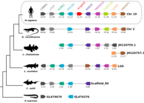
3.4.4. Neighbouring gene analysis ... 73
3.4.5. Morphological and morphometric evaluation of sea bream intact and regenerating skin . 76 3.4.6. Expression of angptl family members during sea bream skin regeneration ... 79
3.5. Discussion ... 83
3.5.1. Angptl members in fish ... 83
3.5.2. ANGPTL emerged early and evolved via gene duplications and deletions ... 85
3.5.3. Wound healing and Angptls expression in sea bream skin ... 86
3.6. Conclusion ... 89
3.7. Acknowledgements ... 89
3.8. Supplementary material ... 90
Teleost osteoglycin duplicates are highly expressed in muscle and skin and involved in late-phase regeneration of skin ...105
4.1. Abstract ... 106
4.2. Introduction ... 107
4.3. Materials and Methods ... 109
4.3.1. Identification and characterization of osteoglycin (ogn) gene(s) in gilthead sea bream .. 109
4.3.2. Phylogenetic analysis and gene environment ... 110
4.3.3. Promoter analysis ... 111
4.3.4. Multiple sequence alignments and protein characterization ... 111
4.3.5. Animal experiments ... 112
4.3.5.1. Ethics Statement ... 112
4.3.5.2. Tissue sampling ... 112
4.3.5.3. Tissue culture experiments ... 112
4.3.5.3.1. Bone-derived gilthead sea bream primary culture ... 112
4.3.5.3.2. Myocyte gilthead sea bream primary culture ... 113
4.3.5.4. OGN in a scale regeneration model ... 114
4.3.5.4.1. Plasma analysis ... 115
4.3.5.4.2. Scale TRAP and ALP activities ... 115
4.3.5.4.3. Calculation of scale area and regeneration rate ... 116
4.3.5.4.4. RNA extraction and cDNA synthesis ... 116
4.3.5.4.5. Quantitative real-time PCR (QRT-PCR) ... 117
4.3.6. Statistical analyses ... 118
X
4.4.4. OGN promoter analysis ... 125
4.4.5. Tissue distributions of ogn1 and ogn2 ... 126
4.4.6. Expression of ogn1 and ogn2 during differentiation of bone-derived and myocyte primary cultures 128 4.4.7. Characterisation of OGN in a scale regeneration model ... 132
4.4.7.1. Plasma parameters ... 132
4.4.7.2. Scale biochemistry and histomorphology ... 133
4.4.7.2.1. Scale growth ... 133
4.4.7.2.2. Enzyme activity ... 134
4.4.7.2.3. Expression profile of ogn1, ogn2 and op during sea bream scale / skin regeneration ... 134
4.5. Discussion ... 136
4.5.1. OGN evolution and structure ... 136
4.5.2. OGN function ... 139
4.6. Conclusion ... 141
4.7. Acknowledgements ... 141
4.8. Supporting material ... 142
The effect of the intermediate metabolite alpha-ketoglutarate (AKG) on skin integrity, function and repair in the teleost sea bream (Sparus aurata) ...147
5.1. Abstract ... 148
5.2. Introduction ... 149
5.3. Material and Methods ... 151
5.3.1. Ethics Statement ... 151
5.3.2. Alpha-ketoglutarate treatment ... 151
5.3.3. Animals experiments ... 151
5.3.3.1. Experiment 1 – Preliminary trial on the influence of AKG treatment in the integument of sea bream 152 5.3.3.2. Experiment 2 – Influence of AKG treatment in sea bream skin regeneration after scale removal ... 152
5.3.3.2.1. Power Analysis ... 152
5.3.3.3. Plasma biochemistry ... 154
5.3.3.4. TRAP and ALP enzymatic assays ... 154
5.3.3.5. Epithelial outgrowth assay ... 155
5.3.3.6. Skin conductivity ... 155
5.3.3.7. Calculation of scale area and regeneration rate ... 156
5.3.3.8. Heart elasticity measurement ... 156
5.3.3.8.1. Force measurements ... 157
5.3.3.8.2. Elastic recoil calculations ... 157
5.3.4. Statistical analysis ... 157
5.4. Results ... 158
5.4.1. Experiment 1 – Preliminary trial on the influence of AKG treatment in the integument of sea bream 158 5.4.1.1. Plasma biochemistry ... 158
5.4.1.2. TRAP and ALP enzymatic activity in sea bream scales ... 159
5.4.1.3. Epithelial outgrowth of sea bream scales-skin ... 160
5.4.1.4. Skin conductivity ... 160
5.4.2. Experiment 2 – Influence of AKG treatment in sea bream skin regeneration after scale removal 161 5.4.2.1. Plasma biochemistry ... 161
5.4.2.2. Enzymatic activity and scale growth in intact and regenerating scales ... 162
5.4.2.3. Epithelial outgrowth of sea bream scales-skin ... 165
5.4.2.4. Skin conductivity ... 166
5.4.2.5. Elasticity of sea bream heart ... 167
5.5. Discussion ... 167
5.5.1. AKG stimulates epithelial outgrowth of sea bream scale-skin ... 167
5.5.2. AKG stimulates sea bream scale biomineralization ... 168
5.5.3. AKG seems to increase elasticity of the sea bream heart muscle ... 169
5.6. Conclusions ... 170
XI
6.2. Future perspectives ... 180 References ...181
XII
TABLE 1.1-DESCRIPTION OF WOUND HEALING…...………..………12
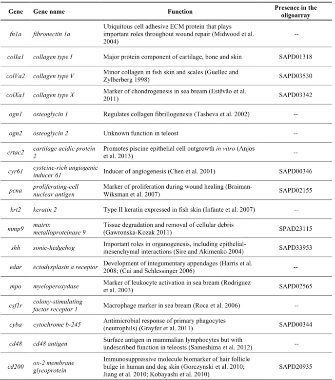
TABLE 2.1-ANNOTATION STATISTICS OF THE DOWN-REGULATED PROBES IN THE MICROARRAY. ... 27
TABLE 2.2-CANDIDATE TRANSCRIPTS SELECTED FOR EXPRESSION ANALYSIS IN SEA BREAM SKIN. ... 28
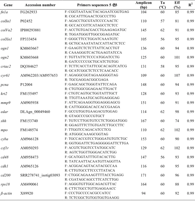
TABLE 2.3-LIST OF THE PRIMERS USED FOR GENE EXPRESSION ANALYSIS BY QUANTITATIVE RT-PCR IN SEA BREAM (SPARUS AURATA) SKIN. ... 31
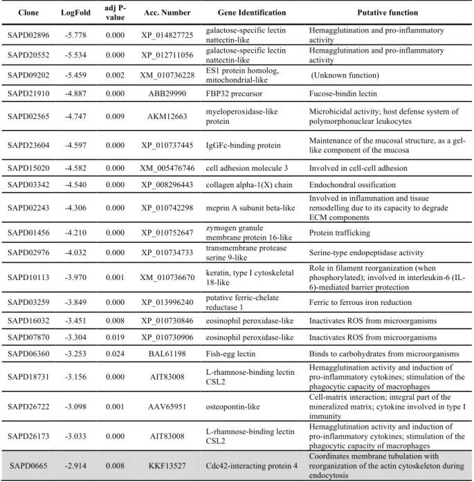
TABLE 2.4-TOP 20 KNOWN GENES DOWN-REGULATED AT 3 AND 7(SHADED ROWS) DAYS AFTER SCALE REMOVAL. ... 34
TABLE 3.1-LIST OF THE PRIMERS USED FOR GENE EXPRESSION ANALYSIS BY QUANTITATIVE RT-PCR IN SEA BREAM (SPARUS AURATA) SKIN. ... 67
TABLE 3.2-DIGITAL EXPRESSION DATA OF ANGPTLS IN THE TELEOST SKIN. ... 80
TABLE 4.1-LIST OF PRIMERS USED FOR GENE EXPRESSION ANALYSIS BY QUANTITATIVE REAL-TIME PCR(QPCR). ... 118
TABLE 4.2-OGNS AND POSITIVE SELECTION. ... 121
TABLE 4.3-COMPARISON OF VERTEBRATE OGNS AND FISH DUPLICATES OGN1 AND 2. ... 123
XIII
FIGURE 1.1-A REPRESENTATION OF SKIN STRUCTURE IN MAMMALS. ... 3
FIGURE 1.2 -AGEING SKIN IN MAMMALS.………..………..………...8
FIGURE 1.3-STAGES OF SKIN WOUND REPAIR IN MAMMALS ………...……….…………..11
FIGURE 1.4-INTEGUMENTARY IMMUNE SYSTEM IN THE SKIN OF MAMMALS AND TELEOSTS……….……….………....13
FIGURE 2.1-VENN DIAGRAM WITH THE DISTRIBUTION OF THE DOWN-REGULATED GENES AT 3(3 WS) AND 7(7 WS) DAYS AFTER SCALE REMOVAL. ... 32
FIGURE 2.2-CHANGES OF PLASMA OSMOLALITY IN SEA BREAM DURING SKIN REGENERATION. ... 35
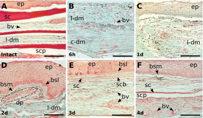
FIGURE 2.3-MORPHOLOGICAL CHARACTERIZATION OF HISTOLOGICAL SECTIONS STAINED WITH MASSON TRICHROME OF INTACT AND REGENERATING SKIN FROM SEA BREAM DURING THE HEALING PERIOD AFTER SCALE REMOVAL. ... 36
FIGURE 2.4-DETAILED IMAGES OF HISTOLOGICAL SECTIONS STAINED WITH MASSON TRICHROME OF INTACT AND REGENERATING SEA BREAM SKIN. ... 37
FIGURE 2.5-RELATIVE EXPRESSION OF ECM AND MATRICELLULAR GENES IN INTACT AND REGENERATING SKIN DURING THE HEALING PERIOD AFTER SCALE REMOVAL IN SEA BREAM. ... 38
FIGURE 2.6-EXPRESSION PROFILE OF CELLULAR PROLIFERATION, REMODELLING AND EPITHELIAL-MESENCHYMAL INTERACTIONS RELATED GENES DURING SEA BREAM HEALING AFTER SCALE REMOVAL IN INTACT AND REGENERATING SKIN. ... 39
FIGURE 2.7-RELATIVE EXPRESSION OF IMMUNE-RELATED GENES DURING SEA BREAM SKIN HEALING AFTER SCALE REMOVAL IN INTACT AND REGENERATING SKIN. . 41
FIGURE 3.1-ANGPTL FAMILY GENE MEMBERS IN FISH.THE NUMBER OF PREDICTED ANGPTL GENES IDENTIFIED IS INDICATED. ... 69
FIGURE 3.2-PHYLOGENETIC TREE OF THE FISH AND OTHER METAZOAN ANGPTL FAMILY MEMBERS CONSTRUCTED WITH THE MAXIMUM-LIKELIHOOD (ML) ALGORITHM.. ... 71
FIGURE 3.3-SCHEMATIC REPRESENTATION OF THE DEDUCED STRUCTURE AND CONSERVED CONSENSUS MOTIFS OF THE FISH AND HUMAN ANGPTLS. ... 73
FIGURE 3.4-COMPARISON OF THE HOMOLOGOUS GENOME REGIONS HARBOURING THE HUMAN ANGPTL8 WITH FISH. ... 74
FIGURE 3.5-COMPARISON OF THE HOMOLOGOUS GENOME REGIONS HARBOURING THE FISH ANGPTL9 WITH HUMAN. ... 75
FIGURE 3.6-MORPHOLOGICAL EVALUATION OF SEA BREAM INTACT AND REGENERATING SKIN (1,2,3 AND 4 DAYS AFTER WOUNDING) STAINED WITH MASSON`S TRICHROME.. ... 77
FIGURE 3.7-MORPHOMETRIC EVALUATION OF SEA BREAM SKIN DURING WOUND HEALING AFTER SCALE REMOVAL.. ... 78
FIGURE 3.8-NUMBER OF BLOOD VESSELS (A) AND BLOOD VESSEL DIAMETER (B) DURING SKIN WOUND HEALING IN SEA BREAM. ... 79
FIGURE 3.9-RELATIVE EXPRESSION OF THE SEA BREAM ANGPTL1B, ANGPTL2B, ANGPTL4A, ANGPTL4B, ANGPTL7 AND VEGFAB IN INTACT AND REGENERATING SKIN.. ... 82
FIGURE 4.1-PHYLOGENETIC ANALYSIS OF OSTEOGLYCINS (OGNS) IN VERTEBRATES. ... 120
FIGURE 4.2-CONSERVED SYNTENY IN VERTEBRATE OSTEOGLYCINS (OGN). ... 122
FIGURE 4.3-DENDROGRAM COMPARING OGN STRUCTURAL FEATURES FROM REPRESENTATIVE ORGANISMS OF THE MAIN VERTEBRATE LINEAGES. ... 125
FIGURE 4.4-PROMOTER ANALYSIS.. ... 126
FIGURE 4.5-EXPRESSION PROFILE OF GILTHEAD SEA BREAM OSTEOGLYCINS (OGN1 AND OGN2) IN ADULT TISSUES. ... 128
FIGURE 4.6-EXPRESSION PROFILE OF OSTEOGLYCINS (OGN1 AND OGN2) IN GILTHEAD SEA BREAM BONE-DERIVED PRIMARY CELL CULTURES IN OSTEOGENIC MEDIUM. ... 130
FIGURE 4.7-EXPRESSION PROFILE OF OSTEOGLYCINS (OGN1 AND OGN2) IN GILTHEAD SEA BREAM BONE-DERIVED PRIMARY CELL CULTURES IN ADIPOGENIC MEDIUM.. ... 131
FIGURE 4.8-EXPRESSION PROFILE OF OSTEOGLYCINS (OGN1 AND OGN2) IN GILTHEAD SEA BREAM MYOCYTE PRIMARY CULTURED CELLS.. ... 132
FIGURE 4.9-CHANGES IN PLASMA CALCIUM, PHOSPHORUS, SODIUM, POTASSIUM, PROTEINS, OSMOLALITY, ASPARTATE-AMINOTRANSFERASE (AST) AND ALANINE -AMINOTRANSFERASE (ALT) DURING SCALE RECOVERY IN GILTHEAD SEA BREAM.. ... 133
FIGURE 4.10-SCALE GROWTH AND ENZYMATIC ACTIVITIES OF TRAP AND ALP IN SEA BREAM SCALES AND GENE EXPRESSION IN THE SKIN OF SEA BREAM ………133
FIGURE 5.1-ROLE OF AKG IN THE PRODUCTION OF COLLAGEN AND ELASTIN. ... 149
FIGURE 5.2-PLASMA PARAMETERS OF CONTROL AND AKG TREATED SEA BREAM AT THE BEGINNING OF THE PRELIMINARY TRIAL (DAY 0).. ... 158
FIGURE 5.3-PRELIMINARY EVALUATION OF THE ENZYMATIC ACTIVITY OF TARTRATE-RESISTANT ACID PHOSPHATASE AND ALKALINE PHOSPHATASE (ALP) IN SEA BREAM SCALES OF CONTROL AND AKG TREATED FISH. ... 159
FIGURE 5.4-PRELIMINARY EVALUATION OF THE EPITHELIAL OUTGROWTH OF CONTROL AND AKG TREATED SEA BREAM SCALES. ... 160
FIGURE 5.5-PRELIMINARY EVALUATION OF SKIN CONDUCTIVITY IN CONTROL AND AKG TREATED SEA BREAM (N =4) SKIN AT THE BEGINNING (DAY 0) AND AFTER TWO WEEKS (DAY 14) OF THE EXPERIMENTAL TRIAL. ... 161
XIV
FIGURE 5.9-EPITHELIAL OUTGROWTH IN INTACT AND REGENERATING SEA BREAM SKIN. ... 165 FIGURE 5.10-SKIN CONDUCTIVITY IN INTACT AND REGENERATING SKIN. ... 166 FIGURE 5.11-ELASTIC RECOIL OF AKG TREATED AND CONTROL SEA BREAM (N =6) HEART (CONUS). ... 167
XV
SUPPLEMENTARY TABLE 2.1-COMPLETE LIST OF THE NEWLY ANNOTATED AND DIFFERENTLY EXPRESSED TRANSCRIPTS AT DAYS 3 AND 7(SHADED ROWS) AFTER
SCALE REMOVAL. ... 49
SUPPLEMENTARY TABLE 3.1-ACCESSION NUMBERS OF THE FISH, TETRAPOD AND CEPHALOCHORDATE ANGPTL GENES AND TRANSCRIPTS. ... 90
SUPPLEMENTARY TABLE 3.2-PERCENT OF SEQUENCE IDENTITY/SIMILARITY OF THE FISH ANGPTL MEMBERS WITH THE HUMAN ORTHOLOGUES.. ... 92
SUPPLEMENTARY TABLE 3.3-LIST OF THE TELEOST ANGPTL ESTS AND THEIR ORIGIN. ... 93
SUPPLEMENTARY TABLE 3.4-ACCESSION NUMBERS OF THE FISH, TETRAPOD AND CEPHALOCHORDATE ANGPT GENES AND TRANSCRIPTS. NI-NOT IDENTIFIED ... 96
SUPPLEMENTARY TABLE 4.1-ACCESSION NUMBERS OF ALL PROTEINS AND NUCLEOTIDES SEQUENCES USED IN THIS STUDY. ... 142
SUPPLEMENTARY TABLE 5.1-POWER ANALYSIS. ... 171
SUPPLEMENTARY TABLE 5.2-BIOLOGICAL PARAMETERS OF THE EXPERIMENTAL FISH. ... 172
List of supplementary figures
SUPPLEMENTARY FIGURE 2.1-FUNCTIONAL ANNOTATION ACCORDING TO GENE ONTOLOGY (GO) LEVEL 2 OF THE DOWN-REGULATED GENES 3 AND 7 DAYS AFTER SCALE REMOVAL. ... 53SUPPLEMENTARY FIGURE 2.2-RELATIVE EXPRESSION OBTAINED BY QPCR OF ECM AND MATRICELLULAR PROTEINS DURING SEA BREAM SKIN HEALING AFTER SCALE REMOVAL IN INTACT AND REGENERATING SKIN. ... 54
SUPPLEMENTARY FIGURE 2.3-RELATIVE EXPRESSION OBTAINED BY QPCR OF CELL PROLIFERATION, REMODELLING AND EPITHELIAL-MESENCHYMAL INTERACTIONS RELATED GENES DURING SEA BREAM HEALING AFTER SCALE REMOVAL IN INTACT AND REGENERATING SKIN. ... 55
SUPPLEMENTARY FIGURE 2.4-RELATIVE EXPRESSION OBTAINED BY QPCR OF IMMUNE-RELATED GENES DURING SEA BREAM HEALING AFTER SCALE REMOVAL IN INTACT AND REGENERATING SKIN. ... 56
SUPPLEMENTARY FIGURE 3.1-EXPANDED PHYLOGENETIC TREE OF THE FISH AND OTHER METAZOAN ANGPTL FAMILY MEMBERS USING BAYESIAN INTERFERENCE (BI). ... 97
SUPPLEMENTARY FIGURE 3.2-SUPPLEMENTARY FIGURE 2:PHYLOGENETIC TREE OF THE FISH AND OTHER METAZOAN ANGPTL FAMILY MEMBERS CONSTRUCTED WITH THE MAXIMUM-LIKELIHOOD (ML) ALGORITHM. ... 98
SUPPLEMENTARY FIGURE 3.3-SEQUENCE ALIGNMENTS OF THE HUMAN, SPOTTED GAR, ZEBRAFISH, SEA BASS AND SEA BREAM ANGPTLS. ... 99
SUPPLEMENTARY FIGURE 3.4-COMPARISON OF THE HOMOLOGOUS GENOME REGIONS HARBOURING THE VERTEBRATE ANGPTL7 WITH THE CEPHALOCHORDATE………..……….…101
SUPPLEMENTARY FIGURE 3.5-AMINO ACID SEQUENCE ALIGNMENT OF THE CEPHALOCHORDATE ANGPTL-LIKE 7WITH THE HUMAN AND SPOTTEDGAR ANGPTL7……….………..………103
SUPPLEMENTARY FIGURE 4.1-PHYLOGENETIC ANALYSIS OF OSTEOGLYCINS (OGNS) IN VERTEBRATES PERFORMED WITH THE NEIGHBOUR JOINING (NJ) METHOD. 143 SUPPLEMENTARY FIGURE 4.2-GILTHEAD SEA BREAM OSTEOGLYCINS 1 AND 2(OGN1 AND OGN2) NUCLEOTIDE SEQUENCES AND ENCODING PROTEINS. ... 144
SUPPLEMENTARY FIGURE 4.3-AMINO ACID SEQUENCE ALIGNMENTS OF HUMAN AND FISH OSTEOGLYCINS (OGN). ... 145
1
1
General introduction
1.1 General introduction
The present thesis is focussed on the skin, the largest organ in the body with an essential role in numerous physiological processes in vertebrates. It has generally been overlooked by anatomists and physiologists, although in recent years it has received increasing attention due to its function as an innate immune barrier and the ease of access for studies of aging and molecular processes. The target organism of this thesis is a teleost fish (sea bream, Sparus
aurata), that belongs to the most successful extant vertebrates with over 28,000 species
identified (Nelson et al. 2001). Studies of skin in fish are of relevance for veterinary medicine, are essential for an important production sector, aquaculture and can also contribute as a biomedical model for regeneration. The advantages of fish skin as an experimental model is the ease of access and experimentation, the epidermis is living and not obscured by a layer of dead cornified cells, it has a high regenerative capacity, does not wrinkle or scar and physiological systems, including the immune system, tend to be less complex in fish, which facilitates interpretation of results. The following introduction is organized to provide a working knowledge about key aspects that are targeted in the experimental studies reported in the thesis and includes considerations about: 1) the anatomy of the skin in vertebrates, 2) the molecular and cellular elements of skin, 3) wound healing and regeneration, 4) the immune system and the skin and 5) model systems for regeneration.
2
1.2 The skin
In vertebrates the integumentary system comprises the skin and its appendages. It is the primary interface between an organism and the environment and provides protection against mechanical insults and physiological stress playing a key role in communication, locomotion and in the detection and reception of external stimulus. Vertebrates possess diverse types of integuments and they have evolved a wide variety of multicellular appendages such as hair, scales, feathers, hooves and nails that perform a wide variety of functions adapted to the different environment that each organism inhabits (Vickaryous and Sire 2009; Hildebrand 1974; Busam 2010). These may include mechanical protection of the soft tissues below, control of water passage in and out of the body to prevent desiccation and disturbance of water balance, regulation of heat transfers to the environment, protection against the entry of injurious organisms and materials, and behavioural aspects such as adaptive coloration and locomotion. The skin may also have a role in respiration, secretion, excretion, sense reception, and also in the storage of fat and glycogen. In vertebrate’s colour is also vested in the skin and different colour pigments exist in the chromatophores (Hyman 1956) and the combined action of the different chromatophores (in fish, xanthophores, erythrophores, iridophores, leucophores, melanophores, and cyanophores and in mammals melanophores) produce distinct colour effects that are tightly regulated by hormones produced in the pituitary (eg: melanocyte-stimulating hormone), thyroid (eg: thyroxine) and adrenals (eg: cortisol) glands (Bentley 1998; Hildebrand 1974; Hyman 1956). Colour is used by various vertebrates for concealment or to make themselves conspicuous (as warning, social releaser or sexual attractant), for control of heat absorption or conservation, and in terrestrial vertebrates for control of vitamin D synthesis.
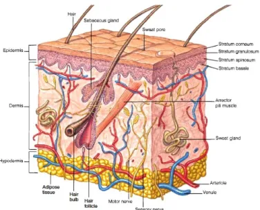
The skin is the largest organ in the body, it is crucial for survival and has an innate ability to regulate its functions but is also coordinated with the rest of the body through the nervous and endocrine systems that mediate its sensory functions and regulate its blood supply (Slominski et al. 2013; Bentley 1998). Skin cells produce hormones, neurotransmitters and neuropeptides and their respective functional receptors having a role as a neuroendocrine organ. They are capable of local immune and steroidogenic activities to protect the body against external environmental and biological factors to maintain local homeostasis (Slominski et al. 2013; Slominski et al. 2008) as reflected by the presence of an HPA (Hypothalamic-Pituitary-Adrenal) (Slominski et al. 2013; Slominski et al. 2008; Slominski et al. 2007) and HPT (Hypothalamic-Pituitary-Thyroid) (Paus 2010) axis in the skin of mammals. Skin is structurally simple but biologically complex and is composed of two principal layers - the epidermis, a superficial layer and a deeper layer - the dermis (figure 1.1) (Hildebrand 1974; Hyman 1956).
3
It is the first organ to emerge in embryonic development and the ectoderm (the outer layer of the gastrula) forms the outer part of the skin, the epidermis and also the nervous system and parts of the eye and ear. The mesoderm (the middle layer of the gastrula) forms the skeletal, muscular, circulatory, urogenital systems and the inner part of the skin, the dermis (Hildebrand 1974).
1.3 General structure of the vertebrate skin 1.3.1 The epidermis and its derivatives
The epidermis is the outer part of the skin, it is relatively thin in most animals and is a stratified epithelium. In the epidermis basal cells are attached to the basement membrane and the most superficial of the supra-basal cell layers form the epithelia. Only the cells in the
stratum basale (figure 1.1), also known as stratum germinativum, are mitotically active and
their function is to replenish the loss of cells on the surface of the superficial stratum. In the intermediate stratum (stratum spinosum and granulosum in figure 1.1) of a stratified epithelium, the cells can undergo various processes of differentiation, such as keratinization (Busam 2010; Bragulla and Homberger 2009).
Figure 1. 1 - A representation of skin structure in mammals. Taken from http://pulpbits.net/7-skin-structure-anatomy-diagrams/structure-of-skin/
In vertebrates, the epidermis is stratified into two or more layers (figure 1.1). In land vertebrates, the outer layer of the epidermis is composed of dead keratinized cells that protect the skin against water loss and bacterial infections. Epidermal cells are continuously replenished by cells produced in the stratum germinativum and as they move toward the upper
4
layer they start producing keratin (a water-insoluble protein) and differentiate from cuboidal cells to flattened keratinized cell that are shed. In humans the epidermis is shed continuously but in the reptiles such as snakes and lizards the whole epidermis is shed at intervals during moulting (Seeley et al. 2001; Hildebrand 1974; Hyman 1956). The epidermis in aquatic vertebrates such as cyclostomes, fish and tailed amphibians differs from terrestrial vertebrates as it is composed of mitotically active cells and contains numerous mucous cells, the secretion of which keeps the skin moist and slimy and confers protection from the aquatic milieu (Hildebrand 1974; Hyman 1956).
1.3.1.1 Epidermal Derivatives 1.3.1.1.1 Glands
In mammals, three types of cutaneous glands of ectodermal origin are present: the sebaceous, sweat (figure 1.1) and mammary glands and they are responsible for lubrication and protection against water loss, thermo-regulation and milk production, respectively. The sebaceous glands are limited to the mammals and are branched alveolar glands that drain into each hair follicle (figure 1.1) and are related to the nipples, lips and genitalia. Sweat glands (figure 1.1) are tubular, simple (not lobulated) and coiled at their inner ends and form from invagination of the epidermis into the dermis. The mammary glands produce and secrete milk to suckle the young and are formed when the epidermis sinks into the dermis and branches into solid cords. In females, these cords enlarge at puberty, pushing under the skin and becoming lobulated and alveolar (Hildebrand 1974; Hyman 1956).
1.3.1.1.2 Keratinized and cornified structures
Vertebrates have a range of divergent keratinized structures, that includes beaks and feathers in birds and epidermal scales in lizards and in the legs of birds. Claws are strong keratinized structures that wrap around the tapering terminal bones of the digits. Hooves are derived from claws. The shells of the turtle and the armadillo have a heavily keratinized epidermis and the baleen plates of whales are outgrowths of the buccal epithelium and serve as strainers for feeding. The horns and antlers of tetrapods come in different shapes and sizes. The horns of the rhinoceros grow continuously from the epidermis, and the keratinized fibres are compacted into a solid structure (cornified material) that is never shed. The antlers of the deer family are bony outgrowths of the skull covered in skin (“velvet”) and they are shed and replaced each year while the in the pronghorns the skin covering the antlers forms horn and not
5
hair. The hairs (figure 1.1) or fur in mammals also have the same epidermal origin and are composed of a keratinized shaft that expands from the root below the skin and is hidden in an epidermal sheath or hair follicle. These keratinized epidermal appendages confer protection and resistance to abrasion, but also provide insulation and protection against water loss (Bragulla and Homberger 2009; Hildebrand 1974).
1.3.2 The dermis and its derivatives
The dermis is usually thicker than the epidermis (figure 1.1). It commonly has an outer, vascular stratum spongiosum and a deeper, thicker stratum compactum that are interconnected to secure the skin to the connective tissue covering the muscles of the body wall. The dermis is composed of numerous collagenous fibres but have fewer elastic fibres that are randomly oriented in a three dimensional network (Hildebrand 1974) that is supported by the extracellular matrix (ECM) which confers support and guidance for the cells.
1.3.2.1 Dermal derivatives: Fish Scales
The scales in modern teleosts are of dermal origin. These specialized dermal appendages are the elasmoid scales (cycloid or ctenoid) that are thin, flexible and transparent structures that are rich in calcium (Ca) and phosphorus (P) and are built by osteoblasts and resorbed and remodelled by osteoclasts (de Vrieze et al. 2011; Rotllant et al. 2005b; Persson et al. 1999; Witten 1997). The bulk of the elasmoid scale consists of the basal layer, an acellular structure that contains collagenous fibres running in various directions and an outer hypermineralized layer devoid of collagen (Sire and Akimenko 2004). The scales are responsible for physical protection and for the hydrodynamic properties of the integument (Elliot 2011b; Hildebrand 1974) and are part of the dermal skeleton, accounting for up to 20 % of the total body calcium (Berg 1968; Flik et al. 1986).
1.4 Major proteins that are part of the skin
Skin is a complex tissue and approximately 63% of all the human proteins are expressed in this organ (Uhlen et al. 2015). Some of the most important proteins involved in skin function are briefly described below.
6
1.4.1 Collagens
Collagens are the major structural components of the extracellular matrix (ECM) of all metazoans and are also the most abundant connective proteins in the skin. Collagens make up 70-80% of the dry weight of the skin, contributing to its stability and structural integrity (Colgravea et al. 2008; Son et al. 2007; Seeley et al. 2001). They can form large stable extracellular fibrils and complex fibrous superstructures that are responsible for the tensile strength of the tissues and are also part of the cytoskeletal system of epithelial cells (Castillo-Briceno et al. 2009; Bragulla and Homberger 2009; Perumal et al. 2008). Fibrillar collagens (I, II, III, V, XI, XXIV and XXVII) are composed of three polypeptide chains with a triple helix arrangement and each collagen α chain is characterized by a unique repeating amino acid structure, Gly-X-Y, where Y is often proline. Hydroxyproline is essential for the formation of the collagen triple helix (Heino et al. 2009; Taşkiran et al. 1999). The deposition of collagen is finely controlled and is dependent on the physiological status of the body. Dysregulation of collagen deposition and remodelling can originate pathological conditions that result from excessive accumulation of type I collagen in the tissue, as in the keloids (Sasaki et al. 1987) or disorders of the connective tissue like the Ehlers-Danlos syndrome, where the fragility and reduced tensile strength of the skin is one of the cutaneous manifestations of the disease. 1.4.2 Keratins
Keratins account for about 25-35% of the extracted proteins in human and murine stratified epidermis (Bowden et al. 1984) and form intermediate filaments found only in vertebrates (Fuchs 1983). The keratins are part of a large multi-gene family that are all characterised by their high cysteine content, which contributes through intermolecular disulphide bond formation to keratins strength and stability (Bragulla and Homberger 2009). The main functions of keratins are to provide resistance to mechanical and non-mechanical stresses and act as a scaffold for the maintenance of cellular architecture (Infante et al. 2007). Evolution of the keratinization of the skin in the tetrapods was accompanied by its association with lipid molecules and these modifications allowed adaptation to the terrestrial environment by providing protection against desiccation. Keratins are also involved in non-structural functions such as cell signalling, proliferation and apoptosis (Gu and Coulombe 2007). Keratins in epithelial cells are not characteristic of entire tissues but of some particular functional properties of cells and tissue regions. In fact, unlike other vertebrates, in teleost fish, hagfishes,
7
lampreys and amphibians keratins are not restricted to epithelial tissues and extracellular keratins have been described (Schaffeld and Schultess 2006).
1.4.3 Elastin
Elastin is a highly elastic protein in the skin, lungs and arteries allowing them to resist deformity and to restoring their shape after stretching or contracting. It is rich in hydrophobic amino acids and in tissue multiple tropoelastin molecules are cross-linked through lysine to desmosine and isodesmosine to generate a massive insoluble and durable complex (Muiznieks and Keeley 2013). In human skin, elastin is a minor component and though it is difficult to identify in histological sections it forms a highly crossed-linked network with the collagens to confer tissue resistance and elasticity.
1.4.4 Matricellular proteins
Matricellular proteins are proteins of the extracellular matrix (ECM) that do not have a structural role (Midwood et al. 2004). Matricellular proteins include galectins, osteopontin, SPARC, tenascins, thrombospondins, vitronectin that act temporally and spatially to provide signals that trigger specific cell activities (Midwood et al. 2004; Bornstein and Sage 2002). They are expressed at high levels during development but not in adult tissues and are up-regulated at sites of tissue remodelling (Midwood et al. 2004). In the skin, some small leucine-rich proteoglycans (SLRPs) have a matricellular function, such as decorin and lumican (Iozzo and Schaefer 2010; Yeh et al. 2010; Merline et al. 2009).
1.5 Skin ageing and skin diseases 1.5.1 Ageing skin
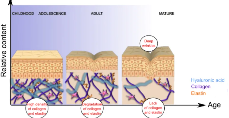
In young skin, the structural properties provided by the cross-linking of the abundant collagens and elastin in the dermis provides an effective support to the epidermis, which is also thinner. Ageing leads to a reduction in the amount and quality of the connective tissue (figure 1.2), which becomes disorganized causing the epidermis to wrinkle, sag and appear lax.
8
Figure 1. 2 - Ageing skin in mammals. Highly crossed-linked collagen and elastin fibres are abundant in young skin. The altered hormonal status with age accompanied by photo-aging of the skin leads to a decrease in
cellular activity so that production of collagen and elastin decreases. The skin starts to lack the necessary tensely strength to keep the epidermis in place, and wrinkles start to form. Image taken from
http://www.ortron.com.au/ortron-blog/latest
Ageing is a complex multifactorial process (Kishi et al. 2009) and age-related changes that occur during cutaneous wound healing are considered to be potentially negative, especially in the elderly population. Disturbances in protein metabolism, one of the main symptoms of ageing and oestrogen deficiency contribute to less efficient wound healing in the elderly (Harrison and Pierzynowski 2008).
A number of recent studies of teleost aging/senescence suggest that homeostasis and repair in their aging skin may give insight into important molecular/structural factors underlying normal skin function. Several ageing markers have been detected in the skin of zebrafish (Danio rerio) namely increased activity of senescence-associated ß-galactosidase and accumulation of oxidized proteins in the muscle (Kishi et al. 2009; Kishi et al. 2003). In the medaka (Oryzias latipes) signs of ageing were identified in the skin, and the dermis of 24-month old individuals had an age-dependent decline in the number of dividing cells and an increase in senescence-associated ß-galactosidase. Other tissues such as the liver (spongiosis hepatitis, steatosis, ballooning degeneration, inflammation and nuclear pyknosis), the heart (fibrosis and lipofucsin accumulation), and the eyes (loss of pigmented cells from the retinal epithelium) had a characteristic age associated change (Ding et al. 2010). These studies provided a set of markers that can be used to trace the process of normal tissue aging and to evaluate the impact of environmental stressors.
Relat
ive
cont
ent
Age
CHILDHOOD ADOLESCENCE ADULT MATURE
Hyaluronic acid Collagen Elastin Deep wrinkles High density of collagen and elastin Degradation of collagen and elastin Lack of collagen and elastin
9
1.5.2 Skin disorders
There is a broad spectrum of lesions that have in common the mechanical fragility of the skin with frequent blister formation and an underlying genetic aetiology (Busam 2010). Mutations of keratins are associated with several disorders that have a clear phenotype in human and murine skins (Pekny and Lane 2007). In epidermis bullosa simplex, an epidermal blister disease, which involves the breakdown of basal cells in stratified epithelia, either KRT5 or
KRT14 is altered. When KRT14 is mutated, the type I keratin K15 can compensate for K14 to
combine with the type II keratin K5, the usual partner of K14. This results in less severe symptoms than when KRT5 is mutated, since there is no compensating keratin (Pekny and Lane 2007) (Steinert 2001). The same is true for K6 and its partners K16/17 (Coulombe and Wong 2004). In the skin disorder epidermolytic hyperkeratosis, another epidermal blister disease (Busam 2010), KRT1 and KRT10, characteristic of keratinizing suprabasal cells in the stratified epidermis are mutated. The disease white sponge nevus, which affects the oral and genital stratified epithelia, is caused by mutations of KRT14 and KRT13 (Pekny and Lane 2007).
Hyperpigmentation skin disorders, such as Acanthosis nigricans, where there is hyperkeratosis and darkening of the skin, are directly associated with insulin resistance (IR), but the most common associated complications of diabetes in the skin are impaired wound healing, foot ulcers and an increased incidence of skin infections (Perez and Kohn 1994). The molecular basis of the condition is unclear but is linked to insulins role in regulating differentiation and glucose transport by keratinocytes and also proliferation as shown by IR mice with diabetes mellitus (Wertheimer et al. 2001).
Several skin disorders are attributed to alterations of the dermal connective tissue (deposition disorders) (Busam 2010). Namely, prolidase deficiency (PD), which is a rare, autosomal recessive, inborn error of amino acid metabolism that affects collagen maturation and is characterized by severe dermatological manifestations, particularly ulcers of the lower extremities (Haywood 2011). There is currently no cure for this condition.
Psoriasis is a chronic, inflammatory skin disease that results from a complex interplay between genetic and environmental factors that stimulates the activation of the dendritic cells. The activated dendritic cells stimulate the differentiation and migration of effector T cells (Th1 and Th17) to the skin, which subsequently leads to the release of inflammatory cytokines that promote further recruitment of immune cells, stimulation of keratinocyte proliferation and sustains a state of chronic inflammation (Nestle et al. 2009). In this condition there is a generalized epidermal barrier dysfunction with immune dysregulation.
10
1.6 Skin repair and regeneration
Skin is in the frontline of defence from external insults and is frequently subject to injuries (Gawronska-Kozak et al. 2014). The efficacy of skin repair is essential to re-establish immune defence since integument homeostasis is essential for health and survival as it confers protection against pathogens. Wound repair also involves two major clinical issues: a) the rate of healing, a prominent problem in the elderly population, and b) the quality of healing, a major concern in cosmetic appearance especially amongst the young population. The outcome of skin trauma can be regarded as a continuum of possible solutions that range from debilitating scars to complete regeneration and restitution of functionality (Seifert and Maden 2014; Seifert et al. 2012b; Redd et al. 2004). Therefore, the primary therapeutic goal of the treatment of wounds involves rapid wound closure with minimal scar formation (Li et al. 2001). The relevance of wound healing for human health has led to many studies to characterise the physical and molecular mechanisms underlying skin repair to control scaring and ameliorate the effects of aging (Schreml et al. 2010; Philips et al. 2011).
1.6.1 Stages of skin wound healing
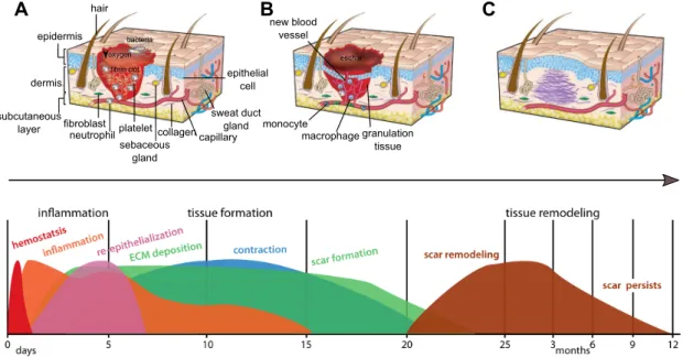
In mammals, the response of skin to damage is a complex cascade of events and includes: inflammation, proliferation and maturation. These events overlap in time and space and lead to restoration of tissue integrity and homeostasis (figure 1.3 and table 1.1). Skin damage triggers an inflammatory response and is rapidly repaired by dermal fibroblasts and keratinocytes leading to scaring (Schreder et al. 2010; Gilliver et al. 2007). Immediately after injury (figure 1.3A and 1.3D), the platelet plug forms a provisional wound matrix composed of fibrin that attracts inflammatory cells like neutrophils and monocytes to the wound. Neutrophils and monocytes differentiate into macrophages and phagocytize foreign particles (such as bacteria and necrotic tissues) and also participate in angiogenesis (blood vessel repair) and matrix deposition. Within hours of injury re-epithelialization of the wound is initiated and (figure 1.3B) epidermal cells migrate to cover the wound, fibroblasts attracted to the injured area proliferate and form granular tissue, and a network of blood vessels are formed by angiogenesis. The remodelling stage is stimulated by matrix metalloproteinases that are released from fibroblasts and macrophages and modulate the extracellular matrix (ECM). In mammals, the initial deposition of collagen III is replaced by collagen I, fibroblasts differentiate into myofibroblasts that contract to decrease the scar surface and later endothelial cells, macrophages and myofibroblasts undergo apoptosis so that a scar (figure 1.3C) is the final
11
product of the tissue healing process (Richardson et al. 2013; Gurtner et al. 2008; Braiman-Wiksman et al. 2007; Midwood et al. 2004; Werner and Grose 2003; Philips et al. 2011).
Figure 1. 3 - Stages of skin wound repair in mammals. There are three major classical stages of wound repair:
inflammation, 0-48 h after injury (A), new tissue formation, 2-21 days after injury (B) and remodelling, up to 1 year (C). A: During inflammation (immediately after wounding), a fibrin clot forms that contains bacteria, degranulated platelets, inflammatory cells and many growth factors. B: Re-epithelialization occurs in 2-6 days and new tissue formation within 2-21 days after injury. These are accompanied by the formation of a scab (eschar) on the surface under which the keratinocytes from the wound margins migrate to re-epithelialize the wound and new blood vessels infiltrate the fibrous granulation tissue of the wound site. C: The dense and disorganized collagen network laid down by the fibroblasts will form a scar which is often raised relative to the surrounding surface. The original architecture of the skin is completely ablated by the scar, there are no hairs, sweat glands or sebaceous glands present in the repair region. Coloured peaks at the bottom represent the range of time periods of individual events during wound healing. The y-axis represents percent maximal response for indicated processes. Adapted from Seifert et al. 2014, 2012 and Gurtner et al. 2008.
Wounds that do not follow the established sequence of events in mammals are not repaired and are called chronic wounds, and are frequently associated with human diseases such as Diabetes (Schreml et al. 2010; Midwood et al. 2004; Perez and Kohn 1994).
Cutaneous repair and regeneration has mainly been explored in tetrapods (mammals and amphibians) but recently the relevance of teleost fish as a valid experimental model to investigative dermatology has been recognized (Rakers et al. 2013; Rakers et al. 2010; Ding et al. 2010; Kishi et al. 2009; Rakers et al. 2011) as the skin of teleosts does not scar, skin damage is rapidly repaired and the bony scales and overlying epithelia are replaced within a few weeks (Guerreiro et al. 2013; Guerra et al. 2008; Ohira et al. 2007b; Fast et al. 2002; Bereiter-Hahn and Zylberberg 1993). granulation tissue macrophage monocyte new blood vessel hair epidermis dermis subcutaneous
layer fibroblastneutrophilplatelet
sweat duct gland epithelial cell collagen sebaceous gland capillary bacteria fibrin clot oxygen eschar A B C
12
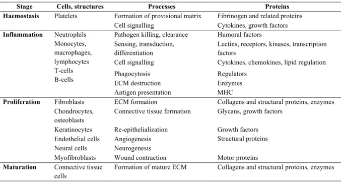
Table 1.1 – Description of wound healing. Overview of the cellular elements and proteins involved in each stage
of wound healing. Wound healing stages are conserved in teleosts and tetrapods however the timing and duration of their occurrence differs between the evolutionary groups and dictates the outcome of the skin repair programme. Adapted from Krasnov et al. 2012.
Stage Cells, structures Processes Proteins
Haemostasis Platelets Formation of provisional matrix Fibrinogen and related proteins Cell signalling Cytokines, growth factors
Inflammation Neutrophils Monocytes, macrophages, lymphocytes T-cells B-cells
Pathogen killing, clearance Humoral factors Sensing, transduction,
differentiation
Lectins, receptors, kinases, transcription factors
Cell signalling Cytokines, chemokines, lipid regulation Phagocytosis Regulators
ECM destruction Enzymes Antigen presentation MHC
Proliferation Fibroblasts ECM formation Collagens and structural proteins, enzymes Chondrocytes,
osteoblasts
Connective tissue formation Glycans, growth factors Keratinocytes Re-epithelialization Growth factors
Structural proteins Endothelial cells Angiogenesis
Neural cells Neurogenesis
Myofibroblasts Wound contraction Motor proteins
Maturation Connective tissue cells
Formation of mature ECM Collagens and structural proteins, enzymes
1.6.2 Role of the immune system in cutaneous repair
The efficacy of skin repair to re-establish immune defense and integument homeostasis is essential for health and survival since it also confers protection against pathogens. The skin is the primary site of defense against external aggressors and the involvement of the immune system in cutaneous repair and regeneration has been described in tetrapods and teleosts fish and in the latter differs significantly from mammals as both the immune response and skin (e.g. it is not keratinized, does not scar and does not develop wrinkles) has substantial differences.
In fact, the evolutionary distance between mammals and teleost fish is reflected in the complexity of their immune systems. While higher vertebrates have a complex innate and acquired immune response that involves a large repertoire of cytokines, growth factors, immunoglobulins, specialized receptors, memory-cells and dedicated sites for their production (such as the bone marrow that produces T- and B-cells and the thymus where T-cell maturation occurs), teleosts possess a less developed immune system with fewer pathways and a limited number of immunoglobulins (figure 1.4). Despite the lower complexity of the immune system in teleosts it also plays a key role during regeneration since immune cells destroy pathogens, damaged cells and extracellular structures, and this process is an essential prerequisite for the onset of tissue repair (Gomez et al. 2013; Richardson et al. 2013; Midwood et al. 2004).
13
Figure 1. 4 - Integumentary immune system in the skin of mammals and teleosts. In mammals the epidermis
is composed of dead keratinized cells while in teleosts the cells are alive and a mucus layer protects the animals from the external environment. Similar cellular components of the innate immune system, like the Langerhans cells, dendritic cells, macrophages, granulocytes and mast cells are found in mammals as well as in teleosts. Differences relate to the localization of B and T cells, the isotype of immunoglobulins and the presence of the secretory component (SC) of the polymeric immunoglobulin receptor (pIgR). The presence of commensal bacteria and anti-microbial peptides (AMPs) is shown in the outer mucosal surface or over the keratin layer. Elements that are suspected to be present in a tissue but have not been studied so far are marked as unknown (?). Adapted from Gomez et al. 2013.
In mammals, cortisol inhibits wound healing and retards reparation of damage (Christian et al. 2006). Increased secretion of cortisol mobilises energy from storage to muscles and shuts down metabolic processes involved in growth and immunity (Barton 2002) and it remains the first-line therapy for suppression of inflammation in human skin (Jackson et al. 2007).
1.7 Animal models of skin regeneration
Skin wound healing with concomitant scar tissue formation has been widely investigated however tissue regeneration is poorly studied due to the lack of suitable animal models. Comparing adult wounds in animals that heal scar free with mammalian wounds that scar may produce new insights particularly with regard to the local environment and how it might relate to dedifferentiation. Adult individuals with regenerative capacities are found in vertebrates such as amphibians and teleosts.
Epidermis
Dermis
Mucous layer
Keratinized epithelial cell
Squamous epithelial cells
Sweat glands / goblet cells
Scale / hair follicle
Langerhans cells
Dendritic cells
Macrophages
Granulocytes
Mast / eosinophilic granule cells
T cells B cells AMPs IgA IgT IgM Commensal bacteria SC of plgR Mammals skin Teleosts skin
14
1.7.1 Mammalian models
Mammalian foetal wounds heal perfectly and represent one of the few examples of tissue regeneration in mammals (Yates et al. 2012; Dang et al. 2003). The foetal immune system is not fully developed and there are fewer neutrophils, monocytes, macrophages and lymphocytes and the length of time the inflammatory cells are present at the wound site is reduced (Cowin et al. 1998). The reduced inflammatory reaction combined with the reticular pattern formed by collagens I and III during foetal wound healing (Cuttle et al. 2005) and the up-regulation of the metalloproteinases (MMPs) relative to the tissue inhibitors of metalloproteinases (TIMPs) are contributing factors that favour the regenerative ability of the foetuses (Gill and Parks 2008; Dang et al. 2003). Nevertheless, the regenerative capacity of the foetus is lost in the second trimester of gestation and thereafter cutaneous repair results in scar formation. The goal of wound healing studies is to identify mechanisms to redirect reparative pathways from debilitating scar formation to regenerative pathways that lead to normal tissue functionality (Gawronska-Kozak et al. 2014). The studies of foetal healing have provided important into the cellular and molecular pathways that lead to the regeneration of tissues and its appendages. Due to the cellular and molecular differences between the foetus and adults, it has been very difficult to develop effective human therapies based on fetal wound healing studies. Adult tissues are fully differentiated and a dedifferentiation process (loss of the differentiated phenotype and re-entry into the cell cycle) is induced after damage while damage in the foetus probably does not have to reactivate the genetic program to repair the damage and instead the excised tissue may simply “carry on” developing, which is may be misinterpreted as scar-free healing (Seifert and Maden 2014).
Few adult mammals are capable of regenerating the skin but some mammalian models have been described. Namely, nude mice and African spiny mice. The nude mice have no hairs, as a consequence of a mutation in the FOXN1 gene, and also lack a thymus and are therefore immune-deficient as a consequence of the of lack T-lymphocytes (Nehls et al. 1994). The timing of the main wound healing stages in the skin of the nude mice is accelerated in comparison to wild type mice. Incisional wounds in the skin of nude mice are completely repaired in 1 to 3 days and granulated tissue is present at day 3 and is largely reduced by 7 days’ post-injury. Thirty-six days’ post injury, the wounded area is virtually absent of collagen fibres while the wild type mice have well-defined and disorganized collagen bundles penetrating the dermis, subcutaneous pads and underlying muscle (Gawronska-Kozak 2011). However, although the inflammatory response plays a role in the regenerative outcome it remains to be categorically demonstrated that it is a determining factor of this process. Particularly since other
15
studies indicate that the regenerative response may be dependent on the bimodal pattern of expression of matrix metalloproteinases (MMPs) MMP9 and MMP13. These proteins are highly expressed in the epidermis during the early phase of repair and in the dermis in the late phase of repair and this pattern is associated with scarless healing (Gawronska-Kozak 2011; Manuel and Gawronska-Kozak 2006).
The African spiny mice (Acomys kempi and Acomys percivali) can shed up to 60% of the total dorsal surface area of their skin as a predator escape behaviour. In the spiny mice the skin regrows and the wound heals without a scar and hairs rapidly regrow (Seifert et al. 2012a) and it is the only mammal capable of skin regeneration. The general structure of the skin in the African spiny mouse is the same as the house mouse (Mus musculus) but the mechanical properties are different. The skin in Acomys is brittle and requires much less energy/force to tear it while the skin of Mus displays elastic properties before breaking and is 20 times stronger than Acomys skin. The ability of Acomys to heal full-thickness excisional skin wounds (4mm and 1.5cm) is characterized by rapid formation of a scab and haemostasis followed by fast re-epithelialization (~64% in 24h, as opposed to 5-7 days in adult rats (Soo 2000). The collagen layers are less densely packed in Acomys during wound contraction and collagen III is the predominant structural component, as opposed to collagen I in Mus musculus (Seifert et al. 2012b). Recently, a similar regenerative capacity was recorded for a third Acomys species,
Acomys cahirinus (Matias Santos et al. 2016).
1.7.2 Amphibians and teleosts
Amputated limbs in salamanders and fins, jaws and the tip of the heart in zebrafish all undergo a regeneration process that results in full functional reconstruction of lost/damaged tissues (Petrie et al. 2014; Seifert et al. 2012b; Brockes and Kumar 2005; Poss et al. 2003). What these experimental models have in common is a low inflammatory response, a short homeostasis period, lack of scab formation and a relatively long phase of new extracellular matrix production and remodelling.
Urodele amphibians (salamanders like the newts and axolots) maintain their ability to repair wounds without scaring, and regenerate a range of organs and tissues throughout their lives. The regeneration of whole adult limbs in the salamander is complex and involves different cell types with distinct embryonic origins and can be divided into 3 distinct phases: the initial wound healing phase, the progenitor cell activation phase and finally the phase of re-development, that is thought to be a recapitulation of embryonic development on a larger scale in an adult context (Godwin and Rosenthal 2014). The Anurans (frogs and toads) developmental
16
stages are characterized by variable capacities to scar-free repair and regeneration is restricted to the early stages of development (Godwin and Rosenthal 2014). The innate and adaptive immune system of amphibians are generally considered similar to mammals but specific differences exist that justify their different regenerative capacity. Urodele amphibians are regarded to have a week immune system (Tournefier et al. 1998) while anurans such as the
Xenopus sp. Have a more robust immune response and less regenerative potential (Murawala
et al. 2012).
Teleosts, in common with amphibians, have the ability to regenerate amputated fins and scales through epimorfic regeneration (Petrie et al. 2014). The caudal fin of zebrafish regenerates fast and several gene markers reviewed by (Poss et al. 2003) have been identified that relate to the different phases of the process, like the wound healing, the blastema formation and the regenerative outgrowth and the link between inflammation and adult regeneration as also been described (Petrie et al. 2014). Teleost skin is specialized and scales form late in ontogeny. Most teleosts loose some of their scales at least once during their lifetime (Bereiter-Hahn and Zylberberg 1993) and when scales are lost, the epidermis is also lost and the superficial dermis and scale pocket are directly exposed to the aquatic environment (Guerreiro et al. 2013; Vieira et al. 2011; Quilhac and Sire 1999) Loss of scales creates a failure in a key barrier of the innate immune system and wounds caused by scale loss are rapidly repaired and re-epithelialization from the wound margins is triggered (Quilhac and Sire 1999; Iger and Abraham 1990). According to Sire and Akimenko (2004), the late development of scales is a disadvantage for studies of their development but facilitates studies of the squamation pattern, scale development and ossification/mineralization processes as they can be easily accessed at the body surface (Metz et al. 2012; Thamamongood et al. 2012). The relevance of scales development for understanding tissue development becomes even more compelling with the identification of several genes known to be involved in the control of organogenesis. Developmental processes such as epidermal-dermal interactions, cell proliferation and differentiation can be easily studied during scale formation since they occur in larger animals (late larval phases) compared to embryos and their development occurs over a relatively short period of time. Also, teleosts skin is rich in collagen and elastin and has regenerative properties which makes them an interesting model to study cutaneous wound repair.
17
1.8 Objectives and Organisation of the thesis
The overall aim of the study was to identify key factors involved in skin homeostasis and repair and through inductive research and hypothesis testing generate a simple model for skin repair integrating metabolic, endocrine and immune considerations. The model species of this study was the gilthead sea bream (Sparus aurata), a perciform fish with high commercial value in South-Atlantic and Mediterranean aquaculture. Due to its economic value numerous biological studies have been conducted to investigate growth and metabolism, energy homeostasis and immune function in sea bream (Pavlidis and Mylonas 2011).
The specific objectives of the thesis were to:
1) determine the molecular, structural and functional modifications of skin repair in fish and identify important target genes/proteins of the skin and immune system; 2) test the hypothesis that angiopoietin-like family members are involved in skin repair
and homeostasis in teleosts, as it has been reported in mammals;
3) test the hypothesis that osteoglycine, a SLRP matrix protein, previously identified in the skin proteome of regenerating sea bream functions as an osteoinductive factor associated with scale eruption;
4) test the hypothesis that intermediates in the tricarboxylic acid cycle (TCA cycle), such as the metabolite alpha-ketoglutarate (AKG) modulate cellular metabolism and that this causes increased skin connective tissue anabolism such that the skins integrity and function is improved.
The first study was directed at characterizing skin and scale regeneration in the sea bream (chapter 2). Analysis of microarray data from a previous study (Vieira et al. 2011) yielded the main biological processes down-regulated during sea bream skin regeneration after scale removal. Even though initial stages of skin regeneration in sea bream are associated with strong down-regulation of gene transcription, most studies have focussed on up-regulated genes. In chapter 2 of the thesis I identify using Gene Ontology (GO) annotation the main processes that were down-regulated 3 days after scale removal in sea bream. Biological markers representative of the modified GO processes was selected and used to build a detailed model of the early stages of skin regeneration in response to superficial damage caused by scale removal. The initial stages of repair (0h – 4 days) in teleosts fish were characterized and particular attention was given to immune system markers and the simultaneous process of extracellular matrix and cytoskeletal remodelling post-injury. A baseline comparison of intact and