Universidade de Lisboa
Faculdade de Medicina de Lisboa
Effects of Dehydroepiandrosterone on Cognition:
an Electrophysiological Approach
Sónia Isabel do Vale Fernandes
Orientadores: Professor Doutor João Maria Martin Martins
Professor Doutor Carles Enric Escera Micó
Doctoral Program in Metabolic Disorders and Eating Behavior
Tese especialmente elaborada para obtenção do grau de
Doutor em
Medicina, Especialidade Endocrinologia
Universidade de Lisboa Faculdade de Medicina de Lisboa
Effects of Dehydroepiandrosterone on Cognition:
an Electrophysiological Approach
Sónia Isabel do Vale Fernandes
Orientadores: Professor Doutor João Maria Martin Martins Professor Doutor Carles Enric Escera Micó
Doctoral Program in Metabolic Disorders and Eating Behavior
Tese especialmente elaborada para obtenção do grau de Doutor em Medicina, Especialidade Endocrinologia
Júri:
Presidente:
Doutor José Luís Bliebernicht Ducla Soares, Professor Catedrático em regime de tenure e Vice-Presidente do Conselho Científico da Faculdade de Medicina da Universidade de Lisboa (por subdelegação do Doutor José Augusto Gamito Melo Cristino, Professor Catedrático e Presidente do Conselho Científico da Faculdade de Medicina da Universidade de Lisboa)
Vogais:
- Professor Carles Enric Escera Micó, Full Professor (Catedràtic d´Universitat) da University of Barcelona; (co-Orientador)
- Doutora Maria Helena Cardoso Pereira da Silva, Professora Associada Convidada do Instituto de Ciências Biomédicas Abel Salazar da Universidade do Porto;
- Doutor Valeriano Alberto Pais Horta Leite, Professor Auxiliar Convidado da Faculdade de Ciências Médicas da Universidade Nova de Lisboa;
- Doutor Rui Manuel Martins Victorino, Professor Catedrático da Faculdade de Medicina da Universidade de Lisboa;
- Doutor Manuel Diamantino Pires Bicho, Professor Catedrático da Faculdade de Medicina da Universidade de Lisboa;
- Doutor Manuel Augusto de Castro Pereira Barbosa, Professor Associado com Agregação da Faculdade de Medicina da Universidade de Lisboa;
- Doutor João Maria Martin Martins, Professor Auxiliar Convidado da Faculdade de Medicina da Universidade de Lisboa; (Orientador).
This work has been supported by the Spanish Ministry of Economy and Knowledge (CDS2007-00012), the Catalan Government (SGR2009-11 and SGR2014-177), ICREA (Catalan Institution for Research and Advanced Studies) Academia Distinguished Professorship awarded to Carles Escera, the Portuguese Calouste Gulbenkian Foundation (116503, 2011), Lisbon Medical School (Metabolic Disorders and Eating Behavior course and Endocrinology University Clinic), Hospital Association of Endocrinology and Diabetes (HEID) and Merck SA.
As opiniões expressas nesta publicação são da exclusiva
responsabilidade do seu autor.
A impressão desta dissertação foi aprovada em Conselho Científico
da Faculdade de Medicina de Lisboa em reunião de 19 de Abril de
2016.
Dissertação apresentada à Faculdade de Medicina de Lisboa para
obtenção do grau de Doutor em Medicina
À minha família: aos que sucedo, aos que precedo e em especial ao meu marido.
Liberdade
Aqui nesta praia onde
Não há nenhum vestígio de impureza, Aqui onde há somente
Ondas tombando ininterruptamente, Puro espaço e lúcida unidade,
Aqui o tempo apaixonadamente Encontra a própria liberdade.
Sophia de Mello Breyner Andresen, in “Mar”
“We know accurately only when we know little; doubt grows with knowledge.”
Acknowledgements
To Professor João Martin Martins for everything he taught me in endocrinology, for showing me the beauty of behavioral endocrinology, for teaching and letting me participate in his research, for the liberty and incentive to pursue the present research, for the active participation in the initial steps of this research and for the supervision of this thesis.
To Professor Carles Escera, who kindly received me at the laboratory and the team he leads, allowing the present research. His outstanding expertise in electrophysiology and psychophysiology were fundamental for the present work. I also thank his invaluable help, support and friendly words along these years.
To Lenka Selinger, whose work and friendship were fundamental. She actively participated in the research performed in Barcelona, taught me many practical aspects regarding electrophysiology research and shared some of my headaches while trying to solve research problems.
To Professor Isabel do Carmo, for challenging me to pursue this doctoral program and for the positive and supportive attitude during its performance.
To Professor Manuel Bicho, who kindly allowed me the use of the Genetics’ laboratory for the performance of the endocrine measurements and kindly followed the development of the present works and thesis.
To Professor Rui Victorino, for his vote of confidence and supportive attitude. To Professor Manuel Barbosa for his support, friendship and incentive words. To Dr. Anabela Oliveira, the head of my emergency room team, for understanding and allowing my absence from that work while in Barcelona and for the positive attitude towards the professional development of the team elements.
To Dr. Nelson de Melo, for the English corrections and his good will and friendship at all times.
To José Valenzuela who programmed the computer sequences used in the experimental protocols and Francisco Diaz, who solved other informatics issues. To the other colleagues at the Barcelona Brainlab, namely to Irene Romero, who made me feel at home while abroad.
To the study participants.
To the unnamed ones who somehow contributed to the present works. To my friends and good colleagues.
And
Especially, to my lovely family, that really allowed my everyday work and happiness, without whom this thesis would not be possible.
This work has been carried out in the Endocrinology University Clinic, led by Professor João Martin Martins and in the Genetics Laboratory, led by Professor Manuel Bicho, at the Lisbon Medical School, University of Lisbon (Lisbon, Portugal); in Santa Maria University Hospital (Lisbon, Portugal) led by Professor Isabel do Carmo; in the Institute for Brain, Cognition and Behavior (IR3C) and the Cognitive Neuroscience Research Group (Centre of Excellence established by the Generalitat de Catalunya) at the Department of Psychiatry and Clinical Psychobiology, Faculty of Psychology, University of Barcelona (Barcelona, Catalonia, Spain), led by Professor Carles Escera.
Index
Summary ... i
Resumo ... v
List of Original Publications in Relation to the Present Thesis ... xi
Abbreviations ... xiii
Introduction/Literature Review: Dehydroepiandrosterone and Dehydroepiandrosterone-sulphate as Neuroactive Steroids ... 1
Synthesis and distribution ... 3
Changes along the life span and regulation ... 20
Mechanisms of action and cellular effects ... 29
Cognitive and neuropsychiatric effects ... 44
Electrophysiological and neuroimaging correlates ... 56
Aims and Hypothesis ... 83
Study I ... 87 Study II ... 107 Study III ... 141 Study IV ... 173 Discussion ... 205 Conclusions ... 233
Future research and clinical perspectives ... 235
References ... 237
Figure index
Figure 1: Cyclopentanoperhydrophenanthrene structure, dehydroepiandrosterone and
dehydroepiandrosterone-sulphate chemical structure. ... 3
Figure 2: Biochemical pathway for DHEA synthesis.. ... 5
Figure 3: Adrenal steroidogenesis.. ... 5
Figure 4: Steroidogenic pathway in the ovary.. ... 6
Figure 5: Steroidogenesis in human testis. ... 7
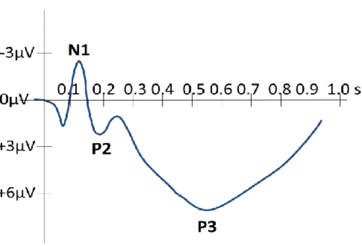
Figure 6: Typical cortical components elicited to an auditory stimulus.. ... 57
Summary
Dehydroepiandrosterone (DHEA) and dehydroepiandrosterone-sulfate (DHEAS) physiologic role remain controversial. Central nervous system and behavioral effects have been described. DHEA and DHEAS have been hypothesised to play a role in cortical organization and brain maturation and suggested to have memory, attention and mood enhancement effects in humans. A neuro-stimulatory action and an anti-cortisol mechanism of action may contribute to those effects. Moreover, the balance between DHEA and DHEAS may modulate their effects in the central nervous system. The objective of this thesis was to explore behavioral correlates of DHEA and DHEAS in humans, in particular regarding personality, stress response, working memory and emotion. Relations at the performance and brain processing levels were explored. DHEA response to the performance of cognitive tasks was also analyzed.
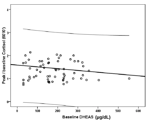
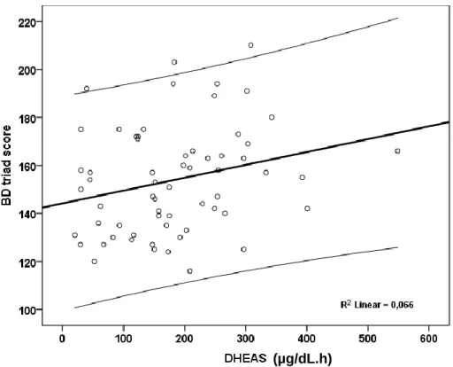
In study I we explored the relation between DHEAS and both pituitary-adrenal axis reactivity and personality in human subjects of both genders. This was a retrospective study of 120 patients assisted at the endocrine outpatient department of a public central Portuguese hospital, before medical treatment. Personality was evaluated with the Minnesota Multiphasic Personality Inventory (MMPI) and the pituitary-adrenal axis reactivity was assessed with the corticotrophin releasing hormone (CRH) test. Baseline serum DHEAS was inversely related to peak/baseline cortisol response to CRH infusion. DHEAS reactivity in the CRH test was directly related to the Deviant Behavior triad and type A personality. These results suggest higher DHEAS levels may be a protective factor against an excessive cortisol response under stressful situations and personality may be related to DHEAS reactivity.
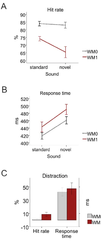
In studies II and III DHEA, DHEAS and cortisol relations to working memory and distraction were studied by recording the electroencephalogram of 23 young women performing a discriminatory (no working memory load) or 1-back (working memory load) task in an audio-visual oddball paradigm. We measured salivary DHEA, DHEAS and cortisol both before each task and at 30 and 60 min intervals. In study II, we showed that under
ii
working memory load, a higher baseline cortisol/DHEA ratio was related to higher distraction as indexed by an enhanced novelty-P3. This suggests that cortisol may lead to increased distraction whereas DHEA may hinder distraction by leading to lower processing of the distractor. An increased DHEA production with consecutive cognitive tasks was found, and higher DHEA responses attributed to working memory load were related to enhanced working memory processing as indexed by an enhanced visual P300. Overall, the results suggest that in women DHEA may oppose cortisol effects, reducing distraction and that a higher DHEA response may enhance working memory at the electrophysiological level. In study III we used that same group of subjects and protocol to analyze the DHEAS/DHEA ratio relation to involuntary attention during the performance of a working memory task. Higher DHEAS/DHEA ratio was related to enhanced auditory novelty-P3 amplitudes during the performance of the working memory task, but there was no significant relation to visual P300 amplitudes, novelty-P3 latencies or visual P300 latencies. Therefore, a relation between higher DHEAS/DHEA ratio and enhanced involuntary attention to the surrounding world without a deleterious effect on working memory processing is suggested. These results also suggest that the balance between DHEAS and DHEA may modulate attentional resources and the importance of the sulfotransferase and sulfatase activity in the modulation of DHEA and DHEAS central nervous system effects.
In study IV we explored DHEAS, DHEA and cortisol relations to the processing of
negative emotional stimuli at behavioral and brain levels by recording the electroencephalogram of 21 young women while performing a visual task with implicit neutral or negative emotional content in an audio-visual oddball paradigm. For each condition, salivary DHEA, DHEAS and cortisol were measured before performing the task, and at 30 and 60 min intervals. DHEA increased after task performance, independently of the implicit emotional content. With implicit negative emotion, higher DHEAS/DHEA and DHEA/cortisol ratios before task performance were related to shorter visual P300 latencies suggesting faster brain processing under a negative emotional context. In addition, higher DHEAS/DHEA ratios were related to reduced visual P300 amplitudes, indicating less processing of the negative emotional stimuli. With this study, we could show that at the electrophysiological level, higher DHEAS/DHEA and DHEA/cortisol ratios
were related to shorter stimulus evaluation times, suggesting less interference of the implicit negative content of the stimuli with the task. Furthermore, higher DHEAS/DHEA ratios were related to reduced processing of negative emotional stimuli which may eventually constitute a protective mechanism against negative information overload.
In conclusion, these studies showed several behavioral correlates of DHEA and DHEAS. Additionally, the results suggest anti-cortisol effects of DHEA and DHEAS and the importance of the balance between DHEAS and DHEA. DHEA levels increased after the performance of cognitive tasks, more so after a working memory load than a no working memory load task, suggesting that cognitive tasks may modulate DHEA levels. Higher baseline DHEAS levels were related to reduced cortisol response to CRH suggesting a protective effect during the stress response. In addition, significant new findings were described regarding DHEA and DHEAS relations to working memory, emotion and attention at the brain processing level. Those results suggest DHEA may eventually oppose cortisol effects reducing distraction while higher DHEAS/DHEA ratios may enhance involuntary attention to the surrounding world during the performance of working memory tasks with no evident deleterious effects on the working memory load task in course. On the other hand, higher DHEA response may enhance working memory at the electrophysiological level. Regarding emotional processing, higher DHEAS/DHEA and DHEA/cortisol ratios may be related to less interference of the implicit negative content of the stimuli and higher DHEAS/DHEA ratios were related to reduced processing of negative emotional stimuli which may eventually protect against negative information overload. In short, results suggest DHEAS levels may be related to personality and reduced cortisol stress response while studies at brain processing level suggest DHEA and/or DHEAS may enhance working memory / attention and reduce the processing of negative information.
Resumo
A desidroepiandrosterona (DHEA) é sintetizada nas suprarrenais, mas também nas gónadas e ao nível do sistema nervoso central. É reversivelmente convertida em desidroepiandrosterona-sulfato (DHEAS) por ação de uma sulfotransferase, quer na periferia, quer no sistema nervoso central. DHEA e DHEAS são hormonas abundantes nos primatas, em especial nos seres humanos e a concentração de ambas é maior no sistema nervoso central que na circulação periférica. A forma como é regulada a sua síntese não está definida, mas as suas concentrações aumentam em situações de stress agudo e reduzem em situações de stress crónico. A partir da quarta década de vida as suas concentrações reduzem-se progressivamente e níveis mais reduzidos relacionam-se com maior morbilidade e mortalidade, independentemente da idade.
Os efeitos fisiológicos e os mecanismos de ação da DHEA e DHEAS são ainda controversos. Contudo, muitos estudos têm sugerido efeitos ao nível do sistema nervoso central. Foi colocada a hipótese de contribuírem para a organização cortical e desenvolvimento cerebral e em particular, têm sido sugeridos efeitos benéficos em relação à memória, atenção e humor. Níveis mais elevados de DHEAS, DHEA ou da razão DHEA/cortisol foram relacionados com melhor desempenho cognitivo, menos sintomas depressivos e níveis mais elevados de bem-estar. Tem sido colocada a hipótese de esses efeitos serem mediados em parte, através de mecanismos de ação anti-cortisol e neuro-estimulantes. De facto, a nível molecular a DHEA e sobretudo a DHEAS são agonistas glutamatérgicos e antagonistas gabaminérgicos. Para além disso, a razão entre níveis de DHEA e DHEAS poderá eventualmente modular os efeitos destas hormonas ao nível do sistema nervoso central. Os efeitos eletrofisiológicos da DHEA e DHEAS em seres humanos são essencialmente desconhecidos e a sua relação com a personalidade ou fenótipo endócrino de resposta ao stress não estão bem definidos.
O objetivo desta tese foi explorar relações da DHEA ou DHEAS com variáveis comportamentais em seres humanos adultos. Em particular foram estudadas relações com a personalidade, resposta ao stress, memória de trabalho e emoções. Foram estudadas relações ao nível do desempenho e do processamento cerebral. Foram
vi
exploradas eventuais relações opostas às do cortisol, bem como os efeitos do balanço entre os níveis de DHEAS e DHEA. Foram ainda analisadas as respostas da DHEA à realização de tarefas cognitivas.
No estudo I explorámos a relação da concentração de DHEAS com a reatividade hipófise-suprarrenal e o perfil de personalidade em adultos de ambos os géneros. Foi um estudo retrospetivo que incluiu 120 doentes assistidos na consulta externa de um hospital público terciário, antes do início de terapêutica médica. Foi avaliada a personalidade utilizando o Inventário de Personalidade Multifásico do Minnesota (MMPI) e a reatividade do eixo hipófise-suprarrenal utilizando a prova da corticoliberina (CRH). A concentração de corticotrofina (ACTH) relacionou-se diretamente com a concentração sérica de DHEAS basal, e em conjunto com a idade e o género explicaram 34% da variabilidade da concentração de DHEAS. A concentração de DHEAS basal relacionou-se inversamente com a resposta pico/basal do cortisol na prova de CRH. A reatividade da DHEAS na prova de CRH relacionou-se diretamente com a pontuação obtida na tríade de Problemas de Comportamento e personalidade tipo A. Estes resultados sugerem que concentrações mais elevadas de DHEAS poderão constituir um fator protetor contra uma resposta excessiva do cortisol em situações de stress. Por outro lado, sugerem a existência de uma relação entre a personalidade e a reatividade da DHEAS.
No estudo II explorámos a relação das concentrações de DHEA, DHEAS e cortisol com a memória de trabalho e distração ao nível do desempenho e do processamento cerebral. Com essa finalidade, registámos o eletroencefalograma de 23 jovens adultas do sexo feminino enquanto realizavam uma tarefa discriminativa (sem utilização da memória de trabalho) e outra com a utilização de memória de trabalho. Essas tarefas eram visuais e antes de cada estímulo visual eram administrados estímulos auditivos, 20% dos quais eram novos e tinham o objetivo de causar distração (paradigma de "oddball"). Medimos a DHEA, DHEAS e o cortisol salivares antes de cada tarefa e aos 30 e 60 min. Durante a tarefa com utilização de memória de trabalho, razões cortisol/DHEA mais elevadas relacionaram-se com deflexões "P3-novidade" (em inglês, novelty-P3) mais amplas, traduzindo maior distração. Este resultado sugere que o cortisol poderá causar maior distração enquanto que a DHEA poderá reduzir essa distração através da redução do
processamento desse estímulo distrativo. Encontrámos uma elevação das concentrações de DHEA com a realização das duas tarefas cognitivas consecutivas, maior com a realização da tarefa com memória de trabalho que com a realização da tarefa discriminativa. Para além disso, uma maior resposta da DHEA devida à realização da tarefa com memória de trabalho, relacionou-se com um maior incremento na amplitude da P300 visual devida à memória de trabalho, traduzindo assim um melhor processamento dessa memória de trabalho. Globalmente, estes resultados sugerem que nas mulheres, a DHEA poderá opor-se aos efeitos do cortisol, reduzindo a distração e que maiores reatividades da DHEA se relacionam com melhor memória de trabalho ao nível eletrofisiológico.
No estudo III utilizámos o protocolo anterior para analisar a relação entre a razão DHEAS/DHEA e a atenção involuntária durante a execução de uma tarefa envolvendo memória de trabalho. Verificámos que razões DHEAS/DHEA mais elevadas se relacionaram com amplitudes maiores da deflexão auditiva P3-novidade durante a realização de uma tarefa visual envolvendo memória de trabalho. Contudo, a razão DHEAS/DHEA não se relacionou com a amplitude da P300 visual, a latência da
P3-novidade ou a latência da P300 visual. Estes resultados sugerem que o balanço entre
DHEAS e DHEA pode modular a atenção involuntária. Especificamente, os resultados sugerem que razões DHEAS/DHEA mais elevadas podem aumentar o processamento involuntário de estímulos auditivos sem um efeito deletério no processamento da memória de trabalho visual em curso. A manutenção da atenção involuntária em relação ao mundo envolvente durante a realização de tarefas envolvendo a utilização de memória de trabalho pode ser um mecanismo protetor importante. Os recursos da atenção são limitados e estes resultados sugerem que o balanço entre DHEAS e DHEA pode modular esses recursos bem como sugerem a importância da atividade da sulfotransferase e sulfatase na modulação desses efeitos.
No estudo IV explorámos as relações da DHEAS, DHEA e cortisol com o processamento de estímulos com conteúdo emocional negativo, ao nível do comportamento e do processamento cerebral. Foi registado o eletroencefalograma de 21 jovens adultas enquanto realizavam uma tarefa com conteúdo emocional implícito neutro
viii
ou negativo, utilizando um paradigma de "oddball" audiovisual. A tarefa alvo era visual e os estímulos auditivos, dos quais 20% eram sons novos, pretendiam causar distração. Em cada condição, foram doseadas a DHEA, DHEAS e cortisol antes da realização da tarefa e aos 30 e 60 min. A concentração de DHEA aumentou após a realização da tarefa, independentemente do conteúdo emocional. Razões DHEAS/DHEA e DHEA/cortisol mais elevadas antes da execução da tarefa com conteúdo emocional implícito negativo relacionaram-se com latências mais curtas da P300 visual, sugerindo um processamento cerebral mais rápido em contexto emocional negativo. Para além disso, razões DHEAS/DHEA mais elevadas relacionaram-se com amplitudes da P300 visual mais reduzidas, indicando menor processamento do estímulo emocional negativo. Com este estudo mostrámos que ao nível eletrofisiológico, razões DHEAS/DHEA e DHEA/cortisol mais elevadas se relacionaram com tempos de avaliação do estímulo mais curtos, sugerindo menor interferência pelo conteúdo implícito negativo dos estímulos. Também, razões DHEAS/DHEA mais elevadas relacionaram-se com menor processamento dos estímulos negativos, o que pode constituir um mecanismo protetor contra o excesso de informação negativa.
Concluindo, estes estudos revelaram relações da DHEA e DHEAS com variáveis comportamentais. Os resultados sugerem efeitos anti cortisol da DHEA e DHEAS e a importância do balanço entre os níveis de DHEAS e DHEA. As concentrações de DHEA aumentaram com a execução de tarefas cognitivas, sugerindo que essas tarefas podem modular as concentrações de DHEA. Concentrações basais mais elevadas de DHEAS relacionaram-se com menor resposta do cortisol à CRH sugerindo um efeito protetor durante a resposta ao stress. Encontrámos também relações entre DHEAS e o perfil de personalidade. Para além disso, encontrámos resultados novos no que respeita às relações da DHEA e DHEAS com o processamento cerebral. Esses resultados sugerem que ao nível eletrofisiológico, a DHEA poderá opor-se aos efeitos do cortisol, reduzindo a distração; que razões DHEAS/DHEA mais elevadas poderão aumentar a atenção involuntária em relação ao meio envolvente, sem um efeito deletério no processamento da memória de trabalho; e que uma maior resposta da DHEA poderá melhorar o processamento dessa memória de trabalho. No que respeita ao processamento emocional, os resultados sugerem que razões DHEAS/DHEA e DHEA/cortisol mais
elevadas se relacionam com menor interferência do conteúdo implícito negativo dos estímulos e que razões DHEAS/DHEA mais elevadas se relacionam com menor processamento do conteúdo emocional negativo, o que poderá constitui um mecanismo protetor contra os efeitos deletérios do excesso de informação negativa. Portanto, os resultados sugerem que a DHEAS se relaciona com a personalidade e menor resposta do cortisol ao stress, enquanto os estudos ao nível do processamento cerebral sugerem que a DHEA e/ou DHEAS poderão melhorar a memória de trabalho / atenção e reduzir o processamento de informação com conteúdo emocional negativo.
Palavras-chave: desidroepiandrosterona (DHEA); personalidade; memória; atenção; processamento emocional.
List of Original Publications in Relation to the Present
Thesis
In agreement with the official edict 388/70, art. 8º, paragraph 2, the results presented in this thesis were published or are currently being prepared for publication.
Dehydroepiandrosterone-sulphate (DHEAS) is related to Personality and Stress
Response. Sónia do Vale, João Martin Martins, Maria João Fagundes, Isabel do Carmo. Neuro Endocrinol Lett. 2011;32(4):442-8.
The Relationship between Dehydroepiandrosterone (DHEA), Working Memory and
Distraction - a Behavioral and Electrophysiological Approach. Sónia do Vale, Lenka Selinger, João Martin Martins, Ana Coelho Gomes, Manuel Bicho, Isabel do Carmo, Carles Escera. PLoS ONE 2014; 9(8): e104869.
Hormonal Modulation of Novelty Processing in Women: enhanced under Working
Memory load with high DHEAS/DHEA ratios. Sónia do Vale, Lenka Selinger, João Martin Martins, Manuel Bicho, Isabel do Carmo, Carles Escera. Submitted for publication.
Dehydroepiandrosterone (DHEA) and Dehydroepiandrosterone-sulphate (DHEAS)
and Emotional Processing – a Behavioral and Electrophysiological Approach. Sónia do Vale, Lenka Selinger, João Martin Martins, Manuel Bicho, Isabel do Carmo, Carles Escera. Hormones and Behavior 2015; 73: 94-103.
Abbreviations
ACh Acetylcholine
ACC Anterior cingulate cortex
AASH Adrenal androgen stimulating hormone ACTH Adrenocorticotropic hormone
AMPA α-amino-3-hydroxy-5-methyl-4-isoxazolepropionic acid BDNF Brain-derived neurotrophic factor
BMI Body mass index
AMPc Cyclic Adenosine Monophosphate CASH Cortical Androgen Stimulating Hormone C/EBP CCAAT/enhancer-binding protein CRH Corticotrophin Releasing Hormone
DA Dopamine
DHEA Dehydroepiandrosterone
DHEAS Dehydroepiandrosterone-sulphate
DHEA(S) Dehydroepiandrosterone and Dehydroepiandrosterone-sulphate DMPC Dorsomedial prefrontal cortex
DNA Deoxyribonucleic acid
EEG Electroencephalogram
EGF Epidermal growth factor
eNOS Endothelial nitric-oxide synthase ERPs Event-Related Potentials
FDG-PET Fluorodeoxyglucose - positron emission tomography fMRI Functional Magnetic Resonance Imaging
GABA-A γ-aminobutyric acid type A
GSH Glutathione
HDLc High density lipoprotein cholesterol HFS High-frequency stimulation
HLA Human leukocyte antigen
xiv
HSD Hydroxysteroid dehydrogenase enzyme IGF Insulin-like growth factor
IL Interleukin
IP3 Inositol triphosphate
Kd Binding affinity
Keq Equilibrium constant
K(m) Michaelis constant
LORETA Low-resolution brain electromagnetic tomography LTP Long-term potentiation
MAP2C Microtubule-associated protein 2C MAPK Mitogen-activated protein kinase MCR Metabolic clearance rate
MMN Mismatch negativity
MMPI Minnesota Multiphasic Personality Inventory
MRI Magnetic resonance imaging
MMSE Mini-Mental State Examination Nov-P3 Novelty-P3
NMDA N-methyl-D-aspartate (NMDA)
OATP Organic Anion Transporting Polypeptide OST Organic Solute Transporter
PAP 3',5'-diphosphoadenosine
PAPS 3'-phosphoadenosine 5'-phosphosulfate PET Positron Emission Tomography
PFC Prefrontal cortex
PPARα Peroxisome proliferator-activated receptor
PRL Prolactin
PTSD Post-traumatic stress disorder r Correlation coefficients
rs Spearman’s Rank Order correlation coefficients
RON Reorienting negativity
SCC Cholesterol side chain cleavage enzyme
SF Steroidogenic factor
SOAT Sodium-dependent Organic Anion Transporter StAR Steroidogenic acute regulatory protein
STAI State-Trait Anxiety Inventory
STS Steroid-sulfatase, steroid sulfohydrolase SULT Hydroxysteroid sulfotransferase enzyme TBPS T-butyl-bicyclo-phosphorothionate TNF Tumor necrosis factor
VPP Vertex positve potential
WM Working memory
Introduction/Literature Review:
Dehydroepiandrosterone and
Dehydroepiandrosterone-sulphate as Neuroactive Steroids
In humans, dehydroepiandrosterone-sulphate (DHEAS) is the most abundant hormone in the peripheral circulation (1; 2). Brain levels of dehydroepiandrosterone (DHEA) and DHEAS are higher than peripheral circulation levels (3; 4) and both forms of the hormone are synthesized in the adrenals, but also in the central nervous system (1). DHEA and DHEAS [DHEA(S)] regulation and its physiological effects are still a matter of debate (5; 6; 7). Nevertheless, stressful stimuli increase DHEA and DHEAS levels in the acute setup (8) whereas chronic stress decreases DHEA and DHEAS levels (9; 10; 11). DHEAS levels are also higher in early adulthood and dramatically decrease with aging (2; 12), reaching a nadir by the time many diseases of aging become more prevalent. Besides, lower DHEAS levels are related to higher morbidity and mortality even after age adjustment (2; 13).
Although DHEA and DHEAS are steroid hormones, no high affinity nuclear receptor has been found for them (5; 6; 7; 14). On the other hand, DHEA and DHEAS effects mediated by binding to cytoplasmic receptors were described. In the brain, these hormones modulate synaptic transmission. They present a general neuro stimulatory effect mainly as glutamatergic agonist and gabaminergic antagonist (1; 3) and general biological actions of DHEA and/or DHEAS involve neuronal survival, neurite growth, neurogenesis, anti-oxidant, anti-inflammatory and anti-glucocorticoid effects (7). It was hypothesised that DHEA and DHEAS may play a role in cortical organization (15) and brain maturation (16). At the behavioral level, higher DHEA, DHEAS or DHEA-to-cortisol ratios were related to improved attention, cognition and mood (17; 18; 19; 20).
The previous findings raised the hypothesis that restoring DHEA to youthful levels might protect the brain from cognitive decline or improve cognition, improve mood and even extend life span. In this regard, DHEA and DHEAS were evaluated in the treatment of
2
neuropsychiatric disorders, with published reports appearing as early as 1952 (21; 22). Since the late 1980s, a growing amount of studies addressing pre-clinical aspects and some controlled clinical trials with DHEA administration were performed (23). Regarding the effects of DHEA administration, until the present date, the studies failed to show consistent beneficial effects on cognition but suggest beneficial effects of DHEA administration on mood. Although the knowledge of neurobiological actions of DHEA and DHEAS is rapidly growing, the neurofunctional correlates of DHEA and DHEAS are mostly unknown. In fact, only a very small number of studies addressed the effects of DHEA on brain processing, both at the electrophysiological level (mostly since the late 1990s) (24) and functional neuroimaging level (since the early 2010s) (25).
This review will address the following DHEA and DHEAS aspects as neurosteroids: 1) molecular structure, synthesis and distribution; 2) concentrations along the life span; 3) molecular mechanisms of action and cellular effects; 4) cognitive and neuropsychiatric effects; 5) electrophysiological and neuroimaging correlates.
Synthesis and distribution
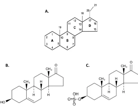
Dehydroepiandrosterone (also designated as prasterone) is a steroid hormone. It is derived from the cyclopentanoperhydrophenanthrene structure, which is composed of three cyclohexane rings and a single cyclopentane ring (figure 1.A). Its International Union of Pure and Applied Chemistry (IUPAC) designation is 3β-hydroxyandrost-5-en-17-one or 5-androsten-3β-ol-17-one (26). DHEA possesses a hydroxyl group at position C3 (figure 1.B) while DHEAS has a sulphate group in that position (figure 1.C).
DHEA and DHEAS are synthesized in the adrenals, gonads and central nervous system. The adrenals are responsible for most of its synthesis in both genders, but while in women, the gonads are a minor source, in adult men, the testes secrete approximately 10-25% of DHEA and 5% of DHEAS (27; 28). The brain is believed to produce only small amounts of peripheral DHEA and DHEAS levels (27; 28; 29).
B. C.
A.
Figure 1: Cyclopentanoperhydrophenanthrene structure (A), dehydroepiandrosterone (B) and dehydroepiandrosterone-sulphate (C) chemical structure.
4
DHEA synthesis in the adrenals and gonads
In the periphery, DHEA is mostly synthesized in the adrenals but also in the gonads. In the adrenals it is synthesized in the adrenal cortex, mostly in the zona reticularis (to a lesser degree it can be synthesized in the zona fasciculata). In the ovary, it is produced in the thecal cells (30) and in the testis it is produced by the Leydig cells.
Cholesterol is the precursor of all adrenal and gonadic steroids, either internalized in steroidogenic cells by receptor mediated endocytosis or synthesized de novo within those cells (31). Within the steroidogenic cells (adrenal cortical cells, theca cells, theca-interstitial cells and Leydig cells), the intracellular cholesterol is transported from the outer to the inner mitochondrial membrane, a process that is mediated by the steroidogenic acute regulatory protein (StAR) (32). This transport of cholesterol across the mitochondrial membrane is the initial rate-limiting step in adrenal steroidogenesis (33; 34). The placental steroidogenic cells are an exception because they do not express StAR and directly utilize the mitochondrial cholesterol side chain cleavage enzyme (a cytochrome P450 enzyme) to initiate steroidogenesis (the protein MLN64 is one candidate for a factor mediating the transport of cholesterol into placental mitochondria) (35). In the mitochondria of adrenal and other steroidogenic cells in general, the cholesterol is converted to pregnenolone by cytochrome P450 side chain cleavage enzyme (P450scc, desmolase or CYP11A1). The steroidogenesis involves the action of other cytochrome P450 enzymes. The cholesterol side-chain cleavage enzyme and the 11β-hydroxylase (CYP11B) enzymes are mitochondrial enzymes and require an electron shuttle system to hydroxylate steroids. The 17α-hydroxylase (CYP17) and 21-hydroxylase (CYP21A2) are microsomal-endoplasmic reticulum enzymes and involve a distinct electron shuttle system (cytochrome b5, a flavoprotein) (34; 35).
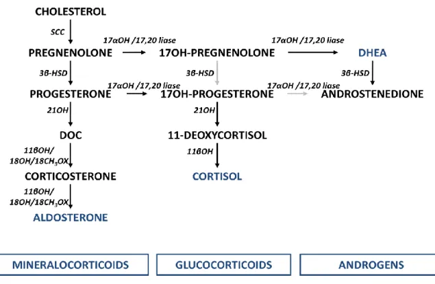
The pathway for DHEA synthesis is mostly identical in the adrenals and gonads. After the synthesis of pregnenolone by the action of the P450scc (CYP11A1) enzyme, CYP 17α-hydroxylase hydroxylates pregnenolone to form 17-hydroxypregnenolone. Then, the CYP17 also possesses 17,20-lyase activity and this activity converts 17-hydroxypregnenolone into DHEA, which is a C19 androgen (figure 2) (34). The synthesis of adrenal androgens occurs mostly in the reticularis zone. Additionally, DHEA is sulfated to
DHEAS in the adrenal zona reticularis. DHEA is liposoluble and can easily cross biological membranes, but DHEAS, having a sulfate group, does not (2; 36).
Besides androgens, the adrenals produce two other main types of hormones: glucocorticoids and mineralocorticoids, see figure 3 (34; 35; 37).
Figure 2: Biochemical pathway for DHEA synthesis. SCC - cholesterol side chain cleavage enzyme (CYP11A1); 17α-OH - 17α-hydroxylase enzyme; 17,20-lyase - 17,20-lyase enzyme.
Figure 3: Adrenal steroidogenesis. Three main types of hormones are produced by the adrenals: glucocorticoids, mineralocorticoids and androgens. SCC - cholesterol side chain cleavage enzyme (CYP11A1); 17α-OH/17,20-lyase - 17α-hydroxylase/17,20-lyase enzyme (CYP17); 3β-HSD - 3β-hydroxysteroid dehydrogenase enzyme (HSD3B); 21OH - 21-hydroxylase enzyme (CYP21A2); DOC – deoxycorticosterone; 11βOH - 11-hydroxylase (CYP11B1); 11βOH/18OH/18CH3OX – aldosterone synthase (CYP11B2), which has
11β-hydroxylase (11βOH), 18-hydroxylase (18OH) and 18-methyl oxidase (18CH3OX) activity; black arrows
6
In the fetal adrenal, because of relative lack of 3β-HSD and high sulfotransferase activity, DHEA and DHEAS are the main steroids produced by the adrenal. The fetal adrenal has considerable sulfotransferase activity but little steroid sulfatase activity, also favoring conversion of DHEA to DHEAS. The resulting DHEAS is secreted, 16α-hydroxylated in the fetal liver (by the hydroxilase CYP3A7) (38; 39), and then acted on by placental 3βHSD1, 17βHSD1, and P450 aromatase to produce estriol. Small amounts of DHEA and DHEAS bypass the liver and are not 16α-hydroxylated, and hence yield estrone and estradiol. Estrogens in turn, further inhibit adrenal 3βHSD activity, providing a feedback system to promote production of DHEAS (40; 41).
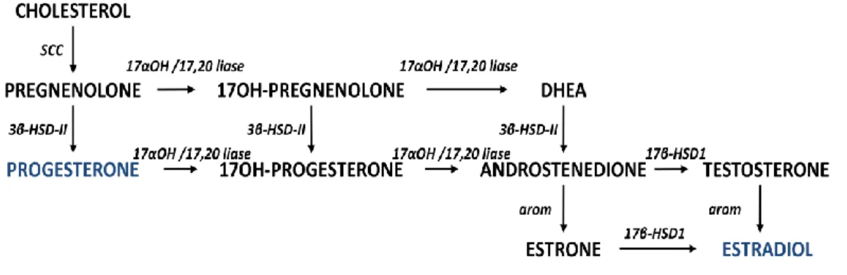
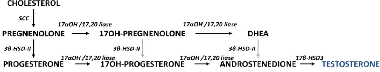
As mentioned, the gonads secrete DHEA although in much lower levels then the adrenals. The main hormonal products of the normal ovaries of reproductive age women are progesterone and estradiol. Accordingly, the preferential pathways are to convert pregnenolone into progesterone and estradiol (figure 4) (30). In the testis, testosterone is the main steroid produced (35; 42; 43), see figure 5.
Figure 4: Steroidogenic pathway in the ovary. DHEA is synthesized as an intermediary metabolite. SCC - cholesterol side chain cleavage enzyme; 17α-OH/17,20-lyase - 17α-hydroxylase/17,20-lyase enzyme; 3β-HSD - 3β-hydroxysteroid dehydrogenase enzyme type II; 17βOH – 17β-hydroxysteroid dehydrogenase type 1 enzyme; arom – aromatase.
Figure 5: Steroidogenesis in human testis. SCC - cholesterol side chain cleavage enzyme; 17α-OH/17,20-lyase - 17α-hydroxylase/17,20-17α-OH/17,20-lyase enzyme; 3β-HSD - 3β-hydroxysteroid dehydrogenase enzyme type II; 17βOH – 17β-hydroxysteroid dehydrogenase type 3 enzyme; black arrows indicate the predominant pathway in humans.
DHEA and DHEAS interconversion, secretion, peripheral metabolism and excretion
As mentioned, DHEA is sulfated to DHEAS in the adrenal reticularis zone. This sulfation reaction is reversibly mediated by the hydroxysteroid sulfotransferase enzyme (SULT2A1), which is a cytosolic enzyme (44; 45). Human cytosolic sulfotransferases transfer the sulfuryl group from 3'-phosphoadenosine 5'-phosphosulfate (PAPS) to the hydroxyl group of DHEA (46). The equilibrium constant for this reaction strongly favours DHEAS synthesis (Keq = 1.3 x 10³), DHEA is the concentration-limiting substrate and nucleotide (3',5'-diphosphoadenosine, PAP) release is the rate-limiting step (46). In the presence of increasing DHEA concentration, there is partial substrate inhibition by DHEA: in that condition, DHEA excess causes it to add more quickly to the enzyme-PAP complex, trapping a greater fraction of PAP in a dead-end complex (DHEA-enzyme-PAP). Therefore, as the DHEA concentration increases, the rate determining step in this reaction shifts from PAP release from the enzyme-PAP complex (K off = 1.2 s-1 )to PAP release from the dead-end complex (DHEA-enzyme-PAP) (K off = 0.48 s-1) (46).
Of note, DHEA-sulfotransferase (SUL2A1) is also expressed in liver (hepatocytes) and a smaller amount is expressed in the gastrointestinal tract (small intestine mucosa and parietal cells of the gastric glands) (47; 48; 49; 50). SUL2A1 expressed in the human liver is responsible for sulfation of bile acids and circulating hydroxysteroids (51; 52; 53; 44), including DHEA. The DHEA-sulfotransferase present in human adrenal and liver show similar physical, biochemical and kinetic properties (47). In humans, the sulfotransferases expression in non-gonadal tissues is identical in both genders (in contrast, rodents show
8
sex-related differences) (48). Several nuclear receptors might regulate SULT2A1 expression in liver (peroxisome proliferator-activated receptor-α; pregnane X receptor; constitutive androstane receptor, vitamin D receptor, liver X receptors, farnesoid receptor, retinoid-related orphan receptors) while Estrogen-Related Receptor-α is suggested to play a major role in the regulation of SULT2A1 expression in the human adrenal (48).
As mentioned, DHEA can easily cross biological membranes. On the contrary, the transport mechanism for adrenal cell export of DHEAS into the circulation is not well understood. The Organic Solute Transporter α - Organic Solute Transporter β (OSTα-OSTβ) is a heteromeric carrier abundantly expressed in the human adrenal gland which may play a role in the export of DHEAS from the adrenal cell (54). It is also expressed in a variety of other tissues, including the small intestine, colon, liver, biliary tract and kidney (54). In polarized epithelial cells, OSTα-OSTβ protein is localized in the basolateral membrane and functions in the export or uptake of bile acids and steroids (54).
DHEAS may be converted back to DHEA by a steroid-sulfatase (STS) (steroid sulfo-hydrolase) enzyme. The steroid-sulfatase gene is X-linked and escapes X inactivation in humans (55). Its expression was observed in the adrenal, liver, adipose tissue, brain, placenta, gonads, uterus, skin and other peripheral tissues (56; 57; 58; 59; 60). STS is localized in the endoplasmic reticulum, with evidence suggesting that its active site is located within the endoplasmic reticulum membrane and that facilitated transport of DHEAS across the ER membrane may not be required for STS activity (61). Therefore, cycling of DHEA and DHEAS (and other steroids and their sulfates) may occur as a result of steroid sulfotransferases and steroid sulfatases activity. Higher concentration of phosphate reduces the velocity of STS reaction and other sulfated steroids (namely pregnenolone sulfate) functioning as competitive inhibitors of this enzyme (58; 62; 63).
In young adults, the adrenals secrete about 4mg of DHEA and 25mg of DHEAS daily (64). DHEA and DHEAS in the peripheral circulation do not bind significantly to sex hormone binding globulin. They bind to albumin, although DHEAS binding to albumin seems non-specific (64). Overall, in the adrenals and other peripheral tissues, 64-74% of the DHEAS produced each day is converted back to DHEA, while only 6-13% of the DHEA
produced is metabolized to DHEAS (28; 64; 65; 66). DHEA and DHEAS can be converted to other steroids in the adrenals, ovaries and testis and released into the peripheral circulation (64; 67). Secreted DHEA and DHEAS can also be converted to other steroids in several peripheral target tissues (like the skin, adipose tissue, liver, bone), although in many cases, probably with no significant release into the general peripheral circulation (intracrinology) (5; 64). In particular, DHEA and DHEAS can be converted into androgens (Δ4-androstenedione, testosterone, 5α-dihydrotestosterone), estrogens (Δ5-androstenediol and those resulting from the aromatization of testosterone and Δ4-androstenedione - estradiol, estrone) and other DHEA derivatives (16α-, and 7α/β-oxygenated DHEA derivatives) (36; 64).
As a result of the previous findings, circulating DHEAS may be seen as a reservoir for DHEA, with conversion by sulfotransferases occurring in several tissues (5). Besides that, the fact that DHEAS and DHEA may be converted to other active steroids gave rise to the hypothesis that DHEA and DHEAS effects could be mediated by its conversion to androgens, estrogens and other metabolites and not direct effects of DHEA or DHEAS (5; 68). To note, the high DHEA and DHEAS concentrations found in human peripheral circulation are not reproduced in other animals. The peripheral concentration of DHEA and DHEAS in monkeys is much lower than that in humans and in most other laboratory animals, particularly rodents, the peripheral concentration of these hormones is very low (7; 64). This is also suggestive that DHEA and DHEAS might play a partcular role in the human species.
Again, although DHEA is expected to freely cross membranes of target cells, another issue was whether DHEAS, a hydrophilic steroid, can enter the specific target cells. To that concern, transport systems were described. The Organic Anion Transporting Polypeptide (OATP) family has broad and overlapping substrate specificities and in particular, some members of the Organic Anion Transporting Polypeptide (OATP) family were described to occur in the liver, kidney, testis, small intestine, thymus, placenta and other tissues and mediate the trans-epithelial / transmembrane uptake of DHEAS and estrone sulfate (69; 70; 71). Besides, a specific steroid-sulfate transporter, the sodium-dependent Organic Anion Transporter (SOAT), can mediate DHEAS cellular inward in the
10
testis (69). SOATs are expressed in other tissues namely in vesicular structures in neurons of the central and peripheral nervous system, but a transport function was not yet detected, namely no transport of DHEAS by SOATs was detected in those cells (72). Besides, OATPs were detected in salivary glands, but no transport of DHEAS by OATPs in those cells was described (73). On the other hand, it was described that DHEAS squeezes through the tight junctions between salivary glands and DHEAS concentrations in saliva are therefore dependent on serum concentration and salivary flow rate (74).
Renal and biliary excretion of DHEA and DHEAS do occur. Renal excretion accounts for 51-73% of the elimination of DHEAS and its metabolites (28). Besides the direct excretion of DHEAS and DHEA, renal excretion products include androsterone sulfate, etiocholanolone sulfate, androsterone glucuronoside and etiocholanolone glucuronoside (75; 76). The half-life of DHEA is similar to cortisol and most other steroid hormones. It is estimated to take 15-30 min, for a metabolic clearance rate (MCR) of approximately 2,000 L / day. On the contrary, the half-life of DHEAS is much longer, 7-10 h, and the MCR is low, 5-20 L/day (36; 64). As a consequence, the plasma concentration of DHEA is not much different from that of other adrenal steroids, but it is several times less abundant than DHEAS. DHEA concentration is in several nmol/L (2-9 μg/dL, 7-31 nmol/L), while the concentration of DHEAS is in several μmol/L (50-250 μg/dL, 1.3-6.8 μmol/L) (64; 77). Of note cortisol molar concentration in the peripheral circulation is about 10 times higher than DHEA concentration (morning cortisol: 5-25 μg/dL, 140-690nmol/L) (77).
DHEAS concentration is 20-30% lower in women, therefore the DHEAS/DHEA ratio is also lower in women (64). The ratios for the conversion of DHEAS and androsterone sulfate to DHEA, are both significantly higher for women than men (78). Plasma levels of DHEA are only a little higher in women than in men [in Bird et al. study (78), levels were 8.50 ± 0.95 and 8.75 ± 1.01 ng/ml for men and women, respectively]. Concluding from the above stated, the X-linked sulfatase activity, which escapes X-inactivation, may explain or contribute to this difference. There is no sex difference in the binding of DHEA and DHEAS to plasma proteins and this is also reflected in the lack of sex difference in the MCRs (78). Nevertheless, women probably excrete more DHEAS and DHEA through urine when compared to men (73±5.5% in women and 51±3.5% in men), while men excrete more
DHEAS and DHEA though the bile than women (79). Circadian rhythm of DHEA is identical to that of cortisol, eventually related to ACTH stimulation of DHEA synthesis. DHEAS levels on the contrary, due to its long half-life, remain stable 24h a day under normal conditions (64).
DHEA synthesis in the brain
Neurosteroids are steroids that can be synthesized de novo in the nervous system from sterol precursors (1). This notion was proposed in 1981. Interestingly, this came to light after the observation that DHEA and DHEAS were present in the adult rat brain. At that time, it was an unexpected finding since the rodents steroidogenic glands do not secrete significant amounts of DHEA. As a sequence to that observation, the nervous system steroidogenesis was discovered (80) and DHEA and DHEAS were known to be neurosteroids. However, it is still not clear where and how DHEA and DHEAS are synthesized in the central nervous system.
Corpéchot et al. (80) performed a series of experiments showing that DHEAS levels in brain were independent of its peripheral synthesis. DHEAS concentrations in the brain tissue largely exceeded DHEA concentrations in the brain tissue and DHEAS concentrations in plasma. Furthermore, there is an anterior to posterior DHEA and DHEAS brain gradient (3; 80; 81). DHEA concentrations are higher in the anterior brain, while DHEAS concentrations are higher in the posterior brain. In a post-mortem study of human brains, subjects 76 to 93 years old (nine women and one man), DHEA concentrations were found to be higher in the prefrontal lobe (29 nmol/kg) than in other brain regions (16 nmol/kg in the parietal lobe, 13 nmol/kg in the temporal cortex, 17 nmol/kg in the cerebellum and 19 nmol/kg in the corpus callosum) (4). Mice also have higher DHEA concentrations in the anterior brain (0.42±0.10 ng/g in anterior brain and 0.12±0.03 ng/g in posterior brain) (80). On the contrary, DHEAS concentration in rats are higher in the posterior (4.89±1.06 ng/g) than in the anterior brain tissue (1.58±0.14 ng/g) (3; 80) and peripheral concentrations of DHEAS in rats are lower than the central ones (0.26±0.13
12
ng/mL) (80). In humans, DHEA levels are also about 6.5 time higher in brain than in the plasma of subjects of similar age (4).
Of note, DHEA concentrations in human brain tissue (homogenates) are higher than plasma concentrations, but cerebrospinal fluid concentrations are lower than plasma concentrations, about 5.4% of plasma concentrations (cortisol levels in the cerebrospinal fluid are also about 5.8% of plasma concentrations) (82). Despite the fact that DHEAS is expected to penetrate less into the cerebrospinal fluid than DHEA (0.03% of circulating DHEAS is expected to cross the blood brain barrier) (83), in adults, the levels of DHEAS in the cerebrospinal fluid are still higher than those of DHEA (82). DHEA-to-DHEAS molar ratio was estimated to be about 0.01 in plasma and 0.52 in the cerebrospinal fluid (in the peripheral circulation DHEAS levels are 100 or more times higher than those of DHEA and about 5-10 times those of cortisol; DHEAS levels in the cerebrospinal fluid are about 1/3 of cortisol levels) (82). Gooddyer et al. studied the relative levels of DHEA, DHEAS and cortisol in the blood and saliva of adolescents (84), and found a similar relationship to that described by Guazzo et al. between blood and cerebrospinal fluid levels (82), hence suggesting that transport into the brain and saliva might be, to this extent, comparable, and that salivary levels may therefore give a reasonable representation to those in the cerebrospinal fluid (82).
Several studies suggest that central nervous system DHEAS levels do not depend on adrenal secretion. Injections for 3 days of long-acting preparations of corticotrophin, to stimulate adrenal steroidogenesis, or of dexamethasone to inhibit endogenous adrenocorticotropic hormone (ACTH) secretion, were not accompanied by clear-cut changes in brain DHEAS (1; 80). Also, brain DHEAS was unchanged one day after castration, whereas testosterone completely disappeared from the brain. Also, no difference was observed in brain DHEAS levels when castrated adrenalectomized male rats were compared 15 days after operation with sham operated controls (1; 80). Taking into account that the rat steroidogenic glands do not secrete significant amounts of DHEA and peripheral concentrations of DHEA and DHEAS are very low (7; 64), these results suggest that in rats, DHEA and DHEAS may be produced mainly in the brain. Moreover, mice have higher DHEA concentrations in brain than in plasma (7).
The same conclusion cannot be directly applied to humans, since DHEA and DHEAS are abundant in brain but also in peripheral circulation. In human beings, brain DHEA might be derived from both local synthesis and peripheral synthesis (7). DHEA may cross the blood-brain barrier and be converted to DHEAS in the brain. On the contrary, DHEAS can enter and leave the brain through specific transport systems but the flux of DHEAS is believed to be mainly from the brain to the peripheral circulation and not the opposite (85). Accordingly, in subjects not taking steroid medication, DHEA and DHEAS levels in blood and cerebrospinal fluid were directly related (82). DHEA levels were shown to decrease in plasma and cerebrospinal fluid in subjects receiving steroid therapy while DHEAS levels decreased in the plasma but not in the cerebrospinal fluid in subjects also receiving steroid therapy (82). Furthermore, in those subjects receiving steroid therapy, cortisol levels in plasma were directly related to cerebrospinal fluid levels, but there was no relation between plasma and cerebrospinal fluid levels of DHEA or DHEAS (82), further suggesting that DHEA and DHEAS may be synthesized in the central nervous system.
The synthesis of DHEA in the central nervous system is believed to follow essentially the same metabolic pathway (3; 86) as the peripheral synthesis. Pregnenolone is derived from cholesterol after side chain cleavage by cytochrome P450scc. Then, cytochrome P450 CYP17, a 17α-hydroxylase with 17,20-lyase activity catalyses the conversion of pregnenolone to 17α-hydroxypregnenolone and DHEA. Finally, hydroxysteroid sulfotransferase enzyme (SULT2A1 and SULT1E1) convert DHEA to DHEAS. This is a reversible reaction, hence, DHEAS can also be converted back to DHEA (by the activity of steroid sulfatase enzyme) in the central nervous system.
Whether or not StAR also participates in brain steroidogenesis, or whether brain steroidogenesis, like that in the placenta, is independent of StAR, remains controversial (35). The mRNAs for StAR and P450scc are co-localized in several regions of the rat brain (87), and StAR mRNA is found in various regions of the human brain (88). In the developing rat, StAR mRNA is found glial and neuronal steroidogenic cells, in the hippocampus, thalamus, cortex, pons and the striatum, with an intracellular pattern, consistent with a mitochondrial localization. In the adult, StAR protein was detected in the same tissues (89). P450scc was also expressed in the same cells (89).
14
Mutations in StAR resulted in adrenal insufficiency (lipoid congenital adrenal hyperplasia) and is lethal (89), but neither StAR knockout mice nor human patients with lipoid congenital adrenal hyperplasia have a phenotype attributable to altered central nervous system (CNS) function, therefore suggesting the existence of a StAR-independent mechanism for steroidogenesis. As mentioned before, the placental steroidogenic cells do not express StAR and utilize a StAR independent pathway. One proposed hypothesis is that a proportion of steroids may be generated from oxysterols in the brain (89). The brain produces significant levels of oxysterols (like 24-hydroxycholesterol) and these can not only regulate StAR expression but also freely diffuse into the mitochondria and be directly converted to steroids (89; 90; 91). This could represent an alternative pathway for the synthesis and regulation of neurosteroid levels (89; 90; 91).
Pregnenolone and its sulfate ester concentrations in the rat brain are about 10 times higher than DHEA concentrations and about 10 times higher than plasma pregnenolone concentrations (92). The presence of immunoreactivity for P450scc protein (which converts cholesterol to pregnenolone) was initially found in the white matter in rat and human brain, and in glial cell cultures of the newborn rat forebrain (93) and later in several regions of the brain, especially neurons in the hippocampus (86; 87), this was established since 1987. N-methyl-D-aspartate (NMDA) stimulation promoted Ca2+ influx and the synthesis of pregnenolone in rat hippocampal neurons, suggesting that pregnenolone synthesis in the hippocampus may be stimulated and regulated by glutamate mediated synaptic communication (86).
P450 CYP17, the key enzyme in the production of DHEA, is found throughout the developing mouse brain (94; 95; 96). Zwain and Yen (97) demonstrated for the first time in 1999 that the neonatal rat brain expressed P450 CYP17 steroidogenic enzyme. More precisely, they demonstrated that hypothalamic and cortical astrocytes in vitro expressed the P450 CYP17 steroidogenic enzyme and were able to synthesize and secrete DHEA and metabolize this hormone to testosterone and estradiol in a dose-dependent manner. These cortical neurons in vitro expressed a very low level of P450 CYP17 mRNA and produced a small amount of DHEA (97). Hypothalamic astrocytes produce DHEA at a level three times higher than that produced by cortical astrocytes (97). On the contrary,
oligodendrocytes neither express the messenger RNA nor produce DHEA (97). In the adult mouse brain, P450 CYP17 was found in the hippocampus (98) and spinal cord (99). In the hippocampus, it was localized to pyramidal neurons and granule cells of the dentate gyrus (in both cases, localized in pre- and post-synaptic locations and in the endoplasmic reticulum) (98). In the spinal cord, cytochrome P450c17 was found in neurons and glial cells (99). Of note, mRNA transcripts of the several steroidogenic enzymes necessary for DHEA synthesis were found in the hippocampus, although in very low levels (86). P450 CYP17 mRNA transcripts in the embryonic mouse brain (94) and in the hippocampus of adult male rats are low, about 1/200th of the expression found in the testis (7; 86; 98). Similar to the stimulation of pregnenolone synthesis, the activity of P450 CYP17 in the hippocampus was enhanced by exposing neurons to NMDA (98). The synthesis of DHEA from pregnenolone in frog brains was also inhibited by ketoconazole, a known inhibitor of the P450 CYP17 (7).
People with P450 CYP17 gene mutations have sexual infantilism in phenotypic females (due to lack of sex steroid precursors and 46,XY subjects also have female infantile external genitalia), hypertension, and hypokalemia (100; 101). There are no reported neonatal neurological problems in those subjects, eventually because they obtain sufficient quantities of 17-hydroxylated steroids from their mothers during prenatal development. Adults with P450 CYP17 gene mutations are not well studied with regards to neuropsychiatric illness, although this could be complicated with the possible psychological effects of disturbed sexual development. In mouse, studies with knock out of this gene were also uninformative, as the P450c17−/− mice died by embryonic day 7, and the cause of this early lethality was unknown (102).
An alternative pathway for DHEA synthesis in the brain might exist. In that respect, Prasad et al. (1994) (103) and Cascio et al. (1998) (104) suggested an alternative pathway for DHEA synthesis in the brain, which was independent of P450scc activity and involved hydroperoxide intermediates. Prasad et al. found evidence of the presence of sterol hydroperoxides or peroxides in brain extracts, that in the presence of FeSO4, increased DHEA concentrations (103). That Fe2+ effect is observed even in the absence of CYP17 activity (105). To a much lesser extent, several reagents like triethylamine, HCl,
16
FeCl3, Pb(OAc)4 also increased DHEA concentrations (103). Prasad et al. proposed a
"hydroperoxide pathway", in which an unknown cholesterol metabolite present in brain
would serve as the precursor for pregnenolone and DHEA synthesis (103; 104).
Prasad et al. also proposed that this transient complex could serve as a precursor of an organic soluble sterol hydroperoxide, sterol cyclic peroxides, and/or other di-substituted sterol peroxides, steroid-O-O-R (where the radical R is not H) (103). Then, the authors proposed that one possibility was that this complex could be converted into the isolable 20-hydroperoxide, the 20,22-cycloperoxide, or the 17,20-cycloperoxide by enzymes that could be specific for hormone synthesis. Possibly through the action of a different enzyme, the 17-hydroperoxide of cholesterol could be the product of the sterol and 17,20-dioxetane, which eventually could be the source of the 17-ketosteroid dehydroepiandrosterone (103). Therefore, in this hypothesized pathway, the peroxidation of cholesterol could be catalyzed by enzymes, different from the cytochrome P450scc. These authors also propose that if the path cholesterol - cholesterol peroxide - dehydroepiandrosterone does exist in the brain or even in the steroid-producing endocrine glands, then it would undoubtedly be associated with its own regulatory system (trophic factors, etc.) and could therefore represent a new aspect of steroidogenesis (103).
Cascio et al. (104) used rat glioma tumor cells and found that FeSO4 induced the synthesis of DHEA (and pregnenolone) in those cells, even in the presence of specific inhibitors of P450scc and/or P450c17. The authors suggested that the synthesis of DHEAS and pregnenolone might result from the fragmentation of in situ-formed tertiary hydroperoxides. When exogenous pregnenolone along with FeSO4 were added to those tumor cell microsomes, the amount of DHEA formed was (5 to 10 times) higher than in controls, indicating that Fe2+ facilitated the conversion of pregnenolone to DHEA. Treatment of those cells with KI, NaBH4 or HIO4 also resulted in increased DHEA synthesis, suggesting that the precursor of DHEA in those cells was a steroid in which C17 and C20 were oxygenated (1; 104). In that alternative pathway, for instance, the precursor hydroperoxide of pregnenolone (hydroperoxi-pregnenlone), would convert to 17-hydroxy-pregnenolone by the addition of KI; then when treated with NaBH4 would result
in pregn-5-en-3β,17,20-triol; and when treated with HIO4 would result in the formation of DHEA (104). Contrary to the observations made in brain extracts, the treatment of rat adrenal or testis extracts with Fe2+ did not increase pregnenolone or dehydroepiandrosterone production (103), suggesting that the effect was tissue specific. Moreover, the enhancement of DHEA formation by Fe2+, was somehow specific, as it was not observed for progesterone (104).
In accordance with the previous alternative pathway, oxidative stress is proposed to contribute to DHEA synthesis in Alzheimer´s Disease and other neurodegenerative diseases linked to oxidative stress (106). DHEA levels in Alzheimer Disease central nervous system and cerebrospinal fluid are significantly higher than in age-matched controls, although serum levels are lower than in the cerebrospinal fluid, and not significantly different from age-matched controls (107). FeSO4 increases DHEA synthesis, more in controls than in Alzheimer Disease patients, suggesting the presence of a precursor of DHEA in controls (107). It is proposed that in the brain with Alzheimer Disease, DHEA is formed by oxidative stress metabolism of that precursor (107). A lower DHEA formation in response to FeSO4 would be a marker of Alzheimer Disease (107). Also, the DHEA variation after oxidation was correlated with the patients' cognitive and mental status (106). Besides ferrous sulfate, beta-amyloid peptide (a pro-oxidant) also enhanced the synthesis of DHEA by CYP17 independent pathway (107; 108; 109). This also points to the hypothesis that DHEA synthesis may involve oxygenated hydroxyperoxides, as free radicals and oxidative stress enhance DHEA synthesis while anti-oxidants reduce that synthesis (109; 110).
Conversion of DHEA into DHEAS has been shown to occur in rats, monkeys and human brains. In fact, a low activity of DHEA sulfotransferase (SULT2A1) was detected in several regions of rat brain (pons, hypothalamus, olfactory bulb, cortex, hippocampus, thalamus, basal ganglia and cerebellum), with the highest level found in the hypothalamus and pons (111; 112; 113). The question remains to be answered, whether this low activity of DHEA sulfotransferase is responsible for the formation of the high DHEAS concentrations found elsewhere in the brain. This sulfotransferase (SULT2A1) catalyzes the conversion of DHEA to DHEAS and it is identical to the one expressed in the