Correspondence to: Nara Lídia Mendes Alencar E-mail: [email protected]
Seed reserve composition and mobilization during germination and early seedling
establishment of
Cereus jamacaru
D.C. ssp.
jamacaru
(Cactaceae)
NARA L.M. ALENCAR1,2, RENATO INNECCO1, ENÉAS GOMES-FILHO2, MARIA IZABEL GALLÃO3,
JUAN C. ALVAREZ-PIZARRO2, JOSÉ T. PRISCO2 and ALEXANDRE B. DE OLIVEIRA1 1Departamento de Fitotecnia, Campus do Pici, Universidade Federal do Ceará,
Av. Humberto Monte, s/n, Caixa Postal 12168, 60356-001 Fortaleza, CE, Brasil 2
Departamento de Bioquímica e Biologia Molecular, Campus do Pici, Universidade Federal do Ceará, Av. Humberto Monte, s/n, Caixa Postal 6039, 60440-970 Fortaleza, CE, Brasil
3Departamento de Biologia, Campus do Pici, Universidade Federal do Ceará, Av. Humberto Monte, s/n, Caixa Postal 6039, 60440-970 Fortaleza, CE, Brasil
Manuscript received on December 17, 2010; accepted for publication on October 28, 2011
ABSTRACT
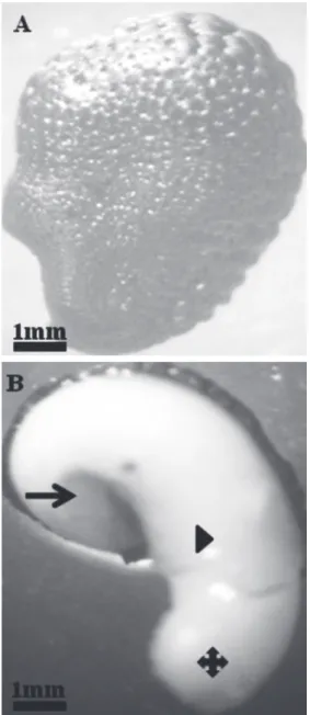
Cereus jamacaru, a Cactaceae found throughout northeast Brazil, is widely used as cattle food and as an ornamental and medicinal plant. However, there has been little information about the physiological and biochemical aspects involved in its germination. The aim of this study was to investigate its reserve mobilization during germination and early seedling growth. For this, C. jamacaru seeds were germinated in a growth chamber and collected at 0, 2, 4, 5, 6, 8 and 12 days after imbibition for morphological and biochemical analyses. Dry seeds had wrinkled seed coats and large, curved embryos. Lipids were the most abundant reserve, comprising approximately 55% and 65% of the dry mass for cotyledons and the hypocotyl-radicle axis, respectively. Soluble sugars and starch were the minor reserves, corresponding to approximately 2.2% of the cotyledons’ dry mass, although their levels showed significant changes during germination. Soluble proteins corresponded to 40% of the cotyledons’ dry mass, which was reduced by 81% at the final period of germination compared to dry seeds. C. jamacaru seed can be classified as an oil seed due to its high lipid content. Moreover, lipids were the main reserve mobilized during germination because their levels were strongly reduced after seed germination, while proteins were the second most utilized reserve in this process. Key words: Cactaceae, carbohydrates, cytochemistry, lipids, morphology, proteins.
INTRODUCTION
Seed germination is composed of two distinct
meta-bolic processes: reserve mobilization by hydrolytic
enzymes and the use of the hydrolysis products
for the formation of new cell structures (Fu et al.
2005, Soltani et al. 2006). The mobilization of food
reserves also provides energy to fuel growth until the
seedling becomes photoautotrophic (Pritchard et al.
2002). Carbohydrates, lipids and proteins, which are
stored during the later stages of seed formation, are
considered the major reserves in most seeds (Bewley
1997, Suda and Giorgini 2000, Lima et al. 2008).
Cactaceae are widely distributed in the
American continent and consist of approximately
1500 to 2000 species (Rojas-Aréchiga and
Vásquez-Yanes 2000). Cactus seeds present considerable
(Annals of the Brazilian Academy of Sciences)
variations in form, size, structure, embryo
characteristics, color of the testa and number
among species and sometimes within the same
species (Rojas-Aréchiga and Vásquez-Yanes 2000).
These authors also reported features of cactus seed
germination such as predation, dissemination,
dormancy, soil seed bank, longevity, propagation and
conservation (Rojas-Aréchiga and Vásquez-Yanes
2000). However, there is no current information
about seed reserve mobilization within the Cactaceae
family. In addition, although the third highest level
of species diversity in the Cactaceae family is found
in Brazil (Taylor and zappi 2004), few studies have
been conducted with cacti inhabiting the Caatinga
vegetation, a semiarid ecosystem that characterizes
northeastern Brazil (Meiado et al. 2008).
Cereus jamacaru
D.C. ssp.
jamacaru
is a
columnar cactus popularly known as ‘mandacaru’
that is widely distributed in the semi-arid tropic of
Brazil, being used as an ornamental plant and as cattle
feed (Taylor and zappi 2004). Farmers also use their
fruit as food, and infusions of their stems and roots
are professed to be efficient for treating respiratory
diseases (Mors et al. 2000, Lorenzi and Matos 2002).
Recently, Meiado et al. (2010) classified C. jamacaru
as a positive photoblastic species and showed that
saline and water stresses negatively affected its
germination. They also reported that the high seed
germination capacity of
C. jamacaru
while under
the influence of different environmental factors was
associated with high fruit production per individual
and the high seed production per fruit. In addition, it
could compensate for the low level of recruitment and
should favor the occurrence and the wide distribution
of the species in the Caatinga vegetation.
As
C. jamacaru
is well adapted to Brazil’s
semi-arid region, knowledge about the reserve of these
seeds is important for understanding how the reserves
can provide enough energy for seed germination.
Moreover, information regarding the way in which an
embryo mobilizes seed reserves during the early stage
of germination and seedling development can provide
insights into the metabolic processes of germination
and the relative nutritional importance of different
reserve compounds, which can be considered crucial
for successful seedling development. Therefore,
the aim of this study was to evaluate the reserve
mobilization process during the seed germination and
early seedling growth of this species and to contribute
with more information about the related biochemical
and physiological aspects of this process.
MATERIALS AND METHODS
PLANT MATERIAL AND GERMINATION
Mature fruits with
C. jamacaru
seeds were collected
in Crateús, Ceará, Brazil. The seeds were harvested
and stored at 4°C in glass pots until use. Seeds were
treated with 5% sodium hypochlorite for 5 min and
washed with distilled water prior to germination on
two paper sheets moistened with distilled water
(4.5 mL) in plastic boxes (14 x 14 x 3.5 cm), which
were placed in a germination chamber at 25°C
under continuous white light from 0.012 W m
-2nm
-1lamps for a 12 h photoperiod. The seeds that were
used had a medium mass of 3 mg per unit. Seed
germination was defined as a radicle protrusion out
of the seed coat that was 1 mm in length. The seeds
were harvested at 0 (dry seed), 2 (imbibed seed),
4 (beginning of seed coat ruptured), 5 (radicle
after protrusion) and 6 days after imbibition (DAI)
(epicotyl elongation), and seedlings were harvested
at 8 and 12 DAI. The radicle protrusion occurred
between 4 and 5 DAI. Four replicates of 100 seeds
were used for each evaluation period.
MORPHOLOGICAL AND CYTOCHEMICAL ANALYSES
buffer (pH 7.2) and 10 g L
-1glutaraldehyde for
24 h at ambient temperature (karnovsky 1965). The
material was then dehydrated in a graded ethanol
series and embedded with a Historesin Embedding
kit (Jung Heidelberg, Germany). The tissue blocks
were sectioned at 5 μm on a Leica RM 2065
microtome (Heidelberg, Germany). Thereafter, the
seed cross sections were subjected to the following
cytochemical stains: xylidine Ponceau (XP) for
proteins (Vidal 1970), periodic acid/Schiff (PAS)
for polysaccharides (Maia 1979), Lugol’s reagent
for starch (Berlyn and Miksche 1976) and Sudan IV
for lipid bodies (Gerlach 1984). The cross sections
were analyzed by light microscopy.
BIOCHEMICAL ANALYSES
Seeds without a seed coat at 0, 2, 4, 5 and 6 DAI
and seedlings at 8 and 12 DAI were separated into
cotyledons and the hypocotyl-radicle axis. These
structures were then freeze-dried, and the tissue
dry mass was obtained. The materials were stored
at 4°C until use.
For total lipid determination, the following
procedure was performed at room temperature.
Initially, 15 mg of dry mass (cotyledon or
hypocotyl-radicle axis) was ground and
homo-genized in 1.5 mL of a chloroform/methanol
mixture (2:1 v/v) according to Bligh and Dyer
(1959). After the separation step, the lipid phase
was collected and dried. Soon after, the lipid
content was determined by gravimetry.
For soluble sugar and free amino acid
deter-minations, 15 mg of cotyledon or hypocotyl-radicle
axis dry mass was ground and homogenized in 1.5
mL of 80% ethanol (v/v), and then the mixture
was warmed at 75°C for 1 h. After centrifugation
at 12,000 x g for 10 min, the supernatant was
collected, and the pellet was re-extracted twice for
30 min each. Supernatants from both extractions
were then combined. The amount of total soluble
sugars was then determined according to Dubois et
al. (1956) using glucose as a standard. Free amino
acids (FAA) were determined using the Yemm and
Cocking (1955) method with glycine as a standard.
Reducing sugars were estimated according to the
Miller (1959) method, whereas non-reducing
sugars were estimated by the difference between
the total soluble and reducing sugars. Starch was
determined according to McCready et al. (1950)
using glucose as a standard.
Proteins were extracted according to their
solubility (Osborne 1924). The material was
subjected to consecutive extractions with distilled
water (albumins), 5% (w/v) sodium chloride
(globulins), 60% (v/v) ethanol (prolamins) and 0.4%
(w/v) sodium hydroxide. The alkali-soluble protein
is referred to in the present study as salt insoluble
protein (SIP). Extracts were centrifuged at 12,000
x g at 4°C for 10 min. An aliquot of each extract
was taken for the quantification of proteins by the
Bradford (1976) method using bovine serum albumin
(BSA) as a standard. Total soluble protein (TSP) was
calculated as the sum of all protein fractions.
Experiments for the biochemical analyses
were set up in a completely randomized design,
and each treatment had four replicates of 100 seeds
each. Differences in biochemical parameters among
treatments were tested for statistical significance using
a one-way ANOVA followed by a Tukey’s honestly
significant difference test. Data are expressed as a
mean of four independent values ± the standard error
(SE). All statistical analyses were carried out using
the program ASSISTAT 7.0 (
P
< 0.05).
RESULTS
MORPHOLOGICAL AND CYTOCHEMICAL ANALYSES
The seeds were evaluated by cytochemical
analyses during germination (Fig. 2). A large
number of lipid bodies were detected in dry
seeds (Fig. 2A); however, after germination at 6
DAI, these structures were reduced (Fig. 2B). A
considerable number of protein bodies present in
dry seeds (Fig. 2C) were reduced or fused after
the radicle had protruded at 6 DAI (Fig. 2D).
Cell walls were thin both in dry seeds (Fig. 2E)
and at 6 DAI (Fig. 2F); moreover, starch grains
were evident in the last period (6 DAI) (Fig. 2F),
which was confirmed by the positive reaction with
Lugol’s reagent (Fig. 2H).
SEED RESERVE MOBILIzATION
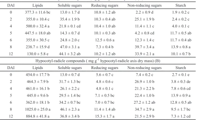
Lipids were the most abundant reserve
com-pounds in dry seeds, accounting for 55% and
65% of the cotyledon and the hypocotyl-radicle
axis dry mass, respectively (Table I). Proteins
were the second most represented compound,
comprising 40% of the cotyledons and 30% of
the hypocotyl-radicle axis. Other compounds
were not as abundant, with 1.9%, 0.5% and
0.3% of the cotyledon dry mass composed of
soluble sugars, free amino acids and starch,
respectively, with similar percentages observed
in the hypocotyl-radicle axis (Table I).
The lipid content was slightly changed in
cotyledons until 6 DAI, except by a significant
increase at 5DAI; however, a strong reduction was
observed at 8 and 12 DAI, corresponding to 37% and
66%, respectively, compared to dry seeds (Table IIA).
On the other hand, the lipid content of the
hypocotyl-radicle axis remained practically unchanged until 6
DAI; thereafter significant increase occurred from 8
DAI (F
(6,21)= 24.13,
P
< 0.001, Table IIB).
The soluble sugar content also showed
significant differences (P
< 0.01), with the highest
values at 8 DAI, showing an increase of 262% in
the cotyledons and (F
(6,21)= 42.96,
P
< 0.01, Table
IIA) 255% in the hypocotyl-radicle axis (F
(6,21)=
42.96,
P
< 0.001, Table IIB), compared to dry seeds.
Reduced sugars were unchanged in the cotyledons
(Table IIA), whereas in the hypocotyl-radicle axis
there were significant changes, with the highest
values at 8 and 12 DAI (F
(6,21)= 17.30,
P
< 0.001,
Table IIB). The non-reducing sugars were highest
at 8 and 12 DAI in cotyledons and at 6 and 8 DAI
in the hypocotyl-radicle axis.
The major reserve proteins in dry seeds were
albumins and SIP, which accounted for 31%
and 53% of the TSP in the cotyledon dry mass,
respectively (Table IIIA). The albumin, globulin
and SIP contents in the cotyledons and the
hypocotyl-radicle axis were significantly reduced
(between 77% to 94%) at 12 DAI compared to dry
seeds (
P
< 0.001, Table III). The prolamins did not
change significantly during the imbibition in the
cotyledons (F
(6,21)= 1.57,
P
> 0.05, Table IIIA),
whereas the total protein content was significantly
reduced in both the cotyledons (F
(6,21)= 99.86,
P
< 0.001, Table IIIA) and the hypocotyl-radicle
axis ( F
(6,21)= 42.35,
P
< 0.001, Table IIIB), and
was accompanied by an increase in free amino acid
levels, which were highest at 8 and 12 DAI.
Seed dry compounds Cotyledons Hipocotyl-radicle axis
Lipids 55.0 65.0
Soluble sugars 1.9 1.9
Starch 0.3 0.4
Soluble Proteins 40.0 30.0
Free amino acids 0.5 0.4
Other compounds 2.3 2.3
TABLE I
Percentage composition (%) of cotyledons and hypocotyl-radicle axis in
Cereus jamacaru dry seeds. Mean values (n=4).
Cotyledon compounds (mg g-1 cotyledon dry mass) (A)
DAI Lipids Soluble sugars Reducing sugars Non-reducing sugars Starch 0 377.3 ± 11.6 bc 13.0 ± 1.7 d 10.8 ± 1.2 ab 2.2 ± 0.9 d 1.9 ± 0.2 c 2 355.0 ± 10.4 c 35.4 ± 1.9 b 10.3 ± 0.4 ab 25.1 ± 1.9 b 2.4 ± 0.2 c 4 500.0 ± 32.4 a 21.8 ± 0.1 cd 10.4 ± 1.0 ab 11.4 ± 1.1 c 4.0 ± 0.1 c 5 447.5 ± 18.0 ab 14.3 ± 0.7 d 10.1 ± 0.3 ab 4.2 ± 0.8 cd 11.7 ± 0.5 ab 6 355.0 ± 30.5 c 24.8 ± 2.0 c 12.5 ± 0.6 a 12.3 ± 1.4 c 11.7 ± 0.4 ab
8 238.7 ± 15.9 d 47.0 ± 3.1 a 7.3 ± 0.4 b 39.7 ± 3.4 a 13.9 ± 0.8 a
12 130.0 ± 5.8 e 44.1 ± 3.2 ab 10.2 ± 1.2 ab 33.9 ± 2.1 a 10.1 ± 0.7 b Hypocotyl-radicle compounds ( mg g-1 hypocotyl-radicle axis dry mass) (B)
DAI Lipids Soluble sugars Reducing sugars Non-reducing sugars Starch
0 454.0 ± 17.7 b 13.0 ± 0.7 d 5.6 ± 0.7 c 7.4 ± 0.2 c 2.7 ± 0.1 e
2 464.3 ± 7.9 b 31.7 ± 1.3 bc 4.8 ± 0.6 c 26.9 ± 1.0 b 3.8 ± 0.3 de
4 461.0 ± 16.1 b 26.1 ± 2.2 c 4.8 ± 0.1 c 21.3 ± 2.2 b 7.8 ± 0.6 cd
5 445.0 ± 9.6 b 29.5 ± 1.4 bc 7.1 ± 0.5 bc 22.4 ± 1.0 b 13.9 ± 0.9 a 6 362.0 ± 18.1 b 34.2 ± 0.7 bc 7.0 ± 0.7 bc 27.2 ± 1.2 ab 12.8 ± 0.5 ab 8 1025.0 ± 25.0 a 46.1 ± 2.3 a 11.4 ± 1.4 ab 34.7 ± 2.9 a 9.5 ± 1.7 bc 12 884.8 ± 41.8 a 36.8 ± 3.4 b 15.3 ± 1.7 a 21.5 ± 2.9 b 7.3 ± 1.2 cd
Values are means ± standard error (n=4). Different letters indicate significant difference at P ≤ 0.05 according to Tukey Test. DAI=Days after imbibition. TABLE II
Changes in lipid, sugars (soluble, reducing and non-reducing) and starch contents in
Cotyledon proteins (mg g-1 cotyledon dry mass) (A)
DAI Albumins Globulins Prolamins SIP TP Free amino acids
0 84.0 ± 2.0 b 41.5 ± 2.9 ab 3.8 ± 0.9ns 145.1 ± 11.0 a 274.4 ± 11.7 ab 3.7 ± 0.2 d 2 124.5 ± 8.4 a 44.1 ± 3.4 ab 4.9 ± 0.4ns 124.5 ± 3.8 ab 298.0 ± 6.8 a 4.3 ± 0.6 d 4 120.1 ± 9.6 a 46.0 ± 1.7 a 4.7 ± 0.7ns 111.0 ± 8.1 ab 281.8 ± 9.2 ab 4.2 ± 0.3 d 5 107.6 ± 8.2 ab 41.8 ± 7.2 ab 6.2 ± 1.1ns 115.2 ± 11.4 ab 270.8 ± 11.5 ab 9.4 ± 0.8 c 6 114.4 ± 7.2 a 28.8 ± 3.0 b 4.5 ± 0.5ns 100.2 ± 11.4 bc 247.9 ± 15.6 b 13.8 ± 0.7 b 8 4.5 ± 0.6 c 6.9 ± 0.5 c 5.6 ± 0.6ns 67.6 ± 9.8 cd 84.6 ± 9.8 c 18.5 ± 1.1 a 12 5.3 ± 0.4 c 6.3 ± 0.5 c 5.9 ± 0.5ns 33.9 ± 3.5 d 51.4 ± 3.9 c 20.7 ± 1.0 a
Hypocotyl-radicle proteins (mg g-1 hypocotyl-radicle dry mass) (B)
DAI Albumins Globulins Prolamins SIP TP Free amino acids
0 26.7 ± 1.9 b 24.5 ± 3.8 c 5.4 ± 0.2 ab 159.8 ± 10.9 a 216.4 ± 15.0 a 2.7 ± 0.2 c 2 27.4 ± 0.8 b 34.1 ± 2.1 abc 6.1 ± 0.4 ab 161.5 ± 17.6 a 229.1 ± 18.6 a 4.7 ± 0.1 c 4 30.9 ± 1.5 ab 40.5 ± 1.5 a 5.5 ± 0.5 ab 112.1 ± 6.7 bc 189.0 ± 7.3 a 6.3 ± 0.5 c 5 32.5 ± 1.0 a 36.1 ± 1.8 ab 6.1 ± 0.1 ab 118.9 ± 10.7 ab 193.6 ± 11.8 a 13.6 ± 0.7 b 6 29.6 ± 0.8 ab 28.5 ± 3.4 bc 6.5 ± 0.1 a 67.2 ± 7.9 cd 131.8 ± 9.5 b 20.4 ± 0.5 a 8 6.7 ± 0.5 c 7.1 ± 0.1 d 5.3 ± 0.1 ab 53.8 ± 6.8 d 72.9 ± 7.2 c 18.6 ± 0.6 a 12 2.8 ± 0.4 c 6.0 ± 0.5 d 5.0 ± 0.3 b 24.2 ± 1.4 d 38.0 ± 1.2 c 17.0 ± 1.6 ab
TABLE III
Changes in albumin, globulin, prolamin, salt insoluble protein (SIP), free amino acids (FAA) and total protein (TP) contents in Cereus jamacaru seed cotyledons (A) and hypocotyl-radicle axis (B). The total
protein (TP) corresponds to the sum of albumins, globulins, prolamins and glutelins.
Values are means ± standard error (n=4). Different letters indicate significant difference at P ≤ 0.05 according to Tukey Test. DAI=Days after imbibition.
DISCUSSION
Similar to
C. jamacaru
, seeds of
Opuntia tomentosa
(Orozco-Segovia et al. 2007) and
Pilosocereus
pachycladus
(Abud et al. 2010) are also
campylotropous and perispermous. Additionally,
Abud et al. (2010) observed seeds with a wrinkled,
black seed coat and an embryo consisting of a
well-developed hypocotyl-radicle axis and reduced
plane-convex cotyledons for
P. pachycladus
.
In the present study, lipid reserves were
laid down in oil bodies, and these structures were
abundant in the cotyledons. Moreover, the high lipid
content (55% in the cotyledon of dry seeds) suggests
that this species is an oil seed, which is supported
by the presence of the high levels of these reserves
until 6 DAI and their strong reduction at 8 DAI. In
addition, although the lipid had increased at 4 DAI
compared to dry seed, it is important to highlight that
this content remained the same at 5 DAI, which in
turn did not differ significantly from that of quiescent
The lipid content was also high in the cactus
species
Pachycereus pringlei
,
Pachycereus
pecten-aboriginum
,
Carnegiea gigantea
,
Stenocereus
thurberi
and
Stenocereus gummosus
, which
ranged from 28 to 31% among these species
(Ortega-Nieblas et al. 2001). In addition, Lim et al.
(2010) also observed that the pitaya (
Hylocereus
cacti) seeds contained a high amount of oil, 18%
(
Hylocereus polyrhizus
) and 28% (
Hylocereus
undatus). It was also verified that a higher content
of lipids (27%) was found in
Opuntia ficus indica
seeds (kossori et al. 1998). It is important to note
that seed reserves during germination and the
mobilization process in Cactaceae species have not
been previously reported in the literature.
As for the other reserves, the soluble sugars and
starch were the less abundant reserve compounds
in this study, indicating that they are not strongly
involved in the seed reserve mobilization of
Cereus
jamacaru
. In seeds of
Euphorbia heterophylla
, soluble
sugars comprised approximately 4% of the seed dry
mass, and starch was not detected in the endosperm
of
E. heterophylla
, which supports the results of this
study in which starch was barely detected. In seeds of
Myracrodruon urundeuva
, an arborescent species of
the Anacardiaceae family, soluble sugars represented
only 3.5% and starch represented 0.1% of dry seeds
(Abdala et al. 2002). In addition, soluble sugar and
starch contents showed low levels in French beans
(Cortelazzo et al. 2005) and
Dalbergia miscolobium
(Silva et al. 1998). Notably, in the kossori et al. (1998)
study on the composition of the cactaceae seeds of
Opuntia ficus indica
, the levels of starch and soluble
carbohydrates corresponded to 21 and 6%, respectively.
Proteins corresponded to the second most
abundant seed reserve used in the heterotrophic
development of this species. These reserves cor-
responded to 31% of the total reserves; consequently,
a large number of protein bodies were observed in
the cotyledons, and a strong protein mobilization was
observed after the radicle protrusion. Protein depletion
was accompanied by an increase in the free amino acid
content, suggesting that, during seed mobilization,
they are transferred to the growing embryo. Although
protein mobilization has not been studied in cactus
seeds, studies involving the composition of
Opuntia
seeds showed that proteins represent a large amount
of the reserves, which corresponded to 46% of the
total dry mass of the seeds (kossori et al. 1998).
In seeds of the columnar cactus from the Sonoran
Desert (
Stenocereus thurberi
,
Carnegiea gigantea
,
Stenocereus gummosus
and
Pachycereus pringlei
),
the protein content varied between 20-22% among
species (Ortega-Nieblas et al. 2001). In addition,
Costa et al. (2001) isolated and characterized a reserve
protein from the seeds of
C. jamacaru
that showed
a similar amino acid composition to the 2S albumin
storage protein family. The authors named the protein
cactin and discussed its potential use as a molecular
marker in the Cactaceae family (Costa et al. 2001).
The total amount of seed reserves available
for a developing seedling and the duration of a
strict dependency of the seed for a given resource
may vary among species in relation to three
characteristics: seed size (total seed mass); seed
quality (concentration of the focal resource); and
the major function of the cotyledons (whether the
cotyledons serve as a photosynthetic or storage
organ of seed reserves after germination).
Light-demanding species tend to have small seeds and
photosynthetic cotyledons (kitajima 2002), which
correspond to characteristics also observed for the
seeds discussed in this study.
and can be stored in a nearly anhydrous form. Their
complete oxidation yields more than twice as much
energy as protein or carbohydrate hydrolysis on a
per unit volume basis (Murphy 2001, Quettier and
Eastmond 2009). In plants, the main site of TAG
storage is in the embryo and/or endosperm tissues of
the seeds, depending on the species (Graham 2008).
When the seeds germinate, the TAGs are degraded to
produce a carbon source that will fuel the embryo’s
postgerminative growth and allow it to become a
photosynthetically active seedling with a root system
and leaves (Graham 2008).
In conclusion, our results suggest that the period
of the most intense mobilization of seed reserves that
were stored in cotyledons occurred at germination,
and that the reserves were strongly reduced at the
seedling growth stage. Moreover, these seeds can be
classified as oil seeds due to the high lipid content
observed in dry and germinating seeds. The lipids,
as the main reserve mobilized, perform an important
role as a fuel source for germination and early
seedling development, and could be crucial for seed
germination and successful seedling establishment
under adverse conditions. Finally, as studies about the
morphology and reserve mobilization of
C. jamacaru
seeds have not yet been reported in the literature,
we can assume that our results provide insights into
the physiological and biochemical mechanisms
involved during the seed germination of this cactus.
ACKNOWLEDGMENTS
The authors are thankful to Brazil`s Conselho
Nacional de Desenvolvimento Científico e
Tecnológico (CNPq) for financial support and for
the scholarship granted to Nara Lídia M. Alencar
during her master studies.
RESUMO
Cereus jamacaru, uma cactácea encontrada comu-mente no nordeste brasileiro, é amplacomu-mente usada como planta forrageira e como ornamental e medicinal. No
entanto, existem poucas informações sobre os aspectos fisiológicos e bioquímicos relacionados à sua germinação. O objetivo desse estudo foi avaliar a mobilização de reservas durante a germinação e o crescimento inicial de plântulas de C. jamacaru. Para isso, as sementes foram germinadas em câmaras de germinação e coletadas aos 0, 2, 4, 5, 6, 8 e 12 dias após a germinação para as análises fisiológicas e bioquímicas. As sementes quiescentes apresentaram tegumento espesso e rugoso e embriões curvados. Os lipídios foram as reservas mais abundantes, correspondendo aproximadamente a 55% e 65% da massa seca dos cotilédones e eixo hipocótilo-radícula, respectivamente. Os açúcares solúveis e o amido foram as reservas menos abundantes, correspondendo aproximadamente a 2,2% da massa seca dos cotilédones, embora suas reservas tenham apresentado mudanças significativas durante a germinação. As proteínas solúveis corresponderam a 40% da massa seca dos cotilédones, que foi reduzida a 81% no período final de germinação comparado a sementes quiescentes. As sementes de C. jamacaru podem ser consideradas oleaginosas devido ao seu alto conteúdo de lipídios. Além disso, os lipídios foram as principais reservas mobilizadas durante a germinação porque seus níveis foram fortemente reduzidos durante esse período, enquanto que as proteínas foram a segunda reserva mais utilizada nesse processo.
Palavras-chaves: Cactaceae, carboidratos, citoquímica, lipídios, morfologia, proteínas.
REFERENCES
ABDALA L, MORAIS LMT, RECHIA CGV, GIORGINI JF, SÁ ME AND POLIzELI MLTM. 2002. Biochemical traits useful for the determination of the genetic variation in a natural population of Myracrodruon urundeuva. Pesq Agrop Bras 37: 909-916.
ABUD HF, GONçALVES NR, REIS RGE, PEREIRA DS AND BEzERRA AME. 2010. Germinação e expressão morfológica de frutos, sementes e plântulas de Pilosocereus pachycladus
Ritter. Rev Ciência Agron 41: 468-474.
BELTRATI CM. 1995. Morfologia e anatomia de sementes. Universidade Estadual Paulista, Departamento de Botâ-nica, Instituto de Biociências, Rio Claro, 112 p.
BEWLEY JD. 1997. Seed Germination and Dormancy. Plant Cell 9: 1055-1066.
BLIGH EG AND DYER WJ. 1959. A rapid method of total lipid
extraction and purification. Can J Bioch Phys 37: 911-917.
BRADFORD MM. 1976. A rapid and sensitive method for the
quantification of microgram quantities of protein utilizing
the principle of dye binding. Anal Biochem 72: 248-254. CORTELAzzO AL, COUTINHO J AND GRANJEIRO PA. 2005.
Storage and ageing of french beans (Phaseolus vulgaris L.): Effect on seed viability and vigor. Braz J Plant Physiol 22: 121-128.
COSTA IR, SOUzA PAS, BLOCH JRC, LLAMOCA-zÁRATE RM AND CAMPOS FAP. 2001. Isolation and characterization of a reserve protein from the seeds of Cereus jamacaru D.C. (Cactaceae). Braz Arch Biol Technol 44: 331-335. DUBOIS M, GILLES kA, HAMILTON Jk, REBERS PA AND SMITH
F. 1956. Colorimetric method for determination of sugars and related substances. Anal Chem 28: 350-356.
FU Q, WANG B-C, JIN X, LI H-B, HAN P, WUEI k-H, zHANG X-M AND zHU Y-X. 2005. Proteomic analysis and
extensive protein identification from dry, germinating
Arabdopsis seeds and young seedlings. J Biochem Mol Biol 38: 650-660.
GERLACH D. 1984. Botanische Microtechnik. Stuttgart: Georg Thieme Verlagh, 311 p.
GRAHAM IA. 2008. Seed storage oil mobilization. Annu Rev Plant Biol 59: 115-142.
kARNOVSkY MJ. 1965. A formaldehyde-glutaraldehyde of high osmolarity for use in electron microscopy. J Cell Biol 27: 137-138.
kITAJIMA k. 1996. Ecophysiology of tropical tree seedlings. In: MULkEY SS, CHAzDON RL and SMITH AP. Tropical forest plant ecophysiology. New York: Chapman & Hall, p. 559-596.
kITAJIMA k. 2002. Do shade-tolerant tropical tree seedlings depend longer or seed reserves? Functional growth analysis of three Bignoniaceae species. Funct Ecol 16: 433-444.
kOSSORI RLE, VILLAUME C, BOUSTANI EE, SAUVAIRE Y AND MéJEAN L. 1998. Composition of pulp, skin and seeds of prickly pears fruit (Opuntia ficus indica sp.). Plant Food for Hum Nutr 52: 263-270.
LIMA RBS, GONçALVES JFC,PANDO SC, FERNANDES V AND SANTOS ALW. 2008. Primary metabolite mobilization during germination in rosewood (Aniba rosaedora Ducke) seeds. Rev Árvore 32: 19-25.
LIM Hk,TAN CP,kARIM R,ARIFFIN AA AND BAkAR J. 2010. Chemical composition and DSC thermal properties of two species of Hylocereus cacti seed oil: Hylocereus undatus
and Hylocereus polyrhizus. Food Chem 119: 1326-1331. LORENzI H AND MATOS FJAM.2002. Plantas medicinais do
Brasil: nativas e exóticas cultivadas. São Paulo: Nova Odessa, 512 p.
MAIA V. 1979. Técnica histológica, 2ª ed., São Paulo: Atheneu, 246 p.
MCCREADY RM,GUGGOLz J,SILVEIRA V AND OWENS HS. 1950. Determination of starch and amylase in vegetables. Anal Chem 22: 1556-1558.
MEIADO MV, ALBUQUERQUE LSC, ROCHA EA, R OJAS-ARéCHIGA M AND LEAL IR. 2010. Seed germination responses of Cereus jamacaru DC. ssp. jamacaru (Cactaceae) to environmental factors. Plant Spec Biol 25: 120-128. MEIADO MV,ROCHA EA,ROJAS-ARéCHIGA M AND LEAL IR.
2008. Comunidad de cactus en la Caatinga: ¿qué influencia la dinámica de semillas en el ambiente semiárido? Boletín de la Sociedad Latinoamericana y del Caribe de cactáceas y otras suculentas 5: 4-6.
MILLER GL. 1959. Use of dinitrosalicylic acid reagent for determination of reducing sugars. Anal Chem 31: 426-428. MORS WB,RIzINNI CT AND PEREIRA NA.2000. Medicinal Plants of Brazil. Michigan: Reference Publications, Inc. Algonac., 501 p.
MURPHY DJ.2001. The biogenesis and functions of lipid bodies in animals, plants and microorganisms. Progr Lipid Res 40: 325-438.
OROzCO-SEGOVIA A, MÁRQUEz-GUzMÁN J, S ÁNCHEz-CORONADO ME, BUEN AG, BASkIN JM AND BASkIN CC.2007. Seed anatomy and water uptake in relation to seed dormancy in Opuntia tomentosa (Cactaceae, Opuntoideae). Ann Bot 99: 581-592.
ORTEGA-NIEBLAS MO, MOLINA-FREANER F, R OBLES-BUGENO MR AND VÁzQUEz-MORENO L.2001. Proximate composition, protein quality and oil composition in seeds of columnar cacti from the sonoran desert. J Food Compos Anal 14: 575-584.
OSBORNE TB.1924. The Vegetable Proteins. Monographs in Biochemistry. Longmans, Green and Co., London, 114 p. PRITCHARD SL,CHARLTON WL,BAkER A AND GRAHAN IA.
2002. Germination and storage reserve mobilization are regulated independently in Arabdopsis. Plant J 31: 639-647. QUETTIER AL AND EASTMOND PJ.2009. Storage oil hydrolysis during early seedling growth. Plant Physiol Biochem 47: 485-490.
ROJAS-ARéCHIGA M AND VÁSQUEz-YANES C.2000. Cactus seed germination: a review. J Arid Environ 44: 85-104. SILVA TRG, CORTELAzzO AL AND DIETRICH SMC. 1998.
Variations in storage compounds during germination and early plantlet growth of Dalbergia miscolobium. Braz J Plant Physiol 10: 119-124.
SOLTANI A,GHOLIPOR M AND zEINALI E.2006. Seed reserve utilization and seedling growth of wheat as affected by drought and salinity. Environ Exp Bot 55: 195-200. SUDA CNk AND GIORGINI JF.2000. Seed reserve composition
and mobilization during germination and early seedling development of Euphorbia heterophylla. Braz J Plant Physiol 12: 226-245.
TAYLOR NP AND zAPPI DC. 2004. Cacti of Eastern Brazil. London: Royal Botanic Gardens, kew, 499 p.
VIDAL BC.1970. Dichroism in collagen bundles stained with xylidine Ponceau 2R. Anal Histochem 15: 289-296. YEMM EW AND COCkING EC.1955. The determination of