Single-port laparoscopic ovariectomy using a pre-tied loop ligature in Santa Ines ewes
Ovariectomia por um portal laparoscópico com aplicação de ligaduras pré-montadas em ovelhas Santa Inês
Felipe Farias Pereira da Câmara BarrosI* Pedro Paulo Maia TeixeiraII
Marco Augusto Machado SilvaIII Cassia Maria Molinaro CoelhoIV
Maristela de Cassia Seudo LopesV Aline Eyko KawanamiI Denise Granato ChungI
Leandro Nassar CoutinhoVI Rachel Bittencourt RibeiroVII Luciana Cristina PadilhaI
Wilter Ricardo Russiano VicenteI
ISSN 0103-8478
ABSTRACT
The aim of the study was to develop and assess the
feasibility, postoperative pain and inflammatory response of the
single-port laparoscopic ovariectomy in ewes, using a simple pre-tied loop ligature technique.Pre-tied Meltzer’s knot was employed for prophylactic hemostasis of the ovarian pedicle. Slipknot was inserted within the abdominal cavity through a 14-gauge needle and tied surrounding the ovarian pedicle. Mean surgical time, manipulation, ligature and resection of each ovary and anesthesia time were 63±20, 20±10 and 91±26 minutes, respectively. No bleeding occurred during the surgeries. Ewes showed low scores
pain (0.5±0.5) at all time-points. Postsurgical plasma fibrinogen
was within the normal range for sheep specie at all time-points.
The ewes showed a significant weight gain in comparison to
the basal scaling (one day before the surgery). Single-port laparoscopic ovariectomy using a pre-tied loop ligature is feasible in the ovine specie and provided minimal postoperative distress and quick weight gain.
Key words: laparoscopy, ovary, ovine, single-site access, slipknot.
RESUMO
Objetivou-se com este trabalho desenvolver e descrever uma técnica de ovariectomia por videolaparoscopia utilizando um portal laparoscópico e um sistema de ligadura pré-montada, avaliando a sua viabilidade, o desconforto doloroso e
o processo inflamatório provocado em ovelhas. O nó de Meltzer pré-montado foi utilizado para hemostasia profilática do pedículo
ovariano. O nó corrediço foi inserido na cavidade abdominal
através de uma agulha 14G e atado em torno do pedículo ovariano.
O tempo médio de cirurgia foi de 63±20min, o de manipulação,
ligadura e ressecção para cada ovário foi de 20±10min, e o de
anestesia 91±26min. Não houve hemorragia durante as cirurgias. As ovelhas apresentaram escores de dor considerados baixos
(0,5±0,5). Todos os valores do fibrinogênio plasmático estiveram dentro do padrão de normalidade, não havendo diferença estatística entre os momentos avaliados. Houve aumento significativo nas médias de peso das fêmeas, quando comparados ao momento
controle (um dia anterior ao experimento). A ovariectomia por um portal laparoscópico com aplicação de ligaduras pré-montadas
é factível para a espécie ovina, provocando mínimo estresse, desconforto doloroso e rápido ganho de peso nos animais.
Palavras-chave: laparoscopia, nó deslizante ovário, ovinos, acesso único.
INTRODUCTION
Gonadectomy is a surgical technique
that inflicts positive impacts on ovine breeding.
Ovariectomy is not only performed to prevent
pregnancy, but also to reduce problems related
to the estrous cycle, such as interference on the weight gain (GARBER et al., 1990; BLEUL et al., 2005). Other purposes of ovariectomy in livestock animals include enhancement of the weight gain performance and improvement of the carcass quality
IDepartamento de Medicina Veterinária Preventiva e Reprodução Animal, Faculdade de Ciências Agrárias e Veterinária (FCAV), Universidade
Estadual Paulista (UNESP), 14884-900, Jaboticabal, SP, Brasil. E-mail: [email protected]. *Corresponding author.
IIPós-graduação em Ciência Animal, Hospital Veterinário, Universidade de Franca (UNIFRAN), Franca, SP, Brasil. IIIUniversidade de Passo Fundo (UPF), Passo Fundo, RS, Brasil.
IVUniversidade Federal Rural do Rio de Janeiro (UFRJ), Seropédica, RJ, Brasil. VUniversidade Federal da Bahia (UFBA), Salvador, BA, Brasil.
VIInstituto da Saúde e Produção Animal (ISPA), Universidade Federal Rural da Amazônia (UFRA), Belém, PA, Brasil.
VIIUniversidade Estadual do Norte Fluminense Darcy Ribeiro (UENF), Campos dos Goytacazes, RJ, Brasil.
2034 Barros et al.
(SILVA et al., 2006), the preservation of gametes of high performance, animals with great genetic value, to sample specimens for advanced researches on
reproductive biotechnologies (PADULA et al., 2002;
TEIXEIRA et al., 2011) or to treat ovarian disorders (MOBINI et al., 2004).
Gonadectomy may present some disadvantages, such as increased costs and risk of surgery-related complications (PEINADO et al., 2008), which can lead to low production performance, especially due to postoperative stress and pain. Therefore, the efforts of the surgical management in
livestock should always be focused on the reduction of cost-benefit and minimal postoperative stress, as
pain can impair weight gain (FITZPATRICK et al., 2006; LUNA, 2008).
One of the most critical steps during the laparoscopic approach for ovariectomy is the achievement of an affective prophylactic hemostasis of the ovarian pedicles. The use of energy surgical sources such as electrosurgical generators, harmonic scalpel and lasers, and automatic stapling/cutting
devices increases the costs-benefit of the procedure and may be expensive and difficult to implement
(AZIZ et al., 2008).
The pre-tied ligature systems, also known as endoscopic slipknots or “endoloops”, are composed of a slipknot and a laparoscopic tool used to guide and tighten the knot, which is known as knot pusher (KATSINELOS et al., 2006).
Hemostasis is achieved by pushing and tightening
the knot around the ovarian pedicle. One of the main advantages of the use of thread ligature is
the absence of thermal injury of adjacent tissue, which is usually caused by electrocoagulation
(LIM et al., 2007).
There are several reports of the use of loop ligature for ovariectomy in the equine specie (BOURÉ et al., 1997; RAGLE & SCHNEIDER, 1998; HANSON & GALLUPO, 1999; RODGERSON & HANSON, 2000). In humans, endoloop ligature was carried out in shorter surgical time and caused less postoperative pain then the electrocoagulation for salpingectomy (LIM et al., 2007). However, no reports regarding the use of pre-tied ligature for ovariectomy in the ovine specie was found in the current literature, furthermore this
technique could also be performed in other females
including women.
The aim of this study was to develop and
assess the feasibility, the postoperative pain, and the inflammatory response of a technique for single-port
laparoscopic ovariectomy in ewes.
MATERIAL AND METHODS
Seven healthy ewes were assessed. The awere fasted for 36 hours prior to surgical
procedure and their body weight was measured
on a digital scale. The animals were premedicated using diazepam (0.5mg kg-1, IM), and tramadol (2mg kg-1, IM). Following anesthetic induction using a mixture of propofol (6mg kg-1, IV) and lidocaine chloride (1mg kg-1, IV), the ovines were
intubated using a cuffed 8-mm tracheal tube, in order to provide humidified oxygen under assisted
ventilation. Anesthesia was maintained with propofol (0.5mg kg-1 min-1, IV) and lidocaine-chloride (1mg kg-1 min-1, IV) in constant rate infusion (CRI). The ewes were positioned in
dorsal recumbence, in Trendelenburg position.
Local anesthesia with lidocaine chloride (2ml) was performed on the incision site.
A single-port laparoscopic approach was carried out. A 11-mm trocar was inserted through
the abdominal wall in the midline, 20cm cranial to the udder. Following establishment of the port,
CO2 pneumoperitoneum was created until
intra-abdominal pressure reached 5-8mmHg, using 5L
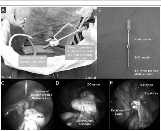
min-1 of flow rate. A 10-mm operating laparoscope,
with a 6mm working channel, was introduced into
the abdominal cavity through the trocar. A 5-mm, 42-cm long Babcock atraumatic forceps was inserted
through the working channel of the laparoscope for manipulation of the ovaries (Figure 1A). A 2-0 nylon pre-tied Meltzer’s knot was introduced into
the abdomen through a 14-gauge needle, using a thin
hollow stainless steel stick as a knot pusher (Figure
1B). The Babcock forceps was passed through the
loop of the slipknot (Figure 1C). Afterwards, the ovary was grasped and pulled through the loop and the slipknot was tighten around the ovarian pedicle (Figure 1D). Two ligatures were applied on each ovarian pedicle in the same fashion. The pedicle was
then raised by the ligature thread, and the atraumatic forceps was replaced by an endoscopic scissors to
resect the ovary. After excision, the hemostasis was checked and the ovary was exteriorized through the
11-mm trocar (Figure 1E). Following removal of both
ovaries, the CO2 pneumoperitonium was completely drained. Skin suture was placed, using the interrupted horizontal mattress pattern with 2-0 nylon. The muscular layer was not sutured. During the early post-operative period, all animals received a single dose of oxytetracycline (20mg kg-1, IM).
ovaries (manipulation of each ovary, ligature and resection), anesthesia time (premedication
to extubation) and recovery of the animals (from extubation to spontaneous standing position and
walking) were measured and expressed as mean ± standard deviation. The postsurgical pain was assessed using a modified visual analogue scale, adapted from other study (MELLOR & STAFFORD, 2004). In such scoring system,
three parameters were considered to establish the
final score: posture, locomotion and food intake. For each parameter, score ranging from 0 to 2
could be assigned. The final score was given by
the sum of scores, which could range from 0 to 6. The postoperative pain assessment was carried
out hourly, for the first six hours following the surgical procedure.
Body weight was scaled weekly, during four weeks after the surgical procedure. Control
weight was measured one day before the surgery. Postsurgical inflammatory response was estimated by daily measurement of plasmatic fibrinogen, for five
days, using a manual refractometer (mg dl-1).
For statistical analysis, data normally
distributed were assessed using repeated
measures one-way ANOVA and Dunnett’s post hoc test were used to assess data regarding weight
and fibrinogen. For non parametrical data, the
Friedman’s test and the Dunn’s post hoc test were performed (P<0.05).
Figure 1 - Position of laparoscope and instrument portals, with a 11-mm trocar inserted through the abdominal wall in the midline, 20cm cranial to the udder, a 10-mm operating laparoscope, with a 6 mm working channel, introduced into the abdominal cavity through
the trocar, and a system of pusher pre-tied Meltzer’s knot (A), System of pusher 2-0 nylon pre-tied Meltzer’s knot which is
2036 Barros et al.
RESULTS
The surgical procedures were carried out
successfully in all animals. There was no bleeding
during surgical procedures. The overall surgical time was 63±20 minutes. Time elapsed for manipulation, ligature and resection of each ovary was 20±10 minutes. Minimum and maximum surgical time in the current study was 36 and 99 minutes respectively.
Regarding the anesthesia protocol, there were no complications that impaired the surgical procedure. Mean anesthesia time was 91 (±26) minutes, and 63 (±44) minutes for total anesthesia recovery. Concerning the post-surgical pain, the ewes demonstrated low pain scores at all time-points
(0.5±0.5). The plasmatic fibrinogen values were also
within normal limits at all time-points (range 100-500mg dl-1). There was no difference among times of
assessment. Mean body weight differed significantly
among days 7, 14, 21 and 28 in comparison to the
control. The ewes had normal body weight gain
during the weeks following the trial.
DISCUSSION
Bilateral ovariectomy using single-port laparoscopic approach and the Meltzer’s knot for prophylactic hemostasis of the ovarian pedicles was successfully performed in all ewes. Reproductive tract was easily visualized using such technique, due to the
pneumoperitoneum, Trendelenburg positioning and amplification of the image using the video system.
Single portal techniques for ovariectomy have also
been reported as feasible and safe in others domestic
species, specially in dogs (DUPRÉ et al., 2009; MANASSERO et al., 2012) and cats (KIM et al., 2011; COISMAN et al., 2014). A 3-port laparoscopic assisted ovariohysterectomy technique using pre-tied loop ligature was assessed in the canine specie,
which resulted in bleeding in three dogs (MAYHEW & BROWN, 2007). Nevertheless, no reports of the use
of a single-port approach for gonadectomy in domestic
animals were found on the literature available.
Intra-operative bleeding was not reported
in the current study, contrasting the results of other study on laparoscopic ovariectomy in mares (RODGERSON & HANSON, 2000) and on laparoscopic-assisted ovariohysterectomy in dogs
(MAYHEW & BROWN, 2007). Therefore, in order
to avoid ligature failure, at least two ligatures should
be applied on each pedicle (FREEMAN, 1999). The affordable, reusable and accessible
nature of the instrument used for ligating the pedicles
in the current study makes such technique simpler and less expensive than other ovariectomy techniques
described in the literature available. In humans,
pre-tied ligatures are indicated in some surgical techniques such as salpingectomy (LIM et al., 2007), appendectomy (BELDI et al., 2004; YILDIZ et al, 2009) and other gastrointestinal surgeries (KATSARELIAS, 2007).
The surgical time obtained in the current
study was longer than other laparoscopic techniques
described in other trial (TEIXEIRA et al., 2011).
Approximately 12 minutes were spent for unilateral
ovariectomy in donkeys, using a specific laparoscope
specially design and manufactured in other study (AZIZ et al., 2008). Longer surgical time (35 minutes) was found in a technique of ovariectomy in llamas using hemostatic clips for pedicular hemostasis
(RODGERSON et al., 1998). In other trial, bilateral
ovariectomy was performed in standing mares using slipknot ligatures in minimum of 50 and maximum of 120 minutes (BOURÉ et al., 1997). In human
patients, obtained shorter surgical time was found
using pre-tied ligatures in comparison to the use of monopolar electrocoagulation for salpingectomy (LIM et al., 2007), and to the use of hemostatic clips in gastrotomy (KATSARELIAS, 2007). The high value for surgical time found in our study may have occurred due to the surgical team’s short learning curve on such surgical technique. Depending on the technique, a minimum of 20 surgical procedures are required in order to achieve
proficiency (SILVA et al., 2011).
Regarding the pain score, results of our study were similar to other laparoscopic techniques, in another trial (TEIXEIRA et al., 2011). In dogs, laparoscopic ovariectomy was compared with conventional open ovariectomy (CULP et al.,
2009). It was observed that bitches undergoing open technique had significant decreases in activity
postoperatively than those undergoing laparoscopy. Furthermore, the use of pre-tied ligatures resulted in
lower post-surgical pain score in women submitted
to salpingectomy compared to electrocoagulation (LIM et al., 2007). In contrast, other studies regarding the feline specie revealed that pain score
was significantly higher in those submitted to
extracorporeal ligature in comparison to bipolar‐
sealing device for hemostasis of the pedicles
(COISMAN et al., 2014). The plasmatic fibrinogen values were within normal limits established for
sheep (JAIN, 1993) at all time-points. The pattern of
raise in post-surgical plasmatic fibrinogen among the
in other studies involving ruminants orchiectomy
(EARLEY & CROWE, 2002; PANG et al., 2006). The ewes gained body weight satisfactorily
following the surgical procedures and during the weeks
following the trial, demonstrating no negative influence of the surgical technique on the body condition. Other study also revealed no negative influence of
laparoscopic ovariectomies on the physical condition of the ewes (TEIXEIRA et al., 2011). The castration of young females is indicated for a faster weight gain in cattle (GARBER et al., 1990). However, other
studies found that flank laparotomy (CARVALHO et
al., 2010) and transvaginal approach using latex ring
for pedicle ligation in cows submitted to ovariectomy (MEIRELLES et al., 2007) caused negative influence
concerning post-surgical weight gain. In another trial,
no benefits were found regarding the influence of flank
ovariectomy on the weight gain in heifers (SILVA et al.,
2006). But, the authors obtained better carcass quality. We highlight that none of those articles that reported negative or no benefits of ovariectomy employed
minimally invasive laparoscopic or video-assisted
techniques, which could have dramatically influenced
the results. Thus, further studies are required in order to support such hypothesis.
CONCLUSION
The proposed technique for single-port
laparoscopic ovariectomy was feasible in ewes and caused no bleeding, surgical stress, postoperative
pain or weight loss.
ACKNOWLEDGMENTS
The authors gratefully thank Conselho Nacional de
Desenvolvimento Científico e Tecnológico (CNPq) and Fundação
de Amparo à Pesquisa do Estado de São Paulo (FAPESP) for
financial support in the study.
BIOETHICS AND BIOSSECURITY COMMITTEE APPROVAL
This study was conducted following approval by the Animal Ethics and Welfare Committee of the School of Agrarian and Veterinary
Sciences of São Paulo State University (protocol N. 025988-08).
REFERENCES
AZIZ, D.M. et al. Laparoscopic ovariectomy in standing donkeys
by using a new instrument. Animal Reproduction Science,
v.107, p.107-114, 2008. Available from: <http://ac.els-cdn.com/
S037843200700214X/1-s2.0-S037843200700214X-main.pdf?_
tid=bda61956-f830-11e3-ab9c-00000aacb360&acdnat=1403237577_ db1326b028fa961688a2adb2160042a7>. Accessed: Jun. 19, 2014. doi: 10.1016/j.anireprosci.2007.06.011.
BELDI, G. et al. Laparoscopic appendectomy using endoloops. A prospective, randomized clinical trial. Surgical Endoscopy, v.18,
p.749-750, 2008. Available from: <http://download.springer.com/
static/pdf/231/art%253A10.1007%252Fs00464-003-9156-z.pdf?a
uth66=1403410259_5548b88c0999c7fb52b323840857ffbd&ext=. pdf>. Accessed: Jun. 19, 2014. doi: 10.1007/s00464-003-9156-z.
BLEUL, U. et al. Laparoscopic ovariectomy in standing cows. Animal Reproduction Science, v.90, p.193-200, 2005. Available from: <http://
ac.els-cdn.com/S0378432005000503/1-s2.0-S0378432005000503-main.pdf?_tid=056cee9a-f831-11e3-972e-00000aacb35d&acdnat=14 03237698_116b279195719f38a764418694edb42b>. Accessed: Jun. 19, 2014. doi: 10.1016/j.anireprosci.2005.01.022.
BOURÉ, L. et al. Paralumbar fossa laparoscopic ovariectomy in
horses with use of endoloop ligatures. Veterinary Surgery, v.26,
p.478-483, 1997. Available from: <http://onlinelibrary.wiley.com/ doi/10.1111/j.1532-950X.1997.tb00520.x/pdf>. Accessed: Jun. 19, 2014. doi: 10.1111/j.1532-950X.1997.tb00520.x.
CARVALHO, C.R.P. et al. Ovariectomy in beef cattle, and their relationship
with the fattening. Revista Acadêmica: Ciências Agrárias e Ambientais,
v.8, n.4, p.405-408, 2010. Available from: <http://www2.pucpr.br/reol/ index.php/academica?dd1=4511&dd99=view>. Accessed: Jun. 19, 2014.
COISMAN, J.G. et al. Comparison of surgical variables in cats
undergoing single-incision laparoscopic ovariectomy using a LigaSure or extracorporeal suture versus open ovariectomy. Veterinary Surgery,
v.43, n.1, p.38-44, 2014. Available from: <http://onlinelibrary.wiley. com/doi/10.1111/j.1532-950X.2013.12073.x/pdf>. Accessed: Dec. 18, 2014. doi: 10.1111/j.1532-950X.2013.12073.x.
CULP, W.T.N. et al. The effect of laparoscopic versus open ovariectomy
on postsurgical activity in small dogs. Veterinary Surgery, v.38, n.7,
p.811-817, 2009. Available from: <http://onlinelibrary.wiley.com/ doi/10.1111/j.1532-950X.2009.00572.x/pdf>. Accessed: Dec. 18, 2014. doi: 10.1111/j.1532-950X.2009.00572.x.
DUPRÉ, G. et al. Laparoscopic ovariectomy in dogs: comparison
between single portal and two-portal access. Veterinary Surgery,
v.38, p.818-824, 2009. Available from: <http://onlinelibrary.wiley. com/doi/10.1111/j.1532-950X.2009.00601.x/pdf>. Accessed: Dec. 18, 2014. doi:10.1111/j.1532-950X.2009.00601.x.
EARLEY, B.; CROWE, M.A. Effects of ketoprofen alone or in combination with local anesthesia during the castration of bull calves on plasma cortisol, immunological, and inflammatory
responses. Journal of Animal Science, v.80, p.1044-1052,
2002. Available from: <http://www.journalofanimalscience.org/ content/80/4/1044.long>. Accessed: Jun. 19, 2014.
FITZPATRICK, J. Assessment of pain and welfare in sheep. Small Ruminant Research, v.62, p.55-61, 2006. Available from: <http://
www.sciencedirect.com/science/article/pii/S0921448805002993#>. Accessed: Jun. 19, 2014. doi: 10.1016/j.smallrumres.2005.07.028.
FREEMAN, L.J. Veterinary endosurgery. Saint Louis: Mosby,
1999. 276p.
GARBER, M.J. et al. Efficacy of vaginal spaying and anabolic implants on growth and carcass characteristics in beef heifers. Journal of Animal Science,
v.68, p.1469-1475, 1990. Available from: <http://www.journalofanimalscience. org/content/68/5/1469.long>. Accessed: Jun. 19, 2014.
2038 Barros et al.
v.28, p.106-112, 1999. Available from: <http://onlinelibrary.wiley. com/doi/10.1053/jvet.1999.0106/pdf>. Accessed: Jun. 19, 2014. doi: 10.1053/jvet.1999.0106.
JAIN, N.C. Essentials of veterinary hematology. Philadelphia:
Lea & Febiger, 1993. 417p.
KATSARELIAS, D. Endoloop application as an alternative method for gastrotomy closure in experimental transgastric surgery. Surgical Endoscopy, v.21, p.1862-1865, 2007. Available
from: <http://download.springer.com/static/pdf/691/art%253A10 .1007%252Fs00464-007-9281-1.pdf?auth66=1403404838_4160
54b74013bcd5aaa6c9d26cfe5afd&ext=.pdf>. Accessed: Jun. 19,
2014. doi: 10.1007/s00464-007-9281-1.
KATSINELOS, P. et al. Endoloop-assisted polypectomy for large pedunculated colorectal polyps. Surgical Endoscopy, v.20, n.8,
p.1257-1261, 2006. Available from: <http://download.springer.com/
static/pdf/104/art%253A10.1007%252Fs00464-005-0713-5.pdf?a
uth66=1403405043_470627ed81af56fdb24dedc555ac64ff&ext=. pdf>. Accessed: Jun. 19, 2014. doi: 10.1007/s00464-005-0713-5.
KIM, Y.K. et al. Feasibility of single-portal access laparoscopic
ovariectomy in 17 cats. Veterninary Record, v.169, n.7,
p.179-182, 2011. Available from: <http://veterinaryrecord.bmj.com/ content/169/7/179.full.pdf+html>. Accessed: Dec. 18, 2014. doi:
10.1136/vr.d4293.
LIM, Y.H. et al. Laparoscopic salpingectomy in tubal pregnancy: prospective randomized trial using endoloop versus electrocautery.
Journal of Obstetrics and Gynaecology Research, v.33, n.6,
p.855-862, 2007. Available from: <http://onlinelibrary.wiley.com/ doi/10.1111/j.1447-0756.2007.00668.x/pdf>. Accessed: Jun. 19, 2014. doi: 10.1111/j.1447-0756.2007.00668.x.
LUNA, S.P.L. Dor, senciência e bem-estar em animais. Ciência
Veterinária nos Trópicos, v.11, n.1, p.17-21, 2008.
MANASSERO, M. et al. Laparoscopic ovariectomy in dogs using a single-port multiple-access device. Veterinary Record, v.171,
n.3, p.69-74, 2012. Available from: <http://veterinaryrecord.bmj.
com/content/171/3/69.full.pdf+html>. Accessed: Dec. 16, 2014. doi: 10.1136/vr.100060.
MAYHEW, P.D.; BROWN, D.C. Comparison of three techniques
for ovarian pedicle hemostasis during laparoscopic-assisted ovariohysterectomy. Veterinary Surgery, v.36, n.6,
p.541-547, 2007. Available from: <http://onlinelibrary.wiley.com/ doi/10.1111/j.1532-950X.2007.00280.x/full>. Accessed: Dec. 22, 2014. doi: 10.1111/j.1532-950X.2007.00280.x.
MEIRELLES, C. et al. Avaliação do ganho de peso de novilhas ovariectomizadas por técnica transvaginal. Revista Acadêmica: Ciências Agrárias e Ambientais, v.5, n.3,
p.303-307, 2007. Available from: <http://www2.pucpr.br/reol/index.php/
ACADEMICA?dd1=1855&dd99=pdf>. Accessed: Jun. 19, 2014.
MELLOR, D.J.; STAFFORD, K.J. Physiological and behavioural
assessment of pain in ruminants: principles and caveats. In:
WORLD CONGRESS, 4., ATLA, v.32, suppl.1A, p.267-271,
2004. Available from: <http://frame.org.uk/atla_article.php?art_ id=637&pdf=true>. Accessed: Jun. 19, 2014.
MOBINI, S. et al. Teriogenologia de ovinos e caprinos. In: PUGH, D.G. Clínica de ovinos e caprinos. São Paulo: Roca, 2004. Cap.6, p.145-208.
PADULA, A.M. et al. Restoration of LH output and 17beta-oestradiol
responsiveness in acutely ovariectomised holstein dairy cows pre-treated with a GnRH agonist (deslorelin) for 10 days. Animal Reproducion Science, v.70, p.49-63, 2002. Available from: <http://ac.els-cdn.
com/S0378432001001920/1-s2.0-S0378432001001920-main.pdf?_
tid=f716f130-f829-11e3-b760-00000aacb35f&acdnat=1403234667_ d91ab87609fa203d47cd4aed118d2e16>. Accessed: Jun. 19, 2014.
doi: 10.1016/S0378-4320(01)00192-0.
PANG, W.Y. et al. Effect of carprofen administration during banding or burdizzo castration of bulls on plasma cortisol, in vitro interferon-g production, acute-phase proteins, feed intake, and growth. Journal of Animal Science, v.84, p.351-359, 2006. Available from: <http://www.
animal-science.org/content/84/2/351.full>. Accessed: Jun. 19, 2014.
PEINADO, J. et al. Influence of sex and castration of females
on growth performance and carcass and meat quality of heavy pigs destined for the dry-cured industry. Journal of Animal Science, v.86, p.1410-1417, 2008. Available from: <http://
www.journalofanimalscience.org/content/86/6/1410.full.pdf>. Accessed: Dec. 16, 2014. doi: 10.2527/jas.2006-807.
RAGLE, C.A.; SCHNEIDER, R.K. Ventral abdominal approach
for laparoscopic ovariectomy in horses. Journal of the Society of Laparoendoscopic Surgeons, v.2, n.4, p.393-394, 1998.
Available from: <http://www.ncbi.nlm.nih.gov/pmc/articles/ PMC3015249/?report=reader>. Accessed: Jun. 19, 2014.
RODGERSON, D.H. et al. Ventral abdominal approach for laparoscopic ovariectomy in llamas. Veterinary Surgery, v.27,
n.4, p.331-336, 1998. Available from: <http://onlinelibrary.wiley. com/doi/10.1111/j.1532-950X.1998.tb00135.x/pdf>. Accessed: Jun. 19, 2014. doi: 10.1111/j.1532-950X.1998.tb00135.x.
RODGERSON, D.H.; HANSON, R.R. Ligature slippage during standing laparoscopic ovariectomy in a mare. Canadian Veterinary Journal, v.41, n.5, p.395-397, 2000. Available from:
<http://www.ncbi.nlm.nih.gov/pmc/articles/PMC1476268/pdf/ canvetj00017-0085.pdf>. Accessed: Jun. 19, 2014.
SILVA, L.A.F. et al. Anel de látex aplicado no pedículo ovariano
de bezerras Nelore. Acta Scientiarum. Animal Sciences, v.28,
n.1, p.97-103, 2006. Available from: <http://periodicos.uem.br/ ojs/index.php/ActaSciAnimSci/article/view/669/416>. Accessed:
Jun. 19, 2014. doi: 10.4025/actascianimsci.v28i1.669.
SILVA, M.A.M. et al. Single-port video-assisted
ovariohysterectomy in bitches: retrospective study of 20 cases.
Ciência Rural, v.41, n.2, p.294-300, 2011. Available from:
<http://www.scielo.br/pdf/cr/v41n2/a871cr4012.pdf>. Accessed:
Jun. 19, 2014. doi: 10.1590/S0103-84782011005000013.
TEIXEIRA, P.P.M. et al. Ovariectomy by laparotomy, a
video-assisted approach or a complete laparoscopic technique in Santa Ines sheep. Small Ruminant Research, v.99, p.199-202, 2011.
Available from: <http://ac.els-cdn.com/S0921448811001908/1- s2.0-S0921448811001908-main.pdf?_tid=29b5111c-f82f-11e3-acef-00000aacb360&acdnat=1403236899_e0f8b6ec04 4bcd53550bba623514cce0>. Accessed: Jun. 19, 2014. doi:
10.1016/j.smallrumres.2011.04.008.
YILDIZ, F. et al. The handmade endoloop technique. A simple and cheap technique for laparoscopic appendectomy. Saudi Medical Journal, v.30, n.2, p.224-227, 2009. Available from: <http://
smj.psmmc.med.sa/index.php/smj/article/view/6521/4295>.