Single-port laparoscopic hysterectomy: preliminary results
RENATA ASSEF TORMENA1*, SÉRGIO CONTI RIBEIRO2, GUSTAVO ARANTES MACIEL3, EDMUND CHADA BARACAT41Assistant Physician – Assistant Physician at the Gynecology Clinic, Hospital das Clínicas, Faculdade de Medicina, Universidade de São Paulo (HC-FMSP), São Paulo, SP, Brazil 2PhD – Head of the sector of Gynecological Endoscopy, HC-FMUSP, São Paulo, SP, Brazil
3PhD – Assistant Physician at the Gynecology Clinic, HC-FMUSP, São Paulo, SP, Brazil 4PhD – Full Professor of Gynecology at FMUSP, São Paulo, SP, Brazil
S
UMMARYStudy conducted at Department of Obstetrics and Gynecology, Universidade de São Paulo, São Paulo, SP, Brazil
Article received: 3/3/2015
Accepted for publication: 3/30/2015
*Correspondence:
Address: Av. Dr. Enéas de Carvalho Aguiar, 255 Postal code: 05403-000 São Paulo, SP – Brazil [email protected]
http://dx.doi.org/10.1590/1806-9282.61.05.446
Objective: to describe the initial results of a laparoscopic single port access hys-terectomy and also to evaluate the feasibility and safety of this access.
Methods: a prospective study was performed at a tertiary university medical center (Hospital das Clínicas, Faculdade de Medicina, Universidade de São Paulo) between March 2013 and June 2014. A total of 20 women, referred for hysterectomy due to benign uterine disease, were included in the study after they had signed an informed consent. Outcome measures, including operat-ing time, blood loss, rate of complications, febrile morbidity, visual analogical pain score and length of hospital stay were registered.
Results: mean patient age and body mass index (BMI) were 47.8 years and 27.15 kg/m2, respectively. Mean operating time was 165.5 min. Blood loss was
minimal, with no blood transfusion. All procedures but one were successful-ly performed via a single incision and no post-operative complications oc-curred. We experienced one conversion to multiport laparoscopic hysterecto-my due to extensive pelvic adhesions. There was no conversion to “open” total abdominal hysterectomy. None of the patients required narcotics or NSAD post-operatively.
Conclusion: single-port hysterectomy is a feasible and safe technique, with no major complications.
Keywords: hysterectomy, laparoscopy, minimally invasive surgical procedures, uterus, gynecologic surgical procedures.
INTRODUCTION
The benefits of laparoscopic surgery over conventional abdominal surgery have been well documented. Reduced postoperative pain, postoperative morbidity, hospital stay and postoperative recovery time have been well demon-strated.1
To optimize the benefits of minimally invasive pro-cedures, surgeons have attempted to reduce the overall abdominal wall trauma by decreasing either the size of the ports or the number of trocars.2
Owing to its nature, the umbilicus offers an exciting site for single port laparoscopy leaving no visible scar.2
In this modality, a 25mm umbilical single incision technique is used to access the peritoneal cavity and all instruments are located in the same incision.
For this reason, single-port access surgery has sever-al limitations including breakdown of triangulation, in-line view, crowding of surgical instruments, “sword-fight-ing” between instruments, and others which are less common in multi-port surgery.3,4
The first laparoscopic hysterectomy was performed by Reich in 1988, and two years later Pelosi reported a to-tal laparoscopic abdominal hysterectomy (LAVH) with bilateral salpingo-oophorectomy (BSO) using only a sin-gle incision.5,6 Although this procedure offers improved
postoperative hemoglobin drop seems to decrease with experience, without increasing complications.8
In this study, we report our initial results with SPA-LH and bilateral salpingectomy.
OBJECTIVES
To report our initial results with SPA-LH, and also to eval-uate the feasibility and safety of this surgical access.
METHODS
In a prospective study, between March 2013 and June 2014, 20 patients between 35 and 65 years old, assigned to undergo hysterectomy due to benign gynecologic con-ditions, were elected to single-port access surgery after signing an informed consent. We limited the SPA to pa-tients with estimated uterine volume smaller than 600cm3. The study was previously approved by the
insti-tutional review board of the Hospital da Clínicas, Uni-versidade de São Paulo, Brazil.
Surgical technique
All surgeries were performed under general anesthesia with patients in semi-gynecological position. Initially, a 2.5cm trans-umbilical longitudinal incision was made until the aponeurosis, which was opened and fixed with a stitch in both sides. Then the peritoneum was opened and a disposable three-channel single-port device was in-serted, either the Triport Access System® (Olympus,
Cen-ter Valley, PA) or the Single Site Laparoscopy (SSL) Ac-cess System® (Ethicon Endo-Surgery, Somerville, NJ, USA);
intra-peritoneal pressure of 15mmHg was kept. A 30°, 5mm obese telescope associated with conventional rigid laparoscopic instruments, including monopolar, bipolar
scissors and the Harmonic Scalpel® (Ethicon
Endo-Sur-gery, Somerville, NJ, USA) were used. The Valtchev
Uter-ine Manipulator® (Conkin, Canada) allowed a complete
range of uterus movements, facilitating different angles to access the uterus.
The hysterectomies were performed as type IV-E laparo-scopic hysterectomies, according to the AAGL classification.9
The utero-ovarian ligament, fallopian tube pedicles and the round ligaments were coagulated and divided with ultracision scissors. The vesicouterine peritoneal fold and bladder were mobilized off the uterus and up-per vagina until the anterior vagina was identified. The broad ligament peritoneum was divided and the uterine artery was coagulated and divided with bipolar and ul-tracision scissors. The cardinal and the uterosacral lig-aments, one each side, were divided. The vagina was en-tered posteriorly near the cervicovaginal junction. A 4 cm
diameter plastic vaginal delineator was placed in the va-gina to outline circumferentially the cervical junction and prevent loss of pneumoperitoneum. A monopolar forceps was used to complete the circumferential cul-dotomy. Although bilateral oophorectomy was performed only in select cases, bilateral salpingectomy was routine-ly done. The specimens were pulled out of the vagina. The vaginal delineator was placed back into the vagina for laparoscopic review of hemostasis and to delineate the vaginal cuff. A 0-Vicryl (Ethicon Endo-Surgery, Somerville, NJ, USA) suture was placed through the right uterosacral ligament and through the posterior and the anterior vaginal fold. The extracorporeal tie technique was used, with a knot-pusher inserted through the sin-gle port device. The same procedure was made on the left side and a third suture completed the vaginal cuff closure. Alternatively, the vaginal access was used to clo-sure the cuff in some cases, using the same stitch, in a running suture technique. The umbilical incision fascia
was closed using 0-vicryl running suture and an intra-dermic 4-0 Monocryl (Ethicon Endo-Surgery, Somer-ville, NJ, USA) suture was placed, ending the procedure. The operative time was analyzed, timing the follow-ing steps: pneunoperoneum installation, includfollow-ing um-bilical incision and single port device insertion; hysterec-tomy until colpechysterec-tomy; colpechysterec-tomy; salpingechysterec-tomy; specimens’ removal; eventual bleeding control; vaginal cuff closure and umbilical incision closure.
Intraoperative bleeding was measured in the vacuum aspirations system right after the surgery, discounting any abdominal fluid infused. The postoperative hemo-globin drop was measured at the end of the procedure, 24 hours, 48 hours and 6 days after surgery, and com-pared to the preoperative level.
Postoperative pain intensity was estimated using a Visual Analog Scale (VAS), on the first, second and sixth postoperative day, applied by different medical residents. On the postoperative days, patients received only sim-ple analgesics and no anti-inflammatory drugs were nec-essary.
Surgical outcomes were evaluated and immediate and late complications were reported.
RESULTS
All procedures underwent successfully through the lapa-roscopy approach. In one patient, due to extensive adhe-sions, two additional suprapubic 5mm-trocars were in-serted to safely complete the hysterectomy.
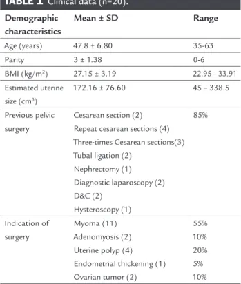
es-timated uterine size and surgical indication, are includ-ed in Table 1. Twelve patients (60%) had previous abdom-inal pelvic surgery.
TABLE 1 Clinical data (n=20).
Demographic characteristics
Mean ± SD Range
Age (years) 47.8 ± 6.80 35-63
Parity 3 ± 1.38 0-6
BMI (kg/m2) 27.15 ± 3.19 22.95 – 33.91
Estimated uterine size (cm3)
172.16 ± 76.60 45 – 338.5
Previous pelvic surgery
Cesarean section (2) Repeat cesarean sections (4) Three-times Cesarean sections(3) Tubal ligation (2)
Nephrectomy (1) Diagnostic laparoscopy (2) D&C (2)
Hysteroscopy (1)
85%
Indication of surgery
Myoma (11) Adenomyosis (2) Uterine polyp (4) Endometrial thickening (1) Ovarian tumor (2)
55% 10% 20% 5% 10% BMI: body mass index; SD: standard deviation.
The surgical time of all procedures is detailed in Table 2. Due to either the inability to keep the pneumoperitone-um or to the lack of suturing expertise, the vaginal vault closure was performed totally or partly through the va-gina in 14 (70%) patients.
Intraoperative bleeding measured in the vacuum as-pirations system right after the surgery ranged from 20 to 500mL (average 194mL), discounting any abdominal fluid infused. No patient had blood cell transfusion or any major intraoperative hemorrhagic complications.
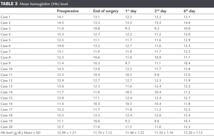
The decrease of mean hemoglobin level from the pre-operative measure to the end of surgery, 1st, 2nd and 6th
postoperative days was respectively 1.25, 1.51, 1.64 and 0.79g/dL (Table 3).
Postoperative reported pain was minimum and no anti-inflammatory or morphine-like drugs were neces-sary. Ordinary routine analgesics were used until the 6th
postoperative day.
The patients were discharged from the hospital on the second day after surgery and returned for evaluation on the 6th postoperative day.
DISCUSSION
As far as we know, this is the first published series of sin-gle port hysterectomy in Brazil. Adopting new techniques is always challenging in medicine, especially in the surgi-cal field. The limitation for triangulation is the main dif-ficulty to be overcome in single port laparoscopy proce-dures. The use of multifunctional instruments helps to reduce this problem. Our preference is to use a dissect-ing bipolar coagulator associated with an ultrasonic in-strument, for dissecting, cutting and coagulating; thus, decreasing the risk of accidents involving the exchange of instruments and reducing the surgical time. In addi-tion, we used a 30o 45cm endoscope for single port access
surgery. It allowed the camera coupler to be positioned 15cm behind the surgeon’s hands, preventing their col-liding with the camera. When it is available, a flexible op-tical system could be alternatively used, placed laterally to the surgeon’s hand, which would also decrease the risk of collision.10-12
In one patient with history of previous severe pelvic inflammatory disease (case 8), we initially tried to access the uterus using only the umbilical incision during 25 min-utes. Unfortunately, we were unable to identify the uterus due to extensive adhesions and we opted to insert two ad-ditional suprapubic 5mm-trocars to safely complete the hysterectomy. Maybe, more experienced teams could man-age this situation through the single port approach.
Although we observed a slight decrease in the hemo-globin level right after the procedure, it was clinically in-significant, and was probably related to the intravascular liquid infusion during the surgery. We observed an in-crease in hemoglobin level on the 6th postoperative day.
Blood transfusion was not necessary in this series of patients; however, in four patients the total amount of blood loss, measured in the vacuum aspirations system right after the surgery, was greater than 300mL. It seems to us that the blood loss increased proportionally to the uterine size. The lack of instrument triangulation reduc-es the accreduc-ess to the uterine blood supply, most signifi-cantly in large uteri. This could explain the greater loss of blood in these cases. Maybe the use of flexible optical systems could facilitate the visualization of all vascular structures in such patients.
nee-TABLE 2 Surgical “step-by-step” time.
Surgery time (min)
A B C D E F G H Total
Case 1 20 66 26 0 1 66 7 18 204
Case 2 10 58 29 17 0 31 5 17 167
Case 3 20 52 28 6 2 43 10 16 160
Case 4 21 86 16 7 3 30 1 24 188
Case 5 15 60 13 5 3 20 6 27 148
Case 6* 20 70 28 0 13 37 3 15 186
Case 7 10 63 29 5 2 29 2 10 150
Case 8 16 69 9 2 1 24 8 10 175
Case 9 37 72 18 4 1 16 6 9 164
Case 10 19 54 33 10 2 46 5 15 184
Case 11* 18 78 7 4 7 27 7 13 161
Case 12 10 42 13 7 2 20 1 16 111
Case 13 9 55 18 26 7 35 4 19 173
Case 14 13 68 31 9 2 12 13 16 164
Case 15 9 56 20 7 3 45 6 12 158
Case 16* 15 70 16 12 8 39 6 19 185
Case 17* 21 24 10 5 1 20 30 16 127
Case 18 17 71 24 10 4 40 9 25 200
Case 19* 11 67 16 9 5 27 8 11 154
Case 20* 28 52 15 4 3 35 9 19 165
Mean ± SD 16.95 ± 6.96 61.65 ± 13.53 19.95 ± 7.95 7.45 ± 5.94 3.55 ± 3.10 32.1 ± 12.59 6.8 ± 6.31 16.35 ± 4.97 166.2 ± 22.60
A: pneumoperitoneum installation, including umbilical incision and single port device insertion; B: hysterectomy until colpectomy; C: colpectomy; D: salpingectomy; E: specimens’ removal; F: va-ginal cuff closure; G: eventual bleeding control and, H: umbilical incision closure; SD: standard deviation. * Laparoscopic vava-ginal vault closure
TABLE 3 Mean hemoglobin (Hb) level.
Preoperative End of surgery 1st day 2nd day 6th day
Case 1 14.1 13.1 12.2 12.2 13.1
Case 2 14.3 13.2 13.5 13.5 14.0
Case 3 11.0 10.8 9.3 9.2 10.0
Case 4 13.3 12.7 12.2 11.2 13.0
Case 5 12.5 11.1 11.7 11.6 12.9
Case 6 14.0 13.2 12.7 11.6 13.3
Case 7 13.1 11.9 11.9 11.7 13.2
Case 8 12.5 10.6 11.0 10.9 11.1
Case 9 11.4 10.3 9.7 11.1 10.4
Case 10 14.3 13.0 12.5 11.7 13.8
Case 11 12.5 10.4 10.2 9.8 12.0
Case 12 13.9 12.7 12.7 12.5 11.9
Case 13 13.6 12.3 11.6 12.4 12.2
Case 14 11.7 11.0 10.5 10.4 11.2
Case 15 13.8 11.1 12.4 12.4 12.7
Case 16 11.4 10.5 10.5 10.4 11.8
Case 17 13.2 11.7 11.8 11.2 12.3
Case 18 15.3 13.5 12.4 12.6 12.4
Case 19 11.1 10.6 9.2 9.6 10.3
Case 20 12.7 11.0 11.5 11.0 12.3
dle and repeated application of the knot-pusher lacerat-ed the plastic part of the single port device in four pa-tients, increasing the gas leakage. Maybe the use of barbed sutures will be helpful to close the vaginal vault, facilitat-ing the intracorporeal runnfacilitat-ing suture technique.13
The average time for the initial setting of the proce-dure was 17 minutes, ranging from 8 to 37 minutes. One obese patient, with previous umbilical incision, suffered accidental bleeding during the dissection of the rectus
fascia, and it took 37 minutes to correctly place the sin-gle port device.
The time spent to free the uterus and to coagulate the blood supply was similar in all cases; however, colpec-tomy on average, took 23 minutes in the first 10 patients and 17 minutes in the last 10 surgeries.
Vaginal vault closure proved to be the most difficult part of the single port hysterectomy. Although intracorpo-real suturing was possible, it was still too time-consuming, increasing significantly the total operative time.10 We
ini-tially tried to close the vaginal vault laparoscopically in 18 patients, using the extracorporeal interrupted knot tech-nique. However we only finished the closure through this approach in 6 cases (30%). In two patients, we started the vaginal vault closure vaginally because we are unable to maintain the pneumoperitoneum after the uterus was re-moved. In 12 patients, we needed to combine laparoscopic and vaginal approaches to carry out the procedure. We spent more than 60 minutes to close the vaginal cuff in the first case, and it was necessary to combine both approaches. On the other hand, we managed to complete the procedure lap-aroscopically in 4 out of the last 5 patients, confirming a clear impact of the learning curve in this type of surgery.
Until now, it seems that the vaginal vault closure is faster when it is done vaginally than through the single port access. However, we believe that with additional ex-perience, the surgical time will be similar to laparoscop-ic multiport hysterectomy, as reported by others.7,10
Due to the multifactorial etiology of postoperative pain, involving neuropraxia of the phrenic nerves, the type of insufflated gas, residual pneumoperitoneum, opera-tive wound pain, direct tissue trauma from electrocoag-ulation and mechanical injury, and mainly due sociocul-tural and individual factors, pain evaluation is subjective and difficult.14,15
We used a VAS to access the intensity of postopera-tive pain, and the results showed a satisfactory evolution using plain analgesics solely. One patient only demand-ed additional analgesic mdemand-edication for neck pain, prob-ably because of inappropriate surgical positioning dur-ing a prolonged procedure.
We observed almost no residual gas in the abdomi-nal cavity after laparoscopic single port access surgeries. We attributed this to the presence of a 2.5cm umbilical
incision that facilitated complete gas extraction. Conse-quently, patients reported almost no shoulder pain. The mean maximum score of pain (a score of 4.5) was ob-served on the 1st postoperative day, probably due to the
peak of inflammatory response as previously demonstrat-ed.1 On the 2nd postoperative day, the mean pain score
re-duced 33%, to a score of 3, with a minimum variation un-til the 6th postoperative day.
Clinical postoperative parameters, including eating, walking and flatus release, would allow an early hospital discharge to all patients. However, due to hospital regu-lations related to the use of new techniques, all patients were discharged only on the second postoperative day.
The inclusion of robotic surgery may be an alterna-tive to reduce the limitations associated with single port access surgery. The capacity of articulation associated with robotic instruments might compensate for the lack of triangulation, showing an improved facility for the dis-section and suturing. Probably it would contribute to de-creasing the learning curve required to master these dif-ficult procedures.16-18
CONCLUSION
We observed that single port hysterectomies are feasible and safe in selected cases, with similar advantages to mul-tiport laparoscopic hysterectomy. Cosmetic benefits may be an advantage of this approach. Additional compara-tive studies with multiport hysterectomy are necessary to further evaluate the benefits of single port access surgery.
RESUMO
Histerectomia laparoscópica de incisão única: resultados preliminares
Objetivo: descrever os resultados iniciais da histerectomia laparoscópica realizada através de punção umbilical úni-ca, além de avaliar a praticabilidade e segurança dessa via de acesso cirúrgico.
ci-rurgia, perda sanguínea, complicações, morbidade febril, dor pós-operatória e tempo de permanência hospitalar. Resultados: a média de idade e índice de massa corpórea das pacientes foi de 47.8 anos e 27.15 kg/m2,
respectiva-mente. O tempo cirúrgico médio foi de 165.5 minutos. A perda sanguínea foi mínima, sem necessidade de transfu-são em nenhuma paciente. Todos os procedimentos foram realizados satisfatoriamente, apenas um caso necessitou de conversão cirúrgica para laparoscopia convencional (com 3 punções abdominais) por múltiplas aderências, po-rém sem necessidade de realização de laparotomia e não houveram complicações pós-cirúrgicas. Nenhuma pacien-te despacien-te estudo solicitou administração de medicação anal-gésica adicional no pós-operatório.
Conclusão: a histerectomia com acesso único umbili-cal é um procedimento factível e seguro, sem maiores complicações.
Palavras-chave: histerectomia, laparoscopia, cirurgia, útero, procedimentos cirúrgicos em ginecologia.
REFERENCES
1. Ribeiro SC, Ribeiro RM, Santos NC, Pinotti JA. A randomized study of total abdominal, vaginal and laparoscopic hysterectomy. Int J Gynecol Obstet. 2003; 83:37-43.
2. Choi YS, Shin KS, Choi J, Park JN, Oh YS, Rhee TE. Single-port access laparoscopy-assisted vaginal hysterectomy: our initial experiences with 100 cases. Minim Invasive Surg. 2012; 2012:543627.
3. Park YS. Current trends of gynecologic surgery in the 21st century: scarless surgery. Korean J Gynecol Endosc Minim Invasive Surg. 2010; 22:69-89. 4. Canes D, Desai MM, Aron M, Haber GP, Goel RK, Stein RJ, et al.
Transumbilical single-port surgery: evolution and current status. Eur Urol. 2008; 54:1020-9.
5. Reich H, DiCaprio J, Mc Glynn F. Laparaoscopic hysterec tomy. J Gynecol Surg. 1989; 5:213-6.
6. Pelosi MA, Pelosi MA 3rd. Laparoscopic hysterectomy with bilateral salpingo-oophorectomy using a single um bilical puncture. N J Med. 1991; 88:721-6. 7. Hart S, Yeung P Jr, Sobolewski CJ. Laparo-endoscopic single site hysterectomy
in gynecologic surgery. Surg Technol Int. 2010; 20:195-206.
8. Paek J, Kim SW, Lee SH, Lee M, Yim GW, Nam EJ, et al. Learning curve and surgical outcome for single-port access total laparoscopic hysterectomy in 100 consecutive cases. Gynecol Obstet Invest. 2011; 72:227-33.
9. Olive DL, Parker W, Cooper JM, Levine RL. The AAGL Classification system for laparoscopic hysterectomy. J Am Assoc Gynecol Laparosc. 2000; 7:9-15.
10. Park D, Kim J, Jun HS, Jeong H, Park Y. Laparoscopic vaginal vault closure with conventional straight instruments in single-port access total laparoscopic hysterectomy. Obstet Gynecol Sci. 2013; 56:389-99.
11. Fanfani F, Monterossi G, Fagotti A, Scambia G. Laparoendoscopic single-site hysterectomy: is it safe and feasible? Curr Opin Gynecol Obstet. 2014; 26:275-80. 12. Jackson T, Einarsson J. Single-port gynecologic surgery. Rev Obstet Gynecol.
2010; 3:133-9.
13. Song T, Lee SH. Barbed suture versus traditional suture in single-port total laparoscopic hysterectomy. J Minim Invasive Gynecol. 2014; 21:825-9. 14. Jung YW, Lee M, Yim GW, Lee SH, Paek JH, Kwon HY, et al. A randomized
prospective study of single-port and four-port approaches for hysterectomy in terms of postoperative pain. Surg Endosc. 2011; 25:2462-9.
15. Eom JM, Choi JS, Choi WJ, Kim YH, Lee JH. Does single-port laparoscopic surgery reduce postoperative pain in women with benign gynecologic disease? J Laparoendosc Adv Surg Tech A. 2013; 23:999-1005.
16. Vizza E, Corrado G, Mancini E, Baiocco E, Patrizi L, Fabrizi L, et al. Robotic single-site hysterectomy in low risk endometrial cancer: a pilot study. Ann Surg Oncol, 2013; 20:2759-64.