Profile and regulation of annexin II expression during early embryogenesis in cattle
[Perfil e regulação da expressão da anexina II durante a embriogênese em bovinos]
L.F.S. Costa, M.S.N Machado, J.F.C. Oliveira, J.C. Silva, R.S. Loguercio, P.B.D.Gonçalves*
Universidade Federal de Santa Maria Faixa de Camobi, Km 9 97105-900 – Santa Maria, RS
ABSTRACT
The presence of annexin II (Ann-II) during the initial stages of bovine embryo development and the regulation of Ann-II expression by retinol and insulin-like growth factor I (IGF-I) were studied. Bovine embryos at different stages of development were produced in vitro on Synthetic Oviductal Fluid (SOF) medium (control group), SOF supplemented with retinol (retinol group; 0.1ng/ml), or IGF-I (IGF-I group; 10ng/ml). The embryos were processed for mRNA extraction, cDNA production and polymerase chain reaction (PCR) using Ann-II-specific oligonucleotides. Ann-II was detected in all stages of early embryo development, except for the 16-cell stage. The blastocyst rates were significantly higher (P<0.05) in the group supplemented with retinol (37.8%, 45/119) during in vitro embryo culture (IVC) than in those cultured in SOF (20.5%, 24/117) or SOF with IGF-I (25.8%, 24/93). Semiquantitative analysis of Ann-II expression in embryos produced in medium supplemented with IGF-I or retinol revealed a lower expression of this gene when compared with embryos cultured in SOF (P<0.05). The Ann-II expression was not different in embryos cultured in the presence of retinol and IGF-I. The presence of retinol increased the production of embryos in vitro by decreasing the expression of Ann-II in early-stage of bovine embryo.
Keywords: cattle, annexin II, embryo development
RESUMO
Foram estudadas a presença de anexina II (Ann-II) durante a fase inicial do desenvolvimento embrionário bovino e sua regulação pelo retinol e pelo fator de crescimento semelhante à insulina (IGF-I). Embriões bovinos em diferentes estádios de desenvolvimento foram produzidos in vitro em fluido sintético de oviduto (SOF) sem suplementação (grupo-controle) ou suplementado com retinol (grupo retinol; 0,1ng/ml medium) ou IGF-I (grupo IGF-I; 10 ng/ml de meio). Os embriões foram processados para extração de mRNA, produção de cDNA e posterior análise por reação em cadeia da polimerase (PCR) com oligonucleotídeos específicos para Ann-II. Em todos os estádios de desenvolvimento embrionário, Ann-II foi detectada, exceto no estádio de 16 células. Os índices de blastocisto foram significativamente maiores (P<0,05) no grupo suplementado com retinol (37,8%, 45/119) durante o cultivo in vitro de embriões (PIV) que aqueles obtidos no grupo controle (20,5%, 24/117) ou no IGF-I (25,8%, 24/93). Análise semiquantitativa da expressão de Ann-II em embriões produzidos em meio suplementado com IGF-I ou retinol revelou uma menor expressão desse gene quando comparado com embriões cultivados somente em SOF (P<0,05). A expressão de Ann-II não foi diferente em embriões cultivados na presença de retinol e IGF-I. A presença de retinol aumentou a produção de embriões in vitro, e diminuiu a expressão de Ann-II em estádios iniciais do desenvolvimento embrionário bovino.
Palavras-chave: bovino, anexina II, desenvolvimento embrionário
INTRODUCTION
In vitro production of bovine embryos has been one of the most widely used biotechnologies in the last decade and represents one of the main tools for the multiplication of genetic material. Rates of oocyte maturation, cleavage and embryo development up to the blastocyst stage have been reported to be approximately 92, 77 and 40%, respectively (Greve et al., 1993; Carolan et al., 1995; Castilho et al., 2007), and continue at these levels, with wide variation being observed due to the use of substances of unknown composition such as estrous cow serum (ECS) or fetal bovine serum (FBS). In addition, pregnancy outcomes are still below the rates obtained with the traditional embryo transfer system. The pregnancy rates obtained with grade 1 embryos produced in vitro and fresh (30-60%) or frozen (5-40%) transferred are lower than those obtained with embryos produced in vivo (fresh 50-80% or frozen 40-70%, Hasler, 1996). In cattle, the embryonic genome is activated during the fourth cycle of cell division, and a large number of both in vivo and in vitro embryos stop their development during this stage. Probably, certain cell division-related genes are involved in this 8-16-cell block.
The regulation of cell cycle progression is a complex process involving kinase cascades and the action of proteases and second messengers, among other factors. Evidence indicates that the intracellular calcium concentration plays a crucial role in these events. Transport-related and calcium-dependent proteins may be important components in the division process and in the control of embryo cleavage block. Modifications in calcium concentrations occur throughout all stages of the cell cycle and are observed together with the resumption of meiosis and in the end of oocyte maturation. Although the mechanism whereby the spermatozoon triggers the activation of the oocyte is still a matter of controversy (Fissore and Vande Woude, 1996), there is no doubt that the onset of embryogenesis depends on oscillations in intracellular calcium ion concentration (Swann and Ozil, 1994; Kline, 1996). A large number of proteins has been indicated as mediators of the Ca2+ effect during the cell cycle, for example the S-100 proteins which regulate the activity of p53, a tumor suppressor that modulates the expression of genes fundamental to cell cycle progression.
Another Ca2+-related protein is annexin II (Ann-II) which, together with Myc Basic Motif Homolog-1 (Mbh1), is responsible for the binding of DNA sequences to replisomes and is considered to be important during DNA replication. Ann-II is a strong candidate to play a role in initial cell division in embryos since this protein has been shown to be present during the process of oocyte cytoplasmic and molecular maturation (unpublished data). However, no data regarding the expression of Ann-II during early embryo development are available.
During embryo development, growth factors produced especially by the endometrium during the preimplantation stage (Paria et al., 2001) play a fundamental role in the regulation of embryo metabolism and exert a mitogenic effect (Simmen and Simmen, 1993). Insulin-like growth factor I (IGF-I) is a low molecular weight growth factor consisting of a single polypeptide chain, with a structure and function similar to insulin (Singh and Armstrong, 1997). The use of a culture medium containing high concentrations of IGF-I in combination with ECS and granulosa cells has been shown to improve the development of bovine embryos in vitro (Palma et al., 1997). In humans, the expression of IGF-I genes is an indicator of embryo quality (Liu and Leroith, 1999).
genes important in cell division and in the control of 8-16-cell block.
The aim of the present study was to investigate the expression of Ann-II during the first stages of embryo development and the regulation of this expression in blastocysts by IGF-I and retinol.
MATERIAL AND METHODS
Bovine ovaries were collected in an abattoir and transported in an isothermic container in 0.9% NaCl for transport. In the laboratory, the ovaries were washed three times in the same solution. Follicles measuring 2 to 8mm in diameter were immediately punctured with a vacuum pump connected to an 18G needle and follicular fluid was collected into a 10-ml test tube. Only cumulus–oocyte complexes (COC), classified as grade I, according to the International Embryo Transfer Society, which are characterized by a compact and complete cumulus and homogenous cytoplasm containing thin granulations, were used.
The oocytes were maintained in follicular fluid until the time for selection to prevent resumption of meiotic division. After selection, 25 COCs in 200µl maturation medium were randomly distributed on Petri dishes (60mm) under silicone oil. The maturation medium consisted of TCM-199 containing Earle salts and L-glutamine supplemented with 25mM HEPES, 100IU/ml penicillin, 50µg/ml streptomycin, 0.2mM sodium pyruvate, 26.19mM sodium bicarbonate, 10% FBS, 0.5µg/ml FSH, and 5.0µg/ml LH. The plates were incubated at 39oC in a 5% CO2
atmosphere with saturated air humidity for 24 hours. A pool of frozen semen of a single lot derived from sires of European breeds was used throughout the experiment. Straws of semen were thawed and washed by centrifugation through a Percoll gradient (45%/90% [v/v]) at 900 x g for 30min. The COC were fertilized by the addition of spermatozoa to the drops at a final concentration of 2x10⁶ spermatozoa/ml. Heparin (10µg/ml) was added for sperm capacitation, and the plates were incubated for 20 hours under the same conditions as described for oocyte maturation. After this period, cumulus cells were removed from presumptive zygotes by mechanical shaking and transferred to embryo development medium. In the first stage of the experiment, presumptive zygotes were cultured
in 200-µl drops of synthetic oviduct fluid (SOF) supplemented with 10% FBS in a 5% O2/5%
CO2 atmosphere at 39oC for different periods of
time according to the stage when development was interrupted, i.e., 2, 4, 8, 16, and 32 cells, morula, blastocyst or expanded blastocyst, with at least 10 embryos per stage in four replicates. In the second part of the experiment, after removal of the cumulus cells, presumptive zygotes were randomly divided into three groups: SOF supplemented with 10ng/ml IGF-I (n=93), SOF supplemented with 0.1ng/ml retinol (n=119), and SOF medium only (control group; n=117), and incubated for six days under the same conditions.
RNA was extracted from the embryos using the RNeasy Mini kit. Briefly, after successive washes in PBS, the embryos were transferred to a 1.5-ml centrifuge tube containing 350µl RLT buffer plus 3.5µl ß-mercaptoethanol and mechanically shaken for 15s, and 350µl 70% ethanol was added. After homogenization, the content of the tube was transferred to an RNA extraction mini-column and centrifuged for 15s. Next, 700µl RW1 buffer were added and the column was centrifuged for 15s, followed by two washes with 500µl RPE buffer (REP supplemented with 100% ethanol, 1:4) and by a last centrifugation for 2min for total drying of the column. The column was then transferred to a new tube, 40µl ultrapure water was added, and the tube was centrifuged for 1min for dilution of the RNA. All centrifugations were performed at 8000g.
SensiscriptTM Reverse Transcriptase kit1 was used to isolate cDNA. Each RT-PCR mixture contained 2.0µl buffer, 2.0µl dNTP, 2.0µl oligoDT-primer, 0.25µl Rnase inhibitor, 1.0µl Sensiscript enzyme, and 12.5µl of the RNA extraction product. The reaction was carried out in a thermocycler at 37oC for 1 hour and the reaction product was stored at -18oC until the time for using.
The primers for Ann-II were synthesized by Invitrogen Life Technologies. The constitutive GAPDH gene was used as control in the RNA extraction reaction. The following primer oligonucleotide sequences were used: forward5’ CCTCCAAGTGCATACGGG 3’ and reward 5’
TCATACTGAGCAGGTGTTTT 3’ for Ann-II,
and forward 5’TGTTCCAGTATGATTCCACCC3’ and
reward 5’ TCCACCACCCTGTTGCTGTA 3’ for GAPDH. The PCR mixture contained 17.5µl ultrapure water, 1.5mM MgCl2, 50mM KCl,
10mM Tris-HCl, pH 8.3, 250µM of each dNTP, 0.4µM of each primer, 2.5U Taq DNA polymerase, and 3µl cDNA, in a final volume of 25µl. Amplification was performed in a thermocycler under the following conditions: 94oC for 5min, 94oC for 1 min, 60oC for 1 min for the Ann-II primers or 54oC for 45s for the GAPDH primers, and 72oC for 1 min. Steps 2-4 were repeated according to the exponential phase, with 34 cycles for Ann-II and 31 cycles for GAPDH, followed by a final extension at 72ºC for 5min. The relative amount of transcript was compared between groups based on the amplification results comparing the constitutive gene (GAPDH) and the Ann-II gene.
The PCR product was visualized on 2% agarose gel in TBE (0.5 M EDTA, pH 8.0, 0.09 M Tris, 0.09 M boric acid) stained with ethidium bromide under ultraviolet light. The data for semiquantitative analysis of gene expression were obtained by capturing the gel images with a digital camera and they recorded with the Alpha
DigiDoc 1000 program to obtain arbitrary values.
Only samples, in which the constitutive GAPDH gene was detected, indicating that mRNA extraction and cDNA isolation were effective, were used in all analyses. The semiquantitative gene expression results were analyzed with one way ANOVA. The data regarding percentage of embryo production were analyzed with the PROC CATMOD program of the SAS statistical software (System…, 2005) and treatments were compared by contrast. Gene expression in embryos at different stages of development was only described regarding the presence or absence of expression, and no statistical analysis was performed.
RESULTS
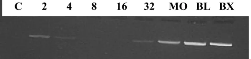
The expression of the Ann-II gene in embryos at different stages of development is shown in Fig. 1. Ann-II was detected in the 2-, 4- and 8-cell stages. No expression of Ann-II was observed in 16-cell embryos, while the transcription of this gene was reinitiated in the 32-cell stage. Ann-II was also detected in the morula, blastocyst and expanded blastocyst stages.
Figure 1. Representative gel showing the expression of the Ann-II gene in embryos at different stages of development. C: control; 2, 4, 8, 16, and 32: 2, 4, 8, 16, and 32 cells, respectively; MO: morula; BL: blastocyst; BX: expanded blastocyst.
The presence of retinol during embryo development led to a significant increase (P<0.05) in blastocyst production determined on day 6 after fertilization compared to the control and IGF-I groups. In the control group, the percentage of blastocysts in relation to the total number of cultured oocytes was 20.5% (24/117), which did not differ from that obtained for the IGF-I group (25.8%, 24/93), whereas a higher blastocyst rate (37.8%, 45/119; P<0.05) was observed in the retinol group (Fig. 2).
b
a
a
0
10
20
30
40
50
control
IGF-I
Retinol
%
o
f
B
la
st
o
cy
st
Figure 2. Percentage of embryos obtained in relation to the total number of cultured oocytes. The embryos were cultured in SOF medium (control) or SOF supplemented with 10ng IGF-I/ml or 0.1ng retinol/ml. The data represent the results of 4 replicates and are reported as mean and standard error of the mean. Different letters indicate a significant difference (P<0.05).
b
b
a
0
0,5
1
1,5
2
2,5
control
IGF-I
Retinol
An
n
I
I/
G
AP
DH
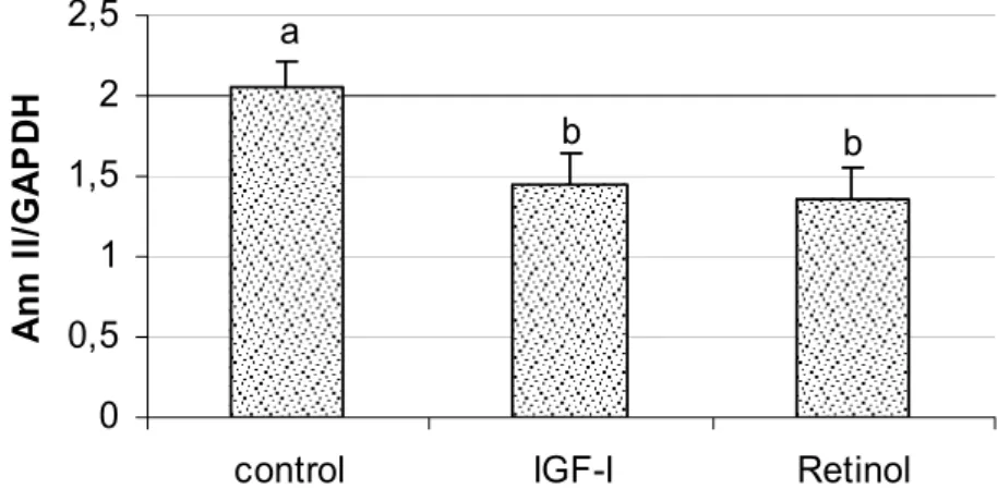
Figure 3. Semiquantitative expression of Ann-II in bovine embryos. The embryos were cultured in SOF medium (control) or SOF supplemented with 10ng IGF-I/ml or 0.1ng retinol/ml. The data are reported as the mean and standard error of the mean of the Ann-II/GAPDH ratio. Different letters indicate a significant difference (P<0.05).
DISCUSSION
This is the first report of the expression of Ann-II in bovine embryos. Data regarding the expression of this gene in animals of commercial interest are very scarce, with studies only reporting the presence of this protein in amphibian embryos (Izant and Brysons, 1991), and of a sequence homologous to a protein associated with Ann-II in fish (Bobe and Goetz,
was still observed even during the block stage (eight cells).
The present study demonstrated the beneficial effect of supplementation of the embryo development medium with retinol, in agreement with previous studies in which the use of retinol promoted a significant increase of blastocyst production in KSOM medium on an oviduct epithelial cell monolayer in an atmosphere of 5% CO2 (Montagner, 1999). The present results
were obtained using SOF medium without oviduct cells in a different atmosphere (5% CO2,
5 % O2, 90% N2). Retinol has antioxidant
activity, minimizing cell membrane lipid peroxidation (Dobreanu and Mody, 1997), which damage the embryonic cells. In addition, retinol acts by preventing apoptosis, thus altering the cascades of events of this process (Koshimizu et al., 1995).
The experiment was performed from April to June in a low-rainfall year with low forage availability. It is possible that the blastocyst production rates were influenced by the nutritional status of the oocyte donor animals and the retinol supplementation minimized these effects. Unfortunately, there was no measurement of pasture availability during this trial that could support this assertion. The use of retinol to improve in vitro embryo production rates seems to be increasingly accepted, with studies demonstrating that retinol is important for embryo development, especially in seasons in which wide variability in the results are observed (Montagner, 1999; Lima et al., 2004).
Semiquantitative analysis of Ann-II expression in blastocysts produced in SOF medium (control) or SOF supplemented with 10ng/ml IGF-I or 0.1ng/ml retinol (IGF-I and retinol groups, respectively) revealed that the expression of this gene was reduced when retinol or IGF-I was present. Ann-II was detected during all initial stages of embryo development. However, the positive results obtained with retinol seem to be related to the modulation of Ann-II expression. Ann-II seems to play a negative role during this stage since embryo production increased qualitatively (data not shown) and quantitatively when the development medium was supplemented with IGF-I or retinol, respectively.
There are few studies about the role of Ann-II on oocyte maturation and embryo development. Ann-II is present during oogenesis and embryonic development in amphibians (Izant and Brysons, 1991) and fishes (Bobe and Goetz, 2000). These findings suggest that there are transcriptional active of Ann-II during oocyte development and at early stages of development in cattle. Ann-II is a member of the annexin family of Ca(2+)- and lipid-binding proteins, which can form a tight heterotetrameric complex with the cellular protein ligand p11, a member of the S100 protein family. In ovarian tissue, the expression of two members of the S-100 protein family (S-100A1 and S-100A10) was high 24 hours before and after the germinal vesicle breakdown (GVBD) while was low after ovulation, suggesting an important role during the periovulatory period in fishes (Bobe and Goetz, 2000). It was observed that Ane-II mRNA of maternal origin is present in bovine embryo until the transition from maternal to embryonic genome control (eight cells). This suggest that Ann-II has an important role in the resumption and progression of meiosis and may be involved in the modulation of intracellular Ca(2+) concentration, during the maturation-promoting factor (MPF) degradation (Kline, 1996). Chelation of intracellular Ca2+ results in the blockage of spontaneous GVBD (Homa, 1995). This hypothesis is based on the fact that Ane-II is calcium channel-associated protein present in oocyte (Dreier et al., 1998).
In conclusion, Ann-II is present in the 2-, 4-, and 8-cell stages but not in 16-cell embryos and is expressed from 32-cell to expanded blastocyst stages, suggesting to be involved in oocyte maturation and embryo development. The presence of retinol increases the production of embryos in vitro by decreasing the expression of Ann-II in early-stage of embryo development in cattle.
References
BOBE, J.; GOETZ, F.W. A S100 homologue mRNA isolated by differential display PCR is down-regulated in the brook trout (Salvelinus fontinalis) postovulatory ovary. Gene, v.257, p.87-194, 2000.
CASTILHO, C.; ASSIS, G.S.; GARCIA, J.M. Influência do diâmetro e da fase folicular sobre a competência in vitro de oócitos obtidos de novilhas da raça Nelore. Arq. Bras. Med. Vet. Zootec., v.59, p.288-294, 2007.
CUI, J.; MICHAILLE, J.; JIANG, W. et al. Retinoid receptors and vitamin A deficiency: differential patterns of transcription during early avian development and the rapid induction of RARs by retinoic acid. Dev. Biol., v.260, p.496-511, 2003.
DOBREANU, M.; MODY, E. Influence of natural antioxidants on in vitro lipoprotein oxidation. Rom. J. Int. Med., v.35, p.55-62, 1997.
DREIER, R.; SCHMID, K.; GERKE, V. et al. Differential expression of annexins I, II and IV in human tissues: An immunohistochemical study. Histochem. Cell. Biol., v.110, p.137-148, 1998.
DUESTER, G.; MIC, F.A.; MOLOTKOV, A. Cytosolic retinoid dehydrogenases govern ubiquitous metabolism of retinol to retinaldehyde followed by tissue-specific metabolism to retinoic acid. Chem. Biol. Inter., v.143, p.201-210, 2003.
FISSORE, R.A.; VANDE WOUDE, G.F. Potential role of mitogen-activated protein kinase during meiosis resumption in bovine oocytes. Biol. Reprod., v.55, p.1261-1270, 1996. GREVE, T.; MADISON, V.; AVERY, B. et al. In vitro
production of bovine embryos: A progress report and the consequences on the genetic upgrading of cattle populations.
Anim. Reprod. Sci., v.33, p.51-69, 1993.
HASLER, J.F. Commercial production of in vitro-derived bovine embryos. Arq. Fac. Vet. UFRGS, v.24, supl., p.117-134, 1996.
HOMA, S. Calcium and meiotic maturation of the mammalian oocyte. Mol. Reprod. Dev., v.40, p.122-134, 1995.
IZANT, J.G.; BRYSONS, L.J. Xenopus annexin II (calpactin I) heavy chain has a distinct amino terminus. J. Biol. Chem.,
v.266, p.18560-18566, 1991.
KAISER, M.E.; MERRILL, R.A.; STEIN, A.C. et al. Vitamin A deficiency in the late gastrula stage rat embryo results in a one to two vertebral anteriorization that extends throughout the axial skeleton. Dev. Biol., v.257, p.14–29, 2003.
KLINE, D. Activation of the mouse egg. Theriogenology, v.45, p.81-90, 1996.
KOSHIMIZU, U.; WATANABE, M.; NAKATSUJI, N. Retinoic acid is a potent growth activator of mouse primordial germ cells in vitro. Dev. Biol., v.168, p.683-685, 1995.
LIMA, P.F.; OLIVEIRA, M.; GONCALVES, P.B.D. et al. Effects of retinol on the in vitro development of Bos Indicus
embryos to blastocysts in two different culture systems.
Reproduction in Domestic Animals. v.39, p.356-360, 2004.
LIU, H.K.; DORÉ, E.J.J.; ROBERTS, P.M. et al. Expression and cellular localization of retinol-binding protein messenger ribonucleic acid in bovine blastocysts and extraembryonic membranes. Biol. Reprod., v.49, p.393-400, 1993.
LIU, J-L.; LEROITH, D. Insulin-like growth factor I is essential for postnatal body growth in response to growth hormone. Endocrinology, v.140, p.5178-5184, 1999.
MONTAGNER, M.M. Produção in vitro de embriões bovinos com meios congelados, hepes e retinol. 1999. 68f. Dissertação (Mestrado) Universidade Federal de Santa Maria, Santa Maria, RS.
PALMA, G.; MÜLLER, M.; BREM, G. Effect of insulin-like growth factor I (IGF-I) at high concentrations on blastocyst development of bovine embryos produced in vitro.J. Reprod. Fertil., v.110, p.347-353, 1997.
PARIA, B.C.; MA, W.; TAN, J. et al. Cellular and molecular responses of the uterus to embryo implantation can be elicited by locally applied growth factors. Proceed. Nat. Acad. Sci. USA, v.98, p.1047-1052, 2001.
SHAW, W.D.; FARIN, P.W.; WASHBURN, S.P. Effect of retinol palmitate on ovulation rate and embryo quality in superovulated cattle. Theriogenology, v.44, p.51-58, 1995.
SIMMEN, R.C.; SIMMEN, F.A. Insulin-like growth factors and blastocyst development. Theriogenology, v.39, p.163-175, 1993.
SINGH, B.; ARMSTRONG, D.T. Insulin-like growth factor-I, a component of serum that enables porcine cumulus cells to expand in response to follicle-stimulating hormone in vitro.
Biol. Reprod., v.56, p.1370-1375, 1997.
STALLINGS-MANN, L.M.; TROUT, E.W.; ROBERTS, M.R. Porcine uterine retinol-binding proteins are identical gene products to the serum retinol-binding protein. Biol. Reprod., v.48, p.998-1005, 1993.
SWANN, K.; OZIL, J.P. Dynamics of the calcium signal that triggers mammalian egg activation. Int. Rev. Cytol., v.152, p.183-222, 1994.
SYSTEM for Microsoft Windows. Release 8.02 TS Level. Disponível em: <http://v8doc.sas.com/sashtml>. Acessado em 21 jun. 2005.
TELFORD, N.A.; WATSON, A.J.; SCHULTZ, G.A. Transition from maternal to zygotic control in early embryonic development: a comparison of several species.
Mol. Reprod. Dev., v.26, p.90-100, 1990.