• Nenhum resultado encontrado

The impact of carbon source in Candida albicans virulence: participation of RLM1 in pathogen host interaction

N/A
N/A
Protected

Academic year: 2020

Share "The impact of carbon source in Candida albicans virulence: participation of RLM1 in pathogen host interaction"

Copied!
109
0
0

Texto

(1)

Tese de Mestrado

Mestrado em Genética Molecular

Trabalho realizado sob a orientação da

P

rofessora Doutora Paula Sampaio

João Miguel Oliveira Pacheco

The impact of carbon source

in Candida albicans virulence:

participation of RLM1 in

pathogen host interaction

e da

(2)

Nome: João Miguel Oliveira Pacheco

Endereço electrónico: jmopacheco@gmail.com Telefone: 925829900

Número do Bilhete de Identidade: 14044233

Título tese: The impact of carbon source in Candida albicans virulence: participation of RLM1 in pathogen host interaction

Orientador(es): Professora Doutora Paula Sampaio e Professora Doutora Célia Pais

Ano de conclusão: 2015 Designação do Mestrado:

Mestrado em Genética Molecular

É AUTORIZADA A REPRODUÇÃO PARCIAL DESTA TESE/TRABALHO, APENAS PARA EFEITOS DE INVESTIGAÇÃO, MEDIANTE DECLARAÇÃO ESCRITA DO INTERESSADO, QUE A TAL SE COMPROMETE;

Universidade do Minho, ___/___/______

(3)

Agradecimentos

Esta secção é dedicada a todas as pessoas que me acompanharam e contribuíram para o sucesso de mais uma etapa da minha vida. A todos desde já o meu MUITO OBRIGADO.

Manifesto de maneira muito especial o meu agradecimento à Professora Doutora Paula Sampaio por me ter acolhido de braços abertos no seu grupo, por toda a dedicação, apoio, determinação, estimulo constante, criticas, sugestões, por ter sempre a palavra certa sempre no momento certo! Pela disponibilidade plena que dispôs para transmitir os seus conhecimentos. Obrigado por tudo Professora, e pela sua presença constante!

À Professora Doutora Célia Pais pela orientação neste trabalho mas sobretudo, por todo o conhecimento, por todo o apoio e todas as sugestões que foram verdadeiramente importantes ao longo deste percurso.

Aos meninos do laboratório, “mana” Ju por ter sempre uma palavra amiga e por me ajudar a tornar-me sempre melhor no laboratório e ter sempre o conselho certo a dar. À Catarina Carneiro, pela ajuda e disponibilidade sempre que era requerida. À Andrea pela incansável disponibilidade para ajudar sempre que era preciso. À Tânia embora tenha tido pouquinho tempo no laboratório mostrou-se sempre disponível para discutir ciência e sempre pronta para ajudar. À Dani pela enorme disponibilidade que teve sempre para me ajudar quando as coisas não corriam bem, e seu contributo foi muito importante para o arranque deste projeto. Ao Ricardo pela ajuda no RT-PCR e pela partilha da sua experiência e sabedoria. Senti-me numa verdadeira família, a nossa família micro II, obrigado meninos!

Aos técnicos do Departamento de Biologia, em especial o Sr. Luís por ser incansável e sempre preocupado em arranjar-me “mais balão”. Muito obrigado Sr. Luís!

À Catarina Vaz um agradecimento muito especial, por tudo aquilo que representa para mim e por estar sempre presente mesmo a uns quilómetros de distância. As palavras são sempre parcas para te agradecer! Obrigado por tudo!

À Joana Cunha pelos momentos de descompressão durante todo este processo, e por ter sempre uma palavra amiga, por estar sempre presente e por todos os sorrisos!

À Ana Xavier que mesmo distante está sempre presente e muitos conselhos importante me têm dado, pela sua amizade e por todos os bons momentos que me proporcionou.

À Samy que nesta etapa final foi muito importante pelo apoio nesta fase árdua que é escrever uma tese e pela luta das formatações! “Hoje foi um belo dia para terminar uma tese”.

(4)

Aos restantes elementos da Happy Family, Mika, Joaninha, Raquel, Nuno, que me acompanharam nesta etapa e que me proporcionaram grandes momentos, e muitos mais estarão para vir amigos! O meu muito obrigado.

Aos marmanjos de lá de casa, Luís Ferraz, Eloy e João Carlos por me fazerem sentir sempre em casa e em família! Vocês são os maiores! Obrigado meus caros!

Aos meu coleguinhas do mestrado que estiveram sempre presentes, sem querer nomear ninguém vocês sabem quem são. Obrigado por toda a vossa amizade e preocupação! “MGM estamos juntos”

Não querendo sendo injusto, mas aproveito agora para fazer um agradecimento mais geral aos meus amigos de bioquímica, não sendo menos importantes, quero vos agradecer pela vossa amizades por se mostrarem sempre disponíveis. Vocês sabem quem são!

À Universidade do Minho, ao Departamento de Biologia e principalmente aos seus professores por todos os conhecimentos facultados.

(5)

Abstract

The survival of all microbes depends upon their ability to respond to environmental

challenges. To establish infection, pathogens such as Candida albicans must support effective

stress responses to counter host defenses while adapting to dynamic changes in nutrient status

within host niches. Studies of C. albicans stress adaptation have generally been performed on

glucose-grown cells, leaving the effects of alternative carbon sources upon stress resistance largely unexplored as well alterations in its virulence.

In a previous work both copies of the RLM1 gene of C. albicans were deleted and phenotypic analysis of ∆rlm1/∆rlm1 mutants showed typical cell wall weakening phenotypes, such as hypersensitivity to Congo Red, Calcofluor White and caspofungin, showing its involvement in the

cell wall remodelling. To understand the role of RLM1 under the influence of different carbon

source, phenotypic characterization against cell wall damaging stress agents, modulation of virulence factors as well as their involvement in host-pathogen interaction was preformed in the present work.

Candida albicans ∆rlm1/∆rlm1 mutant displayed phenotypes associated to cell wall deficiency such as, hypersensitivity to Congo red, caspofungin in glucose- and lactate- grown cells. However the RLM1 mutants were slight more resistant to Congo Red when grown in lactate. On the other hand lactate-grown cells were not able to growth al all in the presence of SDS and presented sensitivity to caffeine, in comparison with glucose-grown cells. The increased transcription of genes already reported to be involved in cell adhesion correlated well with adhesion and biofilm assays, in

which RLM1 mutant presented greater biofilm formation than WT in both growth condition.

However cell adapted to lactate adhered more and biofilm formation was more pronounced. The host-pathogen interaction was accessed by co-incubation with murine macrophages-like cell line (J774). In general, lactate-grown cells were less efficiently killed in comparison to glucose-grown cells. However the mutant presented distinct behaviors, they were more resistant when adapted to glucose than to lactate. The TNF-α and IL-10 were lower in response to Δrlm1/Δrlm1 mutant and the cellular toxicity, measured as extracellular lactate dehydrogenase activity, was significantly lower in comparison with the WT and complemented strains in glucose-grown cells. The effect observed before was reverted when C. albicans cells were grown on lactate.

In conclusion, C. albicans cells adapted to different carbon sources behave differently,

particularly in the interaction with macrophages, in which the RLM1 mutation plays a decisive effect since it affects the cell wall integrity.

(6)
(7)

Resumo

Os microrganismos para sobreviver dependem da capacidade de responder aos desafios apresentados pelo ambiente onde estão inseridos. O sucesso da infecção por parte dos agentes

patogénicos, tais como Candida albicans, assenta na capacidade de responder eficazmente a

stresses causados pelas defesas do hospedeiro e à adaptação a alterações constantes dos nutrientes no hospedeiro. Alguns estudos indicam que o crescimento em fontes de carbono

alternativas tem influência na resistência ao stress e alteram a virulência de C. albicans.

Em trabalhos anteriores, o gene RLM1 de C. albicans foi deletado e a análise fenotípica dos

mutantes Δrlm1/Δrlm1 revelou que estes apresentam hipersensibilidade ao Congo Red, Calcofluor white e Caspofungina, fenótipos típicos de fragilidade na parede celular. De forma a

perceber o papel de RLM1 em células adaptadas a diferentes fontes de carbono, foi feita uma

caracterização fenotípica usando agentes que perturbam a parede celular, avaliada a modulação de alguns fatores de virulência, bem como a interação hospedeiro-patogéno.

Os mutantes Δrlm1/Δrlm1 de Candida albicans crescidos em glucose e láctico exibiram fenótipos associados a deficiências na parede celular, tais como, hipersensibilidade ao Congo Red

e Caspofungina. No entanto, estas revelaram ser ligeiramente mais resistentes ao Congo Red

quando crescidas em láctico. Por outro lado, as células crescidas em ácido láctico apresentaram maior dificuldade em crescer na presença de cafeína, não tendo sido detectado crescimento em SDS, contrariamente às células crescidas em glucose que não apresentaram sensibilidade a estes stresses. O aumento da expressão de genes envolvidos na adesão célular correlacionou-se diretamente com os ensaios de adesão e biofilme, em que os mutantes Δrlm1/Δrlm1 apresentam uma maior formação de biofilme em comparação com a estirpe selvagem em ambas condições de crescimento. Contudo, células crescidas em ácido láctico aderiram mais e a formação de biofilme foi mais evidente. A interação Candida-hospedeiro foi avaliada através da co-incubação com a linha celular de macrófagos J774. Em geral, as células crescidas em ácido láctico foram fagocitadas de forma menos eficiente em comparação com células crescidas em glucose. As estirpes mutante

Δrlm1/Δrlm1 apresentaram comportamentos distintos quando crescidas em diferentes fontes de

carbono; em glucose os níveis de produção de TNF-α e IL-10 foram mais baixos e a toxicidade celular, avaliada pela atividade da lactato desidrogenase, foi significativamente mais baixa, em comparação com as estirpes selvagem e complementadas. O efeito anteriormente observado foi totalmente revertido quando o mutante foi crescido em meio com ácido láctico.

Em conclusão, células de C. albicans a diferentes fontes de carbono apresentam

comportamento diferentes, nomeadamente na interação com macrófagos, onde o gene RLM1

(8)
(9)

Table of contents

Agradecimentos ... iii

Abstract ... v

Resumo ... vii

Table of contents ... ix

Abbreviations and Acronyms list... xi

List of Figures ... xv

1. Background ... 9

1.1. Epidemiology of invasive fungal infection ... 3

1.1.1. Candida albicans, the major model yeast of pathogenicity ... 5

1.1.1.1 Virulence factors of C. albicans ... 5

1.1.2. Host response to fungal infections ... 10

1.1.2.1. The role of innate immune ... 11

1.1.2.2. The role of adaptive immune system ... 13

1.1.3. Cell wall, first barrier or interface for host immune cells ... 16

1.1.4. The RLM1 gene importance ... 18

1.2. Metabolism as a platform for Candida albicans pathogenicity ... 19

1.2.1. Integration of Carbon Metabolism with Virulence ... 22

1.2.1.1. Carbon adaptation modulates stress resistance ... 23

1.2.1.2. Carbon adaptation triggers dynamic changes in the C. albicans cell wall ... 24

1.2.1.3. Metabolic adaptation influences virulence factors ... 26

1.2.1.4. Carbon adaptation modulates immune surveillance ... 27

1.3. Objectives and outline of the thesis ... 30

2. Methods and Materials ... 33

2.1. Carbon source impacts in C. albicans virulence factors Immune Recognition ... 33

2.1.1. Strains and growth conditions ... 33

2.1.2. Quantification of metabolites produced during C. albicans growth ... 33

2.1.3. Susceptibility assays ... 34

2.1.4. Filamentation tests ... 34

2.1.5. Adhesion assay and biofilm formation ... 34

2.1.6. Secreted Aspartyl Proteinases production ... 35

(10)

2.2. Carbon source impacts in C. albicans Immune Recognition ... 38

2.2.1. Macrophage cell cultures ... 38

2.2.2. Macrophage-killing assay ... 38

2.2.3. Determination of lactate dehydrogenase activity and cytokines measurement ... 39

2.2.4. Statistical analyses ... 40

3. Results and Discussion ... 41

3.1. Identification of some metabolites during C. albicans ... 44

3.2. Impact of carbon source in RLM1 mutant response to cell wall stress agents ... 45

3.3. Filamentation tests ... 48

3.4. Adhesion and biofilm formation... 51

3.5. Secreted Aspartyl Proteinases production (SAPs) ... 54

3.6. Influence of carbon source RML1-responsive genes ... 55

3.7. Host-pathogen interaction: Participation of RLM1 in Macrophage-Candida albicans interaction under the influence of different carbon source ... 58

4. Concluding remarks ... 63

5. Perspectives for future work ... 69

(11)

Abbreviations and Acronyms list

A

APCs Antigen Presenting Cells

B

BP Base Pairs

BSA Bovine Serum Albumin

BSIs Bloodstream Infections

C

cDNA Complementary Deoxyribonucleic Acid

CFG Caspofungine

CFU Colny Forming Unit

CFW Calcofluor White

CLRs C-Type Lectin Receptors

COX2 Cyclooxygenase 2

CR Congo Red

CV Crystal Violet

CWI Cell Wall Integrity

CWP Cell Wall Protein

D

DMEM Dulbecco’s Modified Eagle’s Medium E

ELISA Enzyme Linked Immune Sorbent Assay

F

FBS Fetal Bovine Serum

FCM Flow Citometry

(12)

FSC Forward Scatter

G

GDP Glyceraldehyde-3-Phosphate Dehydrogenase

GFP Green Fluorescent Protein

GI Gastrointestinal

GPI Glycosyl Phosphatidyl Inositol

H

HEPES 4-(2-Hydroxyethyl)-1-Piperazineethanesulfonic Acid

HIV Human Immunodeficiency Virus

HSCT Hematopoietic Stem Cell Transplants

I

IFN-γ Gamma Interferon

IgG Immunogloblin G IL Interleucine L LDH Lactate Dehydrogenase LIP Lipases M

MADS Mcm1, Argamous-Deficiens-Serum Response Factor

MAPK Mitogen-Activated Protein Kinase

MHC Major Histocompatibility Complex

N

NLR Nucleotide Leucine-Rich

O

OD Optical Density

(13)

ORF Open Reading Frame

P

PAMPs Pathogen Associated Molecular Patterns

PBS Phosphate Buffered Saline

PCR Polymerase Chain Reaction

PI Propidium iodide

PKC Cell Integrity Or Protein Kinase C

PLs Phospholipases

PRRs Pattern Recognition Receptors

Q

qRT-PCR Real-Time Reverse-Transcription Polymerase Chain Reaction R

RNA Ribonucleic Acid

ROS Reactive Oxygen Species

S

SAP Secreted Aspartyl Proteinases

SBF Swi4-Swi6 Cell Cycle Box Binding Factor

SDS Sodium Dodecyl Sulfate

SEM Scanning Electron Microscope

SSC Side Scatter

T

TCR T-Cell Receptor

TGF-β Transforming Growth Factor Beta

Th T-Helper

TLRs Toll-Like Receptors

(14)

U

USA United States of America

W

WO White Opaque

WT Wild Type

Y

YCB Yeast Carbon Base

YE Yeast Extract

YNB Bacto Yeast Nitrogen Base Without Amino Acids

(15)

List of Figures

Figure 1. Distinctive morphology of C. albicans. Representation of the three different

morphologies that C. albicans can acquired. ... 7

Figure 2. The major pattern-recognition receptors (PRRs) involved in the recognition of specific C. albicans pathogen-associated molecular patterns (PAMPs). ... 12

Figure 3. Recognition of C. albicans blastospores and hyphae by monocytes and macrophages receptors. ... 13

Figure 4. Modulation of T-cell responses by innate immune system. ... 14

Figure 5. The structure of the Candida albicans cell wall. The schematic shows the major components of the cell wall and their distributions. ... 16

Figure 6. Main elements of the MAPK signal transduction network in C. albicans. ... 18

Figure 7. Central carbon metabolism in Candida albicans cells occupying different host niches. 22 Figure 8. The impact of different carbon sources in cell wall architecture. ... 25

Figure 9. Nutrients influence the expression of key virulence factors in Candida albicans. ... 26

Figure 10. Changes in carbon source impact on immune surveillance. ... 28

Figure 11. Growth curve of C. albicans. ... 43

Figure 12. Identification by HPLC of metabolites during C. albicans growth on different carbon sources. ... 44

Figure 13. Sensitivity of C.albicans strains against agents that affect cell integrity. ... 46

Figure 14. Filamentation of C. albicans glucose-grown cells. ... 49

Figure 15. Filamentation of C. albicans on lactate as carbon source. ... 50

Figure 16. In vitro adhesion and biofilm formation.. ... 51

Figure 17. In vitro ability of wild-type, mutant, and complemented strains to adhere to polystyrene surface. ... 53

Figure 18. Secreted aspartyl Proteinases production of C. albicans in different carbon sources.. ... 54

(16)

Figure 20. Yeast Killing. Killing of C. albicans SC5314, SCRLM1M4A, SRLM1M4B, SCRLM1K2A and SCRLM1K2B cells grown on glucose, lactate by macrophages.. ... 58

Figure 21. Host vaibility and immune response to Candida cells grown on glucose or on lactate. ... 59

(17)

Background

Epidemiology of invasive fungal infection

Candida albicans, the major model yeast of pathogenicity Virulence factors of C. albicans Host response to fungal infections Cell wall, first barrier or interface for host immune cells The RLM1 gene importance

Metabolism as a platform for Candida albicans pathogenicity

Integration of Carbon Metabolism with Virulence

(18)
(19)

1. Background

1.1. Epidemiology of invasive fungal infection

Over the last decades, invasive fungal infections, particularly fungal nosocomial infections (FNIs), have assumed a great importance. This phenomenon is essentially due to the increasing population at risk. People with human immunodeficiency virus (HIV) infection, recipients of solid organ or hematopoietic stem cell transplants (HSCT), patients with hematologic malignancies, burns or indwelling medical devices and low-birth weight infants are the most susceptible set of population to these kind of infections (Warnock 2007; Sabino et al. 2010). In the United States of America (USA), from 5231 cases of sepsis in 1979 to 16042 cases in 2000 (207% raze) were caused by fungal infections as reported in the retrospective study of Martin and colleagues (Martin et al., 2003). The advances of medicine, surgery and transplantology is thus one of the major causes of the dramatic increase in the number of immunocompromised individuals who are more susceptible to FNIs, including aspergillosis, candidiasis, cryptococcosis, and zygomycosis (Warnock 2007; Sabino et al. 2010). Currently, many of these patients die not because of their underlying illness but as a result of a deep-seated fungal infection (Tortorano et al. 2004).

Since these infections are among the main cause of morbidity and mortality, leading to an increase in the hospitalization time and, consequently, high costs associated to patients’ treatment, the FNIs are indeed a serious public health problem (Lass-Florl 2009; Hota 2004; Gudlaugsson et al. 2003).

Fungal infections can either be superficial, affecting the skin, hair, nails and mucosal membranes, or systemic, involving major body organs (Ruping et al. 2008). Besides, these kind of infections have been reported as great threat in the intensive care units (ICUs), where the incidence is much superior than in the general population of hospitalized patients (Bassetti et al. 2006; Cheng et al. 2005; Vaz et al. 2011).

In addition to factors related to the hospital unit and underlying disease involving the patient, factors related to the microorganism are of major importance to the progression of hospital acquired infections. In specific situations, such as under immunosuppression, microorganisms of the normal microbiot can be responsible for most of the nosocomial infections (Hota 2004; Eggimann et al. 2003).

A significant cause of infection entails several species of Candida. More than 17 different

species of Candida have been identified as etiologic agents of bloodstream infections (BSIs).

(20)

albicans, Candida glabrata, Candida parapsilosis, and Candida tropicalis (Hajjeh et al. 2004;

Ostrosky-Zeichner et al. 2003; Pfaller et al. 2004). Although C. albicans remains the most

important cause of invasive candidiasis, the non-albicans Candida (NAC) species are gaining clinical importance once they constitute about 50% of the reported cases of candidemia (Krcmery and Barnes 2002).

In Europe, fungal pathogens represent the major eukaryotic agents of serious infection, in which Candida albicans and other emergent Candida species have become the third most frequent cause of nosocomial blood stream infections (Perlroth et al. 2007; Pannanusorn et al. 2013). These yeasts may reside in the host as benign commensals however, they may take advantage of a locally or systemically debilitated immune system to proliferate and cause disease. Moreover, new

extracorporeal reservoirs colonized by Candida species have been identified in recent years, as

natural environments, inanimate surfaces, health professionals’ hands and hospital’s surface. This fact supports and clarifies some recent evidences of the epidemiological cycle of hospital infections

caused by Candida species of exogenous origin (Vaz et al. 2011). In fact, some studies have

showed that the hands of health professionals may be colonized by yeasts and also serve as reservoirs of nosocomial infections (NIs) (Vaz et al. 2011; Ferreira et al. 2013; Sabino et al. 2010). The use of catheters and other medical devices is also related to development of nosocomial

infections (NIs) with Candida species via exogenous routes, because the high prevalence of

Candida isolates in samples from catheter tips may have an important role in the spread, progression and persistence of NIs (Ferreira et al. 2013; Traore et al. 2002; Passos et al. 2005).

However, the Candida species are not related exclusively to hospital candidemia, because

hematogenous infections also occur in the community (Ferreira et al. 2013).

In Portugal there are few studies regarding the epidemiology of candidiasis, but in 2010, a work developed by Sabino et al (Sabino et al. 2010) studied the incidence of candidemia in a Portuguese oncology hospital during 6 years. In this study, it was demonstrated that the most

frequently species found were C. albicans followed by C. parapsilosis, C. tropicalis, C. krusei and

C. glabrata. Furthermore, it was assessed that the mortality rate associated of candidemia was 31,9% (Sabino et al. 2010). Given their increasing frequency and high morbidity and mortality rates, prevention of invasive fungal infections has become of paramount importance. Therefore, host-fungus interaction studies may contribute to understand the epidemiology and pathogenesis of these infections and help develop new strategies to fight fungal infections.

(21)

1.1.1. Candida albicans, the major model yeast of pathogenicity

There are known approximately 200 species known in the genus Candida but only a few

species have been identified as pathogenic. Macroscopically, colonies of Candida spp. are cream-colored to yellowish. All species produces blastoconidia and many of them are dimorphic growing as budding yeast cells, pseudohyphae or true hyphae (Eggimann et al. 2003).

Since the dawn of civilization the growth of the opportunistic fungal pathogen Candida

albicans on the surface of human tissue was noted and described by Hippocrates in the fourth century B.C. (Kumamoto and Vinces 2005).

Candida albicans is an ubiquitous and dimorphic fungus that exists as a commensal in warm-blooded animals, including humans. It colonizes mucosal surfaces of the oral and vaginal cavities and the digestive tract. However, if the balance of the normal flora is disrupted or the immune defenses are compromised, C. albicans can switch from commensal to pathogenic, being capable of infecting a variety of tissues and causing fatal systemic diseases (Sampaio et al. 2010).

The diploid C. albicans has eight pairs of homologous chromosomes, with a genome size of

14.3 Mb, based on genome sequencing of SC5314 strain. The content of GC in C. albicans

genome is about 33.5%, 6438 genes, an average gene size of 1,468 bp and an intergenic average

size of 858 bp (Magee 1993; Butler et al. 2009). Another distinctive feature of C. albicans is an

alteration in its genetic code in which the codon CUG encodes serine rather than leucine (Santos and Tuite 1995). It shares this property with a group of related species that are collectively known as the CUG clade (Butler et al. 2009). This clade includes other pathogenic yeast such as C. paropsilosis and C. tropicalis. This characteristic increases dramatically the number of different proteins encoded by the 6438 C. albicans genes and results in an extensive phenotypic variability considered as among others, an attribute of virulence (Gomes et al. 2007).

1.1.1.1 Virulence factors of C. albicans

Virulence can be defined as the capacity of some microorganism cause disease, and is highly regulated by many factors. In order to establish a variety of infections, pathogens have to evade the immune system, survive, divide in the host environment and spread to new tissues (Yang 2003). Thus, virulence describes the degree of pathogenicity, where some strains can be more or less

virulent than the others. C. albicans can survive in several anatomically distinct sites and the

putative success of conversion from an innocuous commensal organism to an opportunistic pathogen depends on several virulence factors (Jayatilake 2011; Calderone 2004; Gow et al.

(22)

2012). These attribute of virulence include adherence to host cells, morphogenesis, biofilm formation, phenotypic switching and secretion of degradative enzymes (Calderone and Fonzi 2001; Schaller et al. 2005; Chauhan et al. 2006; Lim et al. 2012).

1.1.1.1.1 Adhesion

The adhesion ability of Candida cells to the host cells or to medical-devices is an important

trait to begin the cycle from colonization to the establishment of the infection. The level of adhesion is dependent on host, microbial and abiotic surface proprieties, such as cell wall composition or cell-surface hydrophobicity (Silva et al. 2011).

In Candida species, adhesion is mediated by some well characterized adhesion factors such as the proteins Hwp1, Eap1, all members of the Als family, and others (Calderone and Fonzi 2001). Several of these are glycosylphosphatidylinositol (GPI)-proteins (linked to cell wall glucan) or GPI-anchored proteins (linked to the cell membrane), which are exposed at the cell surface (Gow and Hube 2012b). Mutants lacking these adhesion genes display decreased adherence to host substrates in vitro as well as a corresponding virulence reduction in several in vivo experimental models of candidiasis (Sheppard et al. 2004).

As previously referred, one of the most well-studied adhesins are the agglutinin-like sequence

(Als) proteins family, encoded by eight ALS genes (ALS1-7 and ALS 9) (Yang 2003). Some

differences in N-terminal domain among distinct Als proteins leads to differences in their function. In early stages of infection, Als1p has been reported to mediate binding to human vascular endothelial cells and epithelial cells, while Als5p is responsible for collagen, fibronectin, bovine serum albumin and laminin adhesion (Sheppard et al. 2004).

C. albicans can express all eight ALS genes, but C. parapsilosis and C. tropicalis express only five and three ALS genes, respectively (Silva et al. 2011).

(23)

1.1.1.1.2. Morphogenesis

Another striking feature of C. albicans is its ability to grow as an unicellular budding yeast

(blastopores) and as a filamentous form (hyphae or pseudohyphae) (Sudbery 2011). This ability, represented in Figure 1, is tightly regulated by signal transduction pathways in response to environmental stimuli such as pH, temperature, or different compounds such as N-acetylglucosamine or proline. Perhaps the most critical criterion for pathogenicity is the induction of the mycelial form by serum and during the interaction with macrophage cells (Jacobsen et al. 2012).

Figure 1. Distinctive morphology of C. albicans. Representation of the three different morphologies that C. albicans can acquired. Differential interference contrast (DIC) images of cells with hyphae,

pseudohyphae, and yeast morphologies adapted from (Kim and Sudbery 2011).

Some studies suggest that budding yeast cells are suitable for dissemination while hyphae are essential for adaptation to different host environment conditions and also for tissue and organ invasion (Mavor et al. 2005; Lim et al. 2012).

The ability of hyphae formation is observed in some species of Candida such as C. albicans, C. parapsilosis or C. tropicalis and is considered to be crucial for evasion of host defenses (Lim et

al. 2012). C. glabrata is commonly known and described as unable to form hyphae and

pseudohyphae but still is able to induce infection, indication that other virulence factors may take the lead (Odds et al. 1997; Lachke et al. 2002; Csank and Haynes 2000).

(24)

1.1.1.1.3. Extracellular hydrolytic enzymes

C. albicans as well as other Candida species can secret hydrolytic enzymes. This feature is considered a virulence factor and required during Candida infections. Secreted aspartyl proteinases (Sap), phospholipases and lipases are the most important extracellular enzymes secreted by Candida species (Mavor et al. 2005; Jayatilake 2011). These enzymes are needed to degrade host cell surface molecules to invade host cells by digesting its membranes, to resist to host immunity by attacking immune system cells and molecules, such as heavy chains of IgG, C3 protein, keratin, collagen, albumin, fibronectin, hemoglobin among others, and for nutrient acquisition (Yang 2003; Trofa et al. 2008).

The secreted aspartyl proteinases family is composed by 10 SAP genes that encode the Sap proteins. Some studies carried out by Schaller and co-workers (Schaller et al. 2001) demonstrated that SAPs are differentially expressed in yeast and hyphal form and in phenotypically switched states. For example, SAP1-3 is mainly expressed on cell wall and cytoplasm of blastopores, SAP4-6 is restricted at the tips of hyphae and phenotypically switched cells express SAP1 and SAP3. Furthermore, SAP8 is mostly detected in yeast cells grown at 25ºC and SAP9 is preferentially expressed in later growth phases (Yang 2003). In this sense, the flexibility of SAP genes expression may be contribute to the success of Candida species as an opportunistic pathogen, by allowing the fungus to survive and to cause infections on different host tissues and different growth conditions (Naglik et al. 2003).

Other hydrolytic enzymes responsible for conferring virulence to Candida species are

phospholipases (PLs). These enzymes hydrolyze phospholipids to fatty acids and glycerol. The classification of these enzymes is based on the different ester bonds cleaved being classified as PLs A, B, C and D. However, it has been demonstrated that only proteins encoded by the phospholipase B family (PLB1-5) seem to be extracellular, particularly PLB1 that plays a key role in virulence in animal models of candidiasis (Calderone and Fonzi 2001; Silva et al. 2011). The expression of this enzymes family during infection could lead to host cell membrane impairment and Candida cells adhesion.

Several studies demonstrated that PLs are differentially expressed. For example the PLs can

be expressed at the tips of Candida hyphae and on the initial buds of C. albicans cells during

invasion (Samaranayake et al. 2005; Jayatilake et al. 2005; Ghannoum 2000). These studies confirm PLs involvement in pathogenesis by enhancing tissue penetration. Other studies showed

(25)

that C. tropicalis and C. parapsilosis are also capable of producing extracellular PLs, although in much lower levels than C. albicans. PL activity was not so clear for C. glabrata and C. krusei.

Lipases are involved in the hydrolysis and synthesis of triacylglycerols. Ten LIP genes

(LIP1-10) encode these enzymes, and studies confirmed that they are differentially expressed at different

stages and sites of infection (Trofa et al. 2008). Only in C. albicans and C. tropicalis all ten LIP

genes (LIP1-10) were detected. For C. parapsilosis, only two lipase genes, LIP1-2, have been reported (Trofa et al. 2008) and no studies regarding LIP genes expression in C. glabrata and C. krusei are available. Gácser and co-workers demonstrated that lipases are important for Candida virulence, by showing that with lipase inhibitors the tissue damage observed was significantly reduced during infection in reconstituted human tissue models (Gacser et al. 2007).

1.1.1.1.4. Phenotype switching

Candida species can undergo a phenomenon called phenotypic switching (Slutsky et al. 1985). There are a number of different classes of switching; one of the most well-characterized is white-opaque switching (Slutsky et al. 1987). The most common forms of C. albicans colonies are smooth, white, and dome-shaped, these colonies are called “white”. When a suspension of white phase cells are plated out, about 1 in 1,000 colonies will have a different appearance being flatten and having a grey color. Within these new colonies, cells are larger and have an oblong rather than opaque appearance. Scanning electron micrographs show that the surface of these cells is covered in pimples. These cells are said to be in the “opaque” phase. Opaque cells will switch back to white again with a frequency of about 1:1,000 and this switching process is believed to be under epigenetic control (Kim and Sudbery 2011).

Strain WO-1 of C. albicans is the most studied phenotypic switching to white-opaque phases. Studies involving ultra-structural observations of WO-1 white-opaque phenotypes have showed differences in cell shape, cell surface structures and growth at 37ºC, suggesting that phenotypic switching could affect the virulence behavior of the organisms (Kim and Sudbery 2011).

This ability is also observed in C. glabrata, C. parapsilosis and C. tropicalis. Laffey and

co-workers (Laffey and Butler 2005) identified four main phenotypes in C. parapsilosis colonies,

including the concentric, crepe, smooth and crater phases and confirmed their relation with

biofilms formation. Also, they identified four phenotypes in C. glabrata (white, dark brown, very

(26)

possible phenotypes in C. tropicalis (smooth, rough, ring, semi-smooth) (Franca et al. 2011; Lachke et al. 2002). Phenotypic switching in C. krusei has not been reported yet.

1.1.1.1.6. Biofilm formation

Biofilms are surface-associated communities of cells surrounded by an extracellular matrix

and constitute an increasing problem in the clinical setting (Blankenship and Mitchell 2006). C.

albicans can form biofilms on natural surfaces, such as teeth, and on foreign surfaces, such as implanted devices. Due to the resistance of these biofilms to the common antimicrobial therapy, treatment often requires removal of infected devices to avoid potentially fatal consequences (Nobile and Mitchell 2006; Blankenship and Mitchell 2006; Kojic and Darouiche 2004; Douglas 2003).

In an initial stage, yeast cells adhere to the solid surface. Then, they undergo morphogenesis to produce a dense layer of cells of mixed morphology embedded in an extracellular matrix rich in secreted cell wall polymer ß-1,3 glucan. In vivo, it was shown that the biofilm can protects cells against host defenses (Nobile et al. 2009).

Some of the genes involved in biofilm formation were identified, as being also required for hyphal formation, such as the transcription factor Efg1 and the kinase Yak1. Others are apparently specific to biofilm formation, the zinc finger transcription factor Zap1, that is thought to regulate the production of matrix polysaccharide components and the transcription factor Bcr1, that regulates the formation of adhesens that allow yeast cells to stick to the solid surface (Nobile et al. 2009).

During disease establishment and progression, these virulence factors are assumed to be required in differing extents (Brown and Gow 1999; Huang 2012). Overall, the interaction between C. albicans virulence factors and host defense mechanisms plays a crucial role in determining whether colonization remains harmless or leads to infection of the epithelium and ultimately to systemic (Calderone and Fonzi 2001; Chauhan et al. 2006; Lim et al. 2012; Schaller et al. 2005).

1.1.2. Host response to fungal infections

The human host immune system is sophisticated and efficient being responsible for controlling and eliminating infectious microorganisms. The first line of defense for pathogenic

agents like C. albicans is formed immediately by the innate immune system. However, the

(27)

1.1.2.1. The role of innate immune

The host innate immune system is responsible for a constant and repetitive response that is specific, highly complex, and usually also highly efficient. After pathogen recognition, the immune system is activated and generates effector functions in order to eliminate the infectious microbe, which normally is well succeeded. Providing the first line of immune defense, the innate immune system is both central and critical for the complete host immune response. The innate immune defense affords 3 major effector mechanisms, (1) the complement system, (2) antimicrobial peptides, and (3) immune cells that recognize and respond to foreign microbes. Therefore, in order to survive or invade an immunocompetent host, any pathogen needs to cross the multiple borders of innate immunity (Zipfel et al. 2007).

The cellular mechanism will be further detailed in this thesis.

1.1.2.1.1. A cellular-dependent response: Macrophages and Dendritic cells

Monocytes, macrophages and dendritic cells are the cell types most commonly related with the innate immune response against C. albicans infection. Phagocytic cells have multiple receptors that recognize C. albicans cells.

Phagocytosis is induced and regulated by the pattern recognition receptors (PRRs) on the surface of antigen presenting cells (APCs) that recognize pathogen associated molecular patterns (PAMPs) (Hoebe et al. 2004; Akira and Takeda 2004; Akira et al. 2001). The recognition of PAMPs leads to microbial uptake and, consecutively, to a modulation of gene expression to initiate an adaptive and target-oriented immune response (Gauglitz et al. 2012). So far, three major groups of PRRs have been distinguished: toll-like receptors (TLRs), C-type lectin receptors (CLRs) and nucleotide binding domain, leucine-rich repeat containing (NLR) protein family as shown in Figure 2 (Gauglitz et al. 2012).

(28)

Figure 2. The major pattern-recognition receptors (PRRs) involved in the recognition of specific C. albicans pathogen-associated molecular patterns (PAMPs). Stimulation of host response by C. albicans at the cell membrane is mediated by toll-like receptors (TLRs) and C-type lectin receptors (CLRs) adapted from (Jouault et al. 2003).

The mannose receptor and dendritic cell-specific intercellular adhesion molecule 3-grabbing nonintegrin homolog (SIGN)-related 1 (SIGNR1) are two examples of these receptors (Cambi et al. 2003; Marodi et al. 1991; Filler 2006). Dectin-1 is another known receptor for C. albicans (Brown et al. 2003). This receptor can be found on macrophages and dendritic cells, and recognizes β-glucans, a component of the C. albicans cell wall. Even though, not being specific for macrophages

binding to blastospores, this receptor is required for an optimal phagocytosis of C. albicans

blastospores. Remarkably, when C. albicans cells produce hyphae, they do not express β-glucan

on their surface and so binding to dectin-1 is limited. This evidence suggests that the β-glucans masking on the surface of hyphae may help preventing recognition (Filler 2006).

Incubating macrophages with C. albicans blastospores, results in the blastospores

phagocytosis and secretion of pro-inflammatory mediators. C. albicans blastospores binding to

dectin-1 on macrophages results in the secretion of TNF-α and arachidonic acid, and in increased expression of COX2 (cyclooxygenase 2) (Brown et al. 2003; Suram et al. 2006), which result in a

pro-inflammatory response. Interestingly, phagocyte response to C. albicans hyphae may be

different than to blastospores and a complete anti-inflammatory outcome may be induced (Figure 3). Moreover, members within one family of PRRs can trigger opposite signaling effects, indicating that the ultimate outcome of a pathogen-induced immune response depends on the pathogen signature and the PRRs involved (Gauglitz et al. 2012).

(29)

As depicted in Figure 3, each ligand–receptor system activates specific intracellular signaling pathways that lead to modulation of several components of the host immune response (Jouault et al. 2003).

Figure 3. Recognition of C. albicans blastospores and hyphae by monocytes and macrophages receptors.

Reactive oxygen species (ROS) are produced by macrophages in response to blastospores phagocytosis in order to kill pathogens. However, under most in vitro experimental conditions, human macrophages have limited ability to kill C. albicans, even though they avidly phagocyte the microorganism. Newman et al. (Newman et al. 2005) discovered that one reason for this incomplete killing ability is that living C. albicans blastospores inhibit the phagolysosomal fusion in macrophages. The exact mechanism for this inhibition has not yet been determined. Interestingly, if macrophages are adherent to a collagen matrix the phagolysosomal fusion is not inhibited,

suggesting that the exposed extracellular matrix proteins have an import role on C. albicans

immune evasive strategies (Newman et al. 2005).

1.1.2.2. The role of adaptive immune system

Additionally, fungal cells induce the adaptive immune response which involves antigen-specific T and B cells (Zipfel et al. 2011).

Three signals are crucial for effective stimulation of an adaptive immune response: (1) the presentation of antigens by APCs on the context of MHC molecules/T-cell receptor (TCR) on CD4+

(30)

T-cell, (2) the up-regulation of co-stimulatory molecules, and (3) production of cytokines that determine differentiation of a certain type of helper T-cell (Th) response. Also, it is important to refer that the complete integrated response to a specific pathogen depends on the mosaic of PRRs and receptor complexes that are involved (Jouault et al. 2003; Netea and Marodi 2010). Co-stimulation via multiple PAMPs–PRRs interactions may increase sensitivity and specificity of the immune recognition process as well as the type of adaptive immune response, as demonstrated in Figure 4 (Jouault et al. 2003).

PRRs activation induces production of a cytokine environment that will induce an adaptive immune response directed towards a Th1, Th2 or Th17 phenotype (Kapsenberg 2003; Napolitani et al. 2005; Kubin et al. 1994).

Figure 4. Modulation of T-cell responses by innate immune system. Engagement of pathogen

associated molecular patterns (PAMPs) from Candida albicans by pattern recognition receptors (PRRs) lead to the

induction of specific cytokine profiles regulating the differentiation of naïve CD4+ T-cells in to Th1 helper 1), Th2 (T-helper 2), Th17 (IL-17 secreting T-(T-helper) or Treg (T-regulatory) cells. For activating naïve CD4+ T-cells, presentation of antigens on MHC class II to T-cell receptor (TCR) is necessary in combination.

(31)

1.1.2.2.1. Th1, Th2 a central paradox and the identification of a new subset of T cells: Th17

After cell engulfment fungal antigens are processed by dendritic cells and transported to lymph nodes and presented to T helper (Th) and regulatory T cells (Brown 2011) that ultimately will induce the correct adaptive response.

In 1995, studies in mucosal candidiasis using the mouse model of gastric Candida infection

showed that both, IFN-γ and IL-5 producing CD4+ T cells found in Peyer’s Patches and mesenteric lymph nodes, were correlated to Th1 and Th2, respectively. Additionally, neutralization of IL-4 resulted in amplified IFN-γ production and boosted yeast clearance (Cenci et al. 1995; Hernandez-Santos and Gaffen 2012).

Other studies in oropharyngeal candidiasis (OPC) mouse model demonstrated that T cell-deficient mice were susceptible to infection, but could be protected by transferring CD4+ T cells (Farah et al. 2002). Cytokines related to Th1 cells, including IFN- and TNF- , but not Th2 cytokines in the oral tissue, consistent with a Th1-based response. These results indicated that Th1 cells confer protective antifungal immunity, even though hallmark cytokines of both lineages, Th1 and Th2, were produced during infection (Hernandez-Santos and Gaffen 2012; Cenci et al. 1995; Cenci et al. 1998). However, depletion of CD8 or CD4 T cells in a susceptible mouse strain did not

intensify oral colonization with Candida in the oropharyngeal candidiasis (OPC) mouse model

(Ashman et al. 2003; Hernandez-Santos and Gaffen 2012), Therefore, T cells were involved in immunity to OPC, even though the specific subset and cytokines were still not well elucidated (Hernandez-Santos and Gaffen 2012).

In 1986 the Th1/Th2 paradigm was first described, and immune responses, whether infectious or autoimmune, were considered into these categories for approximately two decades. Over time, it became clear that this model was fraught with discrepancies.

A central paradox was that the signature Th1 effector cytokine, IFN-γ, was significantly less important in various disease settings than was IL-12, the key Th1 inductive cytokine (Steinman 2007). A significant renovation in the prevailing paradigm of CD4-mediated immunity occurred with the identification of Th17 cells, which settled many of these discrepancies. Subsequent studies showed that Th17 cells arise from inductive signals from TGF-β, IL-6, and IL-1b, while IL-23 is an essential maintenance and pathogenic factor for Th17 cells (Stockinger and Veldhoen 2007; McGeachy and Cua 2007).

(32)

Moreover, understanding the facts that regulate whether Th-response adopts a predominantly a Th1, Th2 or a Th17 type is crucial to prevent fungal infections (Chaplin 2006).

Knowing how pathogen and its host interact with each other is a crucial step for future fungal infections therapies.

1.1.3. Cell wall, first barrier or interface for host immune cells

The frontline of the pathogen–host interaction or contact is the fungal cell wall. Cell walls of fungal pathogens have an enormous importance in several biological processes. Being a dynamic structure, fungal cell wall is of great importance once (1) it protects from hostile environments, (2) it confers physical rigidity to maintain cell shape but also control morphogenesis during different stages of the fungus development, (3) it is responsible for the host immune recognition and for immune-avoidance, and (4) it maintains cellular integrity essential for cell growth (Kapteyn et al. 2000; Gow and Hube 2012a).

The fungal cell wall composition differs from each fungal species. In C. albicans example, two major layers constitute its cell wall, a skeletal inner layer (glucan and chitin) and a fibrillar outer

layer (mannoproteins) (Bowman and Free 2006; Klis et al. 2001). The percentage of each C.

albicans cell wall component is around 1–5 % chitin, 60–65 % glucan, and 35–40 % mannoproteins (per dry wall weight) (Klis et al. 2001; Munro et al. 1998) (Figure 5).

Figure 5. The structure of the Candida albicans cell wall. The schematic shows the major components

of the cell wall and their distributions. β-(1,3)-glucan and chitin (poly-β-(1,4)-N-acetylglucosamine) are the main structural components, and these are located towards the inside of the cell wall. The outer layer is enriched with cell wall proteins (CWP) that are attached to this skeleton mainly via glycosylphosphatidylinositol remnants to β-(1,6)-glucan or, for mannoproteins with internal repeat domains (Pir-CWP), via alkali-sensitive linkages to β-(1,3)-β-(1,6)-glucan. The insets show the structure of the glucan and mannan components. Adapted from (Netea et al. 2008)

(33)

Hexose sugars such as glucose, mannose and galactose are used by C. albicans as preferential carbon source to produce energy and to synthesize the main components of cell wall the polysaccharides glucans, mannan and chitin (Lee and Munro 2014).

About a third of the total cellular biomass is constituted by cell wall, but fungal cells have to extremely coordinate the production of new cell wall material to allow cell growth. Several studies have demonstrated the influence of niche-specific metabolic regulation on cell wall remodeling in C. albicans, which implicates alterations in host-pathogen interactions and significant sensitivity to antifungal agents (Ene and Brown 2014; Ene et al. 2012a; Brock 2009).

According to Walker and colleagues, during Candida systemic infections in a rabbit model,

transcriptional profiling shown alterations in the expression of genes associated with alternative carbon source metabolism, glucose assimilation, sugar transporters and cell wall surface remodeling (Walker et al. 2009). Genes associated with gluconeogenesis and the glyoxylate cycles

are up-regulated when C. albicans and C. glabrata are exposed to human blood or neutrophils

(Hube 2006; Fukuda et al. 2013; Lee and Munro 2014). Other studies suggest that starvation is relevant in vivo, for example, fluorescent reporter constructions have shown that the metabolic status of individual C. albicans cells within kidney lesions from a murine infection model can differ with some cells undergoing gluconeogenesis and others glycolysis (Barelle et al. 2006).

But still, the cell wall composition can be adaptably changed under stress conditions such as in response to certain environments or even when exposure to anti-fungal drugs. Several genes that involved in cell wall biosynthesis, remodeling and regulation have been shown to be vital for cell growth viability and pathogenicity (Lee and Munro 2014; Delgado-Silva et al. 2014).

(34)

1.1.4. The RLM1 gene importance

The cell wall is a dynamic structure since it changes with alterations of the surrounding growth conditions and is remodeled as the cell increases in size and during morphogenetic processes such as mating, sporulation, or pseudohyphal growth. Upon cell wall damage, cells activate the cell wall integrity (CWI) mitogen-activated protein (MAP) kinase pathway (also known as the PKC pathway) so that the cell wall is repaired and cell integrity maintained. This response involves several processes (Figure 6).

Figure 6. Main elements of the MAPK signal transduction network in C. albicans. Genes are

represented in normal characters where deletion and/or phenotypic characterization of the corresponding strain has been carried out in C. albicans or, alternatively, in italic, where their position and role are just presumed based on the

S. cerevisiae model and/or inspection of the genome. The existence of additional putative elements that may play a role in the activation of the corresponding MAPK is shown as question marks. The stimuli leading to the activation/repression of the pathways are shown either as stimulating (R) or inhibitory (--|) arrows. It is important to note that the arrows do not establish whether the interaction/crosstalk takes place through a direct or an indirect mechanism. The role of each MAPK in the physiological response is highlighted, but it must be taken into account that the interaction between pathways extends their indicated roles and generates a proper balanced response in the fungal cell. Adapted from (Monge et al. 2006)

The targets of the CWI pathway activation are the transcription factor heterodimer complex

SBF (composed by Swi4 and Swi6), and the MADS-box transcription factor RLM1 that activates

(35)

Ovalle 1998). Sampaio et. al showed that C. albicans RLM1 presents a high allelic diversity, with many variants of the encoded proteins found in surveys of clinical isolates and that isolates with different RLM1 allelic combinations presented different phenotypic characteristics (Sampaio et al.

2009). These studies indicated that RLM1 is required for in vitro viability of C. albicans under

caspofungin, and that inactivating this gene renders a described virulent strain completely avirulent (Delgado-Silva et al. 2014). Furthermore, an increase of cell adhesion binding proteins involved in biofilm formation was observed in C. albicans cells lacking RLM1 gene (Delgado-Silva et al. 2014). The higher expression of adhesion proteins in the mutant strain raised a particular interest on the

protein profile of C. albicans wild-type strain in comparison with the mutant that lacks this RLM1

gene (Delgado-Silva et al. 2014).

1.2. Metabolism as a platform for

Candida albicans

pathogenicity

A few years ago, Brown and Gow suggested that a mechanistically link between the regulation of metabolism and virulence in C. albicans could exist (Brown and Gow 1999; Brown et al. 2000). It was thought that in response to the new microenvironments it encounters during disease establishment and progression, these links might allow this pathogen to adjust its metabolism in parallel with its set of virulence factors (Nowrousian 2014).

A fundamental aspect of infection is the ability of the pathogenic microorganism to assimilate and utilize essential nutrients from the host-niches. For instance, C. albicans that normally thrives as a relatively innocuous commensal organism within diverse niches like the skin, the oral cavity, and the gastrointestinal (GI) and urogenital tracts of most healthy individuals, must have a metabolic flexibility to assimilate and utilize the available nutrients in these distinctive niches (Nowrousian 2014; Brown et al. 2014a; Odds 1988; Calderone and Clancy 2012).

The study of C. albicans metabolism was initiated over fifty years ago (Van Neil and Cohen

1942) and in the 60’s, enzymes of central carbon metabolism were characterized by Rao et al. (Rao et al. 1960).

Either directly or indirectly, studies about C. albicans metabolism have focused on virulence

attributes or this in the mechanisms of action of antifungal drugs (Odds 1988). A good example is the attention that has been paid to the pathways involving ergosterol biosynthesis and cell wall biogenesis, both of them are antifungal targets (Sanglard et al. 2003; Shapiro et al. 2011).

Central and amino acid metabolism have been agreed as potential antifungal targets by many gene deletion studies in C. albicans (Roemer et al. 2003) and by transcript profiling studies (Lorenz

(36)

and Fink 2002, 2001). The main goal of the antifungal therapy is to kill the pathogen fungus rather than making it avirulent. Thus, metabolic process involving growth and survival in the host-niches represent a putative antifungal target and its study is of highly importance (Brock 2009). However, the validity of such targets and efficacy of potential drugs that hit these targets will depend on the extent of cross-reactivity with the host. Pathogenic microbes as C. albicans display a great flexibility in its metabolic program, which will enable it to adapt to different host microenvironments with different nutritional availabilities. So, an increase understanding about this field could provide crucial information that should facilitate the improvement of existing therapies and possibly the development of new therapeutic approaches.

Over the last years, significant efforts have been made to understand the metabolic requirements of C. albicans during infection. However, additional studies are crucial to appreciate the wide-ranging metabolic toolbox that contributes to the fitness of this pathogen and the extent to which metabolic adaptation is coordinated with the regulation of virulence attributes (Calderone and Clancy 2012).

Up until know, Saccharomyces cerevisiae has provided a reasonable metabolic paradigm for C. albicans. Even though, many metabolic pathways are conserved between S. cerevisiae and C. albicans, these organisms display significant differences in their metabolic programmes, which the most obvious is related to their patterns of sugar utilization (Askew et al. 2009; Nowrousian 2014; Brown et al. 2009). It is possible that the higher complexity and plasticity in the regulatory mechanisms that control metabolic processes reflects the evolutionary adaption of yeast to their respective niches and, in the case of C. albicans, to the diverse microenvironments it faces in the host during colonization and disease progression (Nowrousian 2014). These microenvironments are characterized as being complex, dynamic and frequently glucose-limited. The glucose levels are maintained at around 0.06–0.1% (3–5 mM) in the bloodstream, and are around 0.5% in vaginal secretions (Barelle et al. 2006; Owen and Katz 1999). C. albicans induces glycolytic, tricarboxylic acid cycle, and fatty acid β-oxidation genes during mucosal invasion (Zakikhany et al. 2007; Wilson

et al. 2009). During renal infection and in the bloodstream, C. albicans populations are

heterogeneous, and so, individual cells displays glycolytic activity (hexose catabolism) or gluconeogenic activity (hexose anabolism), depending upon their nearby microenvironments (Barelle et al. 2006; Fradin et al. 2003; Fradin et al. 2005). After phagocytosis by macrophages

and neutrophils, C. albicans cells exhibit expression patterns that suggest carbon starvation,

(37)

(Fradin et al. 2005; Lorenz et al. 2004; Rubin-Bejerano et al. 2003). In the GI colonization lactic acid metabolism is essential and this non-fermentable carboxylic acid is present at significant concentrations in the vagina (≈0.4%: 45 mM) (Ueno et al. 2011; Owen and Katz 1999). Hence, C. albicans thrive in host-niches that contain contrasting carbon sources and this metabolic adaptation is controlled by complex transcriptional networks in C. albicans (Ene and Brown 2014; Askew et al. 2009). In a certain way, metabolic adaptation within host-niches is linked to all other virulence factors of the fungus in these niches and metabolic adaptation is integral to C. albicans commensalism and pathogenicity (Brown et al. 2014a).

(38)

1.2.1. Integration of Carbon Metabolism with Virulence

The generation of new biomass is manly obtained by carbon assimilation (i.e. growth). So, in the immunocompromised host the rapid growth of C. albicans depends on the efficient uptake of available carbon sources in vivo. Moreover, the fungal cells must adapt its metabolic programme as it encounters new microenvironments in the host. Various studies have indicated that the

regulation of carbon metabolism is intimately linked to the control of virulence in C. albicans

(Nowrousian 2014; Ene and Brown 2014) (Figure 7).

Figure 7. Central carbon metabolism in Candida albicans cells occupying different host niches.

The right side of the figure indicates central carbon metabolism, with the main pathways highlighted. Glycolysis and glucose are highlighted in blue. Gluconeogenesis, the glyoxylate and tricarboxylic acid (TCA) cycles, and fatty acid b-oxidation are highlighted in red, as are the alternative carbon sources lactate, amino acids and fatty acids. Adapted from (Ene and Brown 2014)

(39)

1.2.1.1. Carbon adaptation modulates stress resistance

Metabolism is indirectly related with the virulence of C. albicans by improving stress

adaptation to different microenvironments. Resistance to stress is required for C. albicans virulence promoting the increase of fungal cells survival in host-niches by reducing their vulnerability to local environmental stresses and to phagocytic killing (Brown et al. 2012; Arana et al. 2007; Patterson et al. 2013).

The response of C. albicans to environmental stress is dependent on the pre-adapted

metabolic state of these fungal cells, and also upon the availability of nutrients encountered in host

microenvironment. The analysis of C. albicans stress responses has been achieved on cells

cultured on rich, glucose-containing media that differ significantly from host microenvironments which are often glucose-limited (Brown et al. 2012).

Recent data indicates dramatic effects upon the stress resistance of C. albicans by changing carbon source utilized (Ene et al. 2012a; Rodaki et al. 2009).

When C. albicans is transiently exposed to glucose some genes involved in oxidative stress

adaptation are expressed, which will enhance cellular resistance to acute oxidative stress (Rodaki et al. 2009). This process is regulated by glucose-sensing pathways and possibly reflects the adaptive prediction (Mitchell et al. 2009), whereby C. albicans has ‘learnt’ over evolutionary time to anticipate the attack of phagocytic cells following entry to the bloodstream (Brown et al. 2014b).

Resistance to osmotic stress and antifungal drugs is also affected by carbon adaptation (Ene et al. 2012a; Ene et al. 2012b). These effects are mediated partly through PKA signaling and may relate to metabolic adaptation upon cellular abundances of osmolytes such as glycerol, and antioxidants such as glutathione and trehalose (Rodaki et al. 2009; Gonzalez-Parraga et al. 2003; Gonzalez-Parraga et al. 2010; Giacometti et al. 2009). For example, some short metabolic branches of the glycolytic pathway are used for synthesize glycerol and trehalose. Though, these

effects can also be involved in cell wall remodeling of C. albicans (Ene et al. 2012b; Ene et al.

2012a). Growth on different carbon sources promotes changes in C. albicans cell wall both

architecturally and biophysically. In this sense, the cell wall of C. albicans suffered a

pre-adaptation, which confers to C. albicans cells different abilities to survive the imposition of osmotic and cell wall stresses (Brown et al. 2014a).

Recent studies comparing cells grown on glucose and on lactate showed that yeast cells grown on lactate presented higher resistance to osmotic stress, amphotericin B, and caspofungin, and reduced resistance to an azole antifungal drug (Miconazole) (Ene et al. 2012a). These studies

(40)

suggest that the metabolic adaptation of C. albicans cells to the availability of nutrients encountered in host-niches have an impact in their ability to overcome local environmental stresses and resist antifungal drug therapy (Brown et al. 2014a).

1.2.1.2. Carbon adaptation triggers dynamic changes in the C. albicans cell wall

The dynamic and complex structure of polysaccharides and proteins that constitutes cell wall of C. albicans suffers remodeling of both composition and architecture depending not only on the growth phase but also on developmental stage and in response to various external signals and also in response to the carbon sources present in the surrounding media (Ene et al. 2012a; Ene et al. 2013; Bowman and Free 2006; Chaffin et al. 1998). As already described above the cell wall structure comprises a relatively thick inner matrix of β-glucan and chitin that is decorated with a dense coat of mannan fibrils (Gow and Hube 2012a; Hall and Gow 2013). These mannans represent heavily N- and O-glycosylated proteins, many of which are covalently crosslinked to the carbohydrate infrastructure (de Groot et al. 2004; Hall et al. 2013). This structure has been defined for C. albicans cells grown on glucose.

In fact, the assimilation of alternative carbon source in vitro, such as lactate, by C. albicans

results in alterations in cell wall architecture, and ultimately changes in the host’s immune

responses (Ene et al. 2013; Ene et al. 2012b; Ene et al. 2012a). These data shows that C.

albicans cell wall is extremely influenced by the cell’s metabolic status, which is depending upon the availability of nutrients within host-niches. And so, this leads to the adjustment of host-pathogen interactions (Lee and Munro 2014).

The relative portions of cell wall components are alike for glucose- and lactate-grown cells (Brown et al. 2014a). Nevertheless, cell wall biomass is significantly reduced after growth on lactate, and the cell walls of lactate-grown cells are thicker than glucose-grown cells (Figure 8) (Ene et al. 2012a).

(41)

Figure 8. The impact of different carbon sources in cell wall architecture. Transmission electron

micrographs of the Candida albicans cell wall from lactate-grown cells and glucose grown cells are shown on the left

(Ene et al. 2012a). On the right, illustration of the structure of the C. albicans cell wall. Reviewed and adapted form

(Brown et al. 2014a).

These dynamic changes have still not been described; perhaps depend upon the construction

of new cell wall. The mechanisms by which carbon source influences C. albicans cell wall

architecture remains unclear, but it seems to involve a combination of metabolism and signaling (Brown et al. 2014a).

Regardless of the mechanisms by which they occur, the effects of carbon source upon cell wall architecture that have been proved in vitro seems to be of extreme relevance in host niches because these changes in cell wall structure were observed in blood cultivation and under vaginal simulating conditions (Sosinska et al. 2008; Lowman et al. 2011). Thus, in host-niches the available carbon sources must have a strong effect on the architecture and functionality of the C. albicans cell wall (Brown et al. 2014a).

(42)

1.2.1.3. Metabolic adaptation influences virulence factors

In addition to promoting cell wall remodeling and stress resistance there are some evidences

in longstanding reports that showed influences of metabolic adaptation in pathogenicity of C.

albicans by modulating the expression of key virulence features (Figure 9) (Brown et al. 2014a).

Figure 9. Nutrients influence the expression of key virulence factors in Candida albicans. Adapted

from (Brown et al. 2014a)

For instance, glucose is one of the many stimuli that can induce hyphal morphogenesis, and glycolytic genes are expressed during the transition between yeast budding form to hyphal form (Hudson et al. 2004; Maidan et al. 2005). During the white–opaque phenotypic switch, the white cells up-regulate glycolytic genes while opaque cells up-regulate genes involved in respiratory metabolism (Lan et al. 2002). It is known that the expression of SAPs is regulated in response to available nitrogen and carbon sources (Hube et al. 1994). For example, SAP2 is expressed at high levels when C. albicans cells are grown on glycerol, at medium levels on glucose or galactose, and at low levels when grown on ethanol (Hube et al. 1994). The ability of adhesion and biofilm

formation of C. albicans cells is also influenced by the growth on different dietary sugars

Imagem

Figure  1.  Distinctive  morphology  of  C.  albicans .  Representation  of  the  three  different  morphologies that  C
Figure 2. The major pattern-recognition receptors (PRRs) involved in the recognition of specific  C
Figure  3.  Recognition  of  C.  albicans   blastospores  and  hyphae  by  monocytes  and  macrophages  receptors
Figure  4.  Modulation  of  T-cell  responses  by  innate  immune  system.  Engagement  of  pathogen  associated  molecular  patterns  (PAMPs)  from  Candida albicans   by  pattern  recognition  receptors  (PRRs)  lead  to  the  induction of specific cytok
+7

Referências

Documentos relacionados

Assim sendo, a região do Alentejo caracteriza-se por ser uma região receptora, ao contrário da região de Lisboa e Vale do Tejo que se apresenta como contribuidora líquida uma vez que

8.5- ANEXO E – Ficha técnica do indicador tático: Taxa de Cobertura Assistencial Total Por Classificação de Prioridade Fisioterapêutica C1, C2, C3, C4, C5... 8.6- ANEXO F –

Após perceber os constantes desafios surgidos da vivência cotidiana da comunidade, o processo acelerado de mudanças urbanas que atinge as favelas da Zona Sul e

As shown in Table 1 and Figure 1, anticancer drugs effectively prevent yeast to hyphal form transition induced by 10% se- rum.. Among the four antimicrotubule anticancer

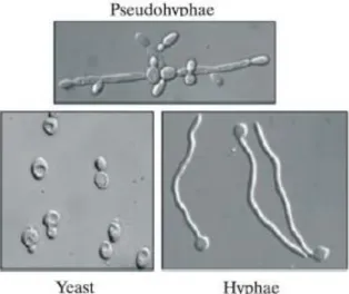
Although Candida albicans is the most frequently-isolated species related to oral opportunistic infections, an increase in the frequency of oral infections caused by non- albicans

Figure 20 Original image (left), guided Grad-CAM (center) and precision-recall (PR) plot (right) for pneumothorax class. The Grad-CAM shows that the CNN is getting a hot region on

Características Ambiente 15 Animais 3 Astrologia 8 Celebridades 146 Cidades 43 Cinema e TV 51 Cultura 3 Economia 91 Educação 2 Desporto 185 Life Style 21 Internacional 37 Política

A empresa está presente em mais de 600 países, o que a torna atraente como opção para facilitar o transporte não apenas de residentes, mas especialmente de turistas que desconheçam