CuO Rapid Synthesis with Different Morphologies by the Microwave Hydrothermal Method
Max Rocha Quirinoa, Guilherme Leocárdio Lucenaa*, Jackson Andson Medeirosa, Ieda Maria Garcia dos Santosb, Matheus José Cunha de Oliveirac
Received: March 27, 2018; Revised: June 29, 2018; Accepted: August 16, 2018
CuO structures were synthesized by microwave hydrothermal treatment using two different
mineralizing agents (NaOH and NH4OH) and were evaluated as photocatalysts. The materials were
characterized by X-ray diffraction (XRD), scanning electron microscopy (SEM), and Brunauer-Emmett-Teller (BET) surface area analysis. The XRD patterns indicated the formation of the monoclinic phase in both samples with 13.78 and 14.23 nm crystallite size. SEM analysis showed different agglomerates
morphologies based on the mineralization agent employed. The CuO nanostructure synthesized with NH4OH presented agglomerated like-plates which results in a spherical shape, whereas the material
synthesized with NaOH presented an agglomerate of larger plates. Both samples showed photocatalytic activity against RNL azo dye. The quasi-spherical shape CuO material reached 93 % of the discoloration.
Keywords: copper oxide, microwave hydrothermal method, photocatalytic.
*e-mail: [email protected]
1. Introduction
One copper oxide (CuO) stable form is the tenorite with
cation valence +21. This oxide is a p-type multifunctional
semiconductor, which has narrow band gap values (1.2 - 1.5 eV)2-4. Its monoclinic structure presents interesting
characteristics, such as, thermal superconductivity, thermal stability5, photovoltaic properties and antimicrobial activity6.
This metal oxide has been extensively investigated in recent years due to several applications such as an electric superconductor7, battery electrodes for lithium-ion cell8,
solar sensors9, gas sensors10, catalysts11 and wastewater
treatments12.
The synthesis of CuO nanostructures has been obtained
by several synthesis methods that apply high temperatures or extensive heat treatments, such as solid state thermal decomposition method13, electrochemical14, thermal oxidation15
and chemical precipitation16. The conventional hydrothermal
method17 applies low temperatures, however, it requires very
long synthesis times, and the microwave hydrothermal (MH) method18 has been receiving greater attention due its higher
heating rate compared to the conventional hydrothermal method. It presents a reaction kinetics increased by one
or two orders of magnitude resulting in energy and time
savings19-21. The reactions are thus completed in minutes or
even seconds22. Lee et al.23 stated that this is viable method
to solve economic and environmental problems because it is a closed system that saves energy and time.
Typically oxides synthesized through the MH method
present differentiated morphologies such as CuO in the form of sea urchin18; the BaMoO
4 in the form of micro octahedra 24
and the CaTiO3 in the form of microtubes25 and multi-linked
ZnO rods26. However, these morphologies were achieved due to the use of so-called shape controlling agents such as templates or surfactants like polyethileneglicol MW 200 or special reactants. Therefore, in this communication, we report a synthesis of CuO with two different morphologies without surfactants or templates, using different hydroxyl source
(NH4OH or NaOH) at 130 ºC for five minutes synthesis.
The degradation of organic pollutants has received special attention in recent years due to the numerous negative effects
on the environment and human health. In particular, industrial dyes have been the most common water pollutants27,28. These
organic dyes have very stable complex structures, which
difficult degradation. Among all processes used in these
dyes degradation, advanced oxidative processes (AOPs)
result in the generation of hydroxyl radicals (•OH), which are used to oxidize organic pollutants to form CO2, H2O or
some less toxic inorganic small molecule29,30. AOPs have been the most effective technology in removing organic pollutants for water treatment.31,32. Currently, semiconductor
photocatalysis is a newly developed advanced oxidation
process followed by the removal of dye pollutants33. Several
experiments were carried out to study the photocatalytic
activity of some semiconductors, such as ZnO, CdS, SnO2
and ZrO2 in the degradation of dyes. Among the range of inorganic semiconductors, CuO has attracted attention due
aLaboratório de Química (LABQUIM), Universidade Federal da Paraíba, Campus III, 58200-000, Bananeiras, PB, Brasil
bNúcleo de Pesquisa e Extensão de Combustíveis e de Materiais (NPE/LACOM), Universidade Federal da Paraíba, Campus I, 58059-900, João Pessoa, PB, Brasil
to its easy production, as well as its high chemical and thermal stability, and adjustable electronic properties33,34. As a photocatalyst for discoloration of organic dyes, CuO was evaluated for the degradation of rhodamine B, methyl
orange, tartrazine and methylene blue3,35-36. Either way, the
CuO has presented high catalytic activity, being attributed
to morphological and superficial aspects.
Thus, in this work, CuO nanoparticles were obtained
by the MH method using two different mineralizing agents aiming to observe their influence on the morphology of the obtained material. It was performed photocatalytic bench tests to analyze the material morphology influence on the degradation of the RNL azo dye.
2. Experimental
2.1 CuO synthesis
Monoclinic copper (II) oxide was synthesized by the
microwave hydrothermal method in a RMW 1 (INOVTEC)
reactor.
The applied reagents were Cu(NO3)23H2O (98%,
PROQUIMS), NaOH (98%, ISOFAR) and NH4OH (28%, PROQUIMS).
Initially, in 90 mL of copper nitrate solution (9 x 10⁻³
mol.L-1) was added 8 mL of the solution (5 mol.L-1) of two different mineralizing agents (NaOH and NH4OH). Both
precursors’ solutions with precipitate were individually
transferred into a sealed Teflon autoclave and placed in a domestic microwave (f = 2450 MHz, maximum power = 900 W). The reaction system was treated at 130 ºC with a heating rate of 10 ºC.min-1 for 5 minutes. The autogenous
pression in the sealed auclave was stabilized at 1.2 kg.cm-2 resulting in a black precipitate. Both samples were washed by centrifugation (400 rpm) for ten min three times with
distilled water until neutral pH and then dried at 110 ºC
for 12 h. The sample synthesized using the mineralizing
agent NaOH was named CuNa05 and another one applying
NH4OH was CuNH05.
2.2 Characterization
Both samples were characterized by X-ray diffraction (XRD), FT-IR spectroscopy (IR), scanning electron microscopy (SEM) and textural analysis.
The XRD patterns were obtained from Shimadzu (XRD 6000 model) operating with KαCu radiation (λ = 1.5406) in the region of 20-80º with a step of 0.02º and a step time of 2 seconds. IR spectra were recorded in the region of 400-800
cm-1 via a Prestige-21 IR Spectrophotometer from Shimadzu. SEM (backscattered-electron) images were obtained on a JEOL-300 microscope. The textural analysis were performed from the adsorption / desorption isotherms of N2 (using the BET equation) on a Bel Japan Belsorp mini II automatic surface area measurement device.
2.3 Photocatalytic and adsorption tests
The CuNa05 and CuNH05 samples were used as
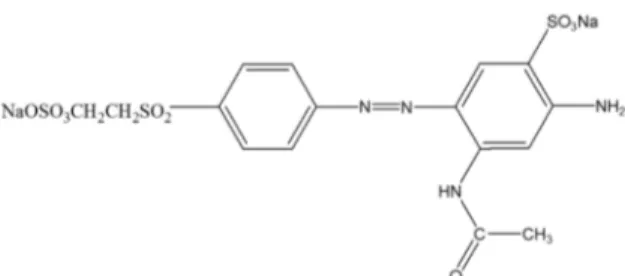
photocatalysts for the degradation of Remazol Golden Yellow dye (RNL). The molecular structure of the RNL dye is shown in Figure 1.
The photocatalytic tests were performed in a homemade reactor with dimensions of 10 x 20 x 100 cm using UVC lamp (λ = 254 nm, 20 W, ILUMISAMPA). The experiments were carried out in triplicate using 10 mg of the photocatalyst and 15 mL of the dye solution with a concentration of 10
mg.L-1 at pH = 6. Petri dishes containing the dye solution and the photocatalysts were photoirradiated for 1 h. After photocatalysis, the mixtures were centrifuged and filtered.
The percentage of discoloration and degradation of the dye solutions were quantified using a UV-vis spectrophotometer (Model UV-2550, Shimadzu) in absorbance mode by measuring the absorbance of the solution at 411 and 240
nm, respectively38. The photocatalytic efficiencies of the
photocatalysts were calculated by calibration curves; the initial
absorption of the untreated dye solution and the respective concentrations before and after photocatalytic treatment were considered. In addition, tests were performed in the dark, using the same conditions of the photocatalytic tests, to evaluate the discoloration solely due to the adsorption of the dye on the surface of the material.
3. Results and Discussion
3.1 Material characterization
Figure 2 presents the XRD patterns of the synthesized samples. The diffraction peaks were indexed according to JCPDS N° 45-0937, indicating that the monoclinic CuO phase was obtained for both samples. No commonly
impurities’ peaks such as Cu(OH)2 or Cu2O were detected
from this pattern, confirming the pure monoclinic phase for both samples.
Lattice constants were calculated using the method of the least squares, starting from the values arranged in the JCPDS Nº 45-0937 card.
Using the values of the diffraction angles and the indexing
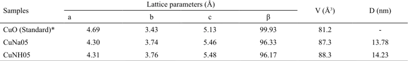
planes, lattice constants, unit cell volume and average crystallite size were calculated. Results are shown in Table 1.
According to the results shown in Table 1, the CuNa05
and CuNH05 samples presented a subtle difference in the
network parameters, whereas in comparison with standard
(CuO JCPDS Nº 45-0937), the values of a and β were reduced and the values of b and c increased. These variations resulted in an increase in unit cell volume of 6.1 Å3 for the CuNa05 sample and 7.1 Å3 for the CuNH05 sample.
This may be occurring due to the experimental conditions
(treatment temperature, heating rate and / or processing time) employed in the synthesis which are capable of promoting structural defects formation, residual stresses and/or minor distortions in the lattice constants for this oxide39. These
small variations may also be due to rapid kinetics in the
formation of nanocrystals synthesized through the HMO
method39,40. Chen et al.41,42 observed a similar result when
synthesizing mixed aluminum and zinc oxides.
Results indicate the differentiation of the mineralizing agents did not affect the crystallographic parameters relevantly. The average crystallite size (D) of particles was determined by the Scherrer's formula43 (equation 1).
(1)
in which λ is the wave-length of the incident x-rays,
L the linear dimension of particle, χ/2 the Bragg angle
and K a numerical constant for which obtained the value
2(ln 2/π)1/2 = 0.93.
For the CuNa05 sample D = 13.78 nm and 14.23 nm for
CuNH05 sample. Yang et al.44 synthesized CuO by the Microwe hydrothermal method at 120 ºC in 5 min using PEG 400 as template resulting in samples with different morphologies, i.e. quasi-spherical nanoplates, willow-leaf-like and rod like. The D parameter ranging from 11.8 to 15 nm. This slightly
variation is also observable in this work.
Low temperatures and short times of synthesis are favorable conditions for the non-growth of particles. Luo
et al.40 reported that in the synthesis of nest like BaMoO 4
nanoparticles; particles grow to a certain extent and then there is re-dissolution in the hydrothermal treatment. This
may also be related to the small size of particles in syntheses
through the hydrothermal microwave assisted method.
Figure 3 shows the IR spectra for both CuNa05 and
CuNH05 samples.
The vibrational modes located at 506 cm-1, 570 cm-1 and
603 cm-1 are characteristic of CuO monoclinic45-47. The high frequency mode at 603 cm-1 and 570 cm-1 refers to the Cu-O
stretch around the direction [-110] while the mode at 506 cm-1 is associated to vibration along the direction [101]48.
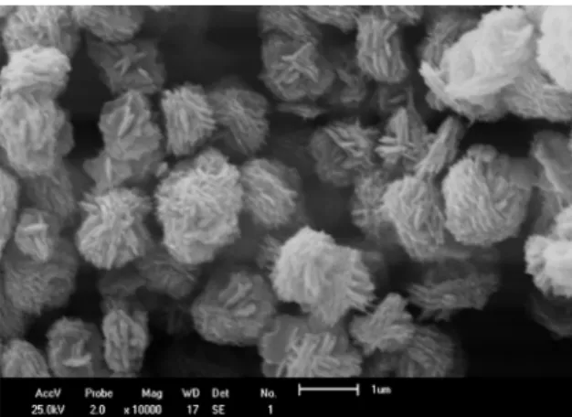
Scanning electron microscopy was used to observe the influence of mineralizing agents on the morphology of materials. SEM images of CuNH05 and CuNa05 samples are presented in Figures 4 and 5.
SEM images for both samples present plate-like particles morphology, but the difference between the two samples is in
the agglomeration. CuNH05 sample the agglomerates present
Figure 2 . XRD patterns of CUNa05 and CuNH05 samples obtained at 130 °C for 5 minutes.
Table 1. Lattice parameters, unit cell volume (V) and crystallite size (D) of both CuNa05 and CuNH05 samples.
Samples Lattice parameters (Å) V (Å3) D (nm)
a b c β
CuO (Standard)* 4.69 3.43 5.13 99.93 81.2
-CuNa05 4.30 3.74 5.46 96.33 87.3 13.78
CuNH05 4.31 3.76 5.48 96.17 88.3 14.23
*JCPDS card nº 45-0937.
Figure 3. IR spectra of the CuNa05 and CuNH05 samples obtained at 130 °C for 5 minutes.
cos D
L K
2
| m
=
quasi spherical form. It is possible to notice that the material
has some smaller quasi-spherical agglomerates and others larger than a micrometer resulting in a small particle size.
For the CuNa05 sample the small plates above large plates resulting in the interconnected channels. With these results it is evident that the agglomeration´s morphologies of the copper oxide synthesized was differentiated as a function of the mineralizing agent used.
Keyson et al.18 synthesized through the HM method
nanostructures similar to sea urchins. In this study the authors synthesized this material with a precursor solution containing polyethylene glycol 400 + CuCO3.Cu(OH)2 + NH4OH at 120°C / 1 h.
They reported that polyethylene glycol (MM = 400 g.mol-1) played a fundamental role in obtaining the oxide with this differentiated morphology. Through the same method, Maul et
al.49 evaluated the influence using or not NaOH and NH 4OH
as mineralizer agents on the CuO morphology. Using NaOH in the precursor solution many small particles and plates were
observed. While NH4OH promoted flower like morphology formed by plates. Without alkali agents small agglomerated particles forming spherical morphologies was observed.
These results were similar to those presented in this work,
although using time of synthesis twelve times shorter. In
another work, Yang et al.50 obtained nanoplates and dendrites of CuO. The precursor solution used consisted of copper nitride, PEG 400, NaOH and urea at a temperature of 120 ºC for five minutes. Shi et al.51 synthesized CuO nanorods from a precursor solution of CuSO4.5H2O + PEG400 + polyethylene glycol (4: 2) + 10 g of Urea at 120 ºC / 5 min.
It was observed that to obtain structures in the micro or
nano scales with differentiated morphologies many authors use to attribute to the role of templates, drivers and/or use of surfactants or same precursor solutions containing chitosan, PEG, Cu2Cl(OH)3 + K2CO3 + CuCl2, copper
glycinatemonohydrate51-55. However in this paper it was possible to obtain differentiated morphology, mainly using the
NH4OH mineralizer, without special reagents and templates. Thus it is observed that this method uses low temperature
and short time to obtain the CuO and with possibility of morphology control being influenced mainly to the type of precursor used for the material synthesis.
In this paper, copper oxide II was synthesized through
the microwave hydrothermal method in two different ways, resulting two samples with different morphologies
distinguishing only two hydroxyl source mineralizing agents: NaOH and NH4OH. These precursor solutions had
pH = 13 and 10, respectively. As no template or driver was used it is suggested that the pH difference as a function of the two distinct bases applied is the main factor responsible for the influence on the final morphology of the two copper
oxide samples obtained. The reactions are presented in equations 2 to 8.
(2)
(3)
51 (4)
(5)
(6)
(7)
(8)
sheets agglomerated
form quasi-spherical
mosphologiy56. Figure 4. Image obtained by SEM for CuNH05 sample ZONH5
10.000 times.
Figure 5. Image obtained by SEM for CuNa05 sample ZONH5
10.000 times.
.
Cu NO
Q
3 2V
3
H O
2 QaqV$
Cu OH
Q
V
2QsV.
+
2
NaNO
3QaqVCu OH
OH
Cu OH
2
hom
orthot bic aq
s Tetrahydroxucupratye II Anion 2 4 2
$
+
-Q
Q
Q Q R QV
V
V V V Wagglomerated sheets
Cu OH
s C minCuO
MWH
s
4 2
130c 5
-Q
V
QV QV.
Cu NO3 23H O2 aq 4NH3aq Cu NH3 4 aq
2
$
+
-Q
V
Q V Q V!
Q
V
$
Q VCu NH3 4 aq 4OH aq Cu OH aq 4NH aq
2 4 2 3 $ + + - -
-Q
V
Q V Q V Q V Q V Q V!
$
Cu OH 4 aq Cu OH aq 2OH aq
2
2
$ +
-
-Q V Q V Q VQ V Q V
Cu OH
aq C minH O
CuO
MWH
s
Different mineralizing agents, due to the basic strength of alkalis, lead to different pH. As result the synthesized materials presented different morphologies. High concentrations of copper cations and hydroxyde anions produces copper
complex ions, such as [Cu(OH)4]2- or [Cu(NH 3)4]
2+ which
are converted to CuO56, in this case by HM. So, the two forms of CuO materials can be synthesized by using different
precursors solutions ions.
Nikan et al.57 synthesized nano-belt like morphology by the microwave-assisted wet chemical synthesis at pH =10 from cupper acetate, 100 µL of NaOH (0.1 mol.L-1) and benzyl alcohol for 1 minute synthesis. Low pH lead to Cu2O
cubes morphology.
Other materials like ZnO varies its the morphology according to solution pH values. Ram et al.58 synthesized ZnO by the microwave hydrotermal method (100 ºC/ 2 min) at different
pH values; 8.5 and 10 using ammonia solution (NH4OH) as mineralizer agent and pH 10 and 13.5 using NaOH, without
using growth agents or templates. The morphology of the materials varies with pH increase, starting from nanoflakes,
than tapered hexagonal rods, and hexagonal nanorods. This
last presenting different size distributions. Also to reinforce the influence of pH on the syntheses of oxides. Wu et al.59 obtained the ZnO oxide by the HC at 200 ºC / 15 h increasing the pH value, at pH = 8.92 rods structure; 11.78 nanorods; 12.81 sea urchin like and 12.89 flower like morphology. The excess of [(OH-)] favors nucleation.
Figure 6 presents the N2 adsorption and desorption isotherms of the CuNa05 and CuNH05 samples.
The plot of adsorbed volume of N2(g) against relative pressure (P/P0) for both samples exhibited Type III isotherm (IUPAC) as show in Fig. 6a and 6b60. The two samples
showed same hysteresis behavior, ie, narrow hysteresis
loop. In set Fig. 5(a) and (b) shows pore size distribution.
The sample CuNa05, agglomerated sheets, exhibited larger pore distribution (max. 34.43 nm) than CuNH05 which
exhibited morphology in form agglomerated sheets resulting
quasi-spherical morphology with max pore size 19.04 nm.
Both materials are nanoporous, the relatively low pore
diameter in both samples may be attributed to the closely
packing of the small particles (sub-units) resulting their final micro-structure. Bhuvaneshwari et al. 201661 synthesized via hydrothermally, ie, without microwaves CuO snowflake, flower, hollowsphere and urchin morphologies. All samples
also exhibited Type III isotherm.
From these results, surface area (SBET), pore volume (Pv) and average pore size (Tp) were estimated and the values are presented in Table 2.
According to Table 2, CuNa05 presented twice the surface area (SBET) than CuNH05 sample. Yang et al.52 synthesized the CuO with different morphologies like tadpole-shaped, spindly, leaf / sphere-like, and fusiform CuO nanoparticles. The specific surface ranging from 18 to 71 m2.g-1. The CuNa05 specific surface area is inside this interval and CuNH05 is out of low limit (11.758 m2.g-1).
Average pore volume (Pv) of the sample alkalinized with NaOH was higher than the sample basified with NH4OH. The average pore size (Ps) was practically uninfluenced by
the mineralizing agent. This pore size non-variation possibly
is associated with the use of inorganic bases (NaOH and
NH4OH) as a mineralizing agent because they are similar chemical substances (bases). According to the literature, variations in pore size were observed in materials synthesized by hydrothermal and hydrothermal microwave methods that
used templates such as PEG 40062 or organic substances, such
as hexane63, decane64, as pore expansion agents.
Figure 6. N2 adsorption / desorption isotherms of CuNa05 (a) and CuNH05 (b).
Table 2. Textural characteristics of CuNa05 and CuNH05 samples. Sample SBET(m2.g-1) Pv (cm3.g-1) Ps(nm)
CuNa05 23.486 5.396 10.561
3.2 Photocatalytic efficiency
The UV-Vis spectra and the percentage discoloration of the RNL dye after the adsorption and photocatalysis processes are shown in Figure 7(a) and (b), respectively.
The quantification of RNL dye degradation is performed
by monitoring the absorbance at 240 nm and 411 nm. The
first mode refers to the transitions of the aromatic groups of
the UV-Vis spectrum, while the latter is associated with the
azo bond (-N=N-). Thus, the decrease in the intensity of the band at 411 nm implies the breakdown of the azo bond or reduction of the dye concentration in solution65.
The UV-vis spectra of the RNL dye after the tests with the CuNa05 and CuNH05 samples in the absence of light (Fig. 7a) showed a decrease in signal intensity at 240 nm and 411 nm, keeping the same profile of the spectrum of pure dye, the discoloration occurred due to adsorption of the dye molecules on the surface of the CuO particles. It was observed that the percentage of discoloration of azo
dye RNL due to adsorption, practically did not change,
maintaining the values of 43% and 45%, for CuNa05 and
CuNH05, respectively. On the other hand, the UV-vis spectra
of the RNL dye after the tests performed in the presence of radiation (Fig. 7b) showed that there was photodegradation and that the discoloration reached 85% for CuNa05 and 93% for CuNH05.
When evaluating the photocatalytic efficiency of the
CuO synthesized in this work, it is noticed that the CuNH05
sample showed an increase of 11% in the RNL discoloration
in relation to the CuNa05 sample. According to literature3,34,36, the change in morphology and surface properties, including
higher specific surface area, may be the cause of the enhanced activity. But in this case the shape of particles also influences
the photodegradation. Although the CuNa05 sample had a
larger surface area than the CuNH05 sample, 23.486 and
11.758 m2.g-1 respectively, but the photodegradation of the
latter sample was slightly higher, demonstrating that the
morphology of the particular agglomerates is more influential than the surface area of the catalyst.
4. Conclusion
The copper oxide (CuO) was synthesized by microwave
hydrothermal method under mild conditions of low temperature and short time synthesis. A significant change in the structures of the CuO was observed for different precursors without surfactants or templates, varying only alkaline mineralizes
NaOH or NH4OH in addition to Cu(NO3)2.3H2O solutions,
separately. The morphologies are affected by the mineralizing
agent, the sample, alkalinized with NH4OH, is in the form of agglomerated plate-like, which results in a quasi-spherical shape. CuNa05 sample, alkalinized with NaOH, which
presents in the form of smaller plates-like on much larger plates. The heterogeneous photocatalysis of these samples was more influenced by the morphology of copper oxide than the surface area, since the alkalinized sample with
NH4OH, quasi-spherical agglomerates and lower surface
area, presented greater degradation of the RNL azo dye.
This material is an candidate to be used in photocatalysis applications.
5. References
1. Richthofen AV, Domnick R, Cremer R. Preparation of cuprite
(Cu2O), paramelaconite (Cu32+Cu 2
1+O
4) and tenorite (CuO) with magnetron sputtering ion plating: characterization by EPMA, XRD, HEED and SEM. Fresenius' Journal of Analytical Chemistry. 1997;358(1-2):312-315.
2. Dong C, Xiao X, Chen G, Guan H, Wang Y. Morphology control of porous CuO by surfactant using combustion method.
Applied Surface Science. 2015;349:844-848.
3. Mageshwari K, Sathyamoorthy R, Park J. Photocatalytic activity of hierarchical CuO microspheres synthesized by facile reflux condensation method. Powder Technology. 2015;278:150-156.
4. Zhang Q, Zhang K, Xu D, Yang G, Huang H, Nie F, et al. CuO nanostructures: Synthesis, characterization, growth mechanisms, fundamental properties, and applications.
Progress in Materials Science. 2014;60:208-337.
5. Zhang L, Feng Q, Nie A, Liu J, Wang H, Fang Y. In Situ Study of Thermal Stability of Copper Oxide Nanowires at Anaerobic Environment. Journal of Nanomaterials. 2014;2014:670849.
6. Tran TH, Nguyen VT. Copper Oxide Nanomaterials Prepared by
Solution Methods, Some Properties, and Potential Applications: A Brief Review. International Scholarly Research Notices. 2014;2014:856592.
7. MacDonald AH. Copper oxides get charged up. Nature. 2001;414:409-410.
8. Wang C, Xu J, Ma R, Yuen MF. Facile synthesis of CuO nanoneedle electrodes for high-performance lithium-ion
batteries. Materials Chemistry and Physics. 2014;148(1-2):411-415.
9. Xia Y, Pu X, Liu J, Liang J, Liu P, Li X, et al. CuO nanoleaves
enhance the c-Si solar cell efficiency. Journal of Materials Chemistry A. 2014;2(19):6796-6800.
10. Kim JH, Katoch A, Choi SW, Kim SS. Growth and sensing properties of networked p-CuO nanowires. Sensors and Actuators B: Chemical. 2015;212:190-195.
11. Sonia S, Poongodi S, Kumar PS, Mangalaraj D, Ponpandian N, Viswanathan C. Hydrothermal synthesis of highly stable CuO nanostructures for efficient photocatalytic degradation of
organic dyes. Materials Science in Semiconductor Processing. 2015;30:585-591.
12. Reddy KJ, McDonald KJ, King H. A novel arsenic removal process for water using cupric oxide nanoparticles. Journal of Colloid and Interface Science. 2013;397:96-102.
13. Shahsavani E, Feizi N, Khalaji AD. Copper Oxide Nanoparticles Prepared by Solid State Thermal Decomposition: Synthesis
and Characterization. Journal of Ultrafine Grained and Nanostructured Materials. 2016;49(1):48-50.
14. Yuan GQ, Jiang HF, Lin C, Liao SJ. Shape- and size-controlled electrochemical synthesis of cupric oxide nanocrystals. Journal of Crystal Growth. 2007;303(2):400-406.
15. Filipic G, Cvelbar U. Copper oxide nanowires: a review of
growth. Nanotechnology. 2012;23(19):194001.
16. Zhu J, Bi H, Wang Y, Wang X, Yang X, Lu L. CuO nanocrystals with controllable shapes grown from solution without any surfactants. Materials Chemistry and Physics. 2008;109(1):34-38.
17. Chakraborty S, Das A, Sood AK. Mixing of Mode Symmetries in Top Gated Bilayer and Multilayer Graphene Field Effect Devices. AIP Conference Proceedings. 2011;1349(1):11-14.
18. Keyson D, Volanti DP, Cavalcante LS, Simões AZ, Varela JA, Longo E. CuO urchin-nanostructures synthesized from
a domestic hydrothermal microwave method. Materials Research Bulletin. 2008;43(3):771-775.
19. Komarneni S, D´Arrigo MC, Leonelli C, Pellacani GC, Katsuki H. Microwave-Hydrothermal Synthesis of Nanophase Ferrites.
Journal of American Ceramic Society. 1998;81(11):3041-3043.
20. Komarneni S, Rajha RK, Katsuki H. Microwave-hydrothermal processing of titanium dioxide. Materials Chemistry and Physics. 1999;61(1):50-54.
21. Komarneni S, Katsuki H. Nanophase materials by a novel
microwave-hydrothermal process. Pure and Applied Chemistry. 2002;74(9):1537-1543.
22. Roberts BA, Strauss CR. Toward Rapid, "Green", Predictable Microwave-Assisted Synthesis. Accounts of Chemical Research. 2005;38(8):653-661.
23. Lee JH, Kumagai N, Watanabe T, Yoshimura M. Direct fabrication of oxide films by a microwave-hydrothermal
method at low temperature. Solid State Ionics. 2002;151(1-4):41-45.
24. Cavalcante LS, Sczancoski JC, Tranquilin RL, Varela JA, Longo E, Orlandi MO. Growth mechanism of octahedron-like BaMoO4 microcrystals processed in microwave-hydrothermal: Experimental observations and computational modeling.
Particuology. 2009;7(5):353-362.
25. Moreira ML, Paris EC, do Nascimento GS, Longo VM, Sambrano JR, Mastelaro VR, et al. Structural and optical properties of CaTiO3 perovskite-based materials obtained by
microwave-assisted hydrothermal synthesis: An experimental and theoretical insight. Acta Materialia. 2009;57(17):5174-5185.
26. Kathalingam A, Chae YS, Rhee JK. Synthesis of
multi-linked ZnO rods by microwave heating. Crystal Research & Technology. 2011;46(5):517-522.
27. Banazadeh A, Salimi H, Khaleghi M, Shafiei-Haghighi S. Highly efficient degradation of hazardous dyes in aqueous phase by
28. Horáková M, Klementová Š, Kříž P, Balakrishna SK, Špatenka P, Golovko O, et al. The synergistic effect of advanced oxidation
processes to eliminate resistant chemical compounds. Surface and Coatings Technology. 2014;241:154-158.
29. Yang Z, Yang Y, Zhu X, Chen G, Zhang W. An Outward Coating Route to CuO/MnO2 Nanorod Array Films and Their Efficient Catalytic Oxidation of Acid Fuchsin Dye. Industrial & Engineering Chemistry Research. 2014;53(23):9608-9615.
30. Cheng M, Zeng G, Huang D, Lai C, Xu P, Zhang C, et al.
Hydroxyl radicals based advanced oxidation processes (AOPs)
for remediation of soils contaminated with organic compounds:
A review. Chemical Engineering Journal. 2016;284:582-598.
31. Wang JL, Xu LJ. Advanced Oxidation Processes for Wastewater Treatment: Formation of Hydroxyl Radical and Application.
Critical Reviews in Environmental Science and Technology. 2012;42(3):251-325.
32. Li H, Liao J, Zeng T. A facile synthesis of CuO nanowires
and nanorods, and their catalytic activity in the oxidative
degradation of Rhodamine B with hydrogen peroxide. Catalysis Communications. 2014;46:169-173.
33. Rao MP, Wu JJ, Asiri AM, Anandan S. Photocatalytic degradation of tartrazine dye using CuO straw-sheaf-like nanostructures.
Water Science and Technology. 2017;75(5-6):1421-1430.
34. Sadollahkhani A, Ibupoto ZH, Elhag S, Nur O, Willander M. Photocatalytic properties of different morphologies of CuO for the degradation of Congo red organic dye. Ceramics International. 2014;40(7 Pt B):11311-11317.
35. Jiang D, Xue J, Wu L, Zhou W, Zhang Y, Li X. Photocatalytic performance enhancement of CuO/Cu2O heterostructures for photodegradation of organic dyes: Effects of CuO morphology.
Applied Catalysis B: Environmental. 2017;211:199-204.
36. Deka P, Hazarika A, Deka RC, Bharali P. Influence of CuO morphology on the enhanced catalytic degradation of methylene
blue and methyl orange. RSC Advances. 2016;6(97):95292-95305.
37. Alvarenga JM, Fideles RA, Silva MV, Murari GF, Taylor JG, Lemos LR, et al. Partition study of textile dye Remazol Yellow Gold RNL in aqueous two-phase systems. Fluid Phase Equilibria. 2015;391:1-8.
38. Muruganandham M, Swaminathan M. Photocatalytic decolourisation and degradation of Reactive Orange 4 by TiO2-UV process.
Dyes and Pigments. 2006;68(2-3):133-142.
39. Rao KJ, Vaidhyanathan B, Ganguli M, Ramakrishnan PA. Synthesis of Inorganic Solids Using Microwaves. Chemistry of Materials. 1999;11(4):882-895.
40. Luo Z, Li H, Shu H, Wang K, Xia J, Yan Y. Synthesis of BaMoO4 Nestlike Nanostructures Under A New Growth Mechanism.
Crystal Growth & Design. 2008;8(7):2275-2281.
41. Chen Z, Shi E, Li W, Zheng Y, Wu N, Zhong W. Particle Size Comparison of Hydrothermally Synthesized Cobalt and Zinc Aluminate Spinels. Journal of the American Ceramic Society. 2002;85(12):2949-2955.
42. Chen Z, Shi E, Zheng Y, Li W, Wu N, Zhong W. Synthesis of mono-dispersed ZnAl2O4 powders under hydrothermal
conditions. Materials Letters. 2002;56(4):601-605.
43. Patterson AL. The Scherrer Formula for X-ray Particle Size Determination. Physical Review. 1939;56(10):978-982.
44. Yang C, Xiao F, Wang J, Su X. Synthesis and microwave modification of CuO nanoparticles: crystallinity and morphological
variations, catalysis, and gas sensing. Journal of Colloid and Interface Science. 2014;435:34-42.
45. Wang H, Xu JZ, Zhu JJ, Chen HY. Preparation of CuO
nanoparticles by microwave irradiation. Journal of Crystal Growth. 2002;244(1):88-94.
46. Zhang L, Lu W, Feng Y, Ni J, Lü Y, Shang X. Facile Synthesis of Leaf-like Cu(OH)2 and its Conversion into CuO with Nanopores.
Acta Physico-Chimica Sinica. 2008;24(12):2257-2262.
47. Sathyamoorthy R, Mageshwari K. Synthesis of hierarchical
CuO microspheres: Photocatalytic and antibacterial activities. Physica E: Low-dimensional Systems and Nanostructures. 2013;47:157-161.
48. Zou GF, Li H, Zhang DW, Xiong K, Dong C, Qian YT. Well-Aligned Arrays of CuO Nanoplatelets. The Journal of Physical Chemistry B. 2006;110(4):1632-1637.
49. Maul J, Brito AS, de Oliveira ALM, Lima SJG, Maurera MAMA, Keyson D, et al. Influence of the synthesis media in the properties of CuO obtained by microwave-assisted hydrothermal method.
Journal of Thermal Analysis and Calorimetry. 2011;106(2):519-523.
50. Yang C, Wang J, Xiao F, Su X. Microwave hydrothermal disassembly for evolution from CuO dendrites to nanosheets
and their applications in catalysis and photo-catalysis. Powder Technology. 2014;264:36-42.
51. Shi L, Yang C, Su X, Wang J, Xiao F, Fan J, et al. Microwave-hydrothermal synthesis of CuO nanorods and their catalytic applications in sodium humate synthesis and RhB degradation.
Ceramics International. 2014;40(3):5103-5106.
52. Yang C, Su X, Wang J, Cao X, Wang SJ, Zhang L. Facile microwave-assisted hydrothermal synthesis of varied-shaped
CuO nanoparticles and their gas sensing properties. Sensors and Actuators B: Chemical. 2013;185:159-165.
53. Raghavendra GM, Jung J, Kim D, Seo J. Chitosan-mediated synthesis of flowery-CuO, and its antibacterial and catalytic
properties. Carbohydrate Polymers. 2017;172:78-84.
54. Xie H, Zhu L, Zheng W, Zhang J, Gao F, Wang Y. Microwave-assisted template-free synthesis of butterfly-like CuO through
Cu2Cl(OH)3 precursor and the electrochemical sensing property. Solid State Sciences. 2016;61:146-154.
55. Huang J, Fu G, Shi C, Wang X, Zhai M, Gu C. Novel
porous CuO microrods: synthesis, characterization, and their photocatalysis property. Journal of Physics and Chemistry of Solids. 2014;75(9):1011-1016.
56. Bozkurt G, Bayrakçeken A, Özer AK. Synthesis and characterization of CuO at nanoscale. Applied Surface Science. 2014;318:244-250.
57. Nikan AV, Arulkashmir A, Krishnamoorthy K, Kulkarni AA,
Prasad BLV. pH-Dependent Single-Step Rapid Synthesis of
58. Ram SDG, Kulandainathan MA, Ravi G. On the study of pH effects in the microwave enhanced rapid synthesis of nano-ZnO.
Applied Physics A. 2010;99(1):197-203.
59. Wu WY, Kung WY, Ting JM. Effect of pH Values on the Morphology of Zinc Oxide Nanostructures and their Photoluminescence Spectra. Journal of American Ceramic Society. 2011;94(3):699-703.
60. Sing KSW, Everett DH, Haul RAW, Moscou L, Pierotti RA, Rouquerol J, et al. Reporting physisorption data for gas/solid systems with special reference to the determination of surface
area and porosity. Pure and Applied Chemistry. 1985;57(4):603-619.
61. Bhuvaneshwari S, Gopalakrishnan N. Hydrothermally synthesized copper oxide (CuO) superstructures for ammonia sensing.
Journal of Colloid and Interface Science. 2016;480:76-84.
62. Quirino MR, Oliveira MJ, Keyson D, Lucena GL, Oliveira JBL, Gama L. Synthesis of zinc oxide by microwave hydrothermal method for application to transesterification of soybean oil
(biodiesel). Materials Chemistry and Physics. 2017;185:24-30.
63. Johansson EM, Córdoba JM, Odén M. The effects on pore size and particle morphology of heptane additions to the synthesis of mesoporous silica SBA-15. Microporous and Mesoporous Materials. 2010;133(1-3):66-74.
64. Zhang H, Sun J, Ma D, Weinberg G, Su DS, Bao X. Engineered Complex Emulsion System: Toward Modulating the Pore Length and Morphological Architecture of Mesoporous Silicas. Journal of Physical Chemistry B. 2006;110(51):25908-25915.
65. Stylidi M, Kondarides DI, Verykios XE. Visible light-induced photocatalytic degradation of Acid Orange 7 in aqueous TiO2