Characterization of ZnAl
2O
4Spinel Obtained by Hydrothermal and Microwave Assisted
Combustion Method: A Comparative Study
Heloísa Pimenta de Macedoa*; Rodolfo Luíz Bezerra de Araújo Medeirosa; Amanda Lucena de
Medeirosa; Ângelo Anderson Silva de Oliveirab; Gilvan Pereira de Figueredoc,d; Marcus Antônio de
Freitas Meloe; Dulce Maria de Araújo Meloa,c
Received: December 11, 2016; Revised: February 10, 2017; Accepted: April 05, 2017.
In this work, zinc aluminate spinel was prepared by two methods of directly synthesis (without calcination): microwave assisted combustion and hydrothermal method. The materials were
characterized by X-ray diffraction (XRD), scanning electron microscopy (SEM) and N2-adsorption/
desorption isotherms. The XRD patterns confirmed the formation of cubic ZnAl2O4 spinel structure
with no secondary phases for both synthesis routes, whereas the hydrothermal method yielded powders with crystallite size 3 times smaller (6.9 nm), as compared to the powders produced by microwave
assisted combustion method (25.6 nm). The micrographs revealed agglomerated powders with plate-like morphologies for both routes. Nitrogen adsorption/desorption isotherms (BET) revealed higher
surface area (183 m2.g-1) and greater pore volume (0.173 cm3.g-1) for ZnAl
2O4 powders prepared by
the hydrothermal method.
Keywords: Zinc Aluminate, Hydrothermal Method, Microwave Assisted Combustion
* e-mail: [email protected]
1. Introduction
Zinc aluminate, ZnAl2O4, is a ternary oxide with spinel
structure that has drawn considerable attention in the past years as an advanced material due to its combination of
desirable properties: high mechanical strength, high thermal and chemical stability, low sintering temperature, low surface acidity, wide band gap and excelent optical properties1-3 with
various applications. Therefore, it is currently being used as high
temperature material, sensors, eletronic and optical materials, as well as catalysts and catalyst support4-8. In general, many
methods of synthesis have been used for the preparation of
ZnAl2O4 oxide, which include co-precipitation
9-10, modifed
citrate sol-gel11, microwave combustion3,12, hydrothermal13-14,
sol–gel15, polymeric precursor16 and solid state route17.
Among the several preparation methods, microwave assisted combustion synthesis is one of the most effective, fast, simple and energy efficiency method for the synthesis of metal oxide based materials, producing high purity and
chemically homogeneous powders3,6. Metal precursors and
fuel (mostly organic compounds like urea, citric acid, glycine,
carbohydrazide or alanine) in an appropriate stoichiometric ratio controls the combustion process in accordance with
the propellant chemistry principles, producing a very fast
and exothermic chemical reaction to form the material12,18.
The hydrothermal method is a wet chemical solution
technique and stands out by using low temperatures to produce directly nanometric powders with high surface areas, narrow size distribution and crystals with great perfection
without the need of subsequent thermal treatments19-20. The
high surface area and a porous structure of ZnAl2O4 are of
great importance for catalytic purposes.
The synthesis method can greatly affect the characteristics
and properties of materials. In this context, the aim of this work was to carry out a comparative study of the synthesis of
ZnAl2O4 prepared without calcination by hydrothermal and
microwave assisted combustion method. Besides, the present work aims to study the influence of the synthesis methods on the structural, morfological and textural parameters of ZnAl2O4 powders. The powders produced were characterized
a Postgraduate Program in Science and Engineering of Materials, Federal University of Rio Grande do
Norte - UFRN, CEP 59078-970, Natal, RN, Brazil
b Postgraduate Program in Science and Engineering of Petroleum, Federal University of Rio Grande do
Norte - UFRN, CEP 59078-970, Natal, RN, Brazil
c Postgraduate Program in Chemistry, Chemistry Institute, Federal University of Rio Grande do Norte -
UFRN, CEP 59078-970, Natal, RN, Brazil
d Chemistry Department, Federal Institute of Education, Science and Technology of Maranhão - IFMA,
CEP 65076-091, São Luís, MA, Brazil
e Department of Chemical Engineering, Federal University of Rio Grande do Norte - UFRN, CEP
by the following techniques: X-ray diffraction (XRD), scanning electron microscopy (SEM) and N2-adsorption/
desorption isotherms.
2. Materials and methods
All chemicals used in the present study were of analytical
grade and used as received without further purification. Al(NO3)3·9H2O (Sigma-Aldrich), Zn(NO3)2·6H2O
(Sigma-Aldrich) and urea CH4N2O (Vetec) were used as starting materials.
2.1. Microwave Assisted Combustion Synthesis
Zinc nitrate and aluminum nitrate were used as precursors
and urea as a fuel in this method. The compounds were
dissolved separately in de-ionized water and mixed together in a glass becker at room temperature under constant stirring to obtain a homogeneous solution. The fuel to oxidizer ratio
(F/O) was equal to 1 as per the concept used in propellant
chemistry. The homogeneous solution was placed inside a
domestic microwave-oven and exposed to irradiation for 5 min at 900 W output power, and frequency of 2.45 GHz. Initially, the solution boiled and underwent dehydration
followed by decomposition with the evolution of gases12.
After ignition started, a rapid flame took place resulting in a solid final product that was denoted as ZnAl2O4_MC.
2.2. Hydrothermal Synthesis
Zinc nitrate and aluminum nitrate were used as precursors
and urea as a basic source in this method. First, zinc nitrate, aluminum nitrate and urea were dissolved in de-ionized
water to form a transparent solution under magnetic stirring. The Zn:Al molar ratio and the Zn:Urea molar ratio
were maintained as 1:2 and 1:10, respectively. Then, the
above solution was transferred into a 100 mL Teflon-lined
stainless steel autoclave, which was further sealed and kept
at 180 °C in an electrical oven for 24 h. The final pH value of the reaction solution was ~10. After being cooled to room temperature, the product was filtered, thoroughly washed with water until the pH value of the filtrate was neutral13.
Then, the resulting precipitate was dried at 80 °C overnight and denoted as ZnAl2O4 –H.
2.3. Characterization
The structural characterization of the ZnAl2O4 spinel
powders were determined by X-ray powder diffraction (XRD) in a Shimadzu XRD 7000 apparatus with Cu-Kα radiation at λ = 1.540 Å for 2θ values ranging from 20° to 80°, operating at 2º.min-1 with 0,02º step. The morphological
characteristics of the powders were analyzed by scanning electron microscopy in a Shimadzu SSX550 microscope,
operating with 15 kV and equipped with tungsten filament.
Previously, the samples were coated with a thin layer of
gold. The textural characteristics of the samples (surface
area, pore size, and pore volume) were determined by the
adsorption and desorption of nitrogen in a Micromeritics ASAP 2020 apparatus using BET and BJH methods. The
pore size distributions were derived from the desorption
branches of the isotherms using Barret–Joyner–Halenda (BJH) method. Prior to measurements samples were degassed
at 200 °C for 10 h.
3. Results and discussions
The XRD patterns of the ZnAl2O4 –H and ZnAl2O4 –MC
samples are shown in Figure 1. Both diffractograms consist of a single crystalline phase, showing characteristic diffraction peaks corresponding to (220), (311), (222), (400), (331), (422), (511), (440), (620) and (533) reflections of cubic ZnAl2O4
spinel structure (JCPDS No. 05-0669). This indicates that there
is a complete formation of the spinel phase in both samples synthesized in the experimental conditions employed in this
work. No diffraction peaks related to secondary phases or impurity were detected. The ZnAl2O4 –MC sample presented
more intensive and sharper diffraction peaks (FWHM311 =
0.2558) revealing its higher crystallinity degree and bigger crystallite size; whereas the ZnAl2O4 –H sample presented
broader and less intense peaks (FWHM311 = 0.2362) indicating
its smaller crystallite size and fine particule nature. The lattice parameters and average crystallite size of the samples are listed in Table 1. The average crystallite size (D), calculated from the most intense X-ray diffraction peak (311) using Scherrer’s equation21 is given by equation (1)
( )
D
=
b0 89.cosmi1
where, λ is the wavelength of the X-ray source, β the
full width at half maximum (FWHW) of the diffraction peak
and 2θ, the diffraction angle. The lattice parameter of cubic
zinc aluminate was calculated based on the X-ray diffraction patterns using equation (2),
(
)
( )
d
hklh
k
l
2
2 2 2 2 2
a
=
+
+
where, a is the lattice parameter, dhkl the interplanar
spacing corresponding to the Miller indices, h, k, and l the
miller indices21. The results show that the crystallite sizes are
in nanometers scale, 25.6 nm and 6.9 nm, for ZnAl2O4 –MC
and ZnAl2O4 –H, respectively. As the working temperature
is relatively low in the hydrothermal synthesis, this method leads to the formation of smaller crystallites22. Whereas,
ZnAl2O4 –MC powders present larger crystallite sizes, probably
due to the large amount of heat released during combustion
Figure 1. XRD patterns of ZnAl2O4 powders.
Table 1. Structural and textural data for ZnAl2O4 powders.
ZnAl2O4 –MC ZnAl2O4 –H
Crystallite size (nm) 25.6 6.9
Lattice parameter (Å) 8.0779 8.1121
Surface area (m2.g-1) 5.3 183.5
Average pore size (nm) 10.9 3.4
Total pore volume (cm3.g-1) 0.011 0.173
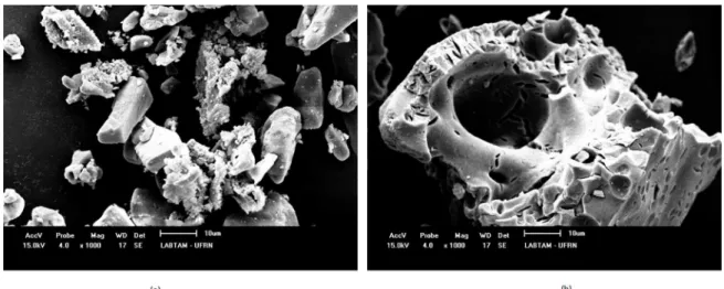
1 and 21 µm. While the powders obtained by microwave
combustion method (Figure 2b) resulted in larger particles, measuring between 3 and 95 µm. The micrography of ZnAl2O4 –H (Figure 2a) revealed agglomerated particules
with shaped plate type morphology and small aggregates on the surface of bigger clusters22. SEM image of ZnAl
2O4 –MC
(Figure 2b) revealed the presence of plate-like aggregates with irregular surface and porous structures. This morphology is typical for combustion synthesized powders due to the large volume of gases released during combustion reaction and the high temperature reached within the reaction mixture3.
The morphology of the powders depends strongly on the
synthesis method used. For example, Du et al.24 obtained
ZnAl2O4 powders with polyhedral morphology prepared by
solid state route. Motloung et al.5 describe zinc aluminate
powders with rod-like-needles morphology prepared by citrate sol-gel. ZnAl2O4 powders with semi spherical morphology
can be obtained by sol-gel25 and co-precipitation26 methods.
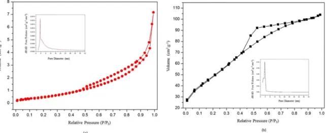
Figure 3 shows the N2 adsorption/desorption isotherms
of the ZnAl2O4 samples. According to IUPAC classification,
both samples have a type IV isotherm and H2 hysteresis, which are typical for mesoporous materials27. The mesoporous
structure was confirmed by the analysis of pore size distribution (see insert in Figure 3), which shows the spectra of the pore diameter in the mesoporous region for both samples. The
pore size distribution curves display a narrow unimodal
distribution with an average pore size of approximately 3.4 nm and 10.9 nm (see Table 1) for sample ZnAl2O4 –H and
ZnAl2O4 –MC, respectively. In addition, ZnAl2O4 –H sample
exhibit higher total pore volume (0.173 cm3.g-1) compared
to ZnAl2O4 –MC sample (0.011 cm
3.g-1) as shown in Table 1.
The surface area was measured via the N2 physisorption
technique calculated by the BET method. The results, listed in Table 1, show that the average area for the ZnAl2O4 –MC
sample was 5.3 m2.g-1, which is compatible with the average
area of powders obtained via microwave-assisted combustion synthesis28. However, BET surface area of only 5 m2.g-1, is
Figure 2. SEM images of (a) ZnAl2O4 –H and (b) ZnAl2O4 –MC powders. obtained by Anand et al. (20 nm)23 and Chen et al. (6–7 nm)13
using the microwave combustion method and hydrothermal method, respectively. The lattice parameters of (8.1121 Å) and (8.0779 Å) for ZnAl2O4 –H and ZnAl2O4 –MC respctively,
are very close to the theoretical value of gahnite (8.0848 Å) mentioned in the PDF file JCPDS 05-0669.
The micrographs of ZnAl2O4 –MC and ZnAl2O4 –H powders
Figure 3. N2 adsorption/desorption isotherms and (insert) pore diameter distribution of (a) ZnAl2O4–MC and (b) ZnAl2O4–H powders.
quite small, especially for catalysis applications14,18. The
most probable explanation for this result might be the large amount of heat released during combustion reaction3. The
BET surface area of ZnAl2O4 –H sample was 183.5 m 2.g-1,
indicating that ZnAl2O4 prepared by hydrothermal method
exibit high surface area, which is in agreement with the average
area of powders obtained via hydrothermal synthesis14,29,30.
Since the temperature is relatively low in the hydrothermal
synthesis, this method leads to the formation of nanometric
powders with high surface area, which is of great importance for catalytic purposes since it allows a greater accessibility
of reactant molecules to the catalyst31. Ballarini et al.9 tested
the catalytic activity of Pt-ZnAl2O4 powders on the n-butane
dehydrogenation reaction. They concluded that the ZnAl2O4
powders with larger BET surface area presented the best
catalytic performance9.
4. Conclusions
Single phase ZnAl2O4 spinel-type powders have been
successfully prepared in a direct procedure without calcination by hydrothermal method and microwave assisted combustion
method. Depending on the method chosen, powders with different physical properties were obtained. Due to the large amount of heat released during the combustion reaction, the resulted ZnAl2O4 –MC powder presented a small BET surface
area (5.3 m2.g-1) and an average crystallite size of 25.6 nm.
Whereas, the hydrothermal method yielded powders with
surface area 30 times higher (183.5 m2.g-1) and crystallite
size 3 times smaller (6.9 nm), as compared to the powders produced by microwave assisted combustion method, once
the working temperature in the hydrothermal synthesis is relatively low. Both samples showed a strong tendency to agglomerate with plate-like morphology powders.
5. Acknowledgments
The authors wish to thank ANP, CAPES and CNPq for financial support, the Postgraduate Program in Science and Engineering of Materials (PPGCEM/UFRN) for support and the Environmental Technology Laboratory (LABTAM/ UFRN) for the characterizations and tests carried out.
6. References
1. Ge D-L, Fan Y-J, Qi C-L, Sun Z-X. Facile synthesis of highly thermostable mesoporous ZnAl2O4 with adjustable pore size. Journal of Materials Chemistry A. 2013;1(5):1651-1658.
2. Tian X, Wan L, Pan K, Tian C, Fu H, Shi K. Facile synthesis of mesoporous ZnAl2O4 thin films through the evaporation-induced
self-assembly method. Journal of Alloys and Compounds. 2009;488(1):320-324.
3. Ianoş R, Borcănescu S, Lazău R. Large surface area ZnAl2O4
powders prepared by a modified combustion technique. Chemical Engineering Journal. 2014;240:260-263.
4. Wang SF, Sun GZ, Fang LM, Lei L, Xiang X, Zu XT. A comparative study of ZnAl2O4 nanoparticles synthesized from
different aluminum salts for use as fluorescence materials.
Scientific Reports. 2015;5:12849.
5. Motloung SV, Dejene FB, Swart HC, Ntwaeaborwa OM. Effects of Zn/citric acid mole fraction on the structure and
luminescence properties of the un-doped and 1.5% Pb2+ doped
ZnAl2O4 powders synthesized by citrate sol–gel method. Journal of Luminescence. 2015;163:8-16.
6. Anand GT, Kennedy LJ, Aruldoss U, Judith Vijaya J. Structural, optical and magnetic properties of Zn1−xMnxAl2O4 (0≤x≤0.5)
spinel nanostructures by one-pot microwave combustion technique. Journal of Molecular Structure. 2015;1084:244-253.
7. Zhang W, Wang Y, Shen Y, Xie M, Guo X. Mesoporous zinc aluminate (ZnAl2O4) nanocrystal: Synthesis, structural
hydroxylation. Microporous and Mesoporous Materials. 2016;226:278-283.
8. Galetti AE, Gomez MF, Arrúa LA, Abello MC. Ni catalysts supported on modified ZnAl2O4 for ethanol steam reforming. Applied Catalysis A: General. 2010;380(1-2):40-47.
9. Ballarini AD, Bocanegra SA, Castro AA, de Miguel SR, Scelza OA. Characterization of ZnAl2O4 Obtained by Different
Methods and Used as Catalytic Support of Pt. Catalysis Letters. 2009;129(3):293-302.
10. Battiston S, Rigo C, Severo EdC, Mazutti MA, Kuhn RC, Gündel A, et al. Synthesis of zinc aluminate (ZnAl2O4) spinel
and its application as photocatalyst. Materials Research. 2014;17(3):734-738.
11. Duan X, Yuan D, Sun Z, Luan C, Pan D, Xu D, et al. Preparation
of Co2+-doped ZnAl
2O4 nanoparticles by citrate sol–gel method. Journal of Alloys and Compounds. 2005;386(1-2):311-314.
12. Anand GT, Kennedy LJ. One-pot microwave combustion synthesis of porous Zn1-xCuxAl2O4 (0 ≤ x ≤ 0.5) spinel nanostructures. Journal of Nanoscience and Nanotechnology. 2013;13(4):3096-3103.
13. Chen XY, Ma C, Zhang ZJ, Wang BN. Ultrafine gahnite (ZnAl2O4)
nanocrystals: Hydrothermal synthesis and photoluminescent properties. Materials Science and Engineering: B. 2008;151(3):224-230.
14. Zhao H, Dong Y, Jiang P, Wang G, Zhang J, Zhang C. ZnAl2O4
as a novel high-surface-area ozonation catalyst: One-step green
synthesis, catalytic performance and mechanism. Chemical Engineering Journal. 2015;260:623-630.
15. Sharma RK, Ghose R. Synthesis and characterization of nanocrystalline zinc aluminate spinel powder by sol–gel method.
Ceramics International. 2014;40(2):3209-3214.
16. Gama L, Ribeiro MA, Barros BS, Kiminami RHA, Weber IT, Costa ACFM. Synthesis and characterization of the NiAl2O4,
CoAl2O4 and ZnAl2O4 spinels by the polymeric precursors method. Journal of Alloys and Compounds. 2009;483(1-2):453-455.
17. Van der Laag N, Snel M, Magusin P, De With G. Structural,
elastic, thermophysical and dielectric properties of zinc
aluminate (ZnAlO). Journal of the European Ceramic Society. 2004;24(8):2417-2424.
18. Alves CT, Oliveira A, Carneiro SAV, Silva AG, Andrade HMC, Vieira de Melo SAB, et al. Transesterification of waste frying oil using a zinc aluminate catalyst. Fuel Processing Technology. 2013;106:102-107.
19. Chen Z, Shi E, Li W, Zheng Y, Wu N, Zhong W. Particle Size Comparison of Hydrothermally Synthesized Cobalt and Zinc Aluminate Spinels. Journal of the American Ceramic Society. 2002;85(12):2949-2955.
20. Mu L, Wan J, Wang Z, Gao Y, Qian Y. Mn-Doped Zinc Aluminate Nanoparticles: Hydrothermal Synthesis, Characterization, and
Photoluminescence Properties. Journal of Nanoscience and Nanotechnology. 2006;6(3):863-867.
21. Cullity BD, Stock SR. Elements of X-Ray Diffraction. 3rd ed.
London: Pearson; 2014. 664 p.
22. Quirino MR, Oliveira MJC, Keyson D, Lucena GL, Oliveira JBL, Gama L. Synthesis of zinc aluminate with high surface
area by microwave hydrothermal method applied in the
transesterification of soybean oil (biodiesel). Materials Research Bulletin. 2016;74:124-128.
23. Anand GT, Kennedy LJ, Vijaya JJ. Microwave combustion synthesis, structural, optical and magnetic properties of Zn1−
xCoxAl2O4 (0≤x≤0.5) spinel nanostructures. Journal of Alloys
and Compounds. 2013;581:558-566.
24. Du X, Li L, Zhang W, Chen W, Cui Y. Morphology and structure features of ZnAl2O4 spinel nanoparticles prepared by
matrix-isolation-assisted calcination. Materials Research Bulletin. 2015;61:64-69.
25. Charinpanitkul T, Poommarin P, Wongkaew A, Kim K-S.
Dependence of zinc aluminate microscopic structure on its synthesis. Journal of Industrial and Engineering Chemistry. 2009;15(2):163-166.
26. Farhadi S, Panahandehjoo S. Spinel-type zinc aluminate (ZnAl2O4)
nanoparticles prepared by the co-precipitation method: A novel,
green and recyclable heterogeneous catalyst for the acetylation
of amines, alcohols and phenols under solvent-free conditions. Applied Catalysis A: General. 2010;382(2):293-302.
27. Sing KSW. Reporting physisorption data for gas/solid systems
with special reference to the determination of surface area and porosity (Recommendations 1984). Pure and Applied Chemistry. 1985;57(4):603-619.
28. Medeiros RLBA, Macedo HP, Melo VRM, Oliveira ÂAS, Barros JMF, Melo MAF, et al. Ni supported on Fe-doped MgAl2O4 for dry reforming of methane: Use of factorial design
to optimize H2 yield. International Journal of Hydrogen Energy.
2016;41(32):14047-14057.
29. Zhu Z, Li X, Zhao Q, Liu S, Hu X, Chen G. Facile solution
synthesis and characterization of porous cubic-shaped superstructure
of ZnAl2O4. Materials Letters. 2011;65(2):194-197.
30. Zhu Z, Zhao Q, Li X, Li Y, Sun C, Zhang G, et al. Photocatalytic performances and activities of ZnAl2O4 nanorods loaded with Ag
towards toluene. Chemical Engineering Journal. 2012;203:43-51.
31. Foletto EL, Battiston S, Simões JM, Bassaco MM, Pereira LSF, de Moraes Flores EM, et al. Synthesis of ZnAl2O4
nanoparticles by different routes and the effect of its pore size