Membrane structural changes support the
involvement of mitochondria in the bile
salt-induced apoptosis of rat hepatocytes
Susana SOLA
;
*
, Maria A. BRITO
*
, Dora BRITES
*
, Jose
!
J. G. MOURA† and
Cecı
!
lia M. P. RODRIGUES
*
*
Centro de Patoge
!
nese Molecular, Faculdade de Farma
!
cia, University of Lisbon, Lisbon, Av. das Forc
:
as Armadas,
1600-083 Lisbon, Portugal, and †Departamento de Quı
!
mica, Faculdade de Cie
#
ncias e Tecnologia, Universidade Nova de Lisboa,
2825 Monte de Caparica, Portugal
A
B
S
T
R
A
C
T
The accumulation of toxic bile salts within the hepatocyte plays a key role in organ injury during liver disease. Deoxycholate (DC) and glycochenodeoxycholate (GCDC) induce apoptosisin vitro
andin vivo, perhaps through direct perturbation of mitochondrial membrane structure and
function. In contrast, ursodeoxycholate (UDC) and its taurine-conjugated form (TUDC) appear to be protective. We show here that hydrophobic bile salts induced apoptosis in cultured rat hepatocytes, without modulating the expression of pro-apoptotic Bax protein, and caused cytochromecrelease in isolated mitochondria. Co-incubation with UDC and TUDC prevented
cell death and efflux of mitochondrial factors. Using spin-labelling techniques and EPR spectroscopy analysis of isolated rat liver mitochondria, we found significant structural changes at the membrane–water surface in mitochondria exposed to hydrophobic bile salts, including modified lipid polarity and fluidity, altered protein order and increased oxidative injury. UDC, TUDC and cyclosporin A almost completely abrogated DC- and GCDC-induced membrane perturbations. We conclude that the toxicity of hydrophobic bile salts to hepatocytes is mediated by cytochromecrelease, through a mechanism associated with marked direct effects
on mitochondrial membrane lipid polarity and fluidity, protein order and redox status, without modulation of pro-apoptotic Bax expression. UDC and TUDC can directly suppress disruption of mitochondrial membrane structure, which may represent an important mechanism of hepatoprotection by these bile salts.
INTRODUCTION
The accumulation of hydrophobic bile salts within the hepatocyte appears to play a key role in organ injury during chronic liver diseases [1–3]. Although the loss of hepatocellular function has been attributed to necrosis, it is now clear that cell death induced by toxic bile salts such as deoxycholate (DC) or glycochenodeoxycholate (GCDC), bothin vitroandin vivo, occurs quite often by
Key words :Bax expression, EPR spectroscopy, hydrophobic bile salts, lipid polarity and fluidity, oxidative injury, protein order,
tauroursodeoxycholate, ursodeoxycholate.
Abbreviations : DC, deoxycholate ; DSA, doxyl stearic acid ; GCDC, glycochenodeoxycholate ; maleimido-TEMPO,
4-maleimido-2,2,6,6-tetramethylpiperidino-oxyl; RT-PCR, reverse transcription–PCR ; TUDC, tauroursodeoxycholate ; UDC, ursodeoxycholate.
Correspondence:Dr Cecı!lia M. P. Rodrigues (e-mail cmprodrigues!ff.ul.pt).
occurs during hepatocyte cell death [11]. Finally, mito-chondrial perturbation due to toxic bile salts may be attributed to increased membrane permeability [7,13–15], whereas an elevation in the mitochondrial cardiolipin content has been shown to occur during cholestasis as an adaptive phenomenon to resist cell death caused by membrane permeabilization [16].
A common event in several models of cell death appears to be the opening of the permeability transition pore at the mitochondrial membrane, which may collapse the mitochondrial transmembrane potential, thus uncoupl-ing the respiratory chain. In addition, several studies suggest that pro-apoptotic members of the Bcl-2 family, such as Bax, can also form pores in the mitochondrial outer membrane [17]. More recently, it was demonstrated that opening of the permeability transition pore signals the redistribution of cytosolic Bax protein to the mito-chondria, where it forms clusters [18]. Although still disputed, cytochrome c release from mitochondria is often regarded as being a consequence of mitochondrial perturbation. Toxic bile salts appear to induce both opening of the permeability transition pore [7,13,14] and translocation of Bax [14,15], culminating in cytochromec release. Further, recent data have implicated Bid cleavage as the initiator of mitochondrial dysfunction in hepato-cytes incubated with toxic bile salts [8]. Following entry into the cytosol, cytochrome c promotes multiprotein complex formation, which induces proteolytic activation of cell death proteases known as caspases [19,20]. In addition, some consequences of increased mitochondrial permeability, such as changes in redox potential and primary activation of caspases, also favour membrane permeability, generating positive feedback loops [21].
In contrast with the biological toxic effects of hydro-phobic bile salts, hydrophilic species such as urso-deoxycholate (UDC) and its amidated conjugates, tauroursodeoxycholate (TUDC) and glycoursodeoxy-cholate, ameliorate symptoms in patients with liver diseases (reviewed in [22]) and protect against the toxicity of hydrophobic bile salts (reviewed in [23]). Apoptotic cell death due to non-bile-salt-inducing agents is also partially prevented by UDC, supporting its unique role in regulating apoptosis through a mechanism involving mitochondrial function [7,14,15]. Indeed, UDC modu-lates the apoptotic threshold, in part, by inhibiting the opening of the mitochondrial permeability transition pore and by preventing translocation of Bax bothin vitro andin vivo. Recent data have also provided evidence that UDC enters the mitochondria, suggesting that its anti-apoptotic effect may be a consequence of a modulation of mitochondrial perturbation [24].
Based on these previous observations, we tested the hypothesis that hydrophobic bile salts act directly on mitochondrial membranes to induce permeabilization and cytochromecrelease. We investigated Bax expression in rat hepatocytes undergoing apoptosis induced by
hydrophobic bile salts. In addition, we used EPR spectroscopy as a sensitive technique to examine changes in the lipid and protein structure of mitochondrial membranes associated with the release of intermembrane proteins, and to evaluate subsequent oxidative injury during exposure of isolated rat liver mitochondria to bile salts. This novel approach uses spin labels to obtain information about the polarity, fluidity and oxidation of the various domains of biomembranes, leading to ac-curate measurements of membrane integrity. The results demonstrate that hydrophobic bile salts induce apoptosis in cultured rat hepatocytes, without modulating pro-apoptotic Bax expression, and cause cytochromecrelease in isolated mitochondria. Moreover, the interaction of hydrophobic bile salts with the mitochondrial membrane resulted in disrupted lipid polarity and fluidity, increased protein order, and oxidative injury. Cytochromecrelease appears to also favour membrane permeability, perhaps generating vicious cycles. UDC, TUDC and cyclosporin A almost completely abrogated DC- and GCDC-in-duced perturbations of mitochondrial membrane struc-ture. Thus the cytotoxicity of hydrophobic bile salts may be mediated, at least in part, by their direct effect of physically disturbing the mitochondrial membrane.
MATERIALS AND METHODS
Isolation and culture of primary rat
hepatocytes
Cells were isolated from male Sprague–Dawley rats (200–250 g) by collagenase perfusion as described pre-viously [25]. Briefly, rats were anaesthesized with pheno-barbitol and their livers were perfused with 0.05 % collagenase. Hepatocyte suspensions were obtained by passing digested livers through 125µm-pore-size gauze and washing the cells in modified Eagle’s medium (Atlanta Biologicals, Inc., Norcross, GA, U.S.A.). Cell viability was determined by Trypan Blue exclusion, and was typically 85–90 %. After isolation, hepatocytes were resuspended in William’s E medium (Life Technologies, Inc., Grand Island, NY, U.S.A.) supplemented with 26 mM sodium bicarbonate, 23 mM Hepes, 0.01 unit}
ml insulin, 2 mM L-glutamine, 10 nM dexamethasone,
5.5 mM glucose, 100 units}ml penicillin and 100 units}ml streptomycin, and then plated on PrimariaTM tissue culture dishes (Becton Dickinson Labware, Lincoln Park, NJ, U.S.A.) at 1.0¬10'cells}ml. Cells were maintained at 37°C in a humidified atmosphere of 5 % CO
#for 3 h.
Induction of apoptosis and morphological
analysis
Freshly isolated hepatocytes were cultured as described above and then incubated with William’s E medium supplemented with either 50µM DC (prepared in water) or 50µM GCDC (prepared in DMSO) for 4 h, with or without 50µM UDC or 50µM TUDC (Sigma Chemical Co., St. Louis, MO, U.S.A.). In co-incubation studies, cells were pretreated for 1 h with hydrophilic bile salts, and these agents were left in the medium when hydro-phobic bile salts were added. Appropriate controls treated with water or DMSO (!1 %) vehicle only were also included. The medium was gently removed at the end of each incubation period and scored for non-viable cells by Trypan Blue dye exclusion, while attached cells were fixed for morphological evaluation of apoptotic changes. In brief, cells were fixed with 4 % formaldehyde in PBS, pH 7.4, for 10 min at room temperature, incu-bated with Hoechst dye 33258 (Sigma Chemical Co.) at 5 mg}ml in PBS for 5 min, washed with PBS and mounted using PBS}glycerol (3 : 1, v}v). Fluorescence was visualized using an Axioskop2fluorescence micro-scope (Carl Zeiss G.m.b.H., Jena, Germany). Fluorescent nuclei were scored by different workers and categorized according to the condensation and staining characteristics of chromatin. Normal nuclei showed non-condensed chromatin dispersed over the entire nucleus. Apoptotic nuclei were identified by condensed chromatin, con-tiguous to the nuclear membrane, as well as by nuclear fragmentation of condensed chromatin. Three random microscopic fields per sample of approx. 200 nuclei were counted, and mean numbers of apoptotic nuclei were expressed as a percentage of total nuclei.
RNA isolation and reverse
transcription–PCR (RT-PCR)
Bile salt-induced changes in Bax expression in rat primary hepatocytes were determined by RT-PCR. Total RNA was extracted from cells exposed to bile salts as described above using TRIZOLTMreagent (Life Technologies Inc.). For RT-PCR, 5µg of total RNA was reverse-transcribed using oligo(dT) (IDT, Inc., Coralville, IA, U.S.A.) and SuperScriptTMII reverse transcriptase (Life Technologies Inc.). Specific oligonucleotide primer pairs were incu-bated with a cDNA template for PCR amplification using the Expand High Fidelity PCR System from Roche Diagnostics (Mannheim, Germany). The following se-quences were used as primers : Bax sense primer, 5«-TGGTTGCCCTTTTCTACTTTG-3«; Bax antisense primer, 5«-GAAGTAGGAAAGGAGGCCATC-3«;β -actin sense primer, 5« -TGCCCATCTATGAGGGTT-ACG-3«;β-actin antisense primer, 5« -TAGAAGCATT-TGCGGTGCACG-3«. The product of constitutively expressedβ-actin mRNA served as a control.
Isolation of rat liver mitochondria and
detection of cytochrome
c
release
Low-calcium liver mitochondria were isolated from male Sprague–Dawley rats (200–250 g) as described previously [13,14]. Following isolation, mitochondria were kept on ice for up to 3 h. Protein concentrations were determined using the Bio-Rad protein assay (Bio-Rad Laboratories, Hercules, CA, U.S.A.).
Isolated mitochondria (1 mg}ml) were incubated in respiration buffer (10 mM Hepes, 10 mM succinate, 215 mM mannitol, 71 mM sucrose, pH 7.4), pretreated with Chelex-100 (Bio-Rad Laboratories), in the presence of 50–500µM DC or GCDC for 5 min, with or without 500µM UDC, 500µM TUDC or 5µM cyclosporin A (Sigma Chemical Co.). In co-incubation studies, mito-chondria were pretreated for 5 min, and all agents were present in the respiration buffer throughout the ex-periment. Appropriate controls treated with water or DMSO (!1 %) vehicle only were also included. Mito-chondrial suspensions were then spun down at 12 000g
for 3 min at 4°C. For detection of cytochromecrelease, aliquots of the supernatant and pellet were subjected to SDS}PAGE. Proteins were separated on a 15 % (w}v) polyacrylamide gel, transferred on to nitrocellulose membranes, and the immunoblots were treated with 15 % H
#O#for 15 min at room temperature. Blots were
then incubated sequentially with 5 % (w}v) milk blocking solution, with primary monoclonal antibody to cyto-chrome c (PharMingen, San Diego, CA, U.S.A.) at a dilution of 1 : 5000 overnight at 4°C, and finally with secondary goat anti-mouse IgG antibody conjugated with horseradish peroxidase (Bio-Rad Laboratories) for 3 h at room temperature. Membranes were processed for cytochrome cdetection using the enhanced chemi-luminescence light (ECL2) reagent from Amersham Life Science, Inc. (Arlington Heights, IL, U.S.A.).
EPR spectroscopy and spin-labelling
techniques
perpendicular components of the hyperfine tensor of the spin label, is the isotropic splitting factora
![27]. Changes
in the parameter of the order of only 2 % are considered, and have been tested, to be significant. Increased polarity is reflected by a higha
!value. In addition, alterations in
the membrane dynamic properties of 5- and 7-DSA spin labels, showing restricted motion in the membrane, were evaluated by measuring the outer width at half-height of the low-field extreme (∆l). The larger the∆l value, the more motion and less order in the local microenvironment reported by the nitroxide group. This parameter is more sensitive than the classical parameterS, and is independent of changes in the polarity of the environment around the nitroxide, rendering corrections unnecessary [29,30]. For the 16-DSA spin label, which showed a higher degree of motional freedom, the ratio of the heights of the low-field and centre-field lines (h
+"}h!)
was used as an empirical measurement of membrane lipid organization [31,32]. An increased ratio reflects a de-crease in membrane organization. Moreover, we used the 4-maleimido-2,2,6,6-tetramethylpiperidino-oxyl (4-maleimido-TEMPO) label, which binds to the thiol groups of proteins, and is therefore suitable for measuring changes in the mobility of protein reactive groups. This spin label is particularly useful for the measurement of changes in the structure of protein-rich membranes [28,33,34]. The ratio of the height of the midline to the height of the high-field line of the spectrum (h
!}h−") gives
the information about the freedom of motion of this spin label at its binding site, which in turn provides a parameter for the mobility of thiol groups within the membrane. A low freedom of motion is indicated by a highh
!}h−"ratio.
All spin labels were dissolved in chloroform, separated into 1µg aliquots, evaporated under nitrogen and stored under vacuum for at least 2 h. The mitochondrial suspension containing 100–150µg of protein was incu-bated in spin-label-coated tubes with gentle shaking for 90 min at 37°C (5-, 7- and 16-DSA), or for 1 min at 22°C (4-maleimido-TEMPO), yielding systems containing ap-prox. 5 mol of spin label per 100 mol of membrane lipid (5 mol % probe). Under these conditions, the incor-poration of the spin label into the membrane was constant, while distortion of the spectra due to spin–spin interaction was not noted. In addition, levels of in-corporation were controlled by double integration of the spectra, ensuring that results were independent of the spin-label concentration. Finally, incorporation of spin labels did not significantly damage liver mitochondria. Release of glutamate dehydrogenase and translocation of cytochrome c in controls did not change appreciably during incubation with spin labels.
Mitochondrial membrane lipid peroxidation was also investigated using EPR spectroscopy and paramagnetic reporter groups incorporated into membranes [35–37]. In short, freshly isolated mitochondria (5–10µg of
protein) were incubated with two membrane-associated, oxidation-sensitive, paramagnetic nitroxyl stearate probes (1µg aliquots), i.e. 5- and 12-DSA, with gentle agitation for 20 min at 22°C. Increased production of reactive oxygen species results in loss of paramagnetism of the spin label and hence loss of signal amplitude, measured as the height of the centre-field line of the spectrum, providing that the shape remains the same.
Mitochondrial membranes thus labelled were exposed to 50–1000µM DC or GCDC in Chelex-100-treated respiration buffer for 5 min at 22°C, yielding a molar ratio of approx. 100 mol of bile salts}100 mol of endo-genous lipids in most experiments. In co-incubation studies, probed mitochondria were pretreated for 5 min with 500µM UDC, 500µM TUDC or 5µM cyclosporin A, and these agents were left in the respiration buffer when the hydrophobic bile salts were added. Appropriate controls treated with water or DMSO (!1 %) vehicle only were also included. The order of lipophilicity of the bile salts using the octanol}water partition coefficient is DC (3α,12α-dihydroxy-5β-cholanoic acid)" cheno-deoxycholate (3α,7α-dihydroxy-5β-cholanoic acid)" UDC (3α,7β-dihydroxy-5β-cholanoic acid) for both the protonated and ionized unconjugated and glycine-amidate bile acids [38].
At the end of the exposure period, the mitochondrial suspension was spun down at 12 000gfor 3 min, and the
pellet was used for EPR spectroscopy analysis. In brief, mitochondrial pellets were resuspended in respiration buffer (60µl) and sucked into 100µl glass capillaries, which were sealed at both ends, and the capillaries were introduced into standard 4-mm quartz tubes containing silicone oil for thermal stability. All spectra were acquired at 9.8 GHz (X-band) on a Bruker EMX EPR spec-trometer (Bruker, Karlsruhe, Germany) using a rec-tangular cavity (model ER 4102ST) and 100 kHz field modulation frequency, 1.05 G modulation amplitude and 20 mW microwave power, at 22°C.
Densitometry and statistical analysis
RESULTS
Is bile salt-induced apoptosis of hepatocytes
associated with altered expression of
pro-apoptotic Bax ?
In initial experiments, we confirmed that significant levels of apoptosis occurred in cultured primary rat hepatocytes after incubation with hydrophobic bile salts [7]. Incubation of cells with 50µM DC resulted in a significant increase in the number of apoptotic cells, from less than 1 % in controls to approx. 5 % and 12 % at 4 and 6 h of incubation respectively (P!0.01). Few hepato-cytes were apoptotic after 4 h of exposure to 50µM GCDC, but almost 8 % displayed nuclear fragmentation and condensed chromatin following incubation with 100µM GCDC (P!0.05). Incubation with UDC or TUDC alone produced no significant apoptotic changes compared with controls. In addition, both of the hydro-philic bile salts almost completely protected against apoptosis induced by the hydrophobic species, reducing cell death by more than 80 % (P!0.01).
We have shown previously that exposure of hepato-cytes to DC resulted in translocation of Bax to the mitochondria, both in vitro and in vivo, while UDC modulated the apoptotic threshold, in part, by preventing relocation of the pro-apoptotic protein [14,15]. Here we investigated whether bile salt-induced apoptosis is related to changes in expression of Bax, and if hydrophilic bile salts mediate cell death by influencing gene transcription. Our results indicate that bile salts did not significantly modulate the expression of Bax, as assessed by RT-PCR in isolated hepatocytes. In fact, more hydrophobic molecules such as DC induced only a marginal 5 % increase in Bax mRNA levels compared with controls, whereas UDC and TUDC resulted in increases of less than 2.5 %. Thus it appears that bile salts markedly influence the translocation of Bax from cytosol to mitochondria during apoptosis without modulating its gene expression.
Do hydrophobic bile salts disturb lipid and
protein structure in isolated liver
mitochondria, and, if so, can this be
prevented by hydrophilic bile salts ?
Although our results indicated that hydrophobic bile salts induce apoptosis in cultured rat hepatocytes, these bile salts did not significantly modulate the expression of pro-apoptotic Bax. In subsequent experiments, we characterized the direct disturbing effects of bile salts on mitochondria, perhaps leading to cell death. The in-teraction with the lipid domain of mitochondrial mem-branes was investigated using EPR spectroscopy and several stearic acid spin labels. Bile salt concentrations were selected based on the general belief that toxicity is
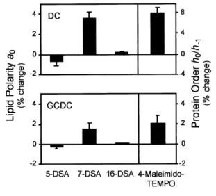
Figure 1 Hydrophobic bile salts influence lipid polarity and protein order, at the surface level, in rat liver mitochondrial membranes
Mitochondria were labelled with 5-DSA, 7-DSA, 16-DSA or 4-maleimido-TEMPO spin probes and exposed to 200µM DC, 200µM GCDC or vehicle only (control) for 5 min as described in the Materials and methods section. Mitochondrial pellets were examined for lipid polarity (a0) and protein order (h0/h
−1) by EPR
spectroscopy analyses. Values are means³S.E.M. from at least four separate experiments of the percentage change relative to controls.
cumulative and dependent on concentration and ex-posure time. Thus higher concentrations are necessary to evaluate toxicity after the short incubation periods required due to the instability of isolated mitochondria. We have chosen to use a 200µM concentration of hydrophobic bile salts for most experiments, because similar concentrations specifically impair mitochondrial respiration and increase membrane permeability in iso-lated organelles [7,12,14,15]. In addition, lower bile salt levels did not significantly change EPR spectra after a 5 min exposure (results not shown).
Table 1 Disruption of lipid fluidity at various depths in isolated rat liver mitochondria exposed to hydrophobic bile salts
Spin-labelled mitochondria were exposed to 200µM DC, 200µM GCDC or vehicle only (control), for 5 min, in respiration buffer as described in the Materials and methods section. Alterations in outer half-width at half-height of the low-field extreme (∆l ) were sensed by the 5-DSA and 7-DSA spin labels, while alterations in the ratio of the heights of the low-field and the centre-field lines (h
+1/h0) were
sensed by the 16-DSA spin label. Percentage decreases in lipid fluidity at C-5 and percentage increases at C-7 and C-16 were calculated based on the corresponding control values. Values are means³S.E.M. of at least four separate experiments.
Change in lipid fluidity (%)
∆l h
+1/h0
Bile salt 5-DSA 7-DSA 16-DSA
DC ®4.2³1.7 8.2³1.5 2.2³0.2 GCDC ®2.6³1.8 5.1³3.3 2.0³0.4
statistical significance at these concentrations (results not shown).
Hydrophobic bile salts at the lowest concentration also increased membrane lipid order in the local micro-environment reported by the 5-DSA spin label. In contrast, using the 7-DSA spin label, DC and GCDC caused a marked elevation in∆l, indicative of increased membrane motion or fluidity, while deeper in the membrane leaflet, h
+"}h! was only slightly raised, as
revealed by the 16-DSA probe. Table 1 shows the values of the motion parameters∆landh
+"}h!expressed as a
percentage of control to identify the membrane region most affected. In fact, lipid fluidity was slightly decreased at C-5 (P!0.05), but markedly enhanced at C-7 (P! 0.01), thus corroborating the information given by the parametera
!.
In parallel experiments, the 4-maleimido-TEMPO spin label was used to gain insight into the protein order structure of mitochondria (Figure 1). After a 5 min exposure to lower concentrations of DC or GCDC, the order of superficial proteins was increased and signifi-cantly different from untreated controls (P!0.01 and P!0.05 respectively). These data are consistent with decreased protein mobility, and provide independent evidence for the proposed interaction of hydrophobic bile salts with the superficial domain of mitochondrial membranes sensed by lipid-oriented spin labels. Further, concentrations of hydrophobic bile salts greater than 200µM markedly decreased protein order (P!0.01), probably reflecting the solubilization of membrane com-ponents.
To determine whether changes in membrane lipid polarity and fluidity could be reversed by hydrophilic bile salts, we analysed membrane perturbations sensed by the 7-DSA spin label using DC in the presence or absence
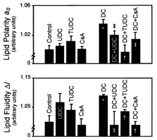
Figure 2 Hydrophilic bile salts and cyclosporin A abrogate disruption of lipid polarity and fluidity induced by DC in rat liver mitochondrial membranes
Labelled mitochondria were exposed to 200µM DC, 500µM UDC, 500µM TUDC, 5µM cyclosporin A (CsA), DC plus UDC, DC plus TUDC, DC plus CsA or vehicle only (control) for 5 min as described in the Materials and methods section. In co-incubation experiments, mitochondria were pretreated with UDC, TUDC or CsA for 5 min. Mitochondrial pellets were examined for lipid polarity (a0) and fluidity (∆l )
sensed by the 7-DSA spin label using EPR spectroscopy analysis. Values are means³S.E.M. from at least four separate experiments, relative to controls. Significance of differences :‡P!0.05,†P!0.01 compared with DC alone ; *P!0.01 compared with control.
of UDC or TUDC (Figure 2). The dynamic properties of mitochondrial membranes at this level were only slightly altered by UDC or TUDC alone compared with untreat-ed organelles. However, markuntreat-ed increases in membrane permeability and motion induced by DC (P!0.01) were significantly prevented by the hydrophilic species, with levels of protection ranging from 40 % to almost 100 %. Similarly, cyclosporin A, an inhibitor of megapore opening, inhibited DC-induced membrane permeabili-zation by at least 60 % (P!0.05). Finally, using the 4-maleimido-TEMPO label, UDC alone decreased protein mobility by"10 % (P!0.001), suggesting a direct effect of the hydrophilic bile salt on the organization of surface membrane proteins. Co-incubation with hydrophilic and hydrophobic bile salts also augmented protein order and, therefore, decreased protein mobility (P!0.05).
Is perturbation of mitochondrial membrane
structure by hydrophobic bile salts
Figure 3 DC and GCDC induce a concentration-dependent release of cytochromecfrom isolated rat liver mitochondria Organelles were exposed to 100, 200 or 500µM DC or GCDC, or to vehicle only (control), for 5 min as described in the Materials and methods section. Mitochondrial pellets were examined for cytochromec levels by Western blot analysis. Following SDS/PAGE and transfer, nitrocellulose membranes were incubated with a monoclonal antibody to cytochromec, and the 15 kDa protein was detected using ECL2.
incubation with various concentrations of DC or GCDC. Cytochromec was abundant in control mitochondria, but levels decreased progressively after exposure to 100, 200 and 500µM DC or GCDC (Figure 3), corresponding to a proportional increase in supernatant levels (P! 0.01). Thus cytochrome c release at concentrations of hydrophobic bile salts slightly lower than those necessary to induce structural changes suggest that the efflux of intermembrane proteins is a primary event that may further disrupt mitochondrial membrane structure, again increasing permeability, in a vicious cycle. Further, these data support a role for a specific injury rather than non-specific effects of these detergents, which was also confirmed by a marked cyclosporin A-sensitive decrease in cytochrome c levels in mitochondria suspensions treated with hydrophobic bile salts (P!0.01). Finally, as shown in previous studies, cytochrome c efflux was reduced by "70 % during pretreatment with either UDC or TUDC (P!0.01).
Of note is the observation that the release of glutamate dehydrogenase from the mitochondrial matrix after a 5 min exposure to DC and GCDC, at lower concen-trations, induced only 5 % and 1 % increases respectively in enzyme release. In addition, these bile salts directly caused mitochondrial release of cytochrome c, but not cytochromecoxidase (results not shown), corroborating the hypothesis that the cytotoxicity of these bile salts may be associated with specific physical perturbation of the mitochondrial membrane, and not simply organelle bursting.
Is the increase in permeability of
mitochondrial membranes induced by
hydrophobic bile salts associated with
altered redox status ?
To determine if exposure to DC or GCDC modifies the levels of mitochondrial membrane oxidation, isolated
Figure 4 DC induces oxidative damage of lipids in rat liver mitochondrial membranes
Mitochondria were labelled with either 5-DSA or 12-DSA spin probes and exposed to DC for 5 min as described in the Materials and methods section. Mitochondrial pellets were examined for free radical production using EPR spectroscopy analysis. (A) Dose–response to DC-induced decrease in signal amplitude sensed by 5- and 12-DSA spin labels. Labelled mitochondria were exposed to either DC (200, 500 or 1000µM) or vehicle only (0µM ; control). (B) Alteration in EPR spectra sensed by the 5-DSA spin label. Labelled mitochondria were exposed to 500µM DC, 500µM UDC, 500µM TUDC, DC plus UDC, DC plus TUDC or vehicle only (control). In co-incubation experiments, mitochondria were pretreated with either UDC or TUDC for 5 min. Values are means³S.E.M. from at least four separate experiments relative to controls. Significance of differences :§P!0.05,*P!0.01 compared with control.
extent. These results indicate less oxidative damage in deeper regions of the bilayer, and support a specific superficial effect. Finally, perturbation induced by GCDC was rapid, but only significant at higher concen-trations (results not shown).
Spin-label signal amplitude in isolated mitochondria was also assessed in the presence of UDC or TUDC (Figure 4B). Treatment of 5-DSA-labelled mitochondria with each hydrophilic bile salt alone did not significantly decrease signal amplitude, while incubation with UDC or TUDC for 5 min prior to exposure to DC completely abrogated oxidative injury (P!0.01). This protection resulted in signal amplitudes that were no different from those of controls, indicating that the hydrophilic bile salts either confer direct protection against lipid peroxidation in isolated mitochondrial membranes or prevent the insertion of hydrophobic bile salts into membranes, indirectly inhibiting free radical production.
DISCUSSION
The mechanisms of hepatocellular damage during liver disease are not entirely understood, but appear to be related to the retention of toxic substances. It is widely recognized that accumulation of hydrophobic bile salts within the hepatocyte plays a key part in liver injury [1–3], where the prominence of hepatocyte-derived acidophilic bodies and cell dropout, rather than extensive necrosis, is supportive of a role for apoptosis [4,5,23]. In addition, exposure of cells to toxic bile salts has been shown to cause apoptosis, bothin vivoandin vitro, in a concentration- and time-dependent manner, at concen-trations that are far lower than the critical micellar concentration associated with cell necrosis [6,7]. Finally, several studies have shown that bile salt-induced cyto-toxicity may be a consequence of mitochondrial dys-function. Short-time incubations of isolated rat liver mitochondria with hydrophobic bile salts causes im-paired mitochondrial respiration [12] as well as mor-phological changes associated with the opening of the mitochondrial permeability transition pore [7,13–15], suggesting a specific membrane perturbation. The nature of membrane changes and the sequence of events that occur in mitochondria remain, however, poorly defined, in part because of the intrinsic difficulty [39] of measuring the dynamic properties of mitochondrial membranes.
The detailed mechanism(s) by which hydrophobic bile salts increase membrane permeability in isolated mito-chondria is not currently understood. Cyclosporin A, which is commonly used to prevent the mitochondrial permeability transition, has been reported to inhibit cytochrome c release from mitochondria treated with hydrophobic bile salts [7,13–15]. This observation raises the possibility that DC and GCDC may interact with membrane components, promoting the release of
inter-membrane proteins through a cyclosporin A-sensitive megapore. The present study demonstrates a direct physical interaction between hydrophobic bile salts and mitochondrial membranes. Indeed, membrane lipid po-larity and fluidity were particularly disturbed near the lipid–water interface, more than in hydrophobic regions, suggesting a specific effect rather than non-specific changes. The decreased mitochondrial membrane po-larity and fluidity reported by the 5-DSA spin label suggests that hydrophobic bile salts intercalated at the C-5 membrane region, increasing phospholipid packing and thus reducing the already low mobility of the phos-pholipid acyl chain in this region of the leaflet. This was confirmed by the order parameter S. The superficial effect described above would thus render inner regions of the membrane more permeable, as indicated by the increaseda
!value sensed by either 7-DSA or, to a much
lower extent, 16-DSA. Further, since perturbation of the mitochondrial membrane induced by hydrophobic bile salts decreased with increasing depth of the leaflet from C-7 to C-16, it is conceivable that this gradient may result from an effect secondary to the superficial accommodation of toxic bile salts. This scenario is not without precedent, since similar effects have been described for other membrane-disturbing agents [40,41]. Finally, exposure to bile salt concentrations greater than 200µM resulted in increased perturbation sensed by all spin labels ; this was, however, proportionally greater in deeper regions, prob-ably reflecting lipid solubilization due to detergent effects. To this end, studies using erythrocyte and hepatocyte membranes also showed marked spectral alterations in apolar regions induced by a different hydrophobic bile salt, unconjugated chenodeoxycholate, at concentrations ranging from 250 to 1000µM [42]. Moreover, the increased membrane polarity after in-cubation with higher bile salt concentrations has been described to correlate with enhanced solubilization of cholesterol and phospholipids [42]. Interestingly, the superficial interaction of hydrophobic bile salts sensed by lipid spin labels may explain the observation that DC and GCDC decreased the mobility of the 4-maleimido-TEMPO label, thus reflecting augmented superficial protein order structure above control values. As expect-ed, higher bile salt concentrations led to increased mobility of this spin label and, therefore, to enhanced membrane fluidity, in accordance with suggestions by other authors [28].
concentrations. It appears that the release of cytochrome c may further disrupt membrane structure, again in-creasing permeability, in a vicious cycle. Moreover, oxidative damage seems to be a consequence of cyto-chromecrelease rather than a promoter of it, whereby the absence of cytochromecmay inhibit electron transfer, disrupting the respiratory chain.
The paramagnetic signal of 5-DSA was markedly lowered in DC-treated mitochondria in particular, re-flecting free radical production, while at C-12 the same effect was slightly attenuated. These findings are support-ed by other studies showing that hydrophobic bile salts promote oxidative stress [7,8,43,44], and are also con-sistent with signs of diminished injury during treatment with antioxidants [45,46].
To determine whether hydrophilic bile salts may play a role in preventing DC- and GCDC-triggered mem-brane perturbation, isolated mitochondria were pre-treated with UDC or TUDC. These hydrophilic bile salts almost completely reversed both the disruption of mitochondrial membrane structure and the oxidative damage caused by the hydrophobic species. The pro-tective role of UDC and TUDC at both the 5 and C-7 levels of the mitochondrial membrane also suggests interaction at superficial membrane domains. However, this explanation does not exclude the possibility that UDC might directly influence protein function, as suggested by the marked effect on protein order reported by the 4-maleimido-TEMPO spin label, or might also interact with deeper regions of the membrane, as reported by others [28]. Most probably, the binding sites of the various bile salts are not the same. As previously suggested, the stabilization of the mitochondrial mem-brane by UDC and TUDC implied here provides an additional explanation for the protective role of these bile salts in preventing apoptosis in several model systems [7,15,47]. Similar observations were made when mito-chondria were preincubated with cyclosporin A. This inhibitor of the megapore opening prevents cytochrome c release induced by several toxic agents [48,49], pre-cluding the involvement of the mitochondrial permea-bility transition during induction of cytotoxicity by hydrophobic bile salts.
Collectively, data from the present study appear to suggest a mechanism of toxicity that involves rapid perturbation of superficial mitochondrial membrane structure during incubation with hydrophobic bile salts, resulting in increased membrane permeability and oxi-dative injury. UDC, TUDC and cyclosporin A almost completely abrogated DC- and GCDC-induced per-turbation of mitochondrial membrane structure. These observations imply that the cytotoxicity of hydrophobic bile salts may be mediated, at least in part, by their effect of physically disturbing the mitochondrial membrane rather than by an influence on the expression of pro-apoptotic Bax. Perturbation of mitochondrial membrane
structure may indeed represent an important component of the apoptotic pathway triggered by accumulated bile salts during liver disease, confirming mitochondria as primary targets of bile salt-induced cytotoxicity. Thus new prospects for the treatment of liver diseases should consider the use of molecules that, like UDC and its conjugated derivatives, stabilize the mitochondrial mem-brane.
ACKNOWLEDGMENTS
We thank Carlos D. Brondino (Departamento de Quı!mica, FCT, Universidade Nova de Lisboa, Monte de Caparica, Portugal) for his expertise in the use of the EPR spectrometer. This work was supported in part by grant PRAXIS}C}SAU}14311}1998 from Fundac:a4o para a Cie#ncia e a Tecnologia, Lisbon, Portugal, and by a Research Fellowship 2000 from the European Association for the Study of the Liver (to C. M. P. R.).
REFERENCES
1 Scholmerich, J., Becher, M. S., Schmidt, K. et al. (1984) Influence of hydroxylation and conjugation of bile salts on their membrane-damaging properties – studies on isolated hepatocytes and lipid membrane vesicles. Hepatology4, 661–666
2 Schmucker, D. L., Ohta, M., Kanai, S., Sato, Y. and Kitani, K. (1990) Hepatic injury induced by bile salts: correlation between biochemical and morphological events. Hepatology12, 1216–1221
3 Heuman, D. M., Mills, A. S., Mccall, J., Hylemon, P. B., Pandak, W. M. and Vlahcevic, Z. R. (1991) Conjugates of ursodeoxycholate protect against cholestasis and hepatocellular necrosis caused by more hydrophobic bile salts. In vivo studies in the rat. Gastroenterology100, 203–211
4 Columbano, A. (1995) Cell death: current difficulties in discriminating apoptosis from necrosis in the context of pathological processes in vivo. J. Cell. Biochem.58, 181–190
5 Patel, T. and Gores, G. J. (1995) Apoptosis and hepatobiliary disease. Hepatology21, 1725–1741 6 Patel, T., Bronk, S. F. and Gores, G. J. (1994) Increases of
intracellular magnesium promote glycodeoxycholate-induced apoptosis in rat hepatocytes. J. Clin. Invest.94, 2183–2192
7 Rodrigues, C. M. P., Fan, G., Ma, X., Kren, B. T. and Steer, C. J. (1998) A novel role for ursodeoxycholic acid in inhibiting apoptosis by modulating mitochondrial membrane perturbation. J. Clin. Invest.101, 2790–2799 8 Yerushalmi, B., Dahl, R., Devereaux, M. W., Gumpricht,
E. and Sokol, R. J. (2001) Bile acid-induced rat hepatocyte apoptosis is inhibited by antioxidants and blockers of the mitochondrial permeability transition. Hepatology33, 616–626
9 Schaffner, F., Bacchin, P. G., Hutterer, F. et al. (1971) Mechanism of cholestasis. 4. Structural and biochemical changes in the liver and serum in rats after bile duct ligation. Gastroenterology60, 888–897
10 Kra$henbu$hl, S., Stucki, J. and Reichen, J. (1992) Reduced activity of the electron transport chain in liver
mitochondria isolated from rats with secondary biliary cirrhosis. Hepatology15, 1160–1166
12 Kra$henbu$hl, S., Talos, C., Fischer, S. and Reichen, J. (1994) Toxicity of bile acids on the electron transport chain of isolated rat liver mitochondria. Hepatology19, 471–479
13 Botla, R., Spivey, J. R., Aguilar, H., Bronk, S. F. and Gores, G. J. (1995) Ursodeoxycholate (UDCA) inhibits the mitochondrial membrane permeability transition induced by glycochenodeoxycholate : a mechanism of UDCA cytoprotection. J. Pharmacol. Exp. Ther.272, 930–938
14 Rodrigues, C. M. P., Fan, G., Wong, P. Y., Kren, B. T. and Steer, C. J. (1998) Ursodeoxycholic acid may inhibit deoxycholic acid-induced apoptosis by modulating mitochondrial transmembrane potential and reactive oxygen species production. Mol. Med.4, 165–178 15 Rodrigues, C. M. P., Ma, X., Linehan-Stieers, C., Fan, G.,
Kren, B. T. and Steer, C. J. (1999) Ursodeoxycholic acid prevents cytochrome c release in apoptosis by inhibiting mitochondrial membrane depolarization and channel formation. Cell Death Differ.6, 842–854
16 Lieser, M. J., Park, J., Natori, S., Jones, B. A., Bronk, S. F. and Gores, G. J. (1998) Cholestasis confers resistance to the rat liver mitochondrial permeability transition. Gastroenterology115, 693–701
17 Degterev, A., Boyce, M. and Yuan, J. (2001) The channel of death. J. Cell Biol.155, 695–698
18 De Giorgi, F., Lartigue, L., Bauer, M. K. A. et al. (2002) The permeability transition pore signals apoptosis by directing Bax translocation and multimerization. FASEB J.16, 607–609
19 Liu, X., Kim, C. N., Yang, J., Jemmerson, R. and Wang, X. (1996) Induction of apoptotic program in cell-free extracts : requirement for dATP and cytochrome c. Cell86, 147–157
20 Zou, H., Henzel, W. J., Liu, X., Lutschg, A. and Wang, X. (1997) Apaf-1, a human protein homologous to C. Elegans Ced-4, participates in cytochrome c-dependent activation of caspase-3. Cell90, 405–413
21 Kroemer, G. (1997) The proto-oncogene bcl-2 and its role in regulating apoptosis. Nat. Med. (N.Y.)3, 614–620 22 Beuers, U., Boyer, J. L. and Paumgartner, G. (1998)
Ursodeoxycholic acid in cholestasis : potential mechanisms of action and therapeutic applications. Hepatology28, 1449–1453
23 Rodrigues, C. M. and Steer, C. J. (2000) Mitochondrial membrane perturbations in cholestasis. J. Hepatol.32, 135–141
24 Azzaroli, F., Mehal, W., Soroka, C. J. et al. (2002) Ursodeoxycholic acid diminishes Fas-ligand-induced apoptosis in mouse hepatocytes. Hepatology36, 49–54 25 Mariash, C. N., Seelig, S., Schwartz, H. L. and
Oppenheimer, J. H. (1986) Rapid synergistic interaction between thyroid hormone and carbohydrate on mRNAS14 induction. J. Biol. Chem.261, 9583–9586 26 Jost, P., Libertini, L. J., Hebert, V. C. and Griffith, O. H.
(1971) Lipid spin labels in lecithin multilayers. A study of motion along fatty acid chains. J. Mol. Biol.59, 77–98 27 Harris, J., Power, T. J., Bieber, A. L. and Watts, A. (1983)
An electron-spin-resonance spin-label study of the interaction of purified mojave toxin with synaptosomal membranes from rat brain. Eur. J. Biochem.131, 559–565 28 Gu$ldutu$na, S., Zimmer, G., Leuschner, M. et al. (1999)
The effect of bile salts and calcium on isolated rat liver mitochondria. Biochim. Biophys. Acta1453, 396–406 29 Mason, R. P., Giavedoni, E. B. and Dalmasso, A. P.
(1977) Complement-induced decrease in membrane mobility : introducing a more sensitive index of spin-label motion. Biochemistry16, 1196–1201
30 Hubbell, W. L. and Mcconnell, M. (1971) Molecular motion in spin-labeled phospholipids and membranes. J. Am. Chem. Soc.93, 314–326
31 Schreier, S., Frezzatti, W. A., Araujo, P. S., Chaimovich, H. and Cuccovia, I. M. (1984) Effect of lipid membranes on the apparent pk of the local anesthetic tetracaine. Spin label and titration studies. Biochim. Biophys. Acta769, 231–237
32 Bianconi, M. L., do Amaral, A. T. and Schreier, S. (1988) Use of membrane spin label spectra to monitor rates of reaction of partitioning compounds: hydrolysis of a local anesthetic analog. Biochem. Biophys. Res. Commun.152, 344–350
33 Lai, C. S., Tooney, N. M. and Ankel, E. G. (1984) Structure and flexibility of plasma fibronectin in solution: electron spin resonance spin-label, circular dichroism, and sedimentation studies. Biochemistry23, 6393–6397 34 Kunicki, T. J., Nugent, D. J., Piotrowicz, R. S. and Lai,
C. S. (1986) Covalent attachment of sulfhydryl-specific, electron spin resonance spin-labels to fab’ fragments of murine monoclonal antibodies that recognize human platelet membrane glycoproteins. Development of membrane protein specific spin probes. Biochemistry25, 4979–4983
35 Bruce-Keller, A. J., Begley, J. G., Fu, W. et al. (1998) Bcl-2 protects isolated plasma and mitochondrial membranes against lipid peroxidation induced by hydrogen peroxide and amyloid beta-peptide. J. Neurochem.70, 31–39 36 Butterfield, D. A., Hensley, K., Harris, M., Mattson, M.
and Carney, J. (1994)β-amyloid peptide free radical fragments initiate synaptosomal lipoperoxidation in a sequence-specific fashion : implications to Alzheimer’s disease. Biochem. Biophys. Res. Commun.200, 710–715 37 Hensley, K., Carney, J., Hall, N., Shaw, W. and
Butterfield, D. A. (1994) Electron paramagnetic resonance investigations of free radical-induced alterations in neocortical synaptosomal membrane protein infrastructure. Free Radical Biol. Med.17, 321–331 38 Roda, A., Minutello, A., Angellotti, M. A. and Fini, A.
(1990) Bile acid structure-activity relationship : evaluation of bile acid lipophilicity using 1-octanol}water partition coefficient and reverse phase HPLC. J. Lipid Res.31, 1433–1443
39 Bernardi, P., Scorrano, L., Colonna, R., Petronilli, V. and Di Lisa, F. (1999) Mitochondria and cell death.
Mechanistic aspects and methodological issues. Eur. J. Biochem.264, 687–701
40 Ondrias, K., Stas)ko, A. and Balgavy!, P. (1987) Spin label study of the perturbation effect of the local anaesthetics tetracaine and dibucaine on synaptosomes at
pharmacological concentrations. Biochem. Pharmacol.36, 3999–4005
41 Brito, M. A., Brondino, C. D., Moura, J. J. G. and Brites, D. (2001) Effects of bilirubin molecular species on membrane dynamic properties of human erythrocyte membranes : a spin label electron paramagnetic resonance spectroscopy study. Arch. Biochem. Biophys.387, 57–65 42 Gu$ldutu$na, S., Zimmer, G., Imhof, M., Bhatti, S., You, T.
and Leuschner, U. (1993) Molecular aspects of membrane stabilization by ursodeoxycholate. Gastroenterology104, 1736–1744
43 Sokol, R. J., Devereaux, M. W., Khandwala, R. and O ’Brien, K. (1993) Evidence for involvement of oxygen free radicals in bile acid toxicity to isolated rat hepatocytes. Hepatology17, 869–881
44 Sokol, R. J., Winklhofer-Roob, B. M., Devereaux, M. W. and McKim, Jr, J. M. (1995) Generation of
hydroperoxides in isolated rat hepatocytes and hepatic mitochondria exposed to hydrophobic bile acids. Gastroenterology109, 1249–1256
45 Sokol, R. J., Devereaux, M. W. and Khandwala, R. (1991) Effect of dietary lipid and vitamin E on mitochondrial lipid peroxidation and hepatic injury in the bile duct-ligated rat. J. Lipid Res.32, 1349–1357
46 Sokol, R. J., McKim, J. M., Goff, M. C. et al. (1998) Vitamin E reduces oxidant injury to mitochondria and the hepatotoxicity of taurochenodeoxycholic acid in the rat. Gastroenterology114, 164–174
48 Pastorino, J. G., Synder, J. W., Serroni, A., Hoek, J. B. and Farber, J. L. (1993) Cyclosporine and carnitine prevent the anoxic death of cultured hepatocytes by inhibiting the mitochondrial permeability transition. J. Biol. Chem.268, 13791–13798
Received 17 July 2002; accepted 8 August 2002