• Nenhum resultado encontrado

Brain and Peripheral Organ Toxicity of "Ecstasy" in Adolescent Rats in Human Relevant Doses

N/A
N/A
Protected

Academic year: 2021

Share "Brain and Peripheral Organ Toxicity of "Ecstasy" in Adolescent Rats in Human Relevant Doses"

Copied!
153
0
0

Texto

(1)
(2)
(3)

Armanda Teixeira Gomes

Brain and peripheral organ toxicity of “ecstasy” in

adolescent rats in human relevant doses

Dissertation thesis for the Master Degree in Analytical

Clinical and Forensic Toxicology

Dissertação do 2º Ciclo de Estudos Conducente ao Grau de

Mestre em Toxicologia Analítica Clínica e Forense

Trabalho realizado sob a orientação de

Elaborated under supervision of

Professor Doutor João Paulo Soares Capela

Professora Doutora Vera Marisa Costa

Professor Doutor Félix Dias Carvalho

(4)
(5)

É AUTORIZADA A REPRODUÇÃO PARCIAL DESTA TESE, APENAS PARA EFEITOS DE INVESTIGAÇÃO, MEDIANTE DECLARAÇÃO ESCRITA DO INTERESSADO, QUE A TAL SE COMPROMETE.

(6)
(7)

The results presented in this thesis led to the following works:

Panel communications:

A. Teixeira-Gomes, V.M. Costa, R. Feio-Azevedo, F.C. Pereira, M. Duarte-Araújo, E. Fernandes, M.L. Bastos, F. Carvalho, and J.P. Capela. ―Differential toxicity in the cerebellum of adolescent and aged rats following a MDMA neurotoxic regimen‖. 7th Meeting of young researchers of U. Porto (IJUP ‗14), Porto, Portugal, February 2014.

R. Feio-Azevedo, V.M. Costa, D. Barbosa, A. Gomes, F.C. Pereira, M. Duarte-Araújo, J.A. Duarte, E. Fernandes, M.L. Bastos, F. Carvalho, and J.P. Capela. ―Aged rats are more prone to ―Ecstasy‖ neurotoxicity than adolescent rats.‖ 50th Congress of the European Societies of Toxicology (Eurotox 2014), Edinburgh, United Kingdom, September 2014.

Papers in international peer-reviewed journals:

A. Teixeira-Gomes, V.M. Costa, R. Feio-Azevedo, M.L. Bastos, F. Carvalho, and J.P. Capela, ―The neurotoxicity of amphetamines during the adolescent period‖. Submitted to International Journal of Developmental Neurosciences.

A. Teixeira-Gomes, V.M. Costa, R. Feio-Azevedo, J.A. Duarte, M.L. Bastos, F. Carvalho, and J.P. Capela, ―Acute brain and peripheral organ toxicity of MDMA to adolescent rats following a human relevant binge dose‖. Submitted to Toxicology Letters.

(8)
(9)

Neste momento tão especial do meu percurso académico, não posso deixar de agradecer a todas as pessoas que tornaram possível a concretização desta dissertação. Sem vocês não teria sido uma etapa alcançável.

Ao Professor Doutor João Paulo Soares Capela, meu orientador, não consigo agradecer o suficiente pela orientação, confiança, incentivos e apoio prestado ao longo da realização desta dissertação. Agradeço por me ter dado a oportunidade de trabalhar neste projecto e agradeço, também, os conhecimentos transmitidos e o interesse com que sempre me auxiliou ao longo desta jornada. Obrigada por tudo!

À Professora Doutora Vera Marisa Costa, minha co-orientadora, agradeço todos os conhecimentos que me transmitiu com tanta competência e todo o apoio prestado em todas as fases de realização desta dissertação. Não podia estar mais grata pela infinita preocupação, confiança, incentivo, conselhos e paciência, sobretudo, pela paciência… Não há palavras suficientes para agradecer todo o auxílio que me prestou!

Ao Professor Doutor Félix Dias Carvalho, meu co-orientador, agradeço os conselhos e saber que me transmitiu ao longo deste trabalho e, também, o seu constante entusiasmo e otimismo.

À Professora Doutora Maria de Lourdes Bastos, agradeço o constante interesse em criar as melhores condições para a realização do Mestrado em Toxicologia Analítica Clínica e Forense. Agradeço, também, a forma atenciosa e disponível como me auxiliou sempre que precisei.

Ao Professor Doutor José Duarte, devo um enorme agradecimento pela oportunidade de realização da componente histológica da minha dissertação na FADEUP. Agradeço, acima de tudo, a preciosa ajuda que me facultou, os conselhos e toda a simpatia que sempre demonstrou.

À Professora Doutora Margarida Araújo, agradeço por ter disponibilizado os serviços do Biotério ICBAS-UP e toda a ajuda prestada durante os ensaios experimentais. Foi crucial para a realização deste trabalho. Este agradecimento é também dirigido à Dra. Bárbara e Dra. Luísa, que foram incansáveis.

(10)

A todos os Professores do Mestrado em Toxicologia Analítica Clínica e Forense, agradeço pelos conhecimentos que me transmitiram com tanta competência e dedicação ao longo destes dois anos de Mestrado. Incutiram em mim o gosto pela Toxicologia e, por isso, lhes estou eternamente grata.

A todos os meus colegas e restantes membros do Departamento de Toxicologia da Faculdade de Farmácia da Universidade do Porto (Engenheira Maria Elisa Soares, Doutora Renata Silva, Mestre Maria João Valente, Doutora Diana Dias da Silva, Doutora Filipa Ponte, e a todos os outros elementos que dele fazem parte) agradeço a forma amável como fui recebida e ajudada sempre que precisei. Obrigada Juliana Garcia e José Luís Sousa por toda a ajuda e companhia. Agradeço, também, à Cátia Faria pela prontidão que sempre revelou na resolução dos nossos problemas. Em especial, agradeço, à Ana Sofia Oliveira, à Mariana Fernandes, à Débora Rios de Oliveira, à Filipa Ferreira, à Ana Margarida Araújo e à Márcia Monteiro as gargalhadas e boa disposição, e por nunca me deixarem sem alimentos durante o trabalho.

À D. Celeste, agradeço a sua ajuda imprescindível com os procedimentos histológicos.

Ao Doutor Daniel Silva, agradeço por todo o auxílio prestado no procedimento experimental. Obrigada pelos inúmeros conselhos, amizade dispensada e pela preocupação demonstrada durante este ano.

À Rita Azevedo, não há palavras suficientes para mostrar o quanto estou grata. Obrigada pela imprescindível ajuda que me deste ao longo de todo este ano! Foi um prazer conhecer-te e trabalhar contigo e, sem dúvida, foi um prazer aprender contigo. Obrigada por tudo!

Aos meus amigos, agradeço toda a amizade e apoio prestado neste último ano em que mais vos faltei, e, por isso, peço desculpa. Um obrigada especial ao Pedro pela ajuda nas formatações!

Ao João, agradeço todo o amor, companheirismo, carinho, dedicação e apoio durante este longo ano! Agradeço a paciência e o facto de nunca duvidares das minhas capacidades e de me incentivares a nunca desistir dos meus objectivos. Sem ti, tudo seria muito mais difícil… Obrigada por tudo!

(11)

Aos meus pais e irmã, tenho tudo a agradecer… Sem a vossa confiança, amor e carinho nada disto teria sido possível. Obrigada por nunca duvidarem de mim, acreditarem sempre que sou capaz e por me apoiarem em todos os meus objectivos. O que mais me encoraja nestes momentos decisivos é saber que tenho os melhores do mundo comigo, sempre. Tenho-vos a vocês. Obrigada!

Obrigada a todos!

Agradeço também o apoio financeiro do trabalho experimental desta dissertação de Mestrado por parte da Fundação para a Ciência e a Tecnologia através do projecto PTDC/SAU-FCF/102958/2008, no âmbito do Programa Operacional Temático Factores de Competitividade (COMPTE) do Quadro Comunitário de Apoio III e do Fundo Comunitário Europeu (FEDER) (FCOMP-01-0124-FEDER-011079).

(12)
(13)

3,4-Methylenedioxymethamphetamine (MDMA or ―ecstasy‖) is a well-known psychoactive drug, with acknowledged neurotoxic properties. It is commonly used in recreational settings by many teenagers and young adults. Despite the great deal of study on MDMA neurotoxicity, the consequences of MDMA exposure during the adolescent period require additional research. Moreover, MDMA effects on the cerebellum and peripheral organs have been scarcely studied. The susceptibility of different aged groups is also poorly known.

This dissertation aimed to assess the acute toxicity of a MDMA binge dose regimen comparable to human usage in four different brain areas (cerebellum, hippocampus, cortex and striatum), and in three peripheral organs (liver, heart and kidneys) in an adolescent rat, 24 h after MDMA binge administration. Another aim was to evaluate the long-term neurotoxicity of the binge MDMA dose regimen in the cerebellum, by assessing its energetic and oxidative stress status in adolescent and old rats, seven days after MDMA administration.

To achieve the aims of the present dissertation, two experiments were conducted. In experiment one, adolescent male Wistar rats (postnatal day 40) were divided in two groups and treated: control saline (three doses of NaCl 0.9%, intraperitoneally, every 2 h, n=7) and MDMA-treated (three doses of MDMA 5 mg/Kg, intraperitoneally, every 2 h, n=7). The temperature of each animal was monitored for 7 h after the first injection. Twenty-four hours after the MDMA administration, brain areas were dissected and the peripheral organs were collected. A significant hyperthermia in the adolescent rats was observed after MDMA exposure. No differences were found between controls and MDMA-treated groups in body weight gain, as well as in food or water consumption, within the experimental period. Adenosine triphosphate (ATP) content was significantly decreased in the cortex of MDMA-treated rats, but not in the other areas. In all brain areas, no alterations were found in glutathione, quinoproteins and protein carbonylation levels in MDMA-treated animals. In liver, heart and kidneys no differences were found in glutathione, protein carbonylation or ATP levels, after MDMA exposure. However, the formation of protein-bound quinones was significantly increased in the liver of MDMA-treated rats, but not in the other two peripheral organs. The histological hepatic examination of adolescent MDMA-treated rats revealed a marked cellular vacuolization in the periportal regions, and sinusoidal dilatation with periportal and centrilobular vascular congestion. In the heart, MDMA promoted punctual signs of cardomyocyte oedema, particularly in the sub-endocardic region. In the kidneys of MDMA-treated adolescent rats,

(14)

scattered interstitial oedema, with dilatation between the nefrotic tubular structures, and signs of vascular congestion were found. The lack of severe cellular damage and/or necrosis in MDMA-treated rats is corroborated by the lack of increase of caspase-3, -8 and -9 activities or changes in the plasma biomarkers of liver or heart injury.

Experiment two was conducted with adolescent (postnatal day 40) and aged (18 to 22 months old) male Wistar rats. Animals were divided in two groups: control saline (n=5) and MDMA group (n=5) that were treated as described for experiment one. Seven days later, animals were sacrificed and the cerebellum was collected. Aged animals did not receive the third dose of MDMA given the high risk of mortality, due to the hyperthermic response verified after the second MDMA administration. Until sacrifice, animal weight and food/water consumption were registered daily. Despite the exposure to a lower cumulative dose the hyperthermic response was more pronounced in aged rats than in adolescents, and the temperature of aged rats remained significantly elevated 24 h after drug exposure. MDMA promoted decreases in adolescent animals‘ body weight at 24 h post-MDMA administration. In aged animals, the food intake suffered a biphasic response after MDMA exposure: at 24 h a decrease was observed, while at 48 h an increase in consumption occurred in MDMA-treated animals, when compared to aged controls. The water intake was significantly increased in aged animals, 24 and 48 h after MDMA exposure. In the cerebellum, all the oxidative stress parameters remained unaltered in both adolescent and aged MDMA-treated rats, whereas decreases in the ATP content were seen in MDMA-treated aged rats.

Altogether, these results suggest that moderate MDMA doses do not promote any significant alterations in the oxidative stress parameters in the adolescents‘ brain areas, 24 h after the exposure. Importantly, the MDMA binge scheme used in adolescent animals promoted morphological tissue alterations to the peripheral organs. Regarding the age factor, a long-term ATP decrease was seen in the cerebellum of aged rats, indicating a higher susceptibility of this group to MDMA. Further investigation is needed to evaluate the impact of moderate MDMA doses either in the brain as in peripheral organs of adolescent animals, and to explore the dissimilar effects of MDMA to different aged animals.

Keywords: Amphetamines, "Ecstasy", Adolescence, Rats, Brain, Peripheral organs,

Oxidative stress, Toxicity.

(15)

A 3,4-metilenodioximetanfetamina (MDMA ou ―ecstasy‖) é uma substância psicoativa bastante conhecida, com propriedades neurotóxicas bem documentadas. É habitualmente consumida em contextos recreativos, principalmente entre os adolescentes e jovens adultos. Apesar de existirem imensos estudos acerca da neurotoxicidade da MDMA, as consequências da exposição a esta substância durante a adolescência requerem, ainda, mais investigação. Para além disso, os efeitos da MDMA no cerebelo e nos órgãos periféricos têm sido alvo de poucos estudos. Acrescente-se que pouco se sabe sobre a suscetibilidade de diferentes faixas etárias a esta droga de abuso.

Esta dissertação teve como objetivo avaliar a toxicidade aguda decorrente de um regime de MDMA, comparável ao utilizado pelos humanos, em quatro áreas do cérebro (cerebelo, hipocampo, córtex e corpo estriado), e três órgãos periféricos (fígado, coração e rins) em ratos adolescentes, 24 h após a administração de MDMA. Outro objetivo centrou-se na avaliação da toxicidade a longo prazo a nível do cerebelo, decorrente da administração repetida de MDMA. Para atingir os objectivos propostos foram avaliados quer parâmetros de stress oxidativo, quer do estado energético, em ratos adolescentes e idosos, sete dias após a administração de MDMA.

De forma a atingir os objetivos traçados nesta dissertação, foram levados a cabo dois trabalhos experimentais distintos. No primeiro, ratos Wistar macho (40 dias de idade) foram divididos em dois grupos e tratados: controlo salino (três doses de NaCl 0.9 %, intraperitonealmente, a cada 2 h, n=7) e tratados com MDMA (três doses de MDMA 5 mg/kg, intraperitonealmente, a cada 2 h, n=7). A temperatura de cada animal foi monitorizada durante 7 h após a primeira injeção. As áreas cerebrais foram dissecadas e os órgãos periféricos recolhidos 24 h após a administração de MDMA. Após a exposição à MDMA foi observada hipertermia nos ratos adolescentes. Não foram encontradas diferenças entre o grupo controlo e o grupo tratado com MDMA no que diz respeito ao ganho de peso, consumo de água e ingestão de alimentos, durante o período experimental. O conteúdo de adenosina trifosfato (ATP) diminuiu significativamente no córtex dos ratos tratados com MDMA, mas não em outras áreas cerebrais. Nos animais tratados com MDMA não foram observadas alterações nos níveis de glutationa, quinoproteínas e carbonilação proteica, em todas as áreas cerebrais estudadas. No fígado, coração e rins dos animais não se encontraram alterações nos níveis de glutationa, carbonilação proteica e ATP, após a exposição à MDMA. No entanto, houve um aumento significativo da formação de quinoproteínas no fígado dos ratos tratados com MDMA, mas não nos restantes órgãos estudados. A observação histológica do

(16)

fígado dos ratos adolescentes tratados com MDMA, revelou vacuolização celular nas regiões periportais e um alargamento dos espaços sinusoidais com congestão sanguínea periportal e centrilobular. No coração dos animais tratados com MDMA, foram observados pontualmente cardiomiócitos edemaciados, particularmente na região sub-endocárdica. Nos rins de ratos adolescentes tratados com MDMA foram encontrados sinais de edema intersticial disperso com alargamento do espaço entre as estruturas tubulares dos nefrónios, assim como sinais de congestão sanguínea. A ausência de dano celular acentuado e/ou necrose em ratos adolescentes tratados com MDMA é corroborada pelo facto de não se ter verificado aumento nas atividades das caspases 3, 8 e 9, ou alteração nos níveis plasmáticos dos biomarcadores de lesão cardíaca ou hepática.

O segundo trabalho experimental foi executado com ratos Wistar macho adolescentes (40 dias de idade) e ratos idosos (18 a 22 meses de idade). Os animais foram, mais uma vez, divididos em dois grupos: controlo salino (n=5) e grupo tratado com MDMA (n=5), administrados como descrito no primeiro trabalho experimental. Sete dias depois, os animais foram sacrificados e o cerebelo foi recolhido. Os animais idosos não receberam a terceira dose de MDMA, dado o elevado risco de mortalidade decorrente da resposta hipertérmica verificada após administração da segunda dose da droga. Até ao sacrifício dos animais, o seu peso, ingestão de água e alimentos foram registados diariamente. Apesar dos ratos idosos terem sido expostos a uma dose total de MDMA menor que a dos ratos adolescentes, a resposta hipertérmica dos ratos idosos foi mais pronunciada e a temperatura dos mesmos manteve-se significativamente elevada 24 h após a exposição à droga. A MDMA levou à perda de peso nos ratos adolescentes, 24 h após a sua administração. Relativamente aos animais idosos, na ingestão de alimentos verificou-se uma resposta bifásica após a exposição à MDMA: foi observada uma diminuição às 24 h, enquanto que às 48 h houve um aumento no grupo de animais tratados com a droga em comparação com os animais controlo. A ingestão de água aumentou significativamente nos animais idosos, 24 e 48 h após a exposição à MDMA. Ao nível do cerebelo, todos os parâmetros de stress oxidativo se mantiveram inalterados nos animais adolescentes e idosos tratados com MDMA, enquanto que houve uma diminuição do conteúdo de ATP nos animais idosos expostos à droga.

Analisando todos os resultados, estes parecem sugerir que doses moderadas de MDMA não levam a alterações notórias nos parâmetros de stress oxidativo nas áreas cerebrais dos ratos adolescentes, 24 h após a exposição à droga. É importante referir que o regime de administração de MDMA usado nos animais adolescentes levou ao aparecimento de alterações morfológicas nos tecidos dos órgãos periféricos. Tendo em conta o fator idade, foi observada uma diminuição dos níveis de ATP, a longo prazo, no

(17)

a doses moderadas de MDMA, quer no cérebro, quer nos órgãos periféricos, de animais adolescentes e explorar os efeitos da MDMA em animais de diferentes faixas etárias.

Palavras-chave: Anfetaminas, "Ecstasy", Adolescência, Ratos, Cérebro, Órgãos

Periféricos, Stress Oxidativo, Toxicidade.

(18)
(19)

PUBLICATIONS AND COMMUNICATIONS ... vii

ACKNOWLEDGEMENTS ... ix

ABSTRACT ... xiii

RESUMO ... xv

INDEX ... xix

FIGURES INDEX ... xxiii

TABLES INDEX ... xxvii

ABBREVIATIONS ... xxix

Part I ... 1

1. INTRODUCTION ... 3

1.1. An historical overview of amphetamines ... 3

1.2. Epidemiology ... 4 1.3. Pharmacology ... 5 1.4. Pharmacokinetics ... 7 1.4.1. Absorption ... 7 1.4.2. Distribution ... 9 1.4.3. Metabolism ... 10 1.4.4. Excretion ... 13

1.5. Acute effects in laboratory animals and humans ... 14

1.5.1. Laboratory animals ... 14

1.5.2. Humans... 16

1.6. Evidences of neurotoxicity ... 19

1.6.1. Laboratory animals in general ... 19

1.6.2. Adolescent animals ... 25

1.6.3. Humans... 35

Part II ... 41

2. AIMS ... 43

Part III ... 45

3. MATERIALS AND METHODS ... 47

3.1. Materials ... 47

(20)

3.3. Experimental protocol ... 48

3.3.1. Experiment 1... 49

3.3.2. Experiment 2... 50

3.4. Blood and tissue collection ... 52

3.5. Measurement of CK-R, CK-MB, AST and ALT ... 53

3.6. Measurement of GSHt, GSH and GSSG ... 53

3.7. Assessment of ATP levels ... 54

3.8. Determination of protein-bound quinones (quinoproteins) ... 55

3.9. Assessment of protein carbonylation ... 55

3.10. Histology treatment and optical microscopy analysis ... 56

3.11. Determination of caspases-3, -8 and -9 activities ... 56

3.12. Protein quantification ... 57

3.13. Statistical analysis ... 57

Part IV ... 59

4. RESULTS ... 61

4.1. Experiment 1 ... 61

4.1.1 MDMA binge administration led to hyperthermia in adolescent rats and had no effect on body weight gain and food or water intake ... 61

4.1.2. The weight of the organs liver, heart and kidneys was not altered by MDMA administration……….. 62

4.1.3. MDMA binge administration to adolescent rats did not promote alterations on levels of GSHt, GSSG, GSH and GSH/GSSG ratio in the brain areas ... 63

4.1.4. ATP content in cortex was decreased 24 hours after the binge MDMA administration to adolescent rats ... 65

4.1.5. MDMA administration to adolescent rats had no influence on quinoprotein levels in the brain areas ... 66

4.1.6. Protein carbonylation was not altered in the brain areas by MDMA binge administration…………. ... 66

4.1.7. MDMA administration to adolescent rats had no effect on plasma AST, ALT, CK-MB and CK-R levels ... 67

4.1.8. MDMA induced vacuolization, oedema and vascular congestion in the peripheral organs, 24 hours after the exposure ... 68

4.1.9. GSHt, GSSG, GSH and GSH/GSSG ratio remained unchanged in liver, heart, and kidneys of adolescent rats after MDMA administration ... 70

(21)

4.1.11. Protein-bound quinones formation in the liver were increased 24 hours after

MDMA administration to adolescent rats ... 72

4.1.12. Protein carbonylation showed a tendency to increase in the adolescent rat kidneys after MDMA administration ... 73

4.1.13. MDMA administration promoted a decrease in caspase-8 activity in rats‘ liver, while caspase-3 and -9 activities remained unaltered ... 74

4.2. Experiment 2 ... 76

4.2.1. MDMA binge administration led to a higher hyperthermic response in aged than adolescent rats ... 76

4.2.2. MDMA treatment induced alterations in the body weight of adolescent animals and food and water intake of aged animals ... 78

4.2.3. GSHt, GSSG, GSH and GSH/GSSG ratio levels in the cerebellum of adolescent and aged rats remained unaltered seven days after the MDMA administration…. ... 80

4.2.4. One week after the MDMA administration aged animals presented significant decreases in the cerebellum ATP content ... 82

4.2.5. Quinoprotein levels were unaltered in the cerebellum of rats one week after MDMA administration ... 82

4.2.6. Seven days after MDMA administration no significant differences were found in cerebellum protein carbonyl levels of rats ... 83

Part V... 85

5. DISCUSSION AND CONCLUSIONS ... 87

5.1. Experiment 1 ... 87

5.1.1. MDMA-induced hyperthermia in adolescent animals ... 87

5.1.2. Acute brain changes induced by MDMA in adolescent animals ... 88

5.1.3. Organs-related changes promoted by acute exposure to MDMA in adolescent animals………… ... 91

5.2. Experiment 2 ... 94

5.2.1. Age related differences in MDMA-induced hyperthermia and in weight, food and water intake ... 94

(22)

5.3. Conclusions ... 98

Part VI ... 99

(23)

Figure 1 – Chemical structures of β-phenylethylamine (numbered), amphetamine

(AMPH), 3,4-methylenedioxymethamphetamine (MDMA or ―Ecstasy‖) and methamphetamine (Meth, ―Ice‖). ... 4

Figure 2 – Major metabolic pathways of amphetamine (AMPH) and methamphetamine

(METH). ... 11

Figure 3 – Major 3,4-methylenedioxymethamphetamine (MDMA or ―Ecstasy‖) metabolic

pathways. ... 12

Figure 4 – Experiment one was conducted with 14 adolescent rats. Three doses of NaCl

0.9% or three doses of MDMA 5 mg/kg were administrated intraperitoneally (i.p.), every 2 h, to controls (n=7) or to MDMA-treated animals (n=7), respectively. MDMA total dose was 15 mg/kg. Temperature was monitored for 7 h, starting at the first administration. Temperature, animal weight, food and water intake were assessed before and 24 h after MDMA exposure. The animals were sacrificed 24 h after the administration and four different brain areas (cerebellum, hippocampus, cortex and striatum), and three peripheral organs (liver, heart and kidneys) were collected. ... 50

Figure 5 – Experiment two was conducted with 10 adolescent rats and 11 aged rats. The

adolescent rats received three doses of NaCl 0.9% (control group, n=5) or MDMA 5 mg/kg (MDMA-treated group, n=5) intraperitoneally (i.p.), every 2 h. MDMA total dose administered to adolescents was 15 mg/kg. The aged rats received two doses of NaCl 0.9% (control group, n=5) or MDMA 5 mg/kg (MDMA-treated group, n=6), i.p., every 2 h. MDMA total dose administered to aged rats was 10 mg/kg. Temperature was monitored for 6 to 7 h, starting at the first administration. Temperature, animal weight, food and water intake were assessed before and during the seven days following MDMA exposure. The animals were sacrificed seven days after the administration and the cerebellum was collected. ... 51

Figure 6 – Temperature monitoring during 7 h of adolescent rats treated with three

administrations of NaCl 0.9% i.p. (control) or 5 mg/kg MDMA i.p. (MDMA-treated). Results in degrees Celsius (ºC) are presented as means ± standard deviation, from seven animals in each group. Statistical comparisons were made using two-way ANOVA repeated measurements followed by the Bonferroni post hoc test (*p < 0.05, **p < 0.01, ***p < 0.001 and ****p < 0.0001 treatment vs. control). ... 61

Figure 7 – Body weight gain per day (A), food (B) or water (C) intake per day and per

(24)

/ g are presented as means ± standard deviation from seven animals in each group. Statistical comparisons were made using the t-test (p > 0.05 treatment vs. control). ... 62

Figure 8 – ATP content in the cerebellum (A), hippocampus (B), cortex (C) and striatum

(D) after MDMA administration to adolescent rats. Results, in nanomol per mg of protein (nmol / mg protein), are presented as means ± standard deviation, and were obtained from seven animals in each group. Statistical comparisons were made using the t-test (**p < 0.01 treatment vs. control). ... 65

Figure 9 – Optical micrographs of liver sections from control (A and B) and MDMA-treated

rats (C and D) stained with hematoxylin/eosin. C and D showed sinusoidal dilatation with a marked cellular vacuolization in the periportal regions. ... 68

Figure 10 – Optical micrographs from heart sections of controls (A and B) and

MDMA-treated rats (C and D) stained with hematoxylin/eosin. In C and D scattered cardiomyocytes with signs of intracellular oedema can be observed, as identified by the reduced cytoplasmic staining. ... 69

Figure 11 – Optical micrographs of kidney sections from controls (A and B) and

MDMA-treated rats (C and D) stained with hematoxylin/eosin. In C and D a slight interstitial oedema, detected by the enlarged space between the tubular structures, can be observed. ... 70

Figure 12 – Quinoprotein levels in liver (A), heart (B) and kidneys (C) of control and

MDMA-treated adolescent rats. Results, in optical density per mg of protein (OD / mg protein), are presented as means ± standard deviation, and were obtained from seven animals in each group. Statistical comparisons were made using the t-test for the quinoprotein levels in the liver and kidneys and the Mann-Whitney Rank Sum test for the quinoprotein levels in the heart (*p < 0.05 treatment vs. control). ... 73

Figure 13 – Protein carbonylation in liver (A), heart (B) and kidneys (C) of control and

MDMA-treated adolescent rats. Results, in percentage of controls (% of control), are presented as means ± standard deviation, and were obtained from six to seven animals in each group. Statistical comparisons were made using the t-test for the protein carbonylation in the liver and heart, and the Mann-Whitney Rank Sum test for the protein carbonylation in the kidneys (p > 0.05 treatment vs. control). ... 74

Figure 14 – Temperature monitoring of adolescent (A) and aged (B) rats for 6 to 7 h after

the administration of either saline (control) or MDMA (MDMA-treated). Results in degrees Celsius (ºC) are presented as means ± standard deviation, from five animals in each group. Statistical comparisons were made using two-way ANOVA followed by the Bonferroni post hoc test (*p < 0.05, **p < 0.01, ***p < 0.001 and ****p < 0.0001 treatment vs. control). ... 77

(25)

in degrees Celsius (ºC) are presented as means ± standard deviation, from five animals in each group. Statistical comparisons were made using two-way ANOVA followed by the Bonferroni post hoc test (***p < 0.001 treatment vs. control). ... 78

Figure 16 – (A and B) Body weight gain per day of adolescent (A) and aged (B) rats,

respectively, for seven days after the treatment day. (C and D) Food intake per day and per weight of animal of adolescent (C) and aged (D) rats, respectively, for seven days after the treatment day. (E and F) Water intake per day and per weight of animal of adolescent (E) and aged (F) rats, respectively, for seven days after the treatment day. Results in g / day or g / day / g are presented as means ± standard deviation, from five animals in each group. Statistical comparisons were made using two-way ANOVA followed by the Bonferroni post hoc test (*p < 0.05, **p < 0.01 and ***p < 0.001 treatment vs. control). ... 79

Figure 17 – (A and B) GSHt levels, in nanomol per mg of protein (nmol / mg protein), in

cerebellum of adolescent (A) and aged (B) rats, respectively, seven days after treatment. (C and D) GSSG levels, in nmol / mg protein, in the cerebellum of adolescent (C) and aged (D) rats, respectively, seven days after treatment. (E and F) GSH levels, in nmol / mg protein, in the cerebellum of adolescent (E) and aged (F) rats, respectively, seven days after treatment. (G and H) GSH/GSSG ratio levels in the cerebellum of adolescent (G) and aged (H) rats, respectively, seven days after treatment. Results are presented as means ± standard deviation, and were obtained from five animals in each group. Statistical comparisons were made using the t-test for GSHt, GSH and GSH/GSSG ratio levels in adolescent and aged rats and for GSSG levels in aged rats, and the Mann-Whitney Rank Sum test for GSSG levels in adolescent rats (p > 0.05 treatment vs. control). ... 81

Figure 18 – ATP content in the cerebellum, seven days after treatment, in adolescent (A)

and aged (B) rats. Results, in nanomol per mg of protein (nmol / mg protein), are presented as means ± standard deviation, and were obtained from five animals in each group. Statistical comparisons were made using the t-test (*p < 0.05 treatment vs. control). ... 82

Figure 19 – Quinoprotein levels in the cerebellum, seven days after treatment, in

adolescent (A) and aged (B) rats. Results, in optical density per mg of protein (OD / mg protein), are presented as means ± standard deviation, and were obtained from five animals in each group. Statistical comparisons were made using t-test for quinoprotein levels in adolescent rats, and Mann-Whitney Rank Sum test for quinoprotein levels in aged rats (p > 0.05 treatment vs. control). ... 83

(26)

Figure 20 – Protein carbonylation in the cerebellum, seven days after treatment, in

adolescent (A) and aged (B) rats. Results, in percentage of control (% of control), are presented as means ± standard deviation, and were obtained from five animals in each group. Statistical comparisons were made using t-test for protein carbonylation in adolescent rats, and Mann-Whitney Rank Sum test for protein carbonylation in aged rats (p > 0.05 treatment vs. control). ... 83

(27)

Table 1 – Relevant findings related with amphetamines pharmacological mechanism of

action. ... 6

Table 2 – Desired, adverse effects, and acute toxicity of amphetamines use. ... 18 Table 3 – Relevant studies related to the neurotoxicity of AMPH to laboratory animals. . 21 Table 4 – Relevant studies related to the neurotoxicity of MDMA to laboratory animals. . 22 Table 5 – Relevant studies related to the neurotoxicity of METH to laboratory animals. .. 23 Table 6 – Liver weight / brain weight ratio, heart weight / brain weight ratio, and kidney

weight / brain weight ratio of control and MDMA-treated rats. ... 63

Table 7 – Total glutathione (GSHt), oxidized glutathione (GSSG), reduced glutathione

(GSH) and GSH/GSSG ratio levels in cerebellum, hippocampus, cortex, and striatum of adolescent rats. ... 64

Table 8 – Quinoprotein levels in the cerebellum, hippocampus, cortex and striatum of

controls and MDMA-treated adolescent rats. ... 66

Table 9 – Protein carbonylation in the cerebellum, hippocampus, cortex and striatum of

controls and MDMA-treated adolescent rats. ... 67

Table 10 – Plasma levels of creatine kinase (CK-R), creatine kinase-MB (CK-MB),

aspartate aminotransferase (AST) and alanine aminotransferase (ALT) of control and MDMA-treated rats. ... 67

Table 11 – Total glutathione (GSHt), oxidized glutathione (GSSG), reduced glutathione

(GSH) and GSH/GSSG ratio in liver, heart, and kidneys of control and MDMA-treated adolescent rats. ... 71

Table 12 – ATP levels in liver, heart, and kidneys of the control and exposed to MDMA

groups. ... 72

Table 13 – Caspase-3, -8 and -9 activities in liver, heart and kidneys of adolescent rats

(28)
(29)

[123I]-β-CIT 123I-labelled 2b-carbomethoxy-3b-(4-iodophenyl)tropane 5-HIAA 5-hydroxyindoleacetic acid

5-HT 5-hydroxytryptamine, serotonin

5-HTT Serotonin transporter

ADHD Attention deficit hyperactivity disorder

ALT Alanine aminotransferase

AMPH Amphetamine

AST Aspartate aminotransferase

ATP Adenosine 5‘-triphosphate

ATS Amphetamine-type psychostimulants

AUC Area under the plasma concentration-time curve

BSA Bovine serum albumin

CK-MB Creatine-kinase MB

CK-R Total creatine kinase

Cmax Peak plasma concentration

CNS Central nervous system

COMT Catechol-O-methyltransferase

CuSO4 Cupper (II) sulphate

CYP Cytochrome P450

DA Dopamine

DAT Dopamine transporter

DIC Disseminated intravascular coagulation

DMSO Dimethyl sulfoxide

DOI 1-(2,5-dimethoxy-4-iodophenyl)-2-aminopropane

DOPAC Dihydroxiphenylacetic acid

DTNB 5,5‘-dithiobis(2-nitrobenzoic acid)

DTT Dithiothreitol

EDTA Ethylenediaminetetraacetic acid

fMRI Functional magnetic resonance imaging GFAP Glial fibrillary acidic protein

GSH Reduced glutathione

GSHt Total glutathione

GSSG Oxidized glutathione

(30)

HEPES 4-(2-hydroxyethyl)piperazine-1-ethanesulfonic acid

HMA 4-hydroxy-3-methoxyamphetamine

HMMA 4-hydroxy-3-methoxymethamphetamine

HVA Homovanillic acid

i.m. Intramuscular

i.p. Intraperitoneal

i.v. Intravenous

KH2PO4 Potassium dihydrogen phosphate

KHCO3 Potassium bicarbonate

MAO Monoamine oxidase

MDA 3,4-methylenedioxyamphetamine

MDMA 3,4-methylenedioxymethamphetamine

α-MeDA α-methyldopamine, 3,4-dihydroxyamphetamine, HHA

METH Methamphetamine

MgCl2 Magnesium chloride

MgSO4 Magnesium sulphate

min Minutes

NA Noradrenaline

Na2CO3 Sodium carbonate

Na2HPO4 Disodium phosphate

NAC N-acetylcysteine

NaCl Sodium chloride

NADPH Nicotinamide adenine dinucleotide phosphate

NaF Sodium fluoride

NaHPO4 Sodium phosphate monobasic

NaOH Sodium hydroxide

NaVO3 Sodium metavanadate

NBT Nitrotetrazolium blue chloride

NET Noradrenaline transporter

N-Me-α-MeDA N-methyl-α-methyldopamine, 3,4-dihydroxymethamphetamine, HHMA

PBS Phosphate buffered saline

PK Pharmacokinetic

PMSF Phenylmethanesulfonyl fluoride

PND Postnatal day

(31)

SDS Sodium dodecyl sulphate

SICAD General-Directorate for Intervention on Addictive Behaviours and Dependencies

SPECT Single photon emission computed tomography

TH Tyrosine hydroxylase

TPH Tryptophan hydroxylase

TUNEL Terminal deoxynucleotidyl transferase-mediated biotin-dUTP nick-end labelling

UNODC United Nations Office of Drugs and Crime

Vd Volume of distribution

(32)
(33)

Part I

(34)
(35)

1. INTRODUCTION

1.1. An historical overview of amphetamines

Amphetamines are psychoactive substances and members of the phenylethylamine family, which include a broad range of substances that may be stimulant, euphoric, anorectic, entactogenic or hallucinogenic agents (Carvalho et al 2012). Amphetamine (AMPH) has a phenyl ring, a two carbon side chain between the phenyl ring and nitrogen, an α-methyl group, and a primary amino group (Figure 1). This basic structural feature is shared by other amphetamine-type psychostimulants (ATS) that enable their typical pharmacological actions (Sulzer et al 2005). AMPH, methamphetamine (METH), and 3,4-methylenedioxymethamphetamine (MDMA, ―ecstasy‖) are widely abused amphetamine-like synthetic drugs, with the basic chemical structure of phenylethylamine. AMPH, METH, and MDMA may be ingested, snorted, and less frequently, injected, and they can be taken in form of tablet, powder, or capsule, and in METH‘s case the crystalline form can also be smoked (EMCDDA 2011).

Their history starts at 1887 in Germany, where AMPH was synthesized for the first time (Greene et al 2008). For thousands of years, humans have used natural amphetamines-like compounds through the consumption of plants, namely Catha edulis (Khat) and various plants of the genus Ephedra (family Ephedraceae), including Ephedra sinica (má huáng). Chewing Khat‘s fresh leaves is a widespread habit in the local populations of East Africa and Arabian Peninsula, and the users report increased levels of energy, alertness, self-esteem, a sensation of elation, enhanced imaginative ability, and a higher capacity to associate ideas. Plant-derived stimulant Ephedra (má huáng), is one of the oldest known medicinal herbs, and has been used in Traditional Chinese Medicine for over 5000 years to treat asthma and the common cold (Kalix 1996).

Over the past decades, MDMA, also known as ―ecstasy‖, ―XTC,‖ ―E,‖ and ―Adam,‖ has become widely used as a recreational drug by young people around the world. It was first synthesized in 1912 by Merck, and patented in 1914, but it was never produced commercially, nor did it achieve clinical use (Burgess et al 2000). MDMA has often been said to have been originally patented for use as an appetite suppressant, but it was actually first patented as a precursor agent for therapeutically active compounds, and was never intended for use as an anorectic drug (Freudenmann et al 2006).

The toxicology of MDMA was examined in the year 1953 for the first time, together with other similar compounds, in a classified research program sponsored by the USA military, presumably as part of a chemical warfare program. In 1976, researchers

(36)

suggested that it might be useful as an adjuvant for psychiatric treatment (Green et al 2003). Recreational use of ―ecstasy‖ began in United States in the late 1960s, and 20 years later in Europe (Montoya et al 2002).

The typical recreational use of AMPH, MDMA or METH is often characterized by a repeated pattern of frequent administrations of the drug during a short time period, also known as a binge administration (Badon et al 2002).

Figure 1 – Chemical structures of β-phenylethylamine (numbered), amphetamine (AMPH),

3,4-methylenedioxymethamphetamine (MDMA or ―Ecstasy‖) and methamphetamine (Meth, ―Ice‖).

1.2. Epidemiology

According to the World Drug Report 2014 of the United Nations Office of Drugs and Crime (UNODC), amphetamines (AMPH and METH) or ―ecstasy‖ are the second most commonly used illicit substances. The illicit drug abuse is commonly related with nightlife, which is more frequently attended by young people, but can also be associated with some specific social contexts and cultural groups (EMCDDA 2014, UNODC 2014). Among ATS, AMPH and MDMA are the most available in Europe (EMCDDA 2014, UNODC 2014).

The 2013 annual report of the General-Directorate for Intervention on Addictive Behaviours and Dependencies (SICAD) states that the use of amphetamines by the Portuguese population became wider after 2007, being more frequent among the young population. Besides, ―ecstasy‖ is the third illegal drug most commonly used in Portugal, after cannabis and cocaine (SICAD 2013).

(37)

1.3. Pharmacology

ATS are psychostimulants known to interact with monoamine transporter sites in the central nervous system (CNS). Amphetamines act as substrates for the membrane transporters of noradrenaline (NET), dopamine (DAT), and serotonin (5-HTT), due to the structural similarity with monoamine neurotransmitters, noradrenaline (NA), dopamine (DA) and serotonin (5-hydroxytryptamine; 5-HT) (Berger et al 1992, Crespi et al 1997, Jones et al 1998, Kegeles et al 1999, Rothman et al 2000, Silvia et al 1996). Thereby, they reduce the uptake of endogenous neurotransmitters to the cytoplasm and favour the reverse transport of endogenous neurotransmitters into the synaptic cleft, resulting in non-exocytotic neurotransmitter release (Berger et al 1992, Crespi et al 1997, Sulzer et al 1995). Amphetamines can also elevate cytoplasmic transmitter concentrations by promoting DA and 5-HT release from storage vesicles via vesicular monoamine transporter (VMAT), while preventing the uptake into vesicles, making neurotransmitters more readily available for reverse transport (Jones et al 1998, Partilla et al 2006, Rothman & Baumann 2003, Sulzer et al 1995).

Although AMPH and its analogues present similar actions at several transporters, its principal mechanism of action is on DAT by increasing release of DA from the presynaptic nerve terminals (Jones et al 1998, Kegeles et al 1999, Silvia et al 1996). Thus, AMPH enters the cell by acting on DA terminals. This entry can occur via DAT, which is the most common route, or, it also can happen by AMPH lipophilic diffusion through the plasma membrane, which is only efficient at higher AMPH concentrations. The presence of the α-methyl group leads to an increased ability to cross membranes due to its amphipathic nature. Moreover, AMPH structure prevents monoamine oxidase (MAO) enzyme ability to oxidize its amine group by the presence of the α-methyl group in its chemical structure (Figure 1) (Carvalho et al 2012, Jones et al 1998). In general, DA is mostly increased by AMPH in the synaptic cleft, by the mechanisms of reverse transport of VMAT and DAT, as referred above (Jones et al 1998, Partilla et al 2006, Sulzer et al 1995).

In normal conditions, DA is metabolized to dihydroxiphenylacetic acid (DOPAC) by MAO (Davidson et al 2001). AMPH also influences DA-metabolic pathways, decreasing the levels of DOPAC, as a result of AMPH-induced MAO inhibition, thus prolonging the monoaminergic transmission (Jones et al 1998, Miller et al 1980, Taylor et al 2013).

Likewise, METH is also an indirect monoaminergic agonist. It is a substrate of NET, DAT and 5-HTT and increases the levels of NA, 5-HT and DA in the synaptic cleft, by acting on their reuptake and also on storage vesicles (Brown et al 2000, Cruickshank & Dyer 2009, Rothman et al 2000). METH also prolongs monoaminergic transmission by

(38)

inhibiting MAO activity, which results in an additional increase of cytosolic DA (Larsen et al 2002).

Table 1 summarizes some of the major findings regarding the pharmacodynamics of AMPH, MDMA, and METH.

Table 1 – Relevant findings related with amphetamines pharmacological mechanism of action.

Findings

References

In vivo studies in rats demonstrate that AMPH acts as a DA releaser.

(Chiueh & Moore 1975, Sulzer & Rayport 1990, Von

Voigtlander & Moore 1973) Studies in the mouse brain indicated that MDMA causes an efflux of

5-HT, DA, and NA.

(Johnson et al 1986, Nichols et al

1982) It was shown that MDMA interacts with monoamine carriers, leading to a

non-exocytic release of 5-HT, DA and NA, in the mouse brain.

(Berger et al 1992, Crespi et al 1997)

In vivo microdialysis showed that MDMA induces DA and 5-HT release in

the rat brain, and the effects of the drug on 5-HT release were predominant.

(Baumann et al 2005, Yamamoto &

Spanos 1988) Experiments in vitro using Planorbis corneus giant dopamine cells

indicated that AMPH decreases vesicular DA content, and redistributes the neurotransmitter to the cytosol, promoting reverse transport, and DA

release.

(Sulzer et al 1995)

Experiments in human embryonic kidney 293 cells indicated that AMPH

is a substrate of the human DAT. (Sitte et al 1998) Studies with DAT knockout (DAT - / -) mice, after AMPH administration,

have shown that despite increased cytosolic DA concentrations through vesicular depletion, the synaptic levels of DA remained unchanged, since

no DAT is available for transporter-mediated release.

(Jones et al 1998)

In vitro studies in rats demonstrated that the 5-HTT uptake blocker

fluoxetine inhibited the calcium-independent release of 5-HT after MDMA/METH, demonstrating that MDMA and METH induce release of

cytoplasmic 5-HT via 5-HTT.

(Berger et al 1992, Wichems et al

1995) Studies with rats demonstrated that the ability of AMPH and METH to

release DA is directly proportional to the dose.

(Kuczenski et al 1995) In rats, METH causes dose-dependent release of DA, NA and 5-HT, and

is more potent in the release than in the reuptake inhibition assay.

(Rothman et al 2000)

(39)

MDMA interaction with the monoaminergic system is similar to that of AMPH and METH, but it has more significant effects on the 5-HT system, namely, inducing the reverse transport of 5-HT in 5-HTT and VMAT (Baumann et al 2005, Berger et al 1992, Crespi et al 1997, Green et al 2003, Partilla et al 2006, Wichems et al 1995). Moreover, it was shown that MDMA also inhibits MAO, mainly MAO type A, the enzyme that preferentially degrades 5-HT. The cytoplasmatic non-vesicular stored 5-HT should, in normal conditions, be degraded by MAO. Therefore, as MDMA is a competitive inhibitor of MAO-A activity, it will contribute to the accumulation of more extracellular 5-HT (Leonardi & Azmitia 1994).

In summary, amphetamines differ in their affinities for monoamine transporters, therefore resulting in the disruption of pathways related to different neurotransmitters. AMPH and METH have more potent actions on DA release rather than in 5-HT release, while MDMA shows greater affinity for 5-HTT over DAT. Accordingly, MDMA causes a higher release of 5-HT instead of DA. Importantly, AMPH, METH, and MDMA are more potent NA releasers, rather than DA and 5-HT (Rothman & Baumann 2003).

1.4. Pharmacokinetics

Amphetamines are weak bases with a pka around 9.9, a low molecular weight, with good oral bioavailability, a high volume of distribution (Vd) (around 4 L/kg), and a low plasma protein binding (ordinarily less than 20%). These properties enable an easy diffusion of these drugs across cellular membranes and lipid layers, reaching high levels in tissues and biological matrices with a pH lower than blood (Carvalho et al 2012, de la Torre et al 2004).

1.4.1. Absorption

AMPH is commonly consumed orally and available either as S-(+)-enantiomer (d-AMPH) or as the racemic mixture. Pizarro and co-workers performed a study in 17 healthy volunteers, in which four different doses of dl-AMPH (25, 30, 35 and 40 mg) were tested. After AMPH administration, they found that the peak plasma concentration (Cmax) was attained between 2 to 3 hours (h). Moreover, when comparing area under the plasma concentration-time curve (AUC) over 24 h and Cmax in the range of doses administrated, investigators were able to conclude that plasma levels can be easily predicted, since variations behave in a linear mode (Pizarro et al 1999). In general, Cmax are reached

(40)

within 4 h after ingestion, and both AUC over 24 h and Cmax are proportional to the dose administered, increasing in a dose-dependent manner. Besides, it has not been reported significant differences of pharmacokinetic (PK) parameters between the two enantiomers (Carvalho et al 2012, de la Torre et al 2004).

PK data reported for MDMA are commonly based on oral and parental administration. Studies showed that after a single per os (p.o.) administration of MDMA (50, 75, 100, 125 or 150 mg) in humans, the Cmax is reached between 1.5 to 3 h. They also showed that for doses of 50, 75, 100 and 125 mg, AUC over 24 h and Cmax increased in a dose-dependent manner (de la Torre et al 2000, Mas et al 1999). Even so and according to de la Torre and co-workers, for the higher 150 mg dose, the increase in MDMA kinetic parameters was not proportional to the dose, suggesting a non-linear PK (de la Torre et al 2000). The non-linear MDMA PK has been confirmed in recent studies, in humans and squirrel monkeys (Mueller et al 2009).

Farré and co-workers studied MDMA PK with the successive administration of this drug in nine healthy male subjects, by administrating 100 mg in two doses, separated by an interval of 24 h. The repeated administration of MDMA led to a disproportionate increase in plasma concentrations, being superior than expected by simple accumulation, which indicated metabolic inhibition (Farré et al 2004). These observations can be related with the non-linear PK of MDMA in humans described above, and both can be explained by a possible saturation of MDMA metabolism, as well as by the inhibitory interaction of metabolites with some MDMA metabolizing enzymes. In vitro studies indicate that MDMA can act as an inhibitor of cytochrome P450 (CYP) 2D6 through a competitive interaction mechanism and/or by forming an enzyme metabolite complex (Carmo et al 2006, Delaforge et al 1999, Heydari et al 2004, Wu et al 1997).

MDMA is commonly consumed orally and available as a racemic mixture. An MDMA enantioselective disposition has been reported in humans. After an oral administration of 40 mg of MDMA racemic mixture, it was observed that the Cmax of (R)-(-)-MDMA enantiomer was significantly higher than the Cmax of (S)-(+)-MDMA, and the more pharmacologically active (S)-(+)-MDMA enantiomer was more rapidly eliminated (de la Torre et al 2004, Fallon et al 1999, Pizarro et al 2004).

METH can be used p.o., intravenously (i.v.), snorted, or inhaled either through vapour or by the smoking of the (S)-(+)-METH hydrochloride salt. Hart et al. were able to conclude that after administration of intranasal METH doses, (0, 12, 25, and 50 mg / 70 kg) the Cmax is observed after 4 h (Hart et al 2007). Schepers and co-workers performed a study of METH PK, administrating oral doses of 10 and 20 mg of (S)-(+)-METH to individuals. They conclude that the Cmax was reached between 5 to 8 h after administration, and that the 24 h AUC and Cmax were significantly dose-dependent

(41)

(Schepers et al 2003). Previous studies in humans, also showed that Cmax is attained after 3 h by oral administration of d-METH hydrochloride (Shappell et al 1996). On the other hand, after i.v. administration in humans of 15 and 30 mg doses of METH, was reported that the Cmax was reached in 0.53 and 0.51 h (Newton et al 2005). Besides, the bioavailability of smoked METH (90%) is significantly superior when compared to the bioavailability of 67% after oral ingestion (Cook et al 1993).

1.4.2. Distribution

As said before, amphetamines in general have low protein binding (< 20%), which means that they are able to diffuse rapidly to the extracellular compartment and to every body tissues (de la Torre et al 2004).

Amphetamine-dependent individuals appear to have larger Vd (6 L/kg) than drug-naive individuals (4 L/kg). They have higher tissue affinity for the drug that leads to AMPH tissue sequestration, as a consequence of the development of tolerance. Protein binding of the two enantiomers, (S)-(+)-AMPH and (R)-(-)-AMPH, is similar, as well as their distribution volumes (de la Torre et al 2004).

There is a lack of clinical studies regarding the MDMA distribution in humans after a controlled administration, in contrast to the various numbers of reported studies in fatal intoxications. It has been observed that the highest MDMA concentrations are found in the brain and liver, suggesting that these drugs preferentially accumulate in the referred organs (De Letter et al 2004, De Letter et al 2006, Garc a-Repetto et al 2003). From the data reported by de la Torre et al., they established a Vd of 6.4 L/kg after the oral administration of 100 mg for MDMA (de la Torre et al 2004). Regarding enantiomers of MDMA, it was also indicated that this drug undergoes an enantioselective disposition in humans, since it was observed that the more pharmacologically active (S)-(+)-MDMA enantiomer had a higher Vd (Vd = 595 ± 204 L) than (R)-(-)-MDMA enantiomer (Vd = 383 ± 97 L) (Fallon et al 1999).

The METH Vd is similar to that of AMPH (4 L/kg) and, according to the literature, is not affected by the time or route of administration (Shappell et al 1996). Volkow and co-workers measured the whole-body distribution and bioavailability of METH in 19 healthy participants (nine Caucasians and ten African Americans), showing that the drug was distributed through several organs. METH appears to be highly accumulated in liver, lungs, and, in a smaller extension, in brain and kidneys. In this study the lung accumulation of METH was reported to be 30% higher for African Americans than Caucasians, though no difference was observed for the other organs (Volkow et al 2010). Another study showed that METH has a fast brain uptake [peak uptake at 9 minutes

(42)

(min)], a widespread (accumulating in cortical and subcortical areas and in white matter), and a long-lasting (slow clearance from brain) distribution in human brain. Besides, no difference among Caucasians and African Americans in METH PK and bioavailability was found in that study (Fowler et al 2008). Moreover, METH accumulates in saliva, hair and nails of drug abusers, which also has clinic and forensic interest (Cook et al 1993, Suzuki et al 1989).

1.4.3. Metabolism

Amphetamines are mainly metabolized in the liver by several enzymes. The main metabolic pathways of AMPH are: (1) N-deamination to phenylacetone, which is converted to benzoic acid by oxidation, followed by conjugation with glycine generating hippuric acid, capable of being excreted; and (2) aromatic hydroxylation in position four of the ring, resulting in 4-hydroxyamphetamine, and further conjugation of the phenol group with sulfate or glucuronic acid (Kraemer & Maurer 2002, Musshoff 2000) (Figure 2). Carvalho and co-workers reported that in the hydroxylation step, AMPH can also be converted to a reactive intermediate that would react with glutathione generating the (glutathion-S-yl)-p-hydroxyamphetamine adduct (Carvalho et al 1996). In vitro studies indicate that N-deamination appears to be catalysed by CYP isoenzymes of the CYP2C subfamily (Yamada et al 1997). On the other hand, hydroxylation of the aromatic ring seems to be catalysed by CYP2D6. Oxidation at the β-carbon of the side chain (aliphatic hydroxylation) is the minor metabolic pathway that, regarding to AMPH, leads to the formation of norephedrine (stereoisomer of phenylpropanolamine), followed by oxidation, in the aromatic ring, to hydroxynorephedrine, a compound that is implicated in the tolerance to peripheral actions of the drug (Lin et al 1997, Musshoff 2000) (Figure 2).

METH is mainly metabolized through N-demethylation into AMPH, and by 4-hydroxylation of the aromatic ring resulting in 4-hydroxymethamphetamine, that can undergo a further conjugation of the phenol group with sulphate or glucuronic acid (Figure 2). Both of the metabolic pathways are catalysed by CYP2D6. Besides, the involvement of the genetic polymorphism associated with CYP2D6 may result in a interindividual variability in metabolism (Lin et al 1997). METH is also metabolized to norephedrine, and hydronorepherine (Caldwell et al 1972, Lin et al 1997, Musshoff 2000). Urinary levels of unchanged METH are much higher than of the methylenedioxy derivatives, like MDMA, suggesting that METH is metabolized in a lower extent (de la Torre et al 2004). According to Cruickshank and Dyer, METH metabolites are formed at low levels and at times where the acute effects are minimal, resulting in a poor contribution of metabolites to the clinical effects of the drug (Cruickshank & Dyer 2009) (Figure 2).

(43)

Figure 2 –Major metabolic pathways of amphetamine (AMPH) and methamphetamine (METH).

MDMA is mainly metabolized by two pathways. Firstly, O-demethylenation of the methylenedioxy group occurs, resulting into a catechol N-methyl-α-methyldopamine (N-Me-α-MeDA, 3,4-dihydroxymethamphetamine, HHMA). This step is catalysed by CYP2D6 and, also, by CYP3A4 and CYP1A2 in humans, and by isoenzymes CYP2D1 and CYP3A2 in rats. That demethylenation is followed by O-methylation of one of the hydroxyl groups to form 4-hydroxy-3-methoxymethamphetamine (HMMA), catalysed by catechol-O-methyltransferase (COMT). Both can be possibly conjugated with glucuronide or sulphate. Secondly, MDMA can be N-demethylated into 3,4-methylenedioxyamphetamine

(44)

(MDA), mainly mediated by CYP2B6 and also by CYP2D6, CYP1A2, and, CYP3A4 in humans and by CYP1A2 and CYP2D1 in rats. This step is followed by O-demethylenation to the catechol α-methyldopamine (α-MeDA, 3,4-dihydroxyamphetamine, HHA), and is catalysed by CYP2D6 and CYP3A4 in humans, and by CYP2D1 and CYP3A2 in rats. Lastly, α-MeDA is methylated into 4-hydroxy-3-methoxyamphetamine (HMA) by COMT. Both, α-MeDA and HMA can be possibly conjugated with glucuronide or sulphate (de la Torre et al 2005, de la Torre et al 2000, Kraemer & Maurer 2002, Segura et al 2001) (Figure 3).

(45)

Studies demonstrated that both catechol metabolites N-Me-α-MeDA and α-MeDA can be oxidized into ortho-quinones, and can conjugate with thiols and glutathione originating adducts (Cho et al 1999). These conjugates formation was also demonstrated in vitro in human liver microsomes, by Easton et al. (Easton et al 2003). Conjugates are formed in the liver, but can be distributed to other organs, namely, to the brain. That was demonstrated by an in vivo study where metabolites were measured in the rat brain (Jones et al 2005).

1.4.4. Excretion

Amphetamines plasma half-life is highly dependent on the urine pH, since the major elimination route is the renal excretion. As weak bases, amphetamines renal excretion is increased by urinary acidification and decreased by urinary alkalinisation. For these reason, some amphetamines consumers ingest bicarbonate, so they can increase the amphetamines plasma half-life, prolonging the drug‘s effect (Cook et al 1993, Quinn et al 1997). The elimination half-life tends to be longer in users dependent of AMPH and METH and in individuals with alkaline urine (de la Torre et al 2004). Data obtained by Poklis et al. demonstrated that about 35 to 44% of AMPH is excreted over 24 h. They also reported that elimination of AMPH increases with increasing urine flow and decreasing urine pH (Poklis et al 1998).

MDMA elimination half-life is between 6 to 9 h, and the majority of the dose is excreted in the 24 h following administration. Most MDMA (80%) is metabolized through the liver, and around 20% is excreted with no changes in urine. Besides, the urinary elimination is independent of the doses administrated, while non-renal elimination appears to be dose-dependent (de la Torre et al 2004, Fallon et al 1999).

Segura et al. reported that the major MDMA metabolite excreted in urine is HMMA (22.7% of the dose), and N-Me-α-MeDA the second with a 17.7% of the total MDMA dose recovery (Segura et al 2001). MDMA elimination in humans is stereoselective, and the more pharmacologically active (S)-(+)-MDMA enantiomer has a shorter half-life (3.6 ± 0.9 h) than the less active (R)-(-)-MDMA enantiomer (5.8 ± 2.2 h) (Fallon et al 1999).

METH is mainly excreted (70% of METH dose) in urine within 24 h after administration. Also, 50% of the METH dose is eliminated unchanged, 15% as the 4-hydroxymethamphetamine, and 10% as AMPH. The percentage of METH excreted unchanged in urine decreases with increasing doses, which can be explained by a lower elimination rate or an increasing in non-renal elimination for higher doses (Carvalho et al 2012, Cook et al 1993).

(46)

1.5. Acute effects in laboratory animals and humans

1.5.1. Laboratory animals

The acute effects of amphetamines in laboratory animals include mainly hyperthermia and hyperlocomotion. These effects are due to the high release of monoaminergic neurotransmitters and the interaction with the specific pre- and postsynaptic receptors (Carvalho et al 2012).

Randrup and Munkvad reported the behaviour of rats, mice, guinea-pigs, cats and squirrel monkeys after the subcutaneous (s.c.) administration of AMPH. All animals performed stereotyped activity. Rats, mice and guinea-pigs presented similar stereotyped sniffing, licking, and biting activity, and in the initial and final phase after the injection was also reported increased locomotion. Cats presented stereotyped head movements, and the squirrel monkeys all performed stereotyped activity, but with individual differences. The behaviour reported for rats was also produced after METH administration (Randrup & Munkvad 1967).

Researchers have studied the DA neurotransmission influence on amphetamine actions, due to its involvement in locomotion, cognition, affect, and neuroendocrine functions. Giros and co-workers showed that AMPH promotes locomotor activity due to its action on DAT, once in mice lacking this transporter, the drug had no effect on locomotor activity or DA release and uptake. They also demonstrated that DAT is the main transporter that mediates hyperlocomotor effects of AMPH (Giros et al 1996). Kuczenski and Segal studied in rats the temporal and dose-related behavioural and striatal monoamine response to AMPH and observed that: (1) a significant relationship between the magnitude and duration of DA release and the increases in behavioural response occurred, (2) the presence of stereotyped movements was not dependent on the quantity of striatal DA, and (3) some of the behavioural responses were related to changes in striatal 5-HT concentrations. These data suggested that the behavioural response to AMPH might be related with levels of both DA and 5-HT release (Kuczenski & Segal 1989). Studies in rats also showed that METH is capable of increasing locomotor activity due to the dopaminergic transmission (Wallace et al 1999).

Several studies in rats showed that MDMA also produces an hyperlocomotor response due to the release of 5-HT in several brain regions, in a dose-dependent manner (McNamara et al 1995, Shankaran & Gudelsky 1999, Souza et al 1997, Yamamoto & Spanos 1988). In addition to the hyperlocomotion, it was also reported the 5-HT syndrome, as a result of excessive 5-5-HT release. The criteria used to determine the

(47)

presence of the 5-HT syndrome in rats is usually based in stereotyped motor behaviours as low body posture, forepaw treading, head weaving, hind limb abduction, piloerection, ejaculation, salivation, followed by dose-dependent convulsions, and possible death (Morley et al 2005, Piper et al 2005, Shankaran & Gudelsky 1999, Souza et al 1997, Spanos & Yamamoto 1989). Callaway and colleagues demonstrated that MDMA produced a dose-dependent increase in rat locomotor activity, due to 5-HT release. They stated that pre-treatment with fluoxetine (a 5-HT uptake inhibitor) inhibited MDMA-induced locomotor hyperactivity, whereas it failed to affect the AMPH-induced hyperactivity. These data suggested that AMPH and MDMA produced locomotor activity by different mechanisms, possible due to the affinity of the drugs to the different drug transporters (Callaway et al 1990).

One of the most dangerous acute effects of amphetamines is hyperthermia, and the most studied amphetamine in this subject is MDMA. Several studies demonstrated that MDMA causes a dose-dependent hyperthermia in various laboratory animals, such as mice, rats, rabbits, guinea pigs, and pigs (Capela et al 2009). Colado and co-workers demonstrated in Lister Hooded rats that an intraperitoneal (i.p.) administration of MDMA (20 mg/kg) induced hyperthermia with a temperature increase of approximately 2.5ºC, that continued for 3 h (Colado et al 1993). Carvalho and colleagues reported an hyperthermic response in mice, after an i.p. injection of MDMA (5, 10, or 20 mg/kg), potentiated by high ambient temperature, since at an ambient temperature of 30 ± 2ºC, the maximum increases were higher than the maximum temperature reached at an ambient temperature of 20 ± 2ºC. MDMA increased mice body temperature, with a maximum at approximately 30 min after injection, and remained for more than 4 h. Besides, the hyperthermic response induced was dependent on MDMA doses, with a largest change of approximately 2ºC at the highest dose (20 mg/kg) (Carvalho et al 2002). Researchers also demonstrated that MDMA administration in rats produced hypothermia at an ambient temperature of 15ºC, and hyperthermia at 19ºC or 30ºC (Green et al 2005, Green et al 2004b). Gordon and co-workers studied the MDMA effects on temperature regulation, and observed that the metabolic rate, evaporative water loss, motor activity, and colonic temperature were augmented and were related to ambient temperature (Gordon et al 1991). Yehuda and Wurtman also reported hyperthermia after AMPH administration in rats kept at ambient temperature of 20ºC and 37ºC and hypothermia in a ambient temperature range of 4 – 15ºC (Yehuda & Wurtman 1972).

The increase in temperature produced by MDMA has been attributed to 5-HT, although, recent studies showed that hyperthermia may be related with the activation of postsynaptic DA D1 receptors that occurs after DA release (Baumann et al 2007). Studies in mice and rats, regarding the hyperthermia induced by amphetamines (MDMA and

Imagem

Figure 2 – Major metabolic pathways of amphetamine (AMPH) and methamphetamine (METH).
Figure 3  –  Major 3,4-methylenedioxymethamphetamine (MDMA or ―Ecstasy‖) metabolic pathways.
Table  2  presented  below  summarizes  the  desired  effects  of  consuming  amphetamines, as well as the most relevant adverse effects and toxicity observed
Table 3 – Relevant studies related to the neurotoxicity of AMPH to laboratory animals
+7

Referências

Documentos relacionados

To determine the relative expression levels of AQP4 throughout the brain, protein was isolated from the cortex, diencephalon, hippocampus, cerebellum, and brainstem, separated

Univariate analysis of size-adjusted shell measurements revealed significant di fferences between the two taxa for shell depth, muscle scar height, and length of ligamental area..

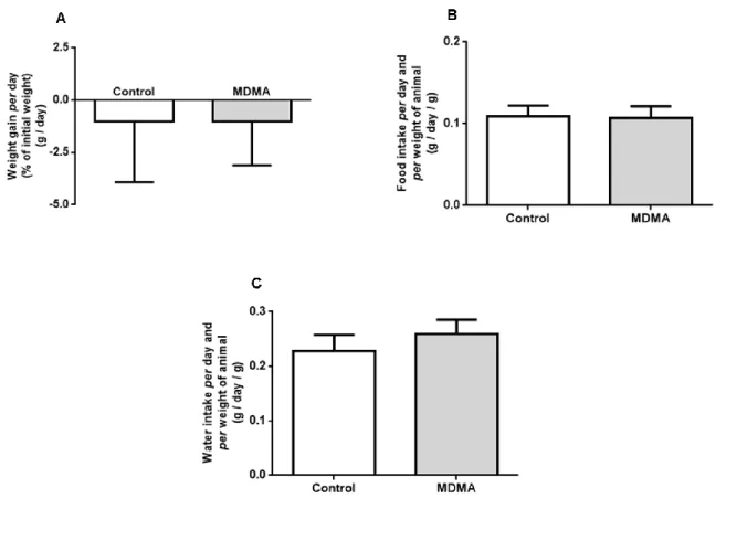
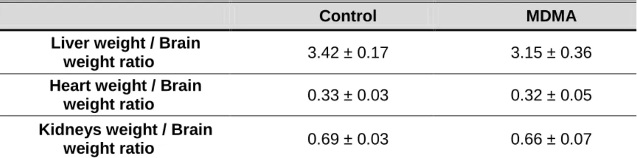
This extract showed no acute or chronic toxicity in rats, as confirmed by no change in the body weight of the treated animals compared to controls and no significant changes in.

Root length (A), root volume (B) and root fresh mass (C) of soybean plants, depending on the application form and concentration of manipueira, in the management of

To evaluate if the ALDH3A2, ALDH3B1, and ALDH4A1 isoforms could to be used as MDS diagnostic markers, we determine the capacity of the gene expression levels of these ALDHs

However, we found that across the monetary and the physical condition subjects showed the same level of trust and reciprocity. Moreover, subjects’ behavior across

Este artigo discute o filme Voar é com os pássaros (1971) do diretor norte-americano Robert Altman fazendo uma reflexão sobre as confluências entre as inovações da geração de

Nota: Envolve dados dos seguintes cursos cadastrados como licenciaturas e com os seguintes nomes: Ciência da Computação, Ciências da Computação, Computação, Computação