ARTIGO DE REVISÃO ARTIGO ACEITE P ARA PUBLICAÇÃO DISPONÍVEL EM WWW .ACT AMEDICAPORTUGUESA.COM
Viscoelastic Tests in the Evaluation of Haemostatic
Disorders in SARS-CoV-2 Infection
Testes Viscoelásticos na Avaliação de Alterações da
Hemostase na Infeção por SARS-COV-2
Anabela RODRIGUES1, Teresa SEARA SEVIVAS2, Carla LEAL PEREIRA1, André CAIADO3, António ROBALO NUNES4
Acta Med Port 2020 xxx;33(AOP):xxx-xxx ▪ https://doi.org/10.20344/amp.14784
ABSTRACT
COVID-19 associated coagulopathy is a dysfunction of severe SARS-CoV-2 infection, characterized by significantly increased fibrino-gen, D-dimer and C reactive protein and normal to near-normal prothrombin time, activated partial thromboplastin time and platelet count. Hypercoagulopathy and hypofibrinolysis coexist and are detected by viscoelastic tests. These features, when associated with immobilization and intrinsic risk factors (age, obesity, comorbidities, drugs) of the patient, can trigger thromboembolic events, despite thromboprophylaxis. The lungs are the first and most severely damaged organ. To date, most patients have exhibited hypercoagulability on viscoelastic tests not detected by standard coagulation tests. A high rate of thrombotic events was reported, suggesting that it should be considered as a cause of clinical deterioration in intensive care and potentially other clinical settings. In advanced stage, COVID-19 associated coagulopathy, fibrinogen and platelet count can decrease significantly, depending on the severity of clinical status resem-bling consumptive coagulopathy. In this stage, bleeding events can occur, especially if the patient is under extracorporeal membrane oxygenation (ECMO). Viscoelastic tests are very useful tools to assess hypercoagulability and hypofibrinolysis (not detectable by standard coagulation tests) in critically ill SARS-CoV-2 patients with COVID-19 associated coagulopathy and look like very promising tools for anticoagulation management. However, further research needs to be carried out to determine whether abnormal viscoelastic tests alone or in combination with other clinical or laboratory findings can identify patients at increased thrombotic risk. Clinical trials to evaluate hypercoagulability using viscoelastic tests and the need for personalized dosage of anticoagulation in SARS-CoV-2 patients are quickly emerging.
Keywords: Blood Coagulation Disorders; Blood Coagulation Tests; Coronavirus Infections; COVID-19; SARS-CoV-2; Thrombosis RESUMO
A coagulopatia associada à COVID-19 é uma disfunção associada à infeção SARS-CoV-2 grave, caraterizada por aumento significa-tivo do fibrinogénio, D-dímeros e Proteína C reativa, e por valores normais/muito pouco alterados do tempo de protrombina, tempo de tromboplastina parcial ativado, e número de plaquetas. A hipercoagulabilidade e a hipofibrinólise coexistem e são detetadas por testes viscoelásticos. Quando associadas à imobilização e aos fatores de risco intrínsecos do doente (idade, obesidade, comorbilidades, drogas) potenciam eventos tromboembólicos, apesar da tromboprofilaxia. Os pulmões são o órgão inicialmente e mais gravemente afetado. Até à data, a maioria dos doentes apresentou hipercoagulabilidade nos testes viscoelásticos, não detetada pelos testes de coagulação de rotina, e foi reportada uma elevada taxa de eventos trombóticos, sugerindo que esta deveria ser considerada uma das causas de deterioração clínica, não só em cuidados intensivos. Na coagulopatia associada à COVID-19 avançada, o número de pla-quetas e o fibrinogénio podem diminuir significativamente, dependendo da gravidade clínica da infeção, assemelhando-se o quadro a uma coagulopatia de consumo. Nesta fase pode haver hemorragia, especialmente se o doente estiver sob extracorporeal membrane oxygenation (ECMO). Os testes viscoelásticos afiguram-se muito úteis para avaliar a hipercoagulabilidade e a hipofibrinólise em doen-tes críticos SARS-CoV-2 com coagulopatia associada à COVID-19, parecendo também promissores para a gestão da anticoagulação. No entanto, é necessária mais investigação para determinar se testes viscoelásticos alterados, individualmente ou quando combi-nados com outros resultados clínicos/laboratoriais, podem identificar os doentes com risco trombótico acrescido. Estão a emergir rapidamente ensaios clínicos para avaliação da hipercoagulabilidade por testes viscoelásticos e da necessidade de personalização da anticoagulação em doentes SARS-CoV-2.
Palavras-chave: COVID-19; Infecções por Coronavírus; Perturbações da Coagulação Sanguínea; SARS-CoV-2; Testes de Coagula-ção Sanguínea; Trombose
INTRODUCTION
The major challenge associated with COVID-19 is severe, often fatal, interstitial pneumonia.1 COVID-19 mortality
bur-den is mainly attributable to a progressive bilateral pneumonia that can progress to acute respiratory distress syndrome
(ARDS), requiring intensive care support.2 While the pulmonary pathophysiology is not fully understood, severe COVID-19
infection is associated with an alveolar inflammatory cell infiltrate, and a systemic cytokine storm.1 Proinflammatory
cy-tokines are modulators of coagulation and fibrinolysis activation and might constitute another trigger to explain the
proco-agulant imbalance in these patients.3 Endothelial injury may play an additional role.3 Post-mortem studies corroborate that
explanation, highlighting marked pathological changes involving lung microvasculature, disseminated micro-thrombi and
haemorrhagic necrosis.4 Severe COVID-19 is also associated with an increased risk of deep vein thrombosis (DVT) and
1. Serviço de Imuno-hemoterapia. Hospital Santa Maria. Centro Hospitalar de Lisboa Norte. Lisboa. Portugal. 2. Serviço de Sangue e Medicina Transfusional. Centro Hospitalar e Universitário de Coimbra. Coimbra. Portugal. 3. Serviço de Imunohemoterapia. Hospital de São José. Centro Hospitalar Universitário de Lisboa Central. Lisboa. Portugal. 4. Serviço de Medicina Transfusional. Hospital das Forças Armadas. Pólo de Lisboa. Lisboa. Portugal.
Autor correspondente: Teresa Seara Sevivas. teresasevivas@hotmail.com
ARTIGO DE REVISÃO ARTIGO ACEITE P ARA PUBLICAÇÃO DISPONÍVEL EM WWW .ACT AMEDICAPORTUGUESA.COM
pulmonary embolism (PE).2
Several studies reported a COVID-19 associated coagulopathy (CAC),5,6 which is one of the most significant poor
prognostic features.7 CAC is characterized by significantly increased fibrinogen, D-dimer and C reactive protein (CRP)
and normal to near-normal prothrombin time (PT), activated partial thromboplastin time (APTT) and platelet count. Hyper-coagulopathy and hypofibrinolysis coexist and are more easily detected by viscoelastic tests (VET), since conventional
coagulation and fibrinolytic tests only reflect a part of the coagulation system.2,8
VET are in vitro point of care (POC) devices, capable of assessing viscoelastic properties of clotting native whole blood
upon activation of hemostasis by added triggers.1 This technology provides a fast and dynamic assessment of
haemo-stasis, and it is a validated device for clinicians to make an early diagnosis of the coagulopathy and to choose the most
appropriate targeted treatment.2 VET also measure hypercoagulability in various clinical scenarios which is not detected by
standard coagulation tests (SCT).9 Global hemostatic tests such as tromboelastography and thromboelastometry are more
reliable in the early identification of a hypercoagulable state (HS) and management of thrombotic scenarios in COVID-19
infection but these are not commonly used yet.10 We may consider using one of the VET as a screening tool or to optimize
anticoagulation therapy, particularly in acute intensive care units (ICU), although this requires further research.11
The aim of this paper is to review all relevant scientific data on the use of VET in detecting the hypercoagulability profile of SARS-CoV-2 infection and its possible role in tailoring antithrombotic therapy.
MATERIAL AND METHODS
A literature search was performed on PubMed, MEDLINE, Google Scholar, Cochrane and Research Gate using the conjugated keywords: ‘SARS-CoV-2’, ‘COVID-19’, ‘Coagulopathy’, ‘Hypercoagulability’, ‘Hypercoagulable’, ‘Thrombosis’,
‘Viscoelastic Testing’, ‘Thromboelastometry’, ‘Thromboelastography’, ‘SEER Sonorheometry’, ‘ROTEM®’, ‘TEG®’,
‘QUAN-TRA®’.
RESULTS
Viscoelastic testing
This review will address two similar technologies, thromboelastography (TEG®) and thromboelastometry (ROTEM®),
and sonic estimation of elasticity via resonance (SEER) sonorheometry (Quantra®).
TEG® and ROTEM® are both established POC VET with a wide range of applications in several scenarios.4,12,13 Both
devices measure coagulation in whole blood under static low shear stress conditions, without blood vessels or flow
con-tribution.14,15 When a clot starts to form inside the cup, it generates a graphically transduced signal.14-16 The magnitude of
displacement is referred to as ‘clot amplitude or firmness’ and relates to the strength of the clot.14 The viscoelastic changes
that occur during coagulation are recorded, providing a graphical representation of the fibrin polymerization.13,17 This
pro-cess depends on platelets, fibrinogen, and the presence of pro and anticoagulants.13,17
These devices showed a good correlation with the assessment of coagulation factors, fibrinogen and platelet count in
critically ill patients.18 Goal-directed transfusion therapy using this technology in different bleeding settings (trauma,
perio-perative and post-partum hemorrhage) are now fully studied and applied with success.12,17,19 This technology has been
successfully applied to areas where conventional testing is inadequate, such as hypercoagulability screening, assessment
of thrombotic risk17,19,20 and the effects of systemic anticoagulants.19 Its implementation requires adequate technical and
interpretation training, and interdisciplinary cooperation.12
All VET parameters are shown in Table 1 and ROTEM® assays and parameters are described elsewhere.12 ROTEM®
and TEG® parameters are comparable but not interchangeable (Table 1).21
The use of thromboelastometry and thromboelastography in the detection of HS has been described in several clinical
conditions.10 A short clotting time (CT) or reaction time (R-time) has been associated with a prothrombotic state.19,22 The
‘thickness’ of the VET tracing [maximum amplitude (MA) on TEG® or maximum clot firmness (MCF)/amplitude at ten
min-utes (A10)- INTEM or FIBTEM on ROTEM®] appears to be the most useful parameter in guiding transfusion and predicting
thrombotic complications.19,20 However, no definitive definition of HS based on VET has been established.
A Chinese expert consensus,18 as well as other authors,23 recommend the use of VET to evaluate severe CAC and
monitor anticoagulation therapy in critically ill patients.
Direct thrombin inhibitors (DTI) (argatroban, bivalirrubin) are used in COVID-19 infected patients with significantly lower
antithrombin levels,24 or if heparin induced thrombocytopenia (HIT) occurs. DTI inhibit thrombin in the Clauss reagent,
un-derestimating fibrinogen quantification, and making VET a valid method for monitoring fibrinogen levels in this setting.24
Emerging studies using VET aim to address the hemostatic changes found in SARS-CoV-2 infection, but no
prospec-tive randomized clinical trial (RCT) data is available.13
ROTEM® (rotational thromboelastometry)
Overview
ARTIGO DE REVISÃO ARTIGO ACEITE P ARA PUBLICAÇÃO DISPONÍVEL EM WWW .ACT AMEDICAPORTUGUESA.COM
cartridge-based fully automated closed system, including four assays (Table 1, Fig. 1).12
Thromboelastometry has shown to be useful in identifying hypercoagulability in various clinical settings not detected
by SCT,25 through the presence of (a) an accelerated clot formation with significantly lower clot formation time (decreased
CFT-EXTEM/INTEM/FIBTEM) due to the increase of plasma fibrinogen and excessive thrombin generation, and/or (b) an
increase in clot strength (increased MCF-EXTEM/INTEM/FIBTEM and A10) and higher α angle.2,25-27 INTEM clot firmness
at 10 minutes (A10) was the best predictor of thromboembolic complications.26 MCF is a reliable marker of
hypercoagulabil-ity.27 CT, CFT, and α angle are useful to assess thrombin generation.28
A hypercoagulability profile is defined by ROTEM® analysis as12,29: CT-EXTEM: < 40s- < 45s; CFT-EXTEM: < 45s - <
50s; MCF-EXTEM: > 68mm; MCF-FIBTEM: > 22 mm-> 24 mm; LI60-EXTEM: ≤ 3%. Clinical trials
Theoretically, thromboelastometry variables may be affected early during the course of SARS-CoV-2 infection
com-pared with D-dimers and may be of great value as a predictor of disease severity (Table 2).2,27,30,31
ROTEM® is the most used device to assess CAC. Initially, two clinical cases32,33 with severe COVID-19 acute
res-piratory distress syndrome (ARDS) requiring mechanical ventilation (MV) were described. Both revealed normal PT and
APTT, very increased D-dimer and fibrinogen levels, and a HS on ROTEM® (increased MCF-EXTEM/INTEM/FIBTEM,32,33
elevated α angle-EXTEM,33 and decreased CFT-EXTEM32/CFT-FIBTEM),33 and therefore a higher dose of unfractionated
heparin (UFH) was started.32,33 No thromboembolic (TE) or hemorrhagic events (HE) were observed and D-dimers have
decreased.32 The authors suggested that VET may have a role in rapidly identifying severe COVID-19 in critically ill patients
with hypercoagulability,32 not detected with SCT,33 and that robust anticoagulation might be needed to prevent TE.32,33
A study by Spieza et al30 included 22 patients admitted to the ICU and showed evidence of hypercoagulability on
ROTEM® with a shorter CFT and high clot firmness (MCF).11,30 Compared with healthy controls, the following results
were observed30: (a) significantly higher fibrinogen and D-dimer levels (p < 0.0001); (b) markedly hypercoagulability on
ROTEM®: shorter CFT-INTEM (p = 0.0002) and CFT-EXTEM (p = 0.01), and higher MCF in INTEM/EXTEM/FIBTEM (p <
0.001). The authors concluded that COVID-19 patients with acute respiratory failure (ARF) presented a severe HS rather than consumptive coagulopathy (CC). Unfortunately, this data did not allow for the assessment of the impact of adequate
dosages of anticoagulants on clotting parameters.11
According to Madathil et al,34 systemic fibrinolysis (SF) was not detected on EXTEM or FIBTEM (maximum lysis-ML:
0%) in 11 critically ill COVID-19 patients receiving MV, despite increased CRP and D-dimer levels. Circulating plasminogen activator inhibitor-1 (PAI-1) was increased and SF is thus unlikely to occur in COVI19 with cytokine storm. Very high dimers along with high CRP levels and A10-FIBTEM were observed, especially in the subset of patients with very high D-dimer levels (> 3245 ng/mL), but without elevated ML-EXTEM (0%), suggesting that SF is unlikely to occur. They propose that critically ill COVID-19 patients have significant elevations in D-dimer levels consistent with microvascular thrombosis,
but only small fractions of fibrin seem to be locally broken down.34
Fibrinolysis shutdown was evidenced in others studies14,27,35 by raised D-dimer levels and complete failure of clot lysis at
30 minutes on thromboelastography (LY30) and thromboelastometry (LI30), which predicts and correlates with TE and the
need for hemodialysis in critically ill COVID-19 patients.14,35 Authors hypothesize that the main source for elevated D-dimer
levels in the presence of hypercoagulability, along with decreased fibrinolysis, could be the lungs.35
Another study36 conducted in 78 COVID-19 patients (48 with ARDS in ICU; 30 in ward), despite substantial heparin
plasma levels (mean anti-Xa activity of 0.35 ± 0.20 IU/mL) and normal levels of antithrombin in 91% of patients, thrombin generation was normal. These findings, along with significantly increased fibrinogen and FVIII levels, suggested high risk
of hypercoagulability, probably related to major inflammatory syndrome not controlled by heparin.36 A TEM-tPA assay was
used to detect both hypercoagulability and hypofibrinolysis simultaneously, and is a promising biomarker of thrombosis
risk.36
The study by Pavoni et al2 included 40 critically ill COVID-19 patients where SCT and ROTEM® were evaluated on ICU
admission day (T0), and days 5 (T5) and 10 (T10). On ICU admission, PT was slightly reduced, and increased significantly at T10 (p = 0.002), while APTT and fibrinogen values were higher at T0 than T10 (p = 0.017; p = 0.002, respectively). Platelet count was normal and increased over time. About 70% of patients had higher D-dimer levels that decreased at
T10 (p = 0.392). ROTEM® profiles were consistent with hypercoagulability (lower CFT, increased MCF) which persisted in
the first five days, decreasing after ten days, without returning to normal values.2 No signs of secondary hyperfibrinolysis
or sepsis-induced coagulopathy were found.2 From the total, 15% and 30% of the patients had DVT and catheter-related
thrombosis, respectively, despite low molecular weight heparin (LMWH) prophylaxis.2 In conclusion, ROTEM® showed
con-sistent evidence of a HS in severe COVID-19 that persisted over time associated with an inflammatory state, rather than
CC.2 The improvement of fibrinogen clot firmness measurements after 10 days of illness suggests a dynamic component
for the changes in coagulation that accompany the rise and fall of inflammatory parameters.37
Almskog et al27 evaluated whether patients with severe disease have more pronounced hypercoagulability compared
ARTIGO DE REVISÃO ARTIGO ACEITE P ARA PUBLICAÇÃO DISPONÍVEL EM WWW .ACT AMEDICAPORTUGUESA.COM
groups depending on care level (regular wards and ICU) and were compared with 89 healthy controls. SCT and ROTEM®
analysis were performed as soon as possible after admission. In total, 80% of patients received prophylaxis with LMWH. The levels of INR, APTT, and platelets were nearly normal, although markedly elevated D-dimer and fibrinogen levels were observed. There was no significant correlation between D-dimer and MCF-EXTEM (p = 0.9), but it was significant between
fibrinogen and MCF-FIBTEM (p < 0.001).27 When compared with healthy controls, the ROTEM® variables of COVID-19
patients were significantly higher in both groups (ward/ICU) (p < 0.0001), and were higher in critically ill patients com-pared with the less severely ill patients in regular wards (p < 0.01 for MCF-EXTEM and p < 0.05 for FIBTEM); CT-EXTEM was significantly longer, particularly in ICU patients (p < 0.001), and CFT-EXTEM significantly shorter in COVID-19 (p <
0.001).27 All these ROTEM® variables determined early after admission were significantly more pronounced in patients
with increased severity.27 This suggested prolonged hemostatic initiation, shortened clot propagation, and pronounced clot
firmness, indicating hypercoagulability.27 The authors concluded that hypercoagulability is present in hospitalized patients
at an early disease course with mild to severe COVID-19 pneumonia and ROTEM® analysis may be a potentially useful
predictor of TE and mortality.27
Two prospective studies38,39 demonstrated that most patients admitted to ICU with severe SARS-CoV-2 infection
showed a pronounced HS, characterized by increased fibrinogen and D-dimer levels, impaired endogenous anticoagula-tion (decreased protein S levels), decreased fibrinolysis, and increased MCF-INTEM/EXTEM/FIBTEM. TE occurred in
20%38 and 33%39 of patients despite appropriate anticoagulation, and HE in 10%.38 The magnitude of coagulation
abnor-malities seemed to correlate with the severity of organ dysfunction according to the sequential organ failure assessment
(SOFA) score being higher if SOFA was over 10.38 Compared with SOFA below 10, COVID-19 patients with SOFA over 10
exhibited higher fibrinogen and D-dimer levels, higher MCF-FIBTEM (p = 0.05), lower ML-INTEM (p = 0.004), and lower
antithrombin, protein C and plasminogen levels.38
TEG® (thromboelastography)
Overview
Thromboelastography parameters include reaction time (R, represents the initiation phase measuring the time from the start of the test to initial fibrin formation), clot formation time (K, represents the amplification phase measuring the time until 20 mm of clot strength is achieved), angle or α (K angle, represents the propagation phase measuring the rate of clot formation), maximum amplitude (MA, represents the overall stability of the clot), and amplitude at 30 minutes (LY30,
rep-resents the fibrinolysis phase and measures the percentage of decrease in amplitude at 30 minutes post-MA).37 The new
TEG®6s system is fully automated.21
In TEG® (Table 1, Fig. 1), hypercoagulability is shown by shorter R-time and K-time, besides increased K/α angle and
MA.17,22,40 In the Mazen et al study,22 hypercoagulability was defined as having at least three abnormal TEG® values.
How-ever, Maatman et al40 defined a hypercoagulable profile as two or more thromboelastographic parameters beyond one
Standard Deviation (SD) of the age-and gender-matched controls. Clinical trials
Panigada et al1 showed hypercoagulability along with a severe inflammatory state based on SCT and
thromboelastog-raphy testing in severe SARS-CoV-2 infection. Escalating from prophylactic to therapeutic LMWH dose requires a careful
approach based on the benefit/risk ratio until clinical trials are available (Table1).1
Similar results were observed by Maatman et al40 and Wright et al,41 showing COVID-19-associated hypercoagulability
and fibrinolysis shutdown (LY30 < 0.8%) 41causing a higher rate of VTE, with both being measured by TEG®41 in 12 and 44
patients admitted to the ICU, respectively.40,41
Similarly, Mortus et al42 found high K/α angle or high MA on TEG® in 90.5% of patients with severe COVID-19, including
increased fibrinogen activity (> 73o angle) plus MA (> 65 mm) in 74% of patients, and MA criteria alone in 26%. TE occurred
at a 62% rate despite thromboprophylaxis in all. In comparison, innate TEG®-MA was significantly higher for the high TE
rate group compared to the low TE rate group (p = 0.01).42 Increased MA was observed in 100% and 45% of patients in the
high and low event rate group respectively. Hypercoagulable innate TEG®-MA yielded 100% sensitivity and 100% negative
predictive value for the occurrence of multiple thrombi.42 In conclusion, in this context, TEG® may be critical in identifying
patients at increased thrombotic risk, where full heparinization is beneficial, and avoiding unnecessary anticoagulation in
those with low thrombosis risk.42
According to Fan et al,10 TEG® appears to be a useful tool in detecting hypercoagulability even in the presence of
hepa-rin or antiphospholipid syndrome in CAC.
QUANTRA® (SEER sonorheometry)
Overview
Quantra® is a new fully automated VET closed system POC,that allows rapid whole blood global hemostasis evaluation
ARTIGO DE REVISÃO ARTIGO ACEITE P ARA PUBLICAÇÃO DISPONÍVEL EM WWW .ACT AMEDICAPORTUGUESA.COM
Quantra® uses ultrasound technology14,16 for direct measurement of physical properties of the clot, without mechanical
clot disruption.14,43 The ultrasound pulses generate a shear wave in the sample and the resulting deformation is
meas-ured.16 The frequency and amplitude of the induced deformation are directly related to the sample’s viscoelastic
proper-ties.16
The following functional parameters can be analyzed14,16,43 (Table 1): clot time (CT) after blood activation with kaolin,44
provides an indication of the functional status of coagulation factors that lead to fibrin formation43; CT with heparinase I to
neutralize heparin (CTH); CT ratio (CTR) (CTR over 1.4 indicates the likelihood of influence of heparin); clot stiffness (CS), measured seven minutes after clot initiation, provides information about fibrin/fibrinogen function and platelet activity in the
presence of thromboplastin, and was compared with A10-EXTEM on ROTEM®.16 It represents the platelet-fibrinogen
inter-action through thrombin and factor XIII, but is also influenced by hematocrit and acidosis14; fibrinogen contribution to CS
(FCS) reflects only fibrinogen’s contribution to the overall CS (hPa),16,43 being compared with A10-FIBTEM on ROTEM®16;
platelet contribution to CS (PCS), and also platelet activation and platelet contraction of the fibrin mesh,15,16,43 is a
param-eter calculated by taking into account the difference between CS and FCS15,16,43; and clot stability to lysis (CSL).
Quantra® enables a goal-directed therapy for bleeding patients in several clinical settings,43 as well as diagnosis and
evaluation of hypercoagulability as seen in COVID-19. Quantra® has a good correlation with other well-established VET
devices and the Clauss assay.14,16,43
Clinical trials
Several publications concerning COVID-19 patients have reported a procoagulant profile with increased clot strength,
platelet and fibrinogen contribution to clot stiffness (PCS,FCS),11,44 which seemed to improve during ICU stay under
antico-agulation.11,44,45 Besides, CT and CTH parameters can be useful to monitor anticoagulation (Table 2).44
The Ranucci et al44 study aimed to characterize the coagulation profile of COVID-19 ARDS patients, and to evaluate
their changes after aggressive thromboprophylaxis. The 16 patients received a complete coagulation profile at ICU admis-sion, as well as mechanical ventilation (MV). Ten patients were monitored in the subsequent seven days, after increased
LMWH dose, antithrombin corrected if lower than 70%, and clopidogrel if platelet count above 400 x 109/L.44 Major
throm-boembolic events (TE) were not observed. At baseline, procoagulant profile was characterized by normal levels of CT, but elevation of CS, PCS, FCS, D-dimer, and fibrinogen levels, which were associated and correlated with increased
interleu-kin-6 levels (R2 = 0.506; p = 0.003),44 confirming the link between inflammation and hypercoagulability.11 After increasing
thromboprophylaxis, and two weeks from the baseline, there was significant time-related decrease of fibrinogen levels (p
= 0.001), D-dimers (p = 0.017), CS (p = 0.013), PCS (p = 0.035), and FCS (p = 0.038).44
Masi et al45 characterized the coagulation and fibrinolysis profiles of 17 COVID-19-associated ARDS patients and
compared them with 11 non-COVID-19-associated ARDS patients. On admission, all received thromboprophylaxis.
Pul-monary embolism (PE) was incidentally diagnosed in 17% of patients.45 Compared to non-COVID-19, COVID-19 patients
exhibited: (a) significantly higher levels of procoagulant factors, mainly: fibrinogen (p = 0.03); Factor V (FV) (p < 0.0001);
FVIII (p = 0.03), and acute phase reactants45. All these parameters were strongly correlated with each other (p < 0.05);
(b) significantly lower thrombin-antithrombin complex (p = 0.03); (c) significantly higher (p = 0.048) t-PA and PAI-1, being
closely correlated (p < 0.001); (d) fibrinolysis shutdown or CC were not observed; (e) Quantra®, exhibited twice higher
levels of CS (p = 0.0077), PCS (p = 0.014), and FCS (p < 0.001).45 CS was strongly correlated with fibrinogen (p = 0.02),
FV (p = 0.043), FVIII (p < 0.001), but not with PAI-1 levels (p = 0.606).45 In conclusion, COVID-19-associated ARDS was
correlated with significant increase in procoagulants, supporting the concept of thromboinflammation.45
DISCUSSION
It is essential to be familiar with the spectrum of CAC11 since all current studies support CAC as a hypercoagulable and
hypofibrinolytic state in the ICU setting.37 Nevertheless, whether this hypercoagulability is due to the invading
microorgan-ism, individual viral load or the massive host inflammatory response, still remains unknown.27 This hypercoagulability can
evolve into consumptive coagulopathy, microthrombosis and multiple organ failure.46 Post-mortem data supports
hyperco-agulability through the presence of micro-thrombi in several systems.4 Activation of coagulation and/or fibrinolysis occurs in
COVID-19 as part of the acute inflammatory response.4 CAC may, in some way, be specific to SARS-CoV-2, representing
new features that need to be clarified through further research.27
All the observational clinical studies described point to the use of VET to assess hypercoagulability and hypofibrinolysis
(not detected by SCT), and probably also for anticoagulation monitoring.1,2,10,27,32,33,35,38-42 Improvements on VET parameters
and patients` clinical status were observed after introduction of tailored thromboprophylaxis.1,2,30,32,44 The use of TEG® or
ROTEM® is recommended by some authors18 and advised by others2,4,10,23,27,32,38 for all COVID-19 patients with severe
pneumonia and coagulation dysfunction.
Hypercoagulable viscoelastic profiles1,20 were identified in several clinical studies in COVID-19 patients: increased A10,
MCF (INTEM/EXTEM/FIBTEM) and α angle and decreased CFT and ML for ROTEM®2,27,30,32,35,36,38,39,46; short R, K or LYS-30
ARTIGO DE REVISÃO ARTIGO ACEITE P ARA PUBLICAÇÃO DISPONÍVEL EM WWW .ACT AMEDICAPORTUGUESA.COM
Quantra® and ROTEM® have shown features of hypercoagulability in COVID-19 patients hinting towards their use in
tailoring treatment, but these non-randomized small studies require confirmation.1,20,44 The development of prospective
tri-als prior to their use in COVID-19 patient care is strongly recommended.13
All intubated ICU patients with low bleeding risk should receive low-intensity prophylaxis with LMWH or UFH.37
Howev-er, due to the very high incidence of thromboembolic complications despite standard low-dose thromboprophylaxis among severe COVID-19 patients with elevated D-dimer levels, intermediate-intensity or full-dose anticoagulation is now routinely
administered.37 VET have already been included in some algorithms to determine anticoagulation needs in COVID-19 ICU
patients, although VET have not yet been validated as appropriate to manage anticoagulation.13
A lower 28-day mortality rate in COVID-19 patients receiving anticoagulation with LMWH was demonstrated.47 Recently,
the International Society of Thrombosis and Hemostasis stressed the need for implementing anticoagulation.48 The latest
recommendations suggest that all hospitalized COVID-19 patients should receive thromboprophylaxis, or full therapeutic
anticoagulation if such a prior indication exists18,49 or depending on patient’s weight and related risk factors.49 Other
strate-gies could include the use of intravenous unfractionated heparin or direct thrombin inhibitors 44 if heparin induced
thrombo-cytopenia occurs 18,24
Currently, data on the usefulness of VET devices in CAC is still limited.50 Further research is needed to investigate
whether ROTEM®/TEG® are useful in identifying COVID-19 patients who might benefit from therapeutic anticoagulation, in
order to guide hemostatic therapy,33,35-37,50 and to determine whether an abnormal VET alone or in combination with other
findings can identify a group of patients with increased thrombotic risk.17,19,37
CONCLUSION
Studies concerning the use of VET to evaluate hypercoagulability in SARS-CoV-2 patients are emerging.
Prospective clinical trials, ideally RCT, could underline the additional value of VET in predicting the clinical course,
guid-ance of anticoagulation, and the risk stratification of COVID-19 patients for CAC.20,47,48
Clinical trials in hospitalized COVID-19 patients are actually underway, such as the Rotterdam cohort study using
ROTEM® (ROHOCO trial)50 and the evaluation of hemostasis by TEG®, platelet function testing, and biomarker analysis
(TARGET-COVID Study37 – ClinicalTrials.gov: NCT04493307).
Several studies have shown that VET are a valuable tool to assess hypercoagulability and hypofibrinolysis in critically ill COVID-19 patients with CAC, as well as an additional contributory tool in anticoagulation management of these
pa-tients.18,27 However, further research needs to be carried out in order to develop evidence-based guidelines.
COMPETING INTERESTS
The authors have declared that no competing interests exist. FUNDING SOURCES
This research received no specific grant from any funding agency in the public, commercial, or not-for-profit sectors.
REFERENCES
1. Panigada M, Bottino N, Tagliabue P, Grasselli G, Novembrino C, Chantarangku V, et al. Hypercoagulability of COVID-19 patients in Intensive Care Unit. A report of thromboelastography findings and other parameters of hemostasis. J Thromb Haemost. 2020;18:1738-42.
2. Pavoni V, Gianesello L, Pazzi M, Stera C, Meconi T, Frigieri FC. Evaluation of coagulation function by rotation thromboelastometry in critically ill patients with severe COVID-19 pneumonia. J Thromb Thromb. 2020;50:281-6.
3. Vazquez-Garza E, Jerjes-Sanchez C, Navarrete A, Joya-Harrison J, Rodriguez D. Venous thromboembolism: thrombosis, inflammation, and immunothrombosis for clinicians. J Thromb Thromb. 2017;44:377-85.
4. Rubulotta F, Soliman-Aboumarie H, Filbey K, Geldner G, Kuck K, Ganau M, Hemmerling TM. Technologies to optimize the care of severe COVID-19 patients for healthcare providers challenged by limited resouces. Anesth Analg. 2020;131:351-64.
5. Tang N, Li D, Wang X, Sun Z. Abnormal coagulation parameters are associated with poor prognosis in patients with novel coronavirus pneumonia. J Thromb Haemost. 2020;18:844-7.
6. Zhou F, Yu T, Du R, Fan G, Liu Y, Liu Z, et al. Clinical course and risk factors for mortality of adult inpatients with COVID-19 in Wuhan, China: a retrospective cohort study. Lancet. 2020;395:1054-62.
7. Guan WJ, Ni ZY, Hu Y, Liang WH, Ou C, He J, et al. China Medical Treatment Expert Group for COVID-19. Clinical characteristics of coronavirus disease 2019 in China. N Engl J Med. 2020;382:1708-20.
8. Brummel KE, Paradis SG, Butenas S, Mann KG. Thrombin functions during tissue factor-induced blood coagulation. Blood. 2002;100:148-52. 9. Akay OM. The double hazard of bleeding and thrombosis in hemostasis from a clinical point of view: a global assessment by rotational thromboelastometry
(ROTEM). Clin Appl Thromb Hemost. 2018;24:850-8.
10. Fan BE, Chia YW, Sum C, Kuperan P, Chan S, Ling LM, et al. Global haemostatic tests in rapid diagnosis and management of COVID-19 associated coagulopathy in acute limb ischaemia. J Thromb Thromb. 2020;50:292–7.
11. Thachil J, Agarwal S. Understanding the COVID-19 coagulopathy spectrum. Anaesthesia. 2020:75:1432-16.
12. Gorlinger K, Pérez-Ferrer A, Dirkmann D, Saner F, Maegele M, Pérez Calatayud AA, et al. The role of evidence-based algorithms for rotational thromboelastometry-guided bleeding management. Korean J Anesthesiol. 2019;72:297-322.
13. American Society of Hematology. Volod O, Reyes Gill M, Selby R, Kreiziger BL, Lee A. COVID-19 and viscoelastic hemostasis assays: frequently asked questions. [accessed 2020 May 11]. Available from: https://www.hematology.org/covid-19/covid-19-and-ve.
ARTIGO DE REVISÃO ARTIGO ACEITE P ARA PUBLICAÇÃO DISPONÍVEL EM WWW .ACT AMEDICAPORTUGUESA.COM
14. Sniecinski R, Tanaka KA. SEER sonorheometry: listening to what the clot has to say. Anesth Analg. 2016;123:1346-7.
15. Solomon C, Ranucci M, Hochleitner G, Schochl H, Schlimp CJ. Assessing the methodology for calculating platelet contribution to clot stregth (platelet component) in thromboelastometry and tThromboelastography. Anesth Analg. 2015;121:868-78.
16. Huffmyer JL, Fernandez LG, Haghighian C, Terkawi AS, Groves DS. Comparison of SEER sonorheometry with rotational thromboelastometry and laboratory parameters in cardiac surgery. Anesth Analg. 2016;123:1390-9.
17. Luddington RJ. Thromboelastography/thromboelastometry. Clin Lab Haem. 2005,27:81-90.
18. Song JC, Wang G, Zhang W, Zhang Y, Li WQ, Zhou Z, et al. Chinese expert consensus on diagnosis and treatment of coagulation dysfunction in COVID-19. Mil Med Res. 2020;7;19.
19. Harahsheh Y, Ho KM. Viscoelastic point-of-care testing to guide transfusion and antithrombotic therapy in perioperative and critically ill patients: are all parameters created equal? Anaesth Intensive Care. 2016;44:11-3.
20. Thachil J, Cushman M, Srivastava A. A proposal for staging COVID-19 coagulopathy. Res Pract Thromb Haemost. 2020;4:731–6.
21. Sahli SD, Rossler J, Tscholl DW, Studt JD, Spahn DR, Kaserer A. Point-of-care diagnostics in coagulation management. Sensors. 2020;20:4254. 22. Toukh M, Siemens DR, Black A, Robb S, Leveridge M, Graham CH, et al. Thromboelastography identifies hypercoagulability and predicts thromboembolic
complications in patients with prostate cancer. Thromb Res. 2014;133:88-95.
23. Allione A, Giamello JD, Paglietta G, Bernardi S, Cavalot G. Switch from oral anticoagulants to parenteral heparin in SARS-CoV2 hospitalized parents: comment. Intern Emerg Med. 2020 (in press). doi: 10.1007/s11739-020-02373-5.
24. Maier CL, Barker NA, Sniecinski R. Falsely low fibrinogen levels in COVID-19 patients on direct thrombin inhibitors. Anesth Analg. 2020;131:e117-9. 25. Akay OM. The double hazard of bleeding and thrombosis in hemostasis from a clinical point of view: a global assessment by rotational thromboelastometry
(ROTEM). Clin Appl Thromb Hemost. 2018;24:850-8.
26. Hincker A, Feit J, Sladen RN, Wagener G. Rotation thromboelastometry predicts thromboembolic complications after major non-cardiac surgery. Crit Care. 2014;18:549.
27. Almskog L, Wikman A, Svensson J, Wanecek M, Bottai M, Van der Linden J, et al. Rotational thromboelastometry predicts care level in COVID-19. medRxiv. doi: 10.1101/2020.06.11.20128710.
28. Gorlinger K, Dirkmann D, Solomon C, Hanke AA. Fast interpretation of thromboelastometry in non-cardiac surgery: reliability in patients with hypo-, normo-, and hypercoagulability. Br J Anaesthesia. 2013;110:222-30.
29. Gorlinger K, Pérez-Ferrer A. Algoritmos basados en test POC para el manejo de la hemorragia aguda. In: Medicina transfusional. Patient blood management. Pérez-Ferrer A, Garcia-Erce JA, Muñoz Gómez M, editors. Madrid: Editorial Medica Panamericana. 2019;9:75-110.
30. Spieza L, Boscolo A, Poletto F, Cerruti L, Tiberio I, Campeloo E, et al. COVID-19-related severe hypercoagulability in patients admitted to intensive care unit for acute respiaratory failure. Thromb Haemost. 2020;120:998-1000.
31. Juffermans N. Diagnosis and management of thrombosis in critically ill patients with COVID-19. Amsterdam: Laboratory of Experimental Intensive Care and Anesthesiology (LEICA) of the Amsterdam Universitair Medische Centra (UMC) and department of Intensive Care medicine, OLVG Hospital; 2020. 32. Raval Jay S, Burnett AE, Rollins-Raval MA, Griggs JR, RosenbaumE, Nielsen ND, et al. Viscoelastic testing in COVID-19: a possible screening tool for
severe disease? Transfusion. 2020;60:1131-2.
33. Iwasaki Y, Shiga T, Konno D, Saito K, Aoyagi T, Oshima K, et al. Screening of COVID-19 associated hypercoagulability using rotational thromboelastometry. J Clin Anesth. 2020;67:1-4.
34. Madathil R, Tabatabai A, Rabin J, Menne AR, Henderson R, Mazzeffi M, et al. Thromboelastometry and D-dimer elevation in coronavirus-2019 (COVID-19). J Cardiothorac Vasc Anesth. 2020 (in press. doi: 10.1053/j.jvca.2020.05.020.
35. Ibañez C, Perdomo J, Calvo A, Ferrando C, Reverter JC, Tassies D, et al. High D dimers and low global fibrinolysis coexist in COVID-19 patients: what is going on in there? J Thromb Thrombolysis. 2020 (in press). doi: 10.1007/s11239-020-02226-0 .
36. Nougier C, Benoit R, Simon M, Desmurs-Clavel H, Marcotte G, Argaud L. et al. Hypofibrinolytic state and high thrombin generation may play a major role in SARS-CoV2 associated thrombosis. J Thromb Hemost. 2020 (in press). doi: 10.1111/jth.15016.
37. Chaudhary R, Kreutz RP, Bliden KP, Tantry US, Gurbel PA. Personalizing antithrombotic therapy in COVID-19: role of thromboelastography and thromboelastometry. Thromb Haemost. 2020 (in press). doi: 10.1055/s-0040-1714217.
38. Corrêa TD, Cordioli RL, Guerra JC, Silva BC, Rodrigues RR, Souza GM, et al. The hypercoagulability state of COVID-19 ICU patients is characterized by impaired endogenous anticoagulation and decreased fibrinolysis. Res Square. 2020 (in press). doi: 10.21203/rs.3.rs-47465/v1
39. Collett LW, Gluck S, Strickland RM, Reddi BJ. Evaluation of coagulation status using viscoelastic testing in intensive care patients with coronavirus disease 2019 (COVID-19): An observational point prevalence cohort study. Aust Crit Care. 2020 (in press). doi: 10.1016/j.aucc.2020.07.003.
40. Maatman T, Jalali F, Feizpour C, Douglas A, McGuire SP, Kinnaman G, et al. Routine venous thromboembolism prophylaxis may be inadequate in the hypercoagulable state of severe Coronavirus Disease 2019. Crit Care Med. 2020;48:e783-90.
41. Wright FL, Vogler TO, Moore EE, Moore HB, Wohlauer MV, Urban S, et al. Fibrinolysis shutdown correlation with thromboembolic events in severe COVID-19 infection. J Am Coll Surg. 2020;231:193-203.e1.
42. Mortus JR, Manek SE, Brubaker LS, Loor M, Cruz MA, Trautner BW, et al. Thromboelastographic results and hypercoagulability syndrome in patients with coronavirus disease 2019 who are critically ill. JAMA Netw Open. 2020;3:e2011192.
43. Ferrante EA, Blasier KR, Givens TB, Lloyd CA, Fischer TJ, Viola F. A novel device for the evaluation of hemostatic function in critical care settings. Anesth Analg. 2016;123:1372-9.
44. Ranucci M, Ballotta A, Di Dedda U, Bayshnikova E, Dei Poli M, Resta M, et al. The procoagulant pattern of patients with COVID-19 acute respiratory distress syndrome. J Thromb Haemost. 2020;18:1747-51.
45. Masi P, Hékimian G, Lejeune M, Chommeloux J, Desnos C, Chambrun MP, et al. Systemic Inflammatory response syndrome is a major contributor to COVID-19 associated coagulopathy: insights from a prospective single-center cohost study. Circulation. 2020;23:1-8.
46. Gorlinger K. Hemostasis management in thrombosis, sepsis, COVID-19 associated coagulopathy and ECMO. EEMEAI ACDx Online Training. doi: 10.13140/RG.2.2.23359.46248.
47. Tang N, Bai H, Chen X, Gong J, Li D, Sun Z, et al. Anticoagulant treatment is associated with decreased mortality in severe coronavirus disease 2019 patients with coagulopathy. J Thromb Haemost. 2020;18:1094-9.
48. Thachil T, Tang N, Ganda S, Falanga A, Cattaneo M, Levi M, et al. ISTH interim guidance on recognition and management of coagulopathy in COVID-19. J Thromb Haemost. 2020;18:1023-6.
49. Fontana P, Casini A, Robert-Ebadi H, Glauser F, Righini M, Blondon M. Venous thromboembolism in COVID-19: Systematic review of reported risks and current guidelines. Swiss Med Wkly. 2020;150:W20301.
ARTIGO DE REVISÃO ARTIGO ACEITE P ARA PUBLICAÇÃO DISPONÍVEL EM WWW .ACT AMEDICAPORTUGUESA.COM
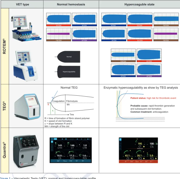
Figure 1 – Viscoelastic Tests (VET): normal and hypercoagulable profile VET type
ROTEM
®TEG
®Quantra
®Normal hemostasis Hypercoaguble state
EXTEM INTEM INTEM EXTEM FIBTEM
FIBTEM APTEM INTEM EXTEM FIBTEM
FIBTEM Normal
INTEM Hypercoagulability
Coagulation Fibrinolysis Patient status: high risk for thrombolic event
Probable cause: rapid thrombin generation
and subsequent clot formation;
Common treatment: anticoagulation
R = time of formation of fibrin strand polymer K = speed of clot formation
α = slope between R and K MA = strength of the clot
MA Time R
K
α
Normal TEG Enzymatic hypercoagulability as show by TEG analysis
EXTEM
ARTIGO DE REVISÃO ARTIGO ACEITE P ARA PUBLICAÇÃO DISPONÍVEL EM WWW .ACT AMEDICAPORTUGUESA.COM
Table 1 – Viscoelastic testing`s corresponding parameters and laboratory tests16,21
ROTEM®
(Werfen, Spain) TEG
®
(Haemonetics, USA) Quantra
®
(Hemosonics, USA) LaboratoryStandard Hemostatic Factors
Clot initiation
CT
EXTEM R
(min) (min)CT
PT Coagulation factors; Anticoagulants; FDP; Tissue
factor-expression on monocytes CT INTEM
(s) APTT Coagulation factors from intrinsic pathway
Clot kinetics
CFT
(s) (min)K Coagulation factors deficit,
Anticoagulants, Fibrinogen, Platelets. α angle (°) α angle (°) Clot strength A5, A10(mm) MCF (mm) A30, A60MA (mm) CS
(hPa) NA Coagulation factors, Fibrinogen, Platelets, FXIII, Colloids
A10 EXTEM
A10 FIBTEM (hPa)FCS FibrinogenClauss
Fibrinogen
(inhibition of platelets to evaluate only the contribution of fibrinogen for clot strength) Difference: A10 EXTEM- A10 FIBTEM PCS (hPa) (PCS = CS - FCS) Platelet
Count Platelet deficit and/ordysfunction
Clot stability (lysis)
LI30, LI60 (%) LY30, LY60
CSL NA Fibrinolytic enzymes,Fibrinolysis inhibitors,
FXIII, Hyperfibrinolysis ML (%) ML EXT > 15% ML APT: NV Unestablished Anticoagulant (heparin) assessment CT HEPTEM
(min) HTEG CTH (min) NA To evaluate heparin presence or FVIII deficit
Ratio:
CT INT/CT HEP HTEG Ratio: CT/CTHCTR NA Heparin presenceFVIII deficit
A: amplitude; A5 / A10: amplitude at 5/10 minutes after CT; APT: APTEM; APTT: activated partial thromboplastin time; CFT: clot formation time; CS: clot stiffness; CSL: clot stiffness lysis; CT: clotting time; s: second; CTH: CT heparinase; CTR: CT ratio; EXT: EXTEM; FCS: fibrinogen contribution to CS; FDP: fibrin degradation products; FIB: FIBTEM; FDP: fibrin degradation products; HEP: HEPTEM; hPa: hector Pascals units; INT: INTEM; K: kinetic time; LI: lysis index; LI30/LI60: lysis index, residual clot firmness, 30 and 60 minutes after CT in % of MCF; MA: maximum amplitude; LY30: lysis 30 minutes after MA in % of MA; MCF, maximum clot firmness; ML: maximum lysis during run time in % of MCF; min: minutes; mm:
millimeters; NA: not applicable; NV: normal value; PCS: platelet contribution to CS; PT: prothrombin time; R: reaction time; VET: viscoelastic tests; HTEG assay: is based on rapid TEG®
ARTIGO DE REVISÃO ARTIGO ACEITE P ARA PUBLICAÇÃO DISPONÍVEL EM WWW .ACT AMEDICAPORTUGUESA.COM Raval et al 32 clinical case 1 63 years M USA ARDS, ICU Shock, MV , vasopressor support (VS) -↑↑ DD -↑ Fibrinogen -NV : PT , APTT Hypercoagulable profile: -↑ MCF-EXT ,INT ,FIB -↓ CFT -EXT -↑ α angle EXTEM
- 7500 IU UFH 8/8h for prophylaxis
- No
TE or HE; ↓DD.
- VET
may have a role in rapidly
identifying severe SARS-CoV
-2. Iwasaki et al 33 clinical case 1 53 years F Japan ARDS, ICU Severe pneumonia -↑↑ DD, CRP -NV : PT , APTT , Platelet. Hypercoagulability: -↑ MCF-EXT/INTEM -↓CFT -FIBTEM - UFH (10 000IU/day)
Hypercoagulability not seen with SCT
but detected with Rotem. Robust
anticoagulation might be needed to
prevent TE in a subset of COVID-19 pts. Spieza et al 30 Single-centre, prospective, observational Study 22 versus healthy control 68 ± 8 years M: 20 F: 2 Padova Univ . Hospital Italy Acute respiratory failure (ARF) ICU Significantly: -↑↑ DD -↑↑Fibrinogen Both: p<0,0001 Marked hypercoagulability (vs healthy controls): ↓CFT -INT (p = 0.0002) ↓CFT -EXT (p = 0.001)) ↑MCF-INT/EXT/FIB (All: p<0,001) - SARS-CoV -2 with
ARF present a severe
hypercoagulability rather than a CC.
- Fibrin formation/polymerization may
predispose to
TE and correlate with worse
outcome. Madathil et al 34 Single -centre, retrospective, observational study 11 53 (45.5 - 65.5) years M: 64% Maryland USA ARDS, ICU MV AH: 54.5% Diab: 45.5% Significantly (sig.): ↑↑ DD ↑↑ Fibrinogen ↑↑ CRP
Despite significantly high CRP+ DD,
systemic fibrinolysis (SF) was not seen
on EXT
or FIB (ML
= 0%). SF is thus
unlikely to occur in SARS-CoV
-2 with
cytokine storm
Critically ill SARS-CoV
-2 pts demonstrate
significantly ↑↑ in DD consistent with
microvascular thrombosis, but only small
fractions of fibrin seem to be locally
broken down, and no SF was observed.
- Fibrinolysis shutdown Ibañez et al 35 Single -centre, prospective, observational study 19 61 (55 - 73) years M: 10 (53%) Hospital Clinic Barcelona, Spain ARDS, ICU MV AH: 47% Diab: 19% SOF A: 4 DIC/SIC :1/1.8 SCT
, DD plus ROTEM:24-48h after ICU admission:
- All pts under thromboprophylaxis
Conclusion
: ROTEM showed
hypercoagulability
with decreased fibrinolytic capacity
despite increased DD, of which main
source could be the lungs
Significantly (sig.): ↑↑ DD ↑↑ Fibrinogen -NV : Platelet, PT , APTT - Hypercoagulability:
↑clot firmness: MCF-EXT/FIB
- Fibrinolysis shutdown: ↓clot lysis:LI30/ LI6 0-E XT /F IB :1 00 % - 9 9%
- No sig. correlation between Rotem,
DD and SOF A score Nougier et al 36 Single -centre, prospective, observational study 78 60.2 ± 14.4 years M: 51 F: 27 Hospital Edouard Herriot Lyon, France ICU: ARDS MV - 66.7% KR T - 14.6% ICU: 48 W ards:30 Significantly (sig.): ↑↑ DD, ( ++ ICU) ↑↑ Fibrinogen ↑↑ Peak Thrombin ( ++ IC U ) ↑ FVIII, ETP ↑ α2antiplamin ↑↑ t-P A,P AI-1 ↑↑ TAFIa/i -NV : A T - Hypercoagulability: ↑↑ MCF ↑↑ TEM t-P A MCF ↑↑ TEM t-P A α angle - Fibrinolysis shutdown: ↓↓ clot lysis:LI30 ( ++ ICU)
- All under thromboprophylaxis
29%-ICU: thrombosis-8 PE,5 DVT
,1 aortic
Conclusion:
↑↑ thrombin generation
capacity which remained within NV
despite heparin, and hypofibrinolysis
mainly associated with ↑↑P
AI-1 levels.
Both contribute to thrombosis risk despite
adequate
AC therapy
.
Modified ROTEM (TEM-t-P
A) is able to
detect HS+ hypofibrinolysis at the same
time in COVID-19 with thrombosis
Pavoni et al 2 Single-centre, retrospective, observational study 40 61 ± 13 years M: 60% F: 40% Florence, Italy ARDS, ICU Severe pneumonia 15% DVT 5% TE 30% CR T SCT
, DD + ROTEM tests performed at admission (T0) and 5
(T5) and 10 (T10) days after hospital admission
- ROTEM analysis showed that an
inflammatory state was associated with a
severe hypercoagulability profile, rather
than a CC, that persisted over time.
Hyperfibrinolysis not found on ROTEM
or SIC
- SCT
fail to highlight the severity of
prothrombotic profile, where ROTEM can
help/be very useful
- PT - slightly ↓ T0 and sig. ↑↑ at T10 (p = 0.002) - APTT/Fibrinogen, were higher at T0 than T10 ( p = 0.017, p = 0.002, respectively) -NV : A T Hypercoagulability: -↓ CFT : INT -40%; EXT -50%pts,
- Sig. higher CS: ↑MCF: INT
-50%,
EXT
-70%, FIB-72.5% pts
-This HS persist in the first 5 days, but
it ↓ 10 days after
, without returning
to NV
Table 2
–
Hypercoagulability evaluation on SARS-CoV
-2 infection using viscoelastic testing (section 1 of 4)
Clinical trial 2020 n Age (mean) Sex M/F Study local Clinical status Standard laboratory ROTEM profile Study conclusion, AC therapy
ARTIGO DE REVISÃO ARTIGO ACEITE P ARA PUBLICAÇÃO DISPONÍVEL EM WWW .ACT AMEDICAPORTUGUESA.COM Almskog et al 27 Single-centre,
prospective, observational study
60
(> 18) versus healthy control
W
ard 61 y
51 - 74 years ICU: 62 y 55 - 66 years
Male Ward: 70% ICU: 60%
Stockholm, Sweden Wards/ ICU ARDS Severe
pneumonia
W
ard/ICU
AH: 48% Diab: 28% AC: 80% (LMWH)
SCT
, DD,
AT
+ ROTEM, just after hosp. admission and compared with healthy controls:
-ROTEM variables (MCF-EXT
, MCF-FIB,
CT
-EXT
, CFT
-EXT) were significantly
dif
ferent in SARS-CoV
-2 pts early after
hospital admission compared with healthy controls. -This pattern was more pronounced in patients with increased disease severity
,
suggesting that ROTEM analysis could be potentially useful predictor of thrombotic complications and mortality in these pts.
-No correlation between DD and MCF-EXTEM (C = 0.02;
p = 0.9)
-Significant correlation between fibrinogen and MCF-FIBTEM (C = 0.84; p < 0.001)
-MCF EXT/FIB were sig. higher in both groups (W
ard/ ICU) (
p < 0,001),and
higher in severely ill compared with those at wards (p < 0.05) -CT
EXT
was sig. longer and
-CFT
EXT
sig. shorter
.
Hypercoagulability is present in mild to severe COVID-19
**
Fibrinolysis (LI30),was not sig. ↑
Corrêa
et al
38
Single-centre,
prospective, observational study
30
61 (52 - 83) years
M: 50%
Hosp. Israelita Albert Einstein, São Paulo, Brazil ARDS, ICU MV/VS: 90% KRT: 33% AH:40% Diab: 36.7% Obesity: 41% 80% of pts: ≥ 1 comorbidity
SCT
, Rotem, Rotem Platelet (ARA/ADPTEM),
Plasma fibrinolysis (DD, Plasminogen, α2-AP),
AT
, PC, PS,
at baseline and D1, D3, D7, D14 analysis according
SOF
A score > 10 or ≤ 10
-22/30 pts (73.3%) prophylactic and 7/30 pts (23.3%) therapeutic heparin. -TE in 20% and HE in10% of pts. -Most COVID-19 pts have a pronounced HS and ↓ fibrinolysis, detected by ROTEM, but not with SCT -The magnitude of coagulation abnormalities seems to correlate with the severity of organ dysfunction.
-↑ Fibr . ( ++ SO FA > 10) ↑↑ DD;↑ PC; ↓ PS -N/↑: AT -NV :PT , APTT ,
Plat. Rotem Platelet, α2-AP
,
Plasminogen
Hypercoagulable state (HS): -↑ MCF-INT/EXT
from D0-D14 -↑ MCF-FIB ( ++ SOF A > 10) -↓ ML-INT/EXT -↓ ML-INT ( ++ SOF A > 10) ( p = 0.004) Collett et al 39 Single-centre,
cohort, retrospective observational study
6
69 (64 - 73) years M: 5 F: 1 Royal Adelaide Hospital, Adelaide, Australia ARDS, ICU MV : 83% KR T: 33% ECMO: 0% -↑ Fibr ., ↑ DD -N: PT ,INR, APTT , Platelet, AT , PC, PS -LA: absent Hypercoagulable state (HS): -↑↑ A10, MCF-EXT/INT/FIB -↓↓ CFT INTEM -↓ ML (< 2%)-EXT/INT/FIB (median in all pts.)
-All under thromboprophylaxis (enoxaparin 40mg/day).
TE: 33%.
-HS: prothrombotic tendency evaluated by VET
, but not by SCT
. ↑fibr
. + ↑ clot
firmness + minimal fibrinolysis are key findings. - should be considered as a cause of clinical deterioration in ICU and perhaps beyond.
Table 2
–
Hypercoagulability evaluation on SARS-CoV
-2 infection using viscoelastic testing (section 2 of 4)
Clinical trial 2020 n Age (mean) Sex M/F Study local Clinical status Standard laboratory ROTEM profile Study conclusion, AC therapy
ARTIGO DE REVISÃO ARTIGO ACEITE P ARA PUBLICAÇÃO DISPONÍVEL EM WWW .ACT AMEDICAPORTUGUESA.COM Panigada et al 1 Single-center , Prospective Observational Study 24 6/24: 30 Observa- tions in 2 days NA NA Hospital Maggiore, Milan, Italy ARDS, ICU MV -↑↑ Fibrinogen -↑↑DD -↑ CRP -↑ FVIII, ↑ vWF -↑ PC -↓ AT , ↓ PS, -NV or ↑ Platelet nº PT , APTT Hypercoagulability state (HS): -↓ R (50% patients) -↓ K (83% patients) -↓ L YS30 (100% patients) -↑ K angle (72% patients) -↑ MA (83% patients)
-Hypercoagulability along with a
severe inflammatory state - may explain
TE (PE/DVT) observed in some and
support thromboprophylaxis.
-Escalating dose from prophylaxis to
treatment needs careful decision based
on risk/benefit ratio, at least until clinical
trials can inform about the best decision.
W right et al 41 Single-center , Prospective Observational Study 44 54 (42 - 59) years M: 63.6% Univ . Colorado, Denver , USA ARDS, ICU MV AH: 47% Diab: 41% -↑↑ DD -DD > 2.6 ng/mL was predictive of
need for dialysis
(p = 0.005) -↑↑ DD, Fibrinogen Hypercoagulability state (HS): -↑ MA; L YS30; ↓ R ;↑ K angle - L Y30 of 0% (57% pts) predicted VTE ( p = 0.021) - L Y30 = 0% + DD > 2.6 ng/mL: with
markedly high risk:
Renal failure (80%, p = 0.004), VTE and TE (50%, p = 0.008) COVID-19-associated HS measured by TEG
+ associated fibrinolysis shutdown
(defined by L
Y30 < 0.8%).
Fibrinolysis shutdown predicts
TE and
need for dialysis in severe COVID-19.
Additional trials are required to ascertain
the need of early
AC and fibrinolytic therapy (t-P A). Maatman et al 40 Multicentre, retrospective, observational study 12/109 with TEG 61 ± 16 (18 - 95) years M: 62% Indianapolis (three Hosp.) USA ARDS, ICU MV (94%) VS (64%) KR T: 15% Comorbidities: AH: 68%; Diab: 39% -↑↑ DD, Fibrinogen -↑↑ CRP , T roponin -DD > 2.6 ng/ml was
the best predictor
of VTE (p < 0.0001) Hypercoagulability state: ≥ 2 and ≥ 1 hypercoagulable TEG Parameters in 58% and 83% pts Respectively . -↓ R (67% pts); ↓ K ; ↓ L YS30 -↑ α angle; ↑ MA; ↑ CI - CI hypercoagulable in 50%pts COVID-19 results in a HS.
HS and/or fibrinolysis shutdown were
found to have higher rate and shorter
time to VTE (40%
vs
5% in pts without
shutdown,
p = 0.013). Routine VTE
prophylaxis may be inadequate in
TE
prevention in severe COVID-19.
Mortus et al 42 Single-centre, retrospective, observational study 21 68 (50 - 89) years M: 57% Baylor College of Medicine, Houston, USA ARDS, ICU MV ECMO: 19% KR T: 86% Comorbidities: 97% (3/pts) -↑↑ DD -↑↑ Fibrinogen -NV : PT , APTT Platelet Hypercoagulable TEG (90%): 74%
TEG defined by fibr
. activity (↑ K/α) + MA criteria; 26% TEG defined only by MA criteria. -Innate TEG MA
: sig. greater for
the high
TE rate group than the
low TE group (75mm vs 61mm; p = 0.01);
providing 100% sensitivity and
100% negative predictive value.
All under thromboprophylaxis
TE: 62% → therapeutic anticoagulation
-TEG may be critical in accurately
Identifying pts at ↑ thrombosis risk
(where full heparinization is needed),
avoiding unnecessary
AC in low
thrombosis risk group.
Eugene Fan et al 10 clinical case 1 39 years M (Bangla-desh) Hospital Tan Tock Seng, Singapore W ard: no AC ↓ D + 13: Acute limb ischaemia + CAC, extensive arterial thrombosis → UFH + vascular surg. → successful endovascular Stent Graft exclusion of aortic thrombus
and right lower
limb embolectomy
On presentation of acute ischaemic limb → HS:
UFH pre-operative → successful vascular
surgery
→ LMWH on post-operative day 1 (mean
anti-Xa 0.61 IU/mL) + aspirin to prevent in
stent thrombosis → warfarin ≥ 12 weeks
Conclusion:
TEG appears to be a useful
tool in detecting HS even in the presence
of heparin or anti-phospholipid syndrome
in CAC. VET
may be useful in the early
identification of a HS and management of
thrombosis in covid-19 infection.
↑ PT , APTT ,↑ CRP Lupus AC (LA) ↑ Ab anticardiolipin IgG/IgM ↑ DD, ↑ Fibrinogen ↑ CF: ↑ FII,FV , FVIII, ↑ FIX, vWF:Ag NV : PC, PS, AT , Homocysteine -↑ CR T-MA -↑ CK-MA -↑ CFF-MA (↑↑ fibrinogen on clot strength)
DIC: score 2 (low DIC risk)
Table 2
–
Hypercoagulability evaluation on SARS-CoV
-2 infection using viscoelastic testing (section 3 of 4)
Clinical trial 2020 n Age (mean) Sex M/F Study local Clinical status Standard laboratory TEG profile Study conclusion, AC therapy
ARTIGO DE REVISÃO ARTIGO ACEITE P ARA PUBLICAÇÃO DISPONÍVEL EM WWW .ACT AMEDICAPORTUGUESA.COM Ranucci et al 44 Single-center ,
Prospective, Observational study
16
61 (55 - 65) years M: 15 F: 1 San Rafaello Hospital, Italy ARDS, ICU MV
At Baseline in ICU: Procoagulant profile
# Increased AC therapy:↑ LMWH (4.000 IU b.i.d to 6.000 IU b.i.d), AT if < 70%, Clopidogrel 300 mg if Platelet > 400 x 109 /L. Conclusion: Procoagulant pattern of
these pts may justify the clinical reports of thromboembolic events (PE) during the course of the disease. CT and CTH: are useful to control anticoagulant therapy
-↑↑ DD -↑↑ Fibrinogen Associated/ correlated with ↑ IL-6 (p = 0.003) -NV of CT -↑ CS -↑ PCS -↑ FCS
After ↑ LMWH dose
# and 2 weeks later
, significant: -↓↓ Fibr .(p = 0.001) -↓↓ DD ( p = 0.017) -↓ CS ( p = 0.013) -↓ PCS ( p = 0.035);-↓ FCS ( p = 0.038) Masi P et al 45 Single-center ,
Prospective, Observational cohort study
17
versus 11 pts non-covid with ARDS
34 (28 - 55) 48 (42 - 58) years
M: 12 M: 7
Hospital Pitié- Salpêtriére, Paris, France ARDS, ICU MV
Compared with non-covid-19, COVID-19 patients
exhibited significantly ↑↑ procoagulant factors,mainly:
Blood collection samples at hospital admission.
All patients received LMWH.
PE: in 17.6% Covid-19 patients. No fibrinolysis shutdown seen, neither CC Conclusion:
Covid-19
ARDS was
associated with a with significantly ↑ in procoagulants, suggesting that the systemic Inflammatory response is a major contributor to CAC, thus supporting the concept of thromboinflammation.
-↑↑ Fibr ., FVIII (p = 0.03 each) -↑↑ FV (p < 0.0001) -↑↑ CRP (p = 0.05)
-↑↑ α1-anti GP (p=0.02) All these parameters: strongly correlated each other (p < 0.05-all) -↑↑ t-P
A, P AI-1 closely correlated (p < 0.001) -↑↑ CS ( p =0.0077) -↑↑ PCS ( p = 0.014) -↑↑ FCS ( p < 0.001)
-CS was strongly correlated with: ▪ Fibrinogen (
p = 0.02),
▪ FV (
p = 0.043),
▪ FVIII (
p < 0,001),
but not with P
AI-1 levels
(p
= 0.606)
**
pneumonia with prolonged hemostatic initiation, shortened clot propagation and, notably
, with a pronounced clot firmness indicating hypercoagulability
. 27 ↑: increase; ↓: decrease; ++ : mainly or more pronounced; AC: anticoagulant; AH: arterial hypertens ion; APTT : activated partial thrombin time; ARDS: acute distress respiratory syndrome; AT : antithrombin; FIB: FIBTEM; C: correlation; CAC: COVID-19-associated coagulopathy; CC: consumptive coagulopathy; CF: coagulation factors; CFF: citrated functional fibrinogen; CFT : clot formation time; CI: coagulation index; CK: citrated kaolin; CRP: C reactive protein; CR T: catheter-related thrombosis; CR T-MA: citrated rapid TEG-MA; CS: clot strength/stif fness; CT : clot time; CTH: clot time heparinase; d: days; DD: D-Dimer; Diab: diabetes; DIC: disseminated intravascular coagulation; DVT : deep venous thrombosis; ETP: endogenous thrombin potential; EXT : EXTEM; F: female; FCS: fibrinogen contribution to the CS; Fibr: fibrinogen; GP: glycoprotein; Hosp: hospital; HS: hypercoagulable state; ICU: intensive care unit; IL-6: interleukin 6; INR: international normalized ratio; HE: hemorrhagic event; IU: international unit; INT : INTEM; K: speed of clot formation; KR T: kidney replaceme nt therapy; LA: lupus anticoagulant; Lab: laboratory; LI30/LI60: lysis at 30/60 minutes; LMWH: low molecular weight heparin; LYS-30: % decrease of clot amplitude at 30 minutes post-MA; M: male; MA: maximal amplitude of the clot; MCF: maximum clot firmness; ML: maximum lysis; MV : mechanical ventilation; nº: number; NA: not available; NV : normal value; PAI-1: plasminogen activator inhibitor; PCS: platelet contribution to the CS;; PE: pulmonary embolism; PC: protein C; PS: protein S; PT : prothrombin time; R: reaction time(= clotting time); SCT : standard coagulation tets (PT , APTT , Fibrinogen, Platelets); SIC: sepsis induced coagulopathy; sig: significantly; st: study; SOF A: sequential organ failure assessment; TAC: thrombin antithrombin complex; TAFIa: thrombin activable fibrinolysis inhibitor activated; TAFIi: thrombin activable fibrinolysis inhibitor inactivated; TE: thrombotic event; t-P A: tissue plasmin ogen activator; VET : viscoelastic test; vs : versus ; vWF:Ag: von
Willebrand factor: antigen; UFH: unfractionated heparin; α: alpha; α2-AP: alpha 2 antiplasmin;
Y: years; W
ards: regular wards
Table 2
–
Hypercoagulability evaluation on SARS-CoV
-2 infection using viscoelastic testing (section 4 of 4)
Clinical trial 2020 n Age (mean) Sex M/F Study local Clinical status Standard laboratory Quantra profile Study conclusion, AC therapy