Evaluation of nutritional status in children with
amblyopia
Avaliação do estado nutricional de crianças com ambliopia
Sevgi Subasi1, Ozgul Altintas2, Selda Mercan3, Filiz Cizmecioglu4, Muge Toprak5, Esra Emre61. Department of Ophthalmology, Körfez State Hospital, Kocaeli, Turkey.
2. Department of Ophthalmology, Acıbadem University Medical Faculty, Istanbul, Turkey.
3. Forensic Toxicology Laboratory, Institute of Forensic Sciences, Istanbul University, Istanbul, Turkey.
4. Division of Pediatric Endocrinology and Diabetes, Department of Pediatrics, Kocaeli University, Kocaeli, Turkey. 5. Department of Ophthalmology, Gebze Fatih State Hospital, Kocaeli, Turkey.
6. Department of Ophthalmology, Çerkezköy State Hospital, Tekirdağ, Turkey.
Submitted for publication: May 31, 2017 Accepted for publication: June 22, 2018
Funding: This study was supported by the Kocaeli University Scientific Research Projects Coordination Unit (KOU-BAP: Project No. 2014/015).
Disclosure of potential conflicts of interest: None of the authors have any potential conflicts of interest to disclose.
Corresponding author: Sevgi Subasi
Gazanfer Bilge Bulvarı Akçakent 2 Sitesi C4/15 - İzmit – Kocaeli - Turkey E-mail: [email protected]
Approved by the following research ethics committee: Kocaeli University Medical Faculty # KOU KAEK 2014/48).
ABSTRACT | Purpose: We aimed to compare the body mass index and vitamin and mineral status of children with and
without amblyopia. Methods: Amblyopic children aged between
5 and 18 years (n=46) and age-matched control children (n=32) were evaluated in terms of anthropometric parameters, including height, weight, body mass index and demographic
features. Serum vitamin B12 and folate were measured using an
Advia Centaur XP (Siemens, Ireland) biochemistry analyzer. We evaluated the inorganic mineral elements from hair samples with inductively coupled plasma-mass spectrometry using a Thermo XSeries 2 analyzer (Thermo Fisher Scientific, Bremen,
Germany). Results: No significant difference was found between
the two groups in terms of height, weight, and body mass index or serum B12 and folate concentrations (p>0.05). Children with
severe amblyopia had lower vitamin B12 and folate and higher
body mass index. The levels of phosphorus (p=0.012), selenium (p=0.002), molybdenum (p<0.001), iodine (p=0.002), chro-mium (p=0.022), boron (p<0.001), and beryllium (p=0.005) were all significantly lower in the amblyopia group compared to the control group. All of these minerals, except phosphorus, were also significantly lower in those with severe amblyopia compared to those with milder amblyopia and controls (p<0.05).
Conclusion: Amblyopic children are significantly deficient in
some inorganic elements. Inorganic elements, vitamin B12, and
folate may play an important role in the visual development of amblyopic children.
Keywords: Amblyopia; Body mass index; Vitamin B12; Folate; Humans; Child; Adolescent
RESUMO | Objetivo: Nosso objetivo foi comparar o índice de massa corporal e o nível de vitaminas e minerais de crianças com
e sem ambliopia. Métodos: Crianças amblióticas com idades
entre 5 e 18 anos (n=46) e crianças controle pareadas por idade (n=32) foram avaliadas quanto a parâmetros antropométricos, incluindo altura, peso, índice de massa corporal e características demográficas. A vitamina B12 e o folato séricos foram medidos utilizando um analisador bioquímico Advia Centaur XP (Siemens, Irlanda). Avaliamos os elementos minerais inorgânicos de amostras de cabelo com espectrometria de massa de plasma indutivamente acoplado usando um analisador Thermo XSeries 2 (Thermo
Fisher Scientific, Bremen, Alemanha). Resultados: Não houve
diferença significativa entre os dois grupos em relação à altura, peso e índice de massa corporal ou concentrações séricas de B12 e folato (p>0,05). Crianças com ambliopia severa tinham menor vitamina B12 e folato e maior índice de massa corporal. Os níveis de fósforo (p=0,012), selênio (p=0,002), molibdênio (p<0,001), iodo (p=0,002), cromo (p=0,022), boro (p<0,001) e berílio (p=0,005) foram todos significativamente menores no grupo com ambliopia em comparação com o grupo controle. Todos esses minerais, exceto o fósforo, também foram significativamente menores naqueles com ambliopia em comparação com aqueles
com ambliopia leve e grupo controle (p<0,05). Conclusão: As
crianças amblíopes são significativamente deficientes em alguns elementos inorgânicos. Elementos inorgânicos, vitamina B12 e folato podem desempenhar um papel importante no desenvol-vimento visual de crianças com ambliopia.
INTRODUCTION
Amblyopia is associated with an increased risk of vision loss in the unaffected eye as well as lower educational and occupational opportunities(1); however, treatment
is generally successful and cost-effective. Traditional amblyopia therapy involves patching or restricting vision in the unaffected, preferred eye, particularly early in life. Recent data show that neuronal plasticity, which is present in children but inhibits amblyopia treatment in adults, is facilitated by calorie restriction(2). Behavioral
interventions such as enhanced sensory motor activity along with healthy diet planning are suggested as com-plementary strategies for current therapies of human amblyopia in adults. Calorie restriction is achieved by reducing caloric intake while maintaining essential nu-tritional requirements and is different from serious hunger(3). Calorie restriction, particularly intermittent
feeding, enhances neuronal plasticity and resistance to oxidative metabolic products(3). Recent reports show that
amblyopic children have higher body mass index (BMI) compared to normal children, suggesting that increased caloric intake may predispose to amblyopia(4).
Ocular tissues contain high concentrations of mineral elements that play an important role in normal visual system functioning. Previous studies have shown that a deficiency of these elements could result in abnormalities in ocular tissues and functions(4-6).
Vitamin B12 and folate are required for DNA synthesis and cell division and support the proliferation of DNA, RNA, and protein synthesis(7). In vivo and in vitro studies
have shown that vitamin B12 is necessary for nerve
myeli-nation and regeneration of degenerating motor nerve terminals, reduces glutamate toxicity, and improves nerve conduction(7). In this context, vitamin B
12 is
neu-roprotective, and studies have shown that both folate and vitamin B12 are essential for brain development(8,9).
In the present study, we aimed to evaluate vitamin B12 and folate concentrations in serum and inorganic elements in hair samples of amblyopic children and age-matched controls and compare anthropometric data, including BMI, between groups to investigate pos-sible body habitus effects on amblyopia development.
METHODS
This prospective case-control study was conducted at Kocaeli University Training Hospital School of Medicine, Department of Pediatric Ophthalmology and Strabis-mus, in Kocaeli, Turkey, from March 2014 to November
2014 and included 32 healthy and 46 amblyopic children aged 5 to 18 years. The purpose and methods described adhered to the principles of the Declaration of Helsinki, and the study was approved by the local institutional review board. Inorganic analysis was performed at Istanbul University, Institute of Forensic Sciences. Pa-rents provided informed consent prior to the ophthal-mologic examination and completed a questionnaire which includes information about the subject’s demo-graphic status, including age, sex, smoke and alcohol exposure, additional vitamin intake, socioeconomic con-ditions (monthly income and living environment), and ocular and medical history.
Height and weight measurements were performed using a stadiometer (Seca 799, Hamburg, Germany). BMI was calculated as weight divided by height in meters squared (kg/m2). All anthropometric measurements were
converted into standard deviation scores (SDS) based on the normative values for Turkish children(10).
All subjects underwent a comprehensive eye exami-nation, including best-corrected visual acuity (BCVA; measured using a Snellen chart at 5 m), intraocular pressure (IOP) measurement, Hirschberg and prism cover test, slit-lamp and indirect fundus examination, and cycloplegic refraction following three applications of cyclopentolate 1% drops.
Amblyopia was defined as a difference of at least two lines on the Snellen chart between eyes on the BCVA test. IOP, anterior segment, and fundus examination were normal in all eyes. Amblyopia depth in the affected eye of each child was categorized as mild (BCVA ≥0.6), moderate (BCVA <0.6 and ≥0.25), or severe (BCVA <0.25). Age-matched children with normal ocular fin-dings were included as the control group.
Subjects were included if they had organic eye disease, history of intraocular surgery, cataract, glaucoma, laser treatment, or retinal or optic nerve disorders. Children with systemic diseases such as diabetes, hypertension, thyroid disease, gastrointestinal disease, and liver and kidney disease, which may interfere with the levels of vitamins and minerals, were excluded from the study.
EDTA blood samples (5 mL) were obtained from all chil dren. Vitamin B12 and folate concentrations were measured using an Advia Centaur XP analyzer (Siemens, Dublin, Ireland) using chemiluminescence.
Inorganic elemental measurements sample
preparation
region of the scalp was used for sampling. Hair samples weighing approximately 0.5 g with a length of 3 cm were collected. The samples were transported sealed in paper to avoid contamination. Next, they were divided into approximately half (0.25 g) and cut into short pieces using sterilized scissors. They were washed in 50 mL of acetone, followed by two washes in 1:200 v/v Triton X-100, then rinsed at least three times with ultra-pure water, re-washed with acetone twice, and dried in an oven at 75°C. Once dry, the samples were weighed again and placed into Teflon perfluoroalkoxy (PFA) microwave vessels (XP-1500) with 10 mL of 65% nitric acid. After 15 min, vessel bombs were prepared in a laminar flow cabinet and placed into the microwave digestion system. The digestion procedure was performed using a one-step temperature program in which the temperature was in-creased to 210°C over 20 min and then maintained for 10 min (maximum power: 1600 W, pressure: 150 PSI). A blank acid sample was separately digested to ensure that there was no contamination from the reaction vessel. A reference standard of hair (NCS ZC81002b) was prepared in the same way as the samples for quality control and method validation. After digestion and cooling, the digests were transferred into volumetric flasks in the laminar flow cabinet and prepared to a volume of 50 mL with ultra-pure water. The final volume was pre-pared by 10-fold dilution using 2% nitric acid and adding internal standards (20 ng mL-1 of gallium and indium)
prior to analysis.
Instrumentation, reagents, and standards
All measurements were made using a Thermo Scien-tific XSeries 2 instrument (Thermo Fisher ScienScien-tific, Bremen, Germany) operating at standard resolution and equipped with a CETAC, ASX-520 Autosampler (CETAC Technologies, Omaha, NE, USA). Sample digestion was performed in a closed vessel microwave digestion sys-tem (Mars, CEM Corporation, Matthews, NC, USA) equi-pped with pressure and temperature control sensors. A Millipore Direct-Q3 UV purification system (Millipore, Molsheim, France) was used to obtain ultra-pure water (18.2 MΩ cm). The argon gas used for the inductively coupled plasma-mass spectrometry (ICP-MS) system was ultra-pure (99.999% purity; Linde, Turkey). Super-pure quality (w/w) nitric acid 65% was purchased from Merck (Darmstadt, Germany). A tuning standard solution con-taining 10 µg/mL of barium (Ba), beryllium (Be), bismuth (Bi), cerium (Ce), cobalt (Co), indium (In), lead (Pb),
li-thium (Li), nickel (Ni), and uranium (U) in 2% nitric acid was obtained from High-Purity Standards (Charleston, SC, USA) and used at a concentration of 10 ng/mL prepared solution. A certificated calibration solution (10 mg/mL-1)
was also purchased from High-Purity Standards. Indium (In) and gallium (Ga; 1000 mg/mL) were used as internal standards (Absolute Standards, Inc., Hamden, CT, USA). Human hair (NCS ZC81002b) was used as a certified reference material and purchased from China National Analysis Center for Iron and Steel (Beijing, People’s Re-public of China).
All solutions were stored in high-density polyethy-lene bottles. Plastic and Teflon flasks were cleaned by soaking in 10% (v/v) nitric acid for 8 h and rinsing twice with ultra-pure water prior to use.
Calibration solutions
The calibration curves were prepared in 2% nitric acid by incremental amounts of 0.0, 0.1, 0.2, 0.5, 1, 2, 5, 10, 20, 50, 100, and 150 ng/mL-1 for Li, Be, aluminum (Al),
chromium (Cr), manganese (Mn), Co, Ni, copper (Cu), arsenic (As), selenium (Se), strontium (Sr), molybdenum (Mo), cadmium (Cd), Ba, Pb, and U. Further calibration curves were prepared at 50, 100, 150, 200, 250, 300, 400, and 500 ng/mL-1 for B, magnesium (Mg), phosphorous
(P), and zinc (Zn). Only potassium (K), calcium (Ca), iodine (I), zirconium (Zr), and tin (Sn) were calculated semi-quantitatively. Internal standards were added to all calibration solutions, which were prepared in triplicate.
Statistical analysis
All statistical analyses were performed using IBM SPSS Statistics for Windows version 20.0 (IBM Corp., Armonk, NY, USA). A Shapiro-Wilk test was used to assess the nor-mality of the data. Normally and non-normally distributed continuous variables were expressed as medians (25th to
75th percentile), and categorical variables were
summa-rized as counts (percentages). Student’s t-test and oneway
RESULTS
Demographic and anthropometric data
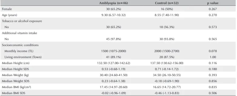
Demographic data, including sex, age, height, and weight, are shown in table 1. The median age values in the amblyopia and control groups were 9.3 years (6.5-10.3) and 8.5 years (7.4-11.9), respectively. There were no significant differences in age, sex, height, height stan-dard deviation score (SDS), weight SDS, BMI, or BMI SDS between groups. The median value of BMI was higher in severe amblyopia (18.00 [16.80-21.05]) and lower in mild amblyopia (16.30 [14.22-19.40]). There was no statistical difference between the amblyopia and control groups in terms of smoke and alcohol exposure, addi-tional vitamin intake, and socioeconomic conditions. Unsurprisingly, BCVA and refractive power showed sig-nificant differences between the amblyopia and control groups (p<0.001).
Vitamin B12 and folate data
Vitamin B12 and folate concentrations were
evalua-ted in blood samples of 42 amblyopic and 32 control subjects. Vitamin B12 concentration demonstrated a statistically significant difference across the amblyopia group when subdivided by severity and the control group (p=0.045). No significant difference was found between folate concentrations in the different degrees of the amblyopia and control groups. The median
vita-min B12 and folate concentrations were lower in those
with severe amblyopia and higher in those with mild amblyopia (Table 2).
Elemental data
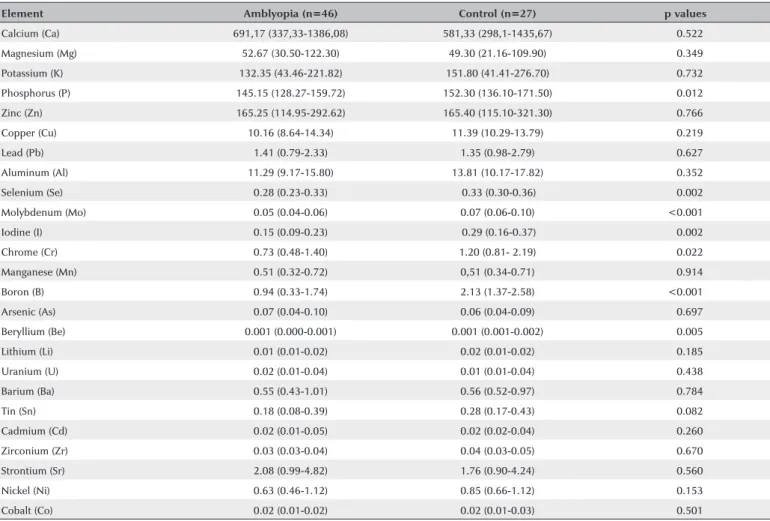
All elemental concentrations were evaluated in the hair samples of 46 amblyopic and 27 control subjects. All linear calibration curves were acceptable, with a correlation coefficient of R >0.999. Acceptable reco-very and precision (RSD%) results were achieved using the certified reference material (80%-110% recovery). All RSD% results were <15%, except Al. The mineral content in the amblyopia and control groups is shown in table 3. The concentrations of P, Se, Mo, I, Cr, B, and Be in the hair samples from amblyopic children were signi-ficantly lower than those in the control group (Table 3). The mineral content in children with differing degrees of amblyopia is shown in table 4. The concentrations of Se, Mo, I, B, and Be were statistically significantly different across the amblyopia group when subdivided by severity and the control group. Table 5 shows the correlations between vitamin B12, folate, BMI SDS, and some ele-mental concentrations. Significant positive correlations were found between Ca and Mg; between I, Mo, and Se; and between Cr, Ca, and Mg. Negative correlations were found between P and Ca, between Mg and I, and between B, Ca, and Mg in the amblyopia and control groups, respectively.
Table 1. Demographic and anthropometric data of the amblyopia and control groups
Amblyopia (n=46) Control (n=32) p value
Female 30 (65.2%) 16 (50%) 0.267
Age (years) 9.30 (6.57-10.32) 8.55 (7.40-11.90) 0.270
Tobacco or alcohol exposure
No 30 (65.2%) 18 (56.3%) 0.573
Additional vitamin intake
No 45 (97.8%) 30 (93.8%) 0.565
Socioeconomic conditions
Monthly income (TL) 1500 (1075-2000) 2000 (1500-2700) 0.078
Living environment (Town) 41 (89.1%) 28 (87.5%) 1.00
Median Height (cm) 132.50 (127.00-142.62) 137.50 (130.62-156.00) 0.116
Median Height SDS 0.53 (-0.68-1.19) 0.71 (-0.14-1.72) 0.180
Median Weight (kg) 30.40 (24.60-41.50) 34.50 (26.10-50.55) 0.393
Median Weight SDS 0.23 (-0.64-1.38) -0.18 (-0.69-1.90) 0.856
Median BMI (kg/cm²) 17.45 (14.97-20.60) 16.65 (14.72-20.77) 0.835
Median BMI SDS -0.02 (-0.96-1.09) -0.46 (-1.13-0.83) 0.506
n= number of subjects; SDS= standard deviation score; TL= Turkish lira.
Table 2. Comparison of BMI, BMI SDS, vitamin B12, and folate in the amblyopia and control groups
BMI (kg/cm²) BMI SDS Vitamin B12 (pmol/L) Folate (nmol/L)
Amblyopia (n=42) 17.45 (14.98-20.60) -0.02 (-0.96-1.09) 278.22 (205.90-374.53) 20.45 (16.37-24.71)
Mild (n=16) 16.30 (14.22-19.40) -0.02 (-1.23-1.31) 330.62 (278.22-422.13) 21.52 (17.73-28.72)
Moderate (n=17) 17.30 (15-23.50) -0.13 (-0.94-1.44) 255.34 (208.11-376.01) 20.97 (17.82-25.05)
Severe (n=9) 18.00 (16.80-21.05) 0.43 (-0.87-0.93) 205.90 (158.67-287.82) 18.89 (14.22-25.62)
Control (n=32) 16.65 (14.73-20.78) -0.46 (-1.13-0.83) 306.27 (224.72-328.77) 20.45 (16.37-24.71)
p values 0.517 0.912 0.045* 0.547
BMI= body mass index; SDS= standard deviation score; n= number of subjects; p values demonstrate comparisons between controls and subjects with differing degrees of amblyopia.
Kruskal-Wallis oneway ANOVA test was used to compare BMI, BMI SDS, vitamin B12, and folate. *Pairwise comparison is shown between mild and severe amblyopia.
Table 3. Mineral contents (µg g-1) in hair samples of amblyopia and control groups
Element Amblyopia (n=46) Control (n=27) p values
Calcium (Ca) 691,17 (337,33-1386,08) 581,33 (298,1-1435,67) <0.522
Magnesium (Mg) 52.67 (30.50-122.30) 49.30 (21.16-109.90) <0.349
Potassium (K) 132.35 (43.46-221.82) 151.80 (41.41-276.70) <0.732
Phosphorus (P) 145.15 (128.27-159.72) 152.30 (136.10-171.50) <0.012
Zinc (Zn) 165.25 (114.95-292.62) 165.40 (115.10-321.30) <0.766
Copper (Cu) 10.16 (8.64-14.34) 11.39 (10.29-13.79) <0.219
Lead (Pb) 1.41 (0.79-2.33) 1.35 (0.98-2.79) <0.627
Aluminum (Al) 11.29 (9.17-15.80) 13.81 (10.17-17.82) <0.352
Selenium (Se) 0.28 (0.23-0.33) 0.33 (0.30-0.36) <0.002
Molybdenum (Mo) 0.05 (0.04-0.06) 0.07 (0.06-0.10) <0.001
Iodine (I) 0.15 (0.09-0.23) 0.29 (0.16-0.37) <0.002
Chrome (Cr) 0.73 (0.48-1.40) 1.20 (0.81- 2.19) <0.022
Manganese (Mn) 0.51 (0.32-0.72) 0,51 (0.34-0.71) <0.914
Boron (B) 0.94 (0.33-1.74) 2.13 (1.37-2.58) <0.001
Arsenic (As) 0.07 (0.04-0.10) 0.06 (0.04-0.09) <0.697
Beryllium (Be) 0.001 (0.000-0.001) 0.001 (0.001-0.002) <0.005
Lithium (Li) 0.01 (0.01-0.02) 0.02 (0.01-0.02) <0.185
Uranium (U) 0.02 (0.01-0.04) 0.01 (0.01-0.04) <0.438
Barium (Ba) 0.55 (0.43-1.01) 0.56 (0.52-0.97) <0.784
Tin (Sn) 0.18 (0.08-0.39) 0.28 (0.17-0.43) <0.082
Cadmium (Cd) 0.02 (0.01-0.05) 0.02 (0.02-0.04) <0.260
Zirconium (Zr) 0.03 (0.03-0.04) 0.04 (0.03-0.05) <0.670
Strontium (Sr) 2.08 (0.99-4.82) 1.76 (0.90-4.24) <0.560
Nickel (Ni) 0.63 (0.46-1.12) 0.85 (0.66-1.12) <0.153
Cobalt (Co) 0.02 (0.01-0.02) 0.02 (0.01-0.03) <0.501
n= number of subject.
All except phosphorus were normally distributed and compared with student’s t test, and for phosphorus Mann-Whitney U Test was used.
Comparisons are significant at the p<0.05 level.
DISCUSSION
Although many contributing factors may lead to am-blyopia, the etiology is not clear. In this study, we in-vestigated the relationship between amblyopia and BMI and vitamin and mineral content. We observed that some mineral element concentrations showed
statisti-cally significant differences in amblyopic children when compared with age-matched healthy controls.
also reported that amblyopic children have increased BMI(4); however, our results are not consistent with this
finding possibly because of the relatively small sample size of our severe amblyopia group. Further larger studies are needed to clarify the effect of BMI on amblyopia development.
Vitamin B12 and folate deficiencies are known to im-pair axonal transport and neuronal regeneration. Visual evoked potential studies have shown demyelination in the white matter of the spinal cord and optic nerve in patients with vitamin B12 deficiency(11-13). In our study,
vi-tamin B12 and folate concentrations in amblyopic chil dren
were similar to those in the control subjects. However, when examining these concentrations in amblyopic children with different degrees of severity, the mean concentrations of vitamin B12 and folate decrease as the disease severity increases. Unfortunately, owing to the small group size, it was not possible to distinguish between amblyopia severity and vitamin B12 and folate concentration differences. Larger sample studies are
ne-eded to determine the relationship between amblyopia severity and vitamin B12 and folate concentrations. We hope that publication of our data will stimulate further work with larger sample sizes to clarify this relationship.
Inorganic elemental concentrations were measured using hair samples to reflect long-term concentrations in the body and thus general dietary intake(14). The Mg
and Ca levels in the hair of amblyopic children were higher than those in the control group but did not reach statistical significance. Mg blocks N-methyl-D-aspartate
(NMDA) receptors, and Ca causes neurotoxicity via the NMDA receptors. Visual system development includes the activation of the NMDA receptors, but this process is inhibited in high Mg concentrations(15,16). The
rela-tionship between Ca, Mg, NMDA, and amblyopia was discussed in a recent study(4).
Boron is vital for some enzymatic reactions as it maintains the integrity and function of cell membranes. It is an important element that increases Ca and Mg me-tabolism while decreasing their urinary excretion(17,18). Table 5. Spearman correlation coefficient matrix for selected variables of the amblyopia group (above diagonal) and the control group (below diagonal)
Folate B12 BMI SDS B P Cr Se Mo I Ca Mg
Folate 0.287 -0.024 -0.198** 0.200 -0.133* -0.190* -0.284 -0.143** -0.190** -0.203**
B12 0.024 -0.037 -0.156** 0.107 -0.044* -0.140* -0.278 -0.034** -0.124** -0.079**
BMI SDS -0.161 -0.058 -0.263** -0.229 -0.110* -0.136* -0.189 -0.172** 0.254** -0.182**
B 0.106 0.185 -0.201 -0.264 -0.047* -0.080* -0.257 -0.089** -0.519** -0.413**
P -0.140 0.154 0.129 -0.418** -0.206* -0.017* -0.172 -0.385** -0.496** -0.423**
Cr -0.350 -0.194 -0.051 -0.090** 0.005 -0.033* -0.167 -0.038** -0.099** -0.082**
Se 0.033 0.228 0.000 -0.210** 0.265 -0.313* -0.103 -0.174** -0.111** -0.031**
Mo -0.065 0.002 -0.219 -0.290** -0.265 -0.346* -0.070* -0.418** -0.034** -0.002**
I 0.183 -0.181 -0.255 -0.236** 0.018 0.067* -0.436* -0.225 -0.428** -0.395**
Ca -0.256 -0.266 0.239 -0.691** -0.280 -0.481* -0.234* -0.116 -0.205** -0.926**
Mg -0.233 -0.279 0.209 -0.662** -0.295 -0.484* -0.162* -0.098 -0.085** -0.950**
SDS= standard deviation score, *= correlation is significant at the 0.05 level, **= correlation is significant at the 0.01 level.
Table 4. Comparison of mineral contents (µg g-1) in hair samples between controls and subjects with differing degrees of amblyopia
Control (n=27) Mild (n=18) Moderate (n=19) Severe (n=9) p value
P 152.30 (136.10-171.50) 143.00 (130.80-161.42) 148.00 (129.40-161.50) 136.80 (117.55-155.95) <0.951,2,3
Se 0.33 (0.30-0.36) 0.28 (0.22-0.32) 0.29 (0.25-0.35) 0.23 (0.20-0.32) <0.0081,3
Mo 0.07 (0.06-0.10) 0.05 (0.04-0.07) 0.05 (0.04-0.06) 0.05 (0.03-0.05) <0.0011,2,3
I 0.29 (0.16-0.37) 0.17 (0.09-0.31) 0.15 (0.07-0.21) 0.15 (0.10-0.23) <0.0161,2,3
Cr 1.20 (0.81-2.19) 0.64 (0.39-1.35) 0.97 (0.55-1.79) 0.85 (0.44-1.26) <0.0591,2,3
B 2.13 (1.37-2.58) 1.36 (0.41-1.82) 0.68 (0.27-1.73) 0.94 (0.09-1.57) <0.0012,3
Be 0.001 (0.001-0.002) 0.001 (0.001-0.001) 0.000 (-0.001-0.002) 0.000 (-0.001-0.001) <0.0082,3
Boron is involved in regulating nicotinamide adenine dinucleotide phosphate, which, in turn, is important for the degradation of oxidative glutathione. Glutathione is an antioxidant substrate; hence, boron is an important mineral element for antioxidant defense mechanisms(19).
In the present study, boron levels were significantly lower in amblyopic patients. As might be expected, the concentrations of Ca and Mg increased in both the amblyopia and control groups as the boron levels incre-ased; this relationship is shown in Table 5 as a negative correlation.
A selenium-deficient diet in rats reduces cell num-bers in the inner retinal nuclear layer, decreases hori-zontal and amacrine cell nucleus, and causes some neuronal injuries(20). Selenium is also an important
antioxidant element, and its effects on the eyes have been demonstrated in many clinical and experimental studies. Selenium deficiency is reported to cause oxida-tive stress in the corneal epithelium, resulting in corneal damage. Thus, selenium has been suggested to help in the treatment of dry eyes(5). In another study, low serum
selenium in myopic children was associated with the development of myopia(6). Therefore, selenium appears
to have an important role in the normal development and function of the visual apparatus. In the present stu-dy, we also found a significantly reduced concentration of Se in amblyopic children compared to controls. Both boron and selenium are important mineral regulators of antioxidant function and may play an important role in maintaining optic nerve health and amblyopia. We suggest that dietary supplements containing selenium and perhaps boron should be considered for amblyopic children.
Molybdenum is a cofactor of the enzymes sulfite oxida-se and xanthine oxidaoxida-se. Molybdenum deficiency is ty-pically considered a metabolic disease and is characteri-zed by a defect in psychomotor functioning. A correlation has been found between molybdenum and both selenium and iodine(21). As a component of both tri-iodothyronine
(T3) and tetra-iodothyronine (T4) thyroid hormones, io-dine is critical for normal thyroid function, and selenium is a cofactor for thyroperoxidase, an enzyme catalyzing the production of these hormones in the thyroid gland. Recent studies have shown that molybdenum-deficient patients have a tendency to develop hypothyroidism(21).
The present study showed that the levels of these ele-ments were decreased in amblyopic children. Further studies are required to investigate any relationship be-tween thyroid function and amblyopia.
A major limitation of this study was its small sample size, especially in the severe amblyopia group; hence, this group needs further study with more subjects, which could affect the statistical analysis of the results. However, we believe that our results are intriguing and that large-scale studies should be performed to further investigate the possible relationships between inorganic elements and amblyopia.
In conclusion, amblyopic children have low levels of selenium, iodine, molybdenum, boron, and beryllium, suggesting that deficiencies of these elements could be related to visual development abnormalities either via excessive oxidative stress, thyroid hypoactivity, or both. Therefore, pediatric ophthalmologists should consi-der the nutritional status when assessing amblyopic children. Further studies are needed to investigate the effects of these elements on the etiology and pathophy-siology of amblyopia.
REFERENCES
1. Leon A, Donahue SP, Morrison DG, Estes RL, Li C. The age-de pen-dent effect of anisometropia magnitude on anisometropic amblyopia severity. JAAPOS. 2008;12(2):150-6.
2. Spolidoro M, Baroncelli L, Putignano E, Maya-Vetencourt JF, Viegi A, Maffei L. Food restriction enhances visual cortex plasticity in adulthood. Nat Commun. 2011;2:320.
3. Maya-Vetencourt JF, Origlia N. Visual cortex plasticity: a complex interplay of genetic and environmental influences. Neural Plast. 2012;2012:631965.
4. Zhu Q, Chen J, Zheng M, Chen L, Liu J, Tian J, et al. Mineral ele-ments in the hair of amblyopic children. Biol Trace Elem Res. 2011; 141(1-3):119-25.
5. Higuchi A, Takahashi K, Hirashima M, Kawakita T, Tsubota K. Se-lenoprotein P controls oxidative stress in cornea. PLoS One. 2010; 5(3):e9911.
6. Fedor M, Socha K, Urban B, Soroczyńska J, Matyskiela M, Borawska MH, et al. Serum concentration of zinc, copper, selenium, manganese, and cu/zn ratio in children and adolescents with myopia. Biol Trace Elem Res. 2017;176(1):1-9.
7. Akaike A, Tamura Y, Sato Y, Yokota T. Protective effects of a vitamin B12 analog, methylcobalamin, against glutamate cytotoxicity in cultured corticalneurons. Eur J Pharmacol. 1993;241(1):1-6. 8. Scalabrino G. The multi-faceted basis of vitamin B12 (cobalamin)
neurotrophism in adult central nervous system: lessons learned from its deficiency. Prog Neurobiol. 2009;88:203-20.
9. McGarel C, Pentieva K, Strain JJ, McNulty H. Emerging roles for folate and related B-vitamins in brain health across the lifecycle. Proc Nutr Soc. 2015;74(1):46-55.
10. Neyzi O, Bundak R, Gökçay G, Günöz H, Furman A, Darendeliler F, et al. Reference values for weight, height, head circumference, and body mass ındex in turkish children.J Clin Res Pediatr Endocrinol. 2015;7(4):280-93.
12. Pandey S, Kalita J, Misra UK. A sequential study of visual evoked potential in patients with vitamin B12 deficiency neurological syndrome. Clin Neurophysiol. 2004;115(4):914-918.
13. Puri V, Chaudhry N, Goel S, Gulati P, Nehru R, Chowdhury D. Vitamin B12 deficiency: a clinical and electrophysiological profile. Electromyogr Clin Neurophysiol. 2005;459(5):273-84.
14. Pasha Q, Malik SA, Shaheen N, Shah MH. Comparison of trace elements in the scalp hair of malignant and benign breast lesions versus healthy women. Biol Trace Elem Res. 2010;134(2):160-73. 15. Murphy KM, Duffy KR, Jones DG. Experience-dependent changes
in NMDAR1 expression in the visual cortex of an animal model for amblyopia. Vis Neurosci. 2004;21(4):653-70.
16. Kato N, Yoshimura H. Reduced Mg2+ block of N-methyl-D-aspar-tate receptor-mediated synaptic potentials in developing visual cortex. Proc Natl Acad Sci USA. 1993;190(15):7114-8.
17. Gümüşderelioğlu M, Tunçay EÖ, Kaynak G, Demirtaş TT, Aydın ST,
Hakkı SS. Encapsulated boron as an osteoinductive agent for bone scaffolds. J Trace Elem Med Biol. 2015;31:120-8.
18. Beattie JH, Peace HS. The influence of a low boron diet and boron supplementation on bone, major mineral and sex steroid meta-bolism in postmenopausal women. Br J Nutr. 1993;69(3):871-84. 19. Doğan A, Demirci S, Apdik H, Bayrak OF, Gulluoglu S, Tuysuz EC,
et al. A new hope for obesity management: Boron inhibits adipo-genesis in progenitor cells through the Wnt/β-catenin pathway. Metabolism. 2017;69:130-42.
20. Amemiya T. Retinal changes in the selenium deficient rat. Int J Vitam Nutr Res. 1985;55(3):233-7.