Characterization of an antagonistic peptide produced by a Bacteroides Fragilis
isolate obtained from a patient with intra-abdominal infection
Caracterização de um peptido antagonístico produzido por Bacteroides
Fragilis isolado obtido em um paciente com infecção intra-abdominal
DOI:10.34117/bjdv5n12-303
Recebimento dos originais: 07/10/2019 Aceitação para publicação: 20/12/2019
Marcela Nascimento Pinheiro Braga
Doutora em Microbiologia
Instituição: Universidade Federal de Minas Gerais
Endereço: Av. Pres. Antônio Carlos 6627, Pampulha, Belo Horizonte- MG, 31270-901 E-mail: marcelanpbraga@yahoo.com.br
Natalia Rocha Guimarães
Mestre em Ciëncias - Microbiologia
Instituição: Universidade Federal de Minas Gerais
Endereço: Av. Pres. Antônio Carlos 6627, Pampulha, Belo Horizonte- MG, 31270-901 E-mail: natyroguiman@yahoo.com.br
Jamil Silvano Oliveira
Especalista em Microbiologia aplicada as Ciëncias da Saúde Instituição: Universidade Federal de Minas Gerais
Endereço- Av. Pres. Antônio Carlos 6627, Pampulha, Belo Horizonte- MG, 31270-901 E-mail: jamil_lefqp@yahoo.com.br
Simone Gonçalves dos Santos
Doutora em Microbiologia
Instituição: Universidade Federal de Minas Gerais
Endereço: Av. Pres. Antônio Carlos 6627, Pampulha, Belo Horizonte- MG, 31270-901 E-mail: simonesantoskey@ufmg.br
Marcelo Porto Bemquerer
Doutor em Bioquímica
Instituição: Embrapa Recursos Genéticos e Biotecnologia
Endereço: Parque Estação Biológica, PqEB, Av. W5 Norte (final). Caixa Postal 02372 – Brasília, DF – CEP 70770-917
E-mail: marcelo.bemquerer@embrapa.br
Paula Prazeres Magalhães
Doutora em Ciências - Microbiologia Instituição: Universidade Federal de Minas Gerais
Endereço: Av. Pres. Antônio Carlos 6627, Pampulha, Belo Horizonte- MG, 31270-901 E-mail: paulaprazeresmagalhaes@gmail.com
Luiz de Macêdo Farias
Doutor em Ciëncias- Microbiologia
Instituição: Universidade Federal de Minas Gerais
Endereço: Av. Pres. Antônio Carlos 6627, Pampulha, Belo Horizonte- MG, 31270-901 E-mail: luizdemacedofarias@gmail.com
ABCTRACT
The indigenous microbiota of humans is extremely rich and diverse, with special emphasis on the intestinal microbiota. As a constituent of this microbiota, it is mentioned the genus Bacteroides, whose members are Gram negative rods, obligate anaerobes, amphibionts, associated to the etiopathogenesis of important infectious diseases, such as intra-abdominal infections. Bacteroides strains have the ability to synthesize antagonistic substances that play an ecological role, especially in densely colonized habitats, giving a competitive advantage to the producing samples. The objective of this study was to evaluate the synthesis capacity of antagonistic substances by 40 samples of
Bacteroides and Parabacteroides isolated from patients with intra-abdominal infections. The
expression of antagonism was evaluated by the overlay diffusion method, using, as well as the test samples, 36 reference samples of Gram negative and Gram-positive bacteria. Subsequently, a production strain (Bacteroides fragilis) was used for extraction, purification and partial characterization of the detected antagonistic substance. As indicator strains, Bacteroides ovatus and
Bacteroides caccae were used. The production strain was submitted to protein extraction, and the
activity of the precipitated intracellular extract was detected with (NH4) 2SO4 in concentrations of 30% (C30) and 50% (C50). C30 and C50 were inactivated by proteases and high temperatures and remained active after exposure to organic solvents and a wide pH range. Both fractions presented antagonistic activity of bacteriostatic nature. The C50 extract was subjected to ion exchange chromatography, and 50 fractions were recovered. Among them, fractions 1 to 4, referring to a single peak, that were not able to bind to the column, presented antagonistic activity. Fractions from the ion exchange chromatography were applied in gel filtration chromatography. Among them, fractions 2 and 3 were able to inhibit the developing sample. These fractions were submitted to reverse phase chromatography, and 50 fractions were collected. One of them, fraction 2C, remained active against the revealing sample. Mass spectrometry, from fraction 2C obtained from reverse phase chromatography, presented ions of approximately 1300.00 Da, which generated a more intense signal. The search performed by similarity between the sequenced peptides and proteins described in the BLASTP database, from fragmentation obtained in reverse phase chromatography, resulted in 100% identity between two peptides. One of the sequenced peptides showed 100% identity to a type VII secretion protein. The search performed by similarity between the sequenced peptides and proteins described in the ANTIMICROBIAL PEPTIDE DATABASE database, from fragmentation obtained with trypsin digestion, resulted in 42% identity with a Streptomyces microcine. Together, the results indicate the production of antagonistic substances by the B. fragilis sample under study. It is plausible to assume that they play a relevant role in interbacterial relationships, as a virulence factor, in a complex environment such as intra-abdominal infection.
Keywords: Bacteroides, Parabacteroides, antagonistic substance, intra-abdominal infection. RESUMO
A microbiota humana indígena é extremamente rica e diversificada, com ênfase especial na microbiota intestinal. Como constituinte dessa microbiota, é mencionado o gênero Bacteroides, cujos membros são bastonetes Gram-negativos, forçando anaeróbios, anfibiontes, associados à etiopatogenia de importantes doenças infecciosas, como infecções intra-abdominais. As cepas de
Bacteroides têm a capacidade de sintetizar substâncias antagônicas que desempenham um papel
ecológico, especialmente em habitats densamente colonizados, dando às amostras produzidas uma vantagem competitiva. O objetivo deste estudo foi avaliar a capacidade de síntese de substâncias
antagônicas por 40 amostras de Bacteroides e Parabacteroides isolados de pacientes com infecções intra-abdominais. A expressão do antagonismo foi avaliada pelo método de difusão por sobreposição, utilizando, bem como as amostras de teste, 36 amostras de referência de bactérias Gram-negativas e Gram-positivas. Posteriormente, uma cepa de produção (Bacteroides fragilis) foi usada para extração, purificação e caracterização parcial da substância antagonista detectada. Como linhas indicadoras, foram utilizados Bacteroides ovatus e Bacteroides caccae. A cepa de produção foi submetida à extração de proteínas e a atividade do extrato intracelular precipitado foi detectada com (NH4) 2SO4 em concentrações de 30% (C30) e 50% (C50). C30 e C50 foram inativados por proteases e altas temperaturas e permaneceram ativos após a exposição a solventes orgânicos e uma ampla faixa de pH. Ambas as frações apresentaram atividade antagonista de natureza bacteriostática. O extrato C50 foi submetido a cromatografia de troca iônica e 50 frações foram recuperadas. Entre elas, as frações 1 a 4, referentes a um único pico, que não foram capazes de se ligar à coluna, apresentaram atividade antagônica. As frações da cromatografia de troca iônica foram aplicadas em cromatografia de filtração em gel. Entre elas, as frações 2 e 3 foram capazes de inibir a amostra em desenvolvimento. Essas frações foram submetidas à cromatografia de fase reversa e foram coletadas 50 frações. Um deles, a fração 2C, permaneceu ativo contra a amostra reveladora. A espectrometria de massa, da fração 2C, obtida por cromatografia de fase reversa, apresentou íons de aproximadamente 1300,00 Da, o que gerou um sinal mais intenso. A busca realizada por similaridade entre os peptídeos e proteínas seqüenciados descritos no banco de dados BLASTP, a partir da fragmentação obtida na cromatografia de fase reversa, resultou em 100% de identidade entre dois peptídeos. Um dos peptídeos sequenciados mostrou 100% de identidade para uma proteína de secreção do tipo VII. A busca realizada por similaridade entre os peptídeos e proteínas seqüenciados descritos no banco de dados ANTIMICROBIAL PEPTIDE DATABASE, a partir da fragmentação obtida com a digestão com tripsina, resultou em 42% de identidade com um microcine Streptomyces. Juntos, os resultados indicam a produção de substâncias antagônicas pela amostra de B.fragilis em estudo. É plausível assumir que eles desempenham um papel relevante nas relações interbacterianas, como fator de virulência, em um ambiente complexo como a infecção intra-abdominal.
Palavras-chave: Bacteroides, Parabacteroides, substância antagonista, infecção intra-abdominal.
1 INTRODUCTION
Bacteroides and Parabacteroides are two important genera that are part of the indigenous
microflora of humans and other animals. This collection of microrganisms inhabiting the human body consists of complex microbial communities such as that present in the digestive tract, particularly in the oral cavity and intestines. As members of the gut microflora, Bacteroides and Parabacteroides offer several advantages to the host, assisting in physiology and normal function of the gastrointestinal tract, helping in digesting complex polysaccharides, and also using nutritional resources, which aids in the intestinal colonization resistance by bacteria potentially harmful (Ishikawa et al., 2013).
B. fragilis is a member of the intestinal microbiota of humans and is involved in a variety of
activities that influence host health, such as polysaccharide digestion, intestinal development and maturation, and modulation of the immune response. In addition to its beneficial role, B. fragilis, as
discussed above, is a classical amphibion, being an opportunistic pathogen associated with several infectious diseases involving anaerobes (Wilson et al., 2015).
The various species that make up the microbial communities are involved in all forms of competition, whether by nutritional resources or space. To overcome their adversaries, microorganisms use all their resources, including secondary metabolites, enzymes and antibiotics. The competitions between bacteria, archaea, fungi and protozoa are often solved by the use of antimicrobial peptides (Wilson et al., 2015).
Among the diseases associated with Bacteroides and Parabacteroides, there is intra-abdominal infection, its severity and high prevalence. After disruption of the intestinal wall, which may occur for different reasons (surgical wound, malignancies, appendicitis or trauma, among others), the indigenous microbiota invade the peritoneal cavity normally sterile site. During the acute phase of infection (approximately 20 h), facultative anaerobes, especially Escherichia coli are established, promoting the fall of the redox potential of the environment. The atmosphere now low in oxygen, then allows bacteria obligate anaerobic, as representatives of Bacteroides and
Parabacteroides, colonize and reproduce, passing to prevail during the chronic phase of infection
(Wexler, 2016;. Nicoletti et al., 2008).
Bacteroides and Parabacteroides, especially the B. fragilis, can express a wide variety of
virulence skills such as synthetic enzymes, toxins secretion and production of antimicrobial substances, known as bacteriocins, which favors microrganism success in colonizing the host and aggression (Diniz et al., 2000; Wexler, 2007; Papaparaskevas et al, 2011). With reference to antagonistic substances produced by anaerobic bacteria, in 1965, Beerens & Baron described for the first time, a bacteriocin synthesized by Bacteroides, but more detailed studies have not been conducted. Until 1983, only a few papers have been published, such as Booth et al (1977) who described a bacteriocin having a molecular mass greater than 300,000 Da, produced by Bacteroides, and the Mossie et al. (1979) have purified a bacteriocin produced by B. fragilis molecular weight between 13,000 and 19,000 Da. In 1981, Mossie et al. described the purification and characterization of another bacteriocin produced by B. fragilis and demonstrated in their ability to inteferir bacterial RNA synthesis by inhibiting RNA polymerase activity.
In 1983, Hayes et al. purified and characterized a bacteriocin produced by B. fragilis, with species-specific activity. The bacteriocin was found in the initial purification step as a higher molecular weight complex with 2 x 10⁷ Da, containing protein, lipid and carbohydrate. Dissociation of the complex allowed the purification of bacteriocin having a molecular mass of 5000 Da. Bacteriocin activity investigated was not altered by RNase, DNase, phospholipase A, pancreatic
lipase, and dextranase but was destroyed by trypsin, proteinase K, heat (80 ° C, 30 min) and pH values below 5 and 8 above.
In 1992, Farias et al reported the ability of producing bacteriocin-like substances for group B.
fragilis isolates the oral cavity and marmosets intestine. Farias et al. (1994) showed the production
of proteins with one or more bacteriocin-like activity for samples B. fragilis recovered primate species Callithrix. The bacteriocin had iso activity and heteroantagonista. The extracellular extracts (S70) and intracellular (C70) showed activity against isolates of B. fragilis from callitrichids gut and against
Streptococcus sanguis reference samples and Streptococcus pyogenes.
The study of the cell surface molecules produced by B. fragilis has deserved prominence because they probably play an important role in colonization, persistence and communication with other microorganisms. The outer membrane components and the B fragilis proteome include components, not yet well characterized, that are used to transport polypeptides to the extracellular space (Wilson et al., 2015). To investigate the expression of antagonistic activity and characterize the substances involved in the phenomenon provide a better understanding of microbial ecological relations, as well as the emergence of new perspectives regarding the use of bacteria producing antagonistic substances.
2 METODOLOGY Samples
To evaluate the expression of antagonism, were included in the study 30 samples test of
Bacteroides strains (11 B. fragilis, 5 B. ovatus, 1 B. thetaiotaomicron, 3 B. uniformis, 1 B. vulgatus,
6 B. caccae, 2 B. capillosus, 1 B. eggherthii) and 10 Parabacteroides distasonis strains isolated from patients with intra-abdominal infections (called test samples). As revealing, were used, in addition to the test samples, a panel of reference samples of bacteria with different types respiratory, Gram positive and Gram negative bacteria. All samples are part of collection of Oral Microbiology Laboratory and Anaerobic and are being kept in Brucella Broth plus 10% glycerol at -80 ° C freezer. The antagonism of expression was evaluated by diffusion method overlying (Booth et al., 1977). Initially, the test samples were inoculated into brain heart infusion, pH 7.2, supplemented with 0.5% porcine hemin, menadione and 0.1% yeast extract 0.5% (BHI-S). The cultivation was performed in the anaerobic chamber with 85% N2, 5% CO2 and 10% H2 at 37°C. After 48 hours, the samples were inoculated into Brain Heart Infusion Agar (BHIA) supplemented with 0.5% porcine hemin, menadione% and 0.1% levedura0,5 extract (BHIA-S) with the aid of a Steers replicator (Steers et al. 1959), in a concentration of approximately 105 cells / spot. After incubation under the same conditions described above as cultures were exposed to chloroform for 30 min and then the plates were kept
open for 30 min to evaporate the residual chloroform. The revealing samples were cultivated in liquid medium and then a 0.2 mL aliquot of the culture was inoculated into 3.5 ml of semisolid medium (0.7% agar). Then the mixture was poured into spots test sample. After incubation at 37oC, the reading is performed by checking the presence or absence of inhibition halos. The experiment was performed in duplicate. For each revealing sample to be tested culture conditions were employed (culture medium, incubation time and atmosphere) indicative of each specific sample.
Factors interference
To assess whether the observed inhibition was due to production of antagonistic substances or other possible factors, the tests were conducted as follows. To evaluate the presence of bacteriophages, possibly responsible for antagonist activity, one agar sample fragment revealing the multiplication zone of inhibition was aseptically removed. The fragment was macerated in saline solution pH 7.2 and then centrifuged at 23,400 g for 20 min. An aliquot of 200 µL of the supernatant was transferred to 3 ml of culture revealing sample. After incubation for 10 min at room temperature to allow adsorption of viral revealing bacteria, an aliquot of 200 μL foi added to 3.5 mL of semisolid agar for revealing specific sample selected and the entire volume was poured on a medium layer solid. The material was incubated at 37° C under appropriate conditions for multiplication of revealing sample. The presence of lysis zones is indicative of the presence of bacteriophages (Turner & Jordan, 1981). The evaluation of the possible inhibition of fatty acids was carried out using S-BHIA with and without addition of 1% soluble starch (Walstad et al., 1974). To exclude the possibility that acids are responsible for antagonism after preparation of S-BHIA plates, the medium pH was measured using a microelectrode. After the test, as described in item 4.2, the zone of inhibition of proliferation of revealing sample was measured. As a control, it was incubated Petri dish containing only BHIA-S and the pH was also measured before and after incubation. The pH values of the inoculated and non-inoculated media were then compared (Apolonio et al., 2007). To evaluate the possibility of interference from the presence of residual chloroform in the multiplication of revealing sample, the following test was performed. The producer sample was inoculated in the spot on the surface of BHIA-S and, after incubation under the conditions set forth above, the revealing sample, previously cultivated suitable liquid medium was inoculated around the producer sample spot, cross-shaped, without that the growth of producer sample was touched. The material was incubated in specific conditions for revealing sample and then evaluated the presence or absence of inhibition halo. (Farias
et al., 1992).
The production H2O2foi investigated by incorporation of catalase 0.03% w / v in BHI-S. As a control, we used the same medium without the addition of the enzyme. The presence of the revealing
sample multiplication inhibition zone only in medium without addition of catalase was considered indicative that the antagonist activity is due to the presence of H2O2 (Ooshima & Hamada, 1975).
PROTEIN EXTRACTION
The experiments of protein fraction extraction were performed according to the protocol proposed by Green & Huges (1955), with some adaptations, according to methodology already standardized in the Laboratory of Oral Microbiology and Anaerobes.
EXTRACTION OF INTRACELLULAR FRACTION
The cell pellet obtained after the first centrifugation step was washed twice with 0.01M Tris-HCl buffer, pH 8.0, by centrifugation at 16,200 g for 30 min. After this procedure, the pellet was suspended in 15 mL of the same buffer and then sonicated at 50 W in an ice bath for 30 cycles of 40 s each, at 40 s intervals. Cell lysis was confirmed by examination under an optical microscope after staining by the Gram method. The resulting sonicated fraction was again centrifuged at 29,829 g at 4 ° C for 30 min. The supernatant was collected, subjected to ammonium sulfate precipitation and dialysed, as described in item 4.5.1, to obtain the intracellular fractions, called C-30, C-50 and C-80.
CHARACTERIZATION OF ACTIVE FRACTIONS
The effects of pH, temperature, proteolytic enzymes and organic solvents on the activity and stability of C30 and C50 fractions were evaluated.
pH
To evaluate the stability of the antagonistic activity against different pH values, a methodology described by Valeff (2011) was used. The Britton-Robinson universal buffer (Britton & Robinson, 1931), which allows pH adjustment to values from 2 to 12, was used. The pH adjustment was performed with 2M NaOH solution and 2M HCl and the buffers were sterilized by filtration. The extracts were diluted 1: 1 (v / v) in the universal buffer and the solution was then incubated at 37 ° C at 15 and 30 min, 1, 2, 3, 4, 5 and 6 h intervals, 1, 2, 3, 6, 10, 20 and 30 days. As a control, the activity of the buffer and the diluted fraction in 0.01 M Tris-HCl, pH 8.0, at the ratio of 1: 1 (v / v), incubated in the same condition as the test tubes was checked.
TEMPERATURE
For the evaluation of the thermal treatment interference in the antagonistic activity of the protein extracts, a methodology described by Ribeiro-Ribas et al. (2009) was used, with some
adaptations. Aliquots of fractions diluted 1: 1 (v / v) in 0.01 M Tris-HCl buffer, pH 8.0, were subjected to the following heat treatments: -86 ° C (freezer), -20 ° C (freezer), 4 ° C (Refrigerator), 25 ° C (room temperature), 37 ° C (oven), 50 ° C, 60 ° C, 80 ° C and 100 ° C (water bath) and 121 ° C (autoclave) , 1, 2, 4 and 8 h, 1, 2, 8, 20, 30 days, 6 and 12 months. As a control, the diluted extract was used without any heat treatment.
PROTEOLYTIC ENZYMES
The effect of proteolytic enzymes on the active fractions was tested according to a methodology described by Valeff (2011). Protein K, α-chymotrypsin, trypsin, papain, and pepsin were used. The α-chymotrypsin and trypsin enzymes were solubilized in 20 mM Tris-HCl Buffer, pH 8.0, proteinase K was diluted in 20 mM Tris-HCl Buffer pH 7.2 to papain in 50 mM phosphate buffer pH 5, And pepsin in 100 mM citrate buffer, pH 3.0, all at the final concentration of 1 mg / ml. All solutions were sterilized by filtration. The tests were performed by diluting aliquots of the active protein extracts in the enzyme solutions in a ratio of 1: 1 (v / v). The material was incubated for 15 min, 30 min and 1 h at 37 ° C. Then, the preservation of the antagonistic activity was evaluated, as described in item 4.6, and the results expressed in UA / mL. As a control, the enzymatic solutions and extracts diluted 1: 1 (v / v) in 20 mM Tris-HCl pH 8.0- and 20-mM Tris-HCl pH 7.2 were used.
ORGANIC SOLVENTS
In order to verify the effect of organic solvents on the antagonistic activity of protein extracts, a methodology described by Apolônio et al. (2008). The solvents used were acetone, acetonitrile, iso-propyl alcohol, butanol, ethanol, hexane and methanol 10% aqueous solutions and 50% of the organic solvents were prepared and then sterilized by filtration. The extracts were diluted 1: 1 (v / v) in these solutions and then incubated at 25 ° C for 1 h. Controls were performed using the aqueous solutions of the organic solvents and the extract diluted 1: 1 (v / v) in 0.01 M Tris-HCl, pH 7.2
STUDY OF THE METHOD OF ACTION OF ACTIVE FRACTIONS
DETERMINATION OF THE MINIMUM INJURY CONCENTRATION (CIM)
MIC is defined as the lowest concentration of the drug that inhibits visible growth of an organism after overnight incubation. This incubation period, however, is extended in tests performed with anaerobic microorganisms, which require more time for multiplication (CLSI, 2012).
For the determination of MIC, the protocol recommended by the Clinical and Laboratory Standards Institute (2012) was used, with some modifications, by means of microdilution in broth. The experiment was performed in duplicate.
For this evaluation, 2 μl of sample culture was added to each well, with the previously adjusted inoculum with 0.85% saline, on the McFarland 0.5 scale, and the final bacterial concentration in each well was 106 CFU / mL. Also, 100 μL of different concentrations of the active extracts were added, in addition to 98 μL of BHI-S. As a positive control, a mixture of 198 μL of BHI-S plus 2 μL of sample culture was used and as a negative control, 98 μL of BHI-S, 100 μL of extract solution and 2 μL of saline. After 48 h of incubation, the MIC was considered to be the lowest concentration of the active extract in UA / mL, which visibly inhibits the bacterial multiplication of the revealing sample (CLSI, 2012).
DETERMINATION OF THE MINIMUM BACTERICIDE CONCENTRATION (CBM)
CBM is defined as the lowest concentration of an antimicrobial agent capable of reducing the microbial count by 99.9% (Levison & Levison, 2009).
This experiment was performed from the microplate used to determine MIC, also in duplicate. Aliquots of 100 μL of the negative control and of each well in which there was no visible bacterial growth were seeded in solid medium with the aid of Drigalski's loop. The positive control was diluted in broth (10-1, 10-3 and 10-5) and 100 μL of each dilution were also seeded in agar. The plates were incubated in an anaerobic chamber at 37 ° C. After incubation, colonies were counted to estimate the number of CFU / mL.
PURIFICATION OF ACTIVE FRACTIONS BY CHROMATOGRAPHY
The active extracts were purified by successive chromatography steps. After each purification step, the fractions were submitted to antagonist activity test. Initially, fast protein liquid chromatography (FPLC) was used on Mono-Q ™ 5/50 GL Tricorn ™ column, pre-equilibrated with 0.02 M Tris-HCl buffer, pH 8.0, and eluted at Even buffer added with 1 M NaCl, in a linear gradient of 0-100%. The obtained fractions were concentrated by lyophilization and diluted in sterile Milli-Q ® water. When necessary (fractions eluted in buffer with high salt concentration), the material was dialyzed against the same buffer, however devoid of salt, prior to lyophilization.
Fractions from ion exchange chromatography with antagonistic activity were subjected to gel filtration (FPLC) on a Superose 12 HR 10/30 column, previously equilibrated with 0.02 M Tris-HCl buffer, pH 8.0, Of 0.2 M NaCl, and eluted in the same buffer. 1.5 mL aliquots were collected in a flow rate of 30 mL / h, lyophilized and diluted in sterile Milli-Q® water. The active fractions obtained
from the above purification step were subjected to high performance liquid chromatography (HPLC on C8 column (4.6 mm in diameter x 25 cm in length) equilibrated with solution A (trifluoroacetic acid (TFA) 31 0.05% v / v in Milli-Q® water) and eluted with solution B (100% acetonitrile + 0.05% TFA) .The eluted fractions were monitored using wavelength 280 and 220 nm. Were concentrated by lyophilization and diluted in sterile Milli-Q® water.
Fractions from ion exchange chromatography with antagonistic activity were subjected to gel filtration (FPLC) on a Superose 12 HR 10/30 column, previously equilibrated with 0.02 M Tris-HCl buffer, pH 8.0, Of 0.2 M NaCl, and eluted in the same buffer. 1.5 mL aliquots were collected in a flow rate of 30 mL / h, lyophilized and diluted in sterile Milli-Q® water. The active fractions obtained from the above purification step were subjected to high performance liquid chromatography (HPLC on C8 column (4.6 mm in diameter x 25 cm in length) equilibrated with solution A (trifluoroacetic acid (TFA) 31 0.05% v / v in Milli-Q® water) and eluted with solution B (100% acetonitrile + 0.05% TFA) .The eluted fractions were monitored using wavelength 280 and 220 nm. Were concentrated by lyophilization and diluted in sterile Milli-Q® water.
3 RESULTS
Evaluation of expression antagonism
None of the Parabacteroides distasonis samples expressed antagonism under the conditions employed in the study. Considering only the Bacteroides samples, heterantagonism was detected in 18 (60.0%) of the 30 samples tested, five (16.7%) of which, all B. fragilis, also expressed isoantagonism. The occurrence of autoantagonism was not detected.
It was observed heteroantagonism expression by 29 (72.5%) and isoantagonism 5 (12.5%) of the 40 samples tested. It was not detected the occurrence of autoantagonismo (Table 1).
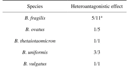
Table 1. Expression of heteroantagonism by Bacteroides samples isolated from patients with intra-abdominal infection.
Species Heteroantagonistic effect
B. fragilis 5/11a
B. ovatus 1/5
B. thetaiotaomicron 1/1
B. uniformis 3/3
B. caccae 6/6
B. capillosus 1/2
B. egghertii 0/1
a, Number of positive samples / number of samples tested.
With respect to the spectrum of action antagonist activity was observed only against phylogenetically related bacteria. Clinical samples used as indicator, B. fragilis, B. ovatus, B. uniformis, B. vulgatus, B. caccae and B. capillosus showed heteroantagonistic ativity to at least three of test samples. B. fragilis expressed isoantagonism for five test samples. All of
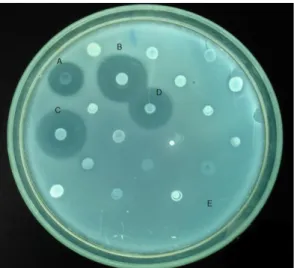
Bacteroides, showed antagonism to at least one sample producer. The sample B. fragilis D 111.4 was
the one who expressed antagonism more often exhibited the broadest spectrum of activity and generated more visible and clear halos. (Figure 1).
Figure 1. Heteroantagonistic activity Bacteroides fragilis samples of patients with intra-abdominal infection. Overlay method, Brain heart infusion agar supplemented with hemin swine, menadione and yeast extract, pH 7.0, incubation at 37 ° C for 48 h in anaerobiosis. producing samples: A, B. fragilis D 51; B, B fragilis D 111.4; C, B. fragilis 57.1; and D,
B fragilis 34.3. Sample revealing: Bacteroides ovatus D 54.1.
FACTORS INTERFERENCE
BACTERIOPHAGES
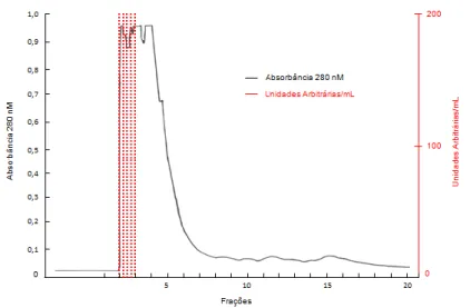
lysis zones due to the presence of bacteriophages were not observed in the test performed (Figure 2A).
FATTY
The addition of starch to the growth medium did not prevent expression of antagonism by the sample production, indicating that fatty acids and other acids were not responsible for antagonism (Figure 2B).
CHLOROFORM
The results of testing for antagonist activity research with or without addition of chloroform were similar, indicating that this substance is not responsible for the inhibition of revealing sample (Figure 2C).
HYDROGEN PEROXIDE
The addition of catalase to the culture medium did not inhibit the antagonism of expression, showing that the production of H2O2 is not responsible for the activity (Figure 2D).
Figure 2. Tests for interfering factors research. Evaluation of activity: A, bacteriophages; B, fatty acids C, chloroform; D H2O2. Overlay method, Brain Heart Infusion agar medium supplemented with swine hemin, menadione and yeast extract, pH 7.0, incubation at 37 ° C for 48 h in anaerobiosis. Sample production: Bacteroides fragilis D 111.4. Sample revealing:
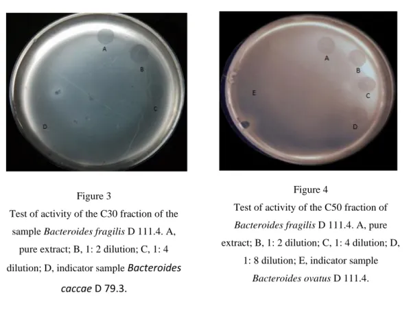
PROTEIN EXTRACTION - ANTAGONIST ACTIVITY TEST OF THE FRACTIONS OBTAINED Only the intracellular protein fractions obtained from the sample B. fragilis D 111.4, precipitated with 30% (NH4) 2SO4 (C30) and 50% (C50) showed antagonistic activity against B.
caccae D 79.3 and B.ovatus D 54.1, respectively Figure 3 and Figure 4). Thus, C30 and C50 were
selected for subsequent steps in the study.
Table 2. Characterization of fractions C30 and C50 obtained from the sample Bacteroides fragilis D 111.4.
Treatment Activity Title (AU/mL)
C30a C50b Temperature Control 200 200 50 ºC/20 min PEc PE 37 ºC/7 dias PE PE 25 ºC/7 dias 200 200 4 ºC/30 dias 200 PE -20 ºC e -80 ºC/12 months 200 200 pH Control 200 200 2, 3, 3,5/0 min 0 0 4, 4,5, 5, 5,5 e 6/3 h PE 0 6,5/ 10 days 200 200 Figure 3
Test of activity of the C30 fraction of the sample Bacteroides fragilis D 111.4. A,
pure extract; B, 1: 2 dilution; C, 1: 4 dilution; D, indicator sample Bacteroides
caccae D 79.3.
Figure 4
Test of activity of the C50 fraction of
Bacteroides fragilis D 111.4. A, pure
extract; B, 1: 2 dilution; C, 1: 4 dilution; D, 1: 8 dilution; E, indicator sample
7, 7,5/7 days PE PE 8, 8,5, 9, 9,5 e 10/5 days PE PE 11, 11,5 e 12/ 0 min 0 0 Enzims (1 mg/mL, 37 ºC) Control 200 200 Tripsin/15 min 0 0 α-quimiotripsin/15 min 0 0 Papain/15 min 0 0 Pepsin/15 min 0 0 Pepsin/1 h 0 0 Pepsin/2 h 0 0 Proteinase K/15 min 200 0 Proteinase K/1 h PE 0 Proteinase K/ 2h 0 0 Organic Solvents (10% e 50%, 25ºC, 2 h) Control 200 200 Solventsd 200 200
Indicator strain a, Bacteroides caccae D 79.3; b, Bacteroides ovatus D 54.1; C, activity of undiluted extract only; D, acetone,
acetonitrile, isopropyl alcohol, butanol, ethanol, hexane, methanol.
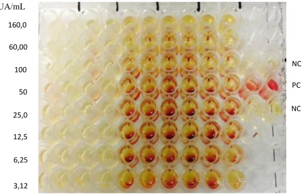
CIM and CBM
The values of CIM and CBM of C30 were 3.125 AU / mL and 160 AU / mL, respectively, which demonstrates the mode of bacteriostatic action of C30 Figure 5. As regards C50, MIC and CBM values were 1.56 AU / mL and 160 AU / mL, respectively, which also demonstrates the mode of bacteriostatic action of C50 (Figure 6).
FIG 5. Determination of MIC of C30, precipitated intracellular extract with 30% ammonium sulfate.
UA/mL
Producing strain, Bacteroides fragilis D 111.4, indicator strain, Bacteroides caccae D 79.3; NC, negative control; PC, positive control.
FIG 6. Determination of MIC of C50, precipitated intracellular extract with 50% ammonium sulfate.
UA/mL
Producing strain, Bacteroides fragilis D 111.4, indicator strain, Bacteroides ovatus D 54.1; NC, negative control; PC, positive control. NC PC NC 160,0 100 50 25,0 12,5 6,25 3,12 60,00 NC PC NC 160,0 60,0 100,0 50,0 25,0 12,5 6,25 3,12
PURIFICATION OF C30 AND C50 FRACTIONS
The application of the extract C30 in ion exchange chromatography did not generate active fractions. With regard to the C50 extract, 50 fractions were generated, among which fractions 1 to 4, referring to a single peak, were not able to bind to the column and presented antagonistic activity (Figure 7). The chromatogram obtained in this purification step is shown in Figure 8.
FIG 7. Inhibition of the indicator strain by fractions 1, 2, 3 and 4 resulting from the ion exchange chromatography of C50, precipitated intracellular extract with 50% of ammonium sulfate.
Producing strain: Bacteroides fragilis D 111.4; Indicator strain: Bacteroides ovatus D 54.1.
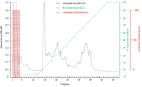
FIG 8. Ion exchange chromatographic of C50, intracellular fraction precipitated with 50% of ammonium sulfate, in Mono-Q TM column, coupled in FPLC system.
Sensitivity 2.0, reading at 280 nm. Fraction volume: 2.0 mL. Producing strain: Bacteroides fragilis D 111.4; Indicator strain: Bacteroides ovatus D 54.1.
Fractions 1 to 4, from ion exchange chromatography, constituted pool 1, which, applied in gel filtration chromatography, generated 26 fractions. Of these, fractions 2 and 3 were able to inhibit the telltale sample (Figure 9). The chromatogram resulting from the gel filtration chromatography is shown in Figure 10.
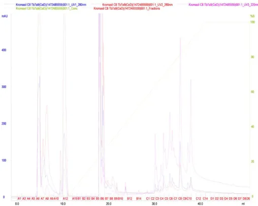
Pool 2, consisting of fractions 2 and 3 from the gel filtration chromatography, was subjected to reverse phase chromatography. Fifty fractions were generated (Figure 11), the fraction 2C being eluted with approximately 60-65% acetonitrile, active against the developer sample (Figure 12).
FIG 9. Inhibition of the producing strain by fractions 2 and 3 resulting from gel filtration chromatography.
Producing strain: Bacteroides fragilis D 111.4; Indicator strain: Bacteroides ovatus D 54.1.
FIG. 10. Gel filtration profile of pool 1 (fractions 1 to 4) from ion exchange chromatography using Superose 12 column coupled to FPLC system.
Sensitivity 2.0, reading 280 nm. Fraction volume 1.5 mL. Producing strain: Bacteroides fragilis D 111.4, indicator strain:
FIG 11. Reverse phase chromatographic profile of pool 2 (fractions 2 and 3) from gel filtration chromatography using C8 column coupled to HPLC system.
Reading at 280 nm. Producing strain: Bacteroides fragilis D 111.4; Indicator strain: Bacteroides ovatus D 54.1.Leitura a 280 nm.
FIG 12. Inhibition of the producing strain by fraction 2C resulting from reverse phase chromatography.
SDS-PAGE
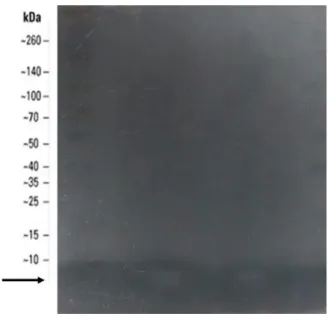
The result of the in situ development of the crude extract activity performed after separation by electrophoresis is shown in Figure 13. Inhibition of the developmental microorganism B. ovatus D 54.1 was verified by all the extracts tested, in a region corresponding to the band with molecular mass <10 kDa.
FIG 13. SDS-PAGE pure extract C50.
Pre-stained molecular mass marker 10-260 kDa (Spectra multicolor broad range protein ladder). Indicator strain:
Bacteroides ovatus D 54.1.-260 kDa (Spectra multicolor broad range protein ladder).
DETERMINATION OF THE MOLECULAR MASS OF ANTAGONIST SUBSTANCE
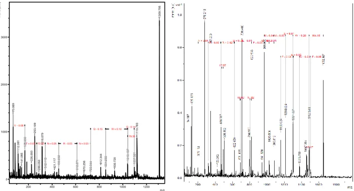
The results of the experiments performed to verify the purity and determination of the molecular mass of the active substances present in fraction 2C obtained after fractionation by reverse phase chromatography Figure 14 and after digestion with trypsin are summarized in Figure 15 and figure 16. In the fraction analyzed by chromatography Of reverse phase, the ions of approximately 1300 Da generated a more intense signal. In the fraction analyzed by trypsin digestion, the fragments were analyzed and sequenced. Trypsin digestion allowed good fragmentation of the peptide.
FIG 14. Mass spectrum from fraction 2C from reverse phase chromatography.
FIG 15 e FIG 16. Mass spectra obtained after fragmentation of strains.
Relative intensity versus mass-on-charge function (m / z). Producing strain: Bacteroides fragilis D 111.4. Left: Fraction 2C, obtained on C8 column reverse phase chromatography. Right: strain extracted from gel and digested with trypsin.
ANALYSIS OF AMINO ACID SEQUENCES
The search performed by similarity between the sequenced fragments and proteins described in the BLASTP database, from fragmentation obtained in reverse phase chromatography, showed 100% identity with a type VII secretory protein (Figure 17). The sequence was VTANRNQWG. The search performed by similarity between the sequenced fragments and proteins described in the Antimicrobial Peptide Database, from fragmentation obtained with trypsin digestion, resulted in 42% identity with a microcine of Streptomyces (Figure 18). The sequence found was GMAAFKSIFGGMSWY. 1 3 0 9 .7 9 5 7 0 .0 6 9 1 1 1 9 .7 8 3 2 5 3 .1 0 9 3 1 6 .0 7 9 1 6 9 .0 6 0 1 1 2 .0 8 1 2 2 6 .0 9 3 1 0 4 0 .4 3 7 8 1 2 .3 4 4 1 0 9 7 .3 2 1 1 4 1 .0 8 5 8 5 4 .2 5 5 4 5 6 .2 0 2 9 3 0 .7 2 0 3 4 2 .1 1 3 4 2 1 .1 1 7 6 1 2 .2 7 1 2 8 8 .0 5 0 6 7 0 .2 5 6 7 2 6 .3 4 4 G - 0.14 W + 0.10 R - 0.03 N + 0.05 72.00 T + 0.01 N + 0.03 Q - 0.15 79.35 V - 0.08 0 1000 2000 3000 In te n s. [ a .u .] 200 400 600 800 1000 1200 m/z
FIG 18. Data for a microcine synthesized by samples of Streptomyces with 42% identity to the antimicrobial peptide of Bacteroides fragilis D 111.4.
1 Antimicrobial
Peptide AP02720
APD ID: AP02720
Name/Class: Sungsanpin (a class 2 lasso peptide; class 1 microcin, bacteriocins; Gram-positive bacteria, prokaryotes; XXJ; UCSB1a)
Source: a Marine Streptomyces species
Sequence: GFGSKPIDSFGLSWL
Length: 15
Net charge: 0
Hydrophobic
residue%: 40%
Boman Index: -0.09 kcal/mol
3D Structure: Unknown
Method: NMR
SwissProt ID: Reference ID: Ref
Activity: Cancer cells
Crucial residues:
Additional info: APD analysis reveals that the sequence of THE PEPTIDE shows 73.3% similarity to Chaxapeptin.
Title: Sungsanpin, a lasso peptide from a deep-sea streptomycete
Author: Um S, Kim YJ, Kwon H, Wen H, Kim SH, Kwon HC, Park S, Shin J, Oh DC.2013
Reference: J Nat Prod. 2013 May 24;76(5):873-9. PubMed
4 DISCUSSION
The most frequently isolated anaerobic of these infectious processes is B. fragilis, involved in the development of almost one-third of the intra-abdominal abscesses that occur in the peritoneal cavity, although it accounts for only 0.5% of the human colon microbiota. While intra-abdominal infections are typically polymicrobial and are generally associated with the indigenous microbiota,
B. fragilis is one of the few bacteria known to be capable of inducing abscess formation as the sole
infecting organism in experimental animal models (Cao et al., 2014).
Species that develop in dense and diverse microbial communities must have mechanisms to establish, persist and thwart competitors (Coyne et al., 2016). To the extent that microorganisms that colonize the intestines of humans evolve competitively in environments where successful species can colonize, the bacterial composition in this ecosystem should be greatly influenced by factors that promote competitive interference such as the production of antimicrobial proteins As well as small microbial peptidic bacteria such as microcines and lantibiotics (Cotter et al., 2014) and larger molecules, such as colicins (Cascales et al., 2007) and piocines (Michel-Briand & Baysse, 2002). These molecules are structurally and functionally diverse in their ability to inhibit or kill cells by various mechanisms, including nuclease activity, pore formation, enzymatic inhibition, and destruction of the cell wall.
In this study, the expression of antagonism by Bacteroides and Parabacteroides samples isolated from patients with intra-abdominal infections was evaluated. Only Bacteroides exhibited antagonistic activity under the experimental conditions employed. In the group, 43.33% of the samples tested showed heteroantagonism only and 16.67% expressed heteroantagonism and isoantagonism, exclusively, B. fragilis. A similar result was reported by Avelar et al. (1999) found that 57% of B.
fragilis samples, including clinical specimens, were bacteriocin-producing.
In the present study, autoantigen activity was not observed. Several reports describe that samples producing antagonistic substances synthesize an immunity protein for their protection (Coyne et al., 2016; Lobo et al., 2016).
However, in a dense and diversified microbiota environment, the bacteriocin produced not only exerts its antagonistic effect, but can also trigger the production of bacteriocins by neighboring cells, "setting the competitor on the counterattack." In this study, the researchers observed that bacteriocins induce the expression of inhibitory substances by neighboring samples and that the potency of a bacteriocin and its induction ability go together. In a competition environment between antagonistic samples, the most "toxic" competitor will prevail (Majeed et al., 2013).
Concerning the incubation time, the absence of expression of antagonism by the sample grown for 24 h and the activity observed for the culture incubated for 48 h suggests that the production of
the antagonist substance occurs mainly at a later stage of the growth curve of B. fragilis D 111.4. The use of oxidative stress, acid precipitation and lysis in the French press led to the total loss of antagonistic activity. On the other hand, the use of guanidine hydrochloride initially showed promising results. However, the impossibility of a satisfactory result of the dialysis process, an essential step for subsequent purification by chromatography, represented an impediment to the use of the methodology. This methodology was successfully employed by Hayes et al. (1983), which purified a protein with antagonistic activity from the B. fragilis sample.
Protein extracts C30 and C50 were tested for characterization. These tests are important for assessing the activity of the antagonist substance in different environments, for example, when challenged by extreme pH and temperature values.
Regarding the evaluation of thermotolerance, loss of antibacterial activity was observed by exposure to temperatures above 50 ° C. Similar results were reported by Farias et al. (1994), which also characterized a bacteriocin of B. fragilis that lost its activity when the extract was treated with temperatures above 60 ° C. The C30 and C50 antagonist activity was preserved at lower temperatures, such as -20 ° C and -86 ° C, for up to 12 months, the last time the test was performed. At 37 ° C and 25 ° C, its activity was maintained for 7 days.
For the purification of the antagonist substance present in the crude protein extract, successive chromatographic steps were performed in this study. The application of the extract C30 in ion exchange chromatography did not generate active fractions. As a result of the above, it was decided to use the C50 fraction in the later stages of the investigation. Intracellular extracts have been successfully used by other authors of our group (Miranda et al., 1993; Farias et al., 1994; Ribeiro-Ribas et al., 2009).
For the purification of the antagonist substance present in the C50 intracellular protein extract, Mono-Q HR 5/5 column ion exchange chromatography having anion exchangers and -CH 2 -N + (CH 3) is charged as the charged group. Fractions 1 to 4, referring to a single peak, which were not able to bind to the column, exhibited antagonistic activity. These fractions were eluted before the start of the gradient performed with 1 M NaCl, indicating that the active substance present in the fraction was not able to interact with the charged group of the column. Subsequent to ion exchange chromatography, the active fractions were subjected to filtration chromatography. The column used was Superose 12 HR 10/30, formed by an agarose matrix, with an optimal separation range ranging from 1000 Da to 300,000 Da. The active fractions originated (2 and 3) were located at a peak at the beginning of the Chromatogram, indicating particles with larger molecular weights. In the next purification step, the reverse phase liquid chromatography on a C8 column (HPLC) was used, and 50 fractions were collected. One of them, fraction 2C, remained active against the revealing sample.
The result of the mass spectrometry experiment, from fraction 2C, presented ions of approximately 1300 Da, which generated a more intense signal. An antagonist substance with molecular mass less than 10 kDa was also detected in the in situ development experiment performed after SDS-PAGE. Differently, the test performed using Amicon tubes, showed activity in fractions with molecular mass greater than 100 kDa. Apparently, the estimate of the molecular mass of the substance was overestimated. Such a fact can be explained by the possibility that a protein extract may include substances of a non-protein nature, such as carbohydrates and lipids, which can bind to proteins. Still, some proteins may remain as aggregates, with high molecular mass, around 30 to 300 kDa. These aggregates eventually lead to incorrect determination of the molecular weight of the protein during gel filtration chromatography (Hayes et al., 1983).
The search performed by similarity between one of the sequenced fragments (VTANRNQWG) and proteins described in the BLASTP database, from fragmentation obtained on reverse phase chromatography, resulted in 100% identity with a type VII secretion protein. The search performed by similarity with proteins described in the Antimicrobial Peptide Database, from fragmentation obtained with trypsin digestion (GMAAFKSIFGGMSWY), resulted in 42% identity with a Streptomyces microcine.
Bacteroides secrete antibacterial proteins (BSAPs), which act on phylogenetically related
samples. Bacteria evolved a remarkable variety of sophisticated nanomachines to export various pathogenicity factors through the bacterial cell envelope. Because bacteria use different mechanisms to compete in microbial communities and the intestinal microbiota is an extremely dense ecosystem, cell-cell contact, which depends on SST6, presents as a very prevalent antagonistic mechanism in the gut. Only recently, SST6 has been identified in Bacteroides strains. SST6 from Bacteroides do not share similar sequences with those from Proteobacteria and therefore are not detectable by methods that depend on protein-protein comparisons, for example BLASTP, which make comparisons of known structural protein profiles. Coyne et al. (2016) identified an SST6 locus in an integrated conjugate element (ICE). In this study, the researchers showed that this locus was transferred between four different species of Bacteroides while corresponding in the intestines of humans. This was the first demonstration of an SST6 locus being transferred between members of a natural microbial community (Coyne et al., 2016).
Relatively little is known about the ecology of the human intestinal microbiota and the combination of factors regulating the composition of an intestinal microbial community. The competition for bacterial interference is beginning to be studied and appreciated as an important contribution to understanding the dynamics of intestinal bacterial populations. According to Roelofs
study, the researchers showed that intestinal samples of BSAPs that antagonize isogenic sensitive samples, suggesting that BSAPs shape the composition and formation of intestinal communities. In summary, the data demonstrate the need to continue studies on the production of antagonistic proteins by B. fragilis, as well as the analysis of extraction methods that allow the optimization of the purification protocol. Future studies are desirable to allow a better understanding of the influence of these proteins on the ecological relationships that occur in the intestinal tract of humans and their potential of application in the inhibition of samples associated with the etiopathogenesis of intra-abdominal infections.
5 CONCLUSIONS
The expression of antagonistic activity was detected only for the genus Bacteroides. Hetero-antagonistic activity was observed only against phylogenetically related samples and isoHetero-antagonistic activity was detected only for B. fragilis. No sample expressed self-starvation. The exclusion of factors that could interfere in the interpretation of phenotypic test results reinforces the hypothesis that the expressed antagonism is due to the production of proteinaceous substance (s), possibly bacteriocin (s). Intracellular extract of B. fragilis D111.4 sample with 30% (C30) and 50% (C50) saturated ammonium sulfate shows antagonistic activity against B. ovatus and B. caccae, respectively. C30 and C50 presented low stability when subjected to heat treatments, they were active against different pH values and several organic solvents and their protein nature was confirmed by inactivation by proteases. CIM and CBM values of C30 and C50 demonstrate that extracts exert a bacteriostatic action. C30 purification was not possible by chromatographic steps. Data obtained by SDS-PAGE indicates that the C50-detected antagonist substance has molecular mass less than 10 kDa and therefore is a peptide. Analysis by mass spectrometry, from fraction 2C obtained from reversed phase chromatography, revealed ions of approximately 1300 Da, which generated a more intense signal. The search performed by similarity with proteins described in the BLASTP database resulted in 100% identity of a fragment (VTANRNQWG) with a type VII secretion protein. The search performed by protein similarity described in the Antimicrobial Peptide Database resulted in 42% identity of a fragment (GMAAFKSIFGGMSWY) with a microcine of Streptomyces.
ACKNOWLEDGMENTS
This study was supported by Fundação de Amparo à Pesquisa do Estado de Minas Gerais (FAPEMIG APQ-00941-13), Conselho Nacional de Desenvolvimento Científico e Tecnológico (CNPq), Coordenação de Aperfeiçoamento de Pessoal de Nível Superior (CAPES) and Pro-reitoria de Pesquisa da Universidade Federal de Minas Gerais (PRPq/UFMG).
REFERENCES
APOLÔNIO, A. C. M.; CARVALHO, M. A. R.; BEMQUERER, M. P.; SANTORO, M. M.; PINTO, S. Q.; OLIVEIRA, J. S.; SANTOS, K. V.; FARIAS, L. M. Purification and partial characterization of a bacteriocin produced by Eikenella corrodens. Journal of Applied of Microbiology, v. 104, p. 508 - 514, 2008.
AVELAR, K. E. S.; PINTO, L. J. F.; LOBO, L. A.; BASTOS, M. C. F.; DOMINGUES, R. M. C. P.; FERREIRA, M. C. Production of bacteriocin by Bacteroides fragilis and partial characterization.
Letters in Applied Microbiology, v. 29, p. 264 - 268, 1999.
BEERENS, E. L.; BARON, G. Demonstration of bacteriocins elaborated by anaerobic gram negative bacteria belonging to the Eggerthella genus. Annales Institut Pasteur Paris, v. 108, p. 255 - 256, 1965.
BOOTH, S. J.; JOHNSON, J. L.; WILKINS, T. D. Bacteriocin production by strains of Bacteroides isolated from human feces and the role of theses strains in the bacterial ecology of the colon.
Antimicrobial Agents and Chemotherapy, v. 11, p. 718 - 724, 1977.
BRITTON, H. T. S.; ROBINSON, R. A. Journal of the Chemical Society, 1931.
CAO, Y.; ROCHA, E. R. ; JEFFREY, C. S. Efficient utilization of complex n-linked glycans is a selective advantage for Bacteroides fragilis in extraintestinal infections. Proceedings of the National
Academy of Sciences, v. 111, p. 12901 - 12906, 2014.
CASCALES, E.; BUCHANAN, S. K.; DUCHE, D.; KLEANTHOUS, C.; LLOUBES, R.; POSTLE, K.; RILEY, M.; SLATIN, S.; CAVARD, D. Colicin biology. Microbiology and Molecular Biology
Reviews, v. 71, p.158 - 229, 2007.
CLINICAL AND LABORATORY STANDARDS INSTITUTE.CLINICAL AND LABORATORY STANDARDS INSTITUTE. Methods for dilution antimicrobial susceptibility testing of
anaerobic bacteria. Approved Standard - Seventh Edition, v. 27 (2). CLSI document M11-A7
(ISBN 1-56238-626-3). CLSI, 940 West Valley Road, Suite 1400, Wayne, Pennsylvania 19087-1898, USA, 2012.
COTTER, P. D. An ‘Upp’-turn in bacteriocin receptor identification. Molecular Microbiology, v. 92, p.1159 - 1163, 2014.
COYNE, M. J.; ROELOFS, K. G.; COMSTOCK, L. E. Type VI secretion systems of human gut Bacteroidales segregate into three genetic architectures, two of which are contained on mobile genetic elements. BMC Genomics, v. 17, p. 1 - 21, 2016.
DINIZ, C. G.; CARA, D. C.; NICOLI, J. R.; FARIAS, L. M.; DE CARVALHO, M. A. R. Effect of metronidazole on the pathogenicity of resistant Bacteroides strains in gnotobiotic mice.
Antimicrobial Agents and Chemotherapy, v. 44, p. 2419 - 2423, 2000.
FARIAS, L. M.; CARVALHO, M. A. R.; DAMASCENO, C. A. V.; CISALPINO, E.O.; VIEIRA, E. C. Bacteriocin-like Activity of Bacteroides fragilis group isolated from marmosets. Research in
Microbiology, v.143, p.151 - 59, 1992.
FARIAS, L. M.; TOTOLA, A. H.; MIRANDA, C. M. S.; CARVALHO, M. A. R.; DAMASCENO, C. A. V.; TAVARES, C. A. P.; CISALPINO, E. O.; VIEIRA, E. C. Extraction, partial purification and characterization of a bacteriocin (fragilicin) produced by a strain of Bacteroides fragilis isolated from Callithrix penicillata. Research in Microbiology, v. 145, p. 9 - 16, 1994.
GREEN, A. A.; HUGES, W. Protein fractionation on the basis of solubility in aqueous solutions of salts and organic solvents. Methods in Enzymology, v.1, p. 67 - 90, 1955.
HAMADA, S.; OOSHIMA, T. Production and properties of bacteriocins (mutacins) from
Streptococcus mutans. Archives of Oral Biology, v. 20, p. 641 - 648, 1975.
HAVLIS, J. et al. Fast-response proteomics by accelerated in-gel digestion of proteins. Analytical
Chemistry., v. 75, p. 1300 - 1306, 2003.
HAYES, T. J.; CUNDY, K. R.; FERNANDES, P. B.; HOOBER, J. K. Purification and characterization of a bacteriocin from Bacteroides fragilis. Journal of Bacteriology, v. 155, p. 1171 - 1177, 1983.
ISHIKAWA, E. ; MATSUKI, T.; KUBOTA, H.; MAKINO, H.; SAKAI, T.; OISHI, K.; KUSHIRO, A.; FUJIMOTO, J.; WATANABE, K.; WATANUKI, M.; TANAKA, R. Ethnic diversity of gut microbiota: species characterization of Bacteroides fragilis group and genus Bifidobacterium in healthy Belgian adults, and comparison with data from Japanese subjects. Journal of Bioscience and
Bioengineering, v. 116, p. 265 - 270, 2013.
LEVISON, M. E.; LEVISON, J. H. Pharmacokinetics and pharmacodynamics of antibacterial agents.
LOBO, L. A.; BENJAMIM, C. F; OLIVEIRA A. C. The interplay between microbiota and inflammation: lessons from peritonitis and sepsis. Clinical & Translational Immunology, v. 5, p. 1 - 8, 2016.
MACFARLAND, L. V. Normal flora: diversity and functions. Microbial Ecology in Health and
Disease, v. 12, p.193 - 207, 2000.
MAJEED, H.; LAMPERT, A.; GHAZARYAN, L.; GILLOR, O. The Weak Shall Inherit: Bacteriocin-mediated interactions in bacterial populations. PLoS ONE, v. 8, p. 6383 - 6387, 2013.
MICHEL-BRIAND, Y.; BAYSSE, C. The pyocins of Pseudomonas aeruginosa. Biochimie, v. 84, p. 499 - 510, 2002.
MIRANDA, C. M.; FARIAS, L. M.; CARVALHO, M. A.; DAMASCENO, C. A.; TOTOLA, A. H.; TAVARES, C. A.; CISALPINO, E. O.; VIEIRA, C. E. Purification and partial characterization of a bacteriocin isolated from Bacteroides ovatus H47. Canadian Journal of Microbiology, v. 39, p. 169 - 174, 1993.
MOSSIE, K. G.; JONES, D. T.; ROBB, F. T.; WOODS, D. R. Characterization and mode of action of a bacteriocin produced by a Bacteroides fragilis strain. Antimicrobial Agents and
Chemotherapy, v. 16, p. 724 - 730, 1979.
MOSSIE, K. G.; JONES, D. T.; ROBB, F. T.; WOODS, D. R. Inibition of ribonucleic acid polymerase by a bacteriocin from Bacteroides fragilis. Antimicrobial Agents and Chemotherapy, v. 20, p. 437 - 442, 1981.
NAKANO, M. M.; MARAHIEL; M. A.; ZUBER, P. Identification of a genetic locus required for biosynthesis of the lipopeptide antibiotic surfactin in Bacillus subtilis. Journal Bacteriology, v. 170, p. 5662 - 5668, 1988.
NICOLETTI, G.; NICOLOSI, D.; ROSSOLINI, G. M. Eziologia, epidemiologia e diagnostic microbiologica delle infezion iintraddominali. Intra-abdominal infections: etiology, epidemiology, microbiological diagnosis and antibiotic resistance issues. Journal Le Infezioni in Medicina, v.1, 2008.
PAPAPARASKEVAS, J.; KATSANDRI, A.; PANTAZATOU, A.; STEFANOU, I.; AVLAMIS, A.; LEGASKIS, N. J.; TSAKRIS, A. Epidemiological characteristics of infections caused by
Bacteroides, Prevotella and Fusobacterium species: a prospective observacional study. Anaerobe,
v. 3; p. 113 - 117, 2011.
RIBEIRO-RIBAS, R. N.; VIEIRA, M. A. R.; APOLONIO, C. A.; MAGALHAES, P. P.; OLIVEIRA, J. S.; FARIAS, L. M. Purification and partial characterization of a bacteriocin produced by an oral
Fusobacterium nucleatum isolated. Journal of Applied Microbiology, v. 7, p. 145 - 152, 2009.
ROELOFS, K. G.; COYNE, M. J.; GENTYALA, R. R.; CHATZIDAKI-LIVANIS, M.; COMSTOCK, L. E. Bacteroidales secreted antimicrobial proteins target surface molecules necessary for gut colonization and mediate competition in vivo. MBio, v. 7, p. 1055 - 1066, 2016.
SHEVCHENKO, A. et al. Mass spectrometric sequencing of proteins from silver-stained polyacrylamide gels. Analytical Chemistry., v. 68, p. 850 - 858, 1996.
STEERS, E.; FOLTZ, E.; GRAVES, B. An inocula replicating apparatus for routine testing of bacterial susceptibility to antibiotics. Antimicrobial Agents and Chemotherapy, v. 9, p. 307 - 311, 1959.
VALEFF, Carolina Nicolai. Atividade antagonista de extrato intracelular obtido de
Fusobacterium necrophorum: modo de ação e caracterização bioquímica. 2011. 144f. Dissertação
(Mestrado em Ciências Biológicas – Microbiologia) – Instituto de Ciências Biológicas, Universidade Federal de Minas Gerais, Belo Horizonte, 2011.
WEXLER, A. G.; BAO, Y.; WHITNEY, J. C.; BOBAY, L. M.; XAVIER, J. B.; SCHOFIELD, W. B. Human symbionts inject and neutralize antibacterial toxins to persist in the gut. Proceedings of
the National Academy of Sciences of the United States of America, 2016.
WILSON, M. M.; ANDERSON, D. E.; BERNSTEIN, H. D. Analysis of the Outer Membrane Proteome and Secretome of Bacteroides fragilis Reveals a Multiplicity of Secretion Mechanisms.
PLoS ONE, v. 10, 2015.
Relative intensity versus mass-on-charge function (m / z). Producing strain: Bacteroides fragilis D 111.4. Left: Fraction 2C, obtained on C8 column reverse phase chromatography. Right: strain extracted from gel and digested with trypsin.