online | memorias.ioc.fiocruz.br
The redox potential interferes with the expression of laminin binding
molecules in
Bacteroides fragilis
Eliane de Oliveira Ferreira/+, Edwin Alexander Yates1, Morris Goldner2, Rossiane Cláudia Vommaro3,
Fernando Costa e Silva Filho3, Débora Barreiros Petrópolis3, Regina MC Pilotto Domingues
Laboratório de Biologia de Anaeróbios, Departamento de Microbiologia Médica, IMPPG, 3Instituto de Biofísica Carlos Chagas Filho, Centro de Ciências da Saúde, Universidade Federal do Rio de Janeiro, Avenida Chagas Filho 373 2o andar, 21941-902 Rio de Janeiro, RJ, Brasil
1School of Biological Sciences, University of Liverpool, Liverpool, UK 2Departament de Biologie Medicale, Brock University, Ontario, Canada
The Bacteroides fragilis ATCC strain was grown in a synthetic media with contrasting redox potential (Eh) lev-els [reduced (-60 mV) or oxidised (+100mV)] and their adhesion capacity to extracellular matrix components was evaluated. The strain was capable of adhering to laminin, fibronectin, fibronectin + heparan sulphate and heparan sulphate. A stronger adherence to laminin after growing the strain under oxidising conditions was verified. Electron microscopy using ruthenium red showed a heterogeneous population under this condition. Dot-blotting analyses confirmed stronger laminin recognition by outer membrane proteins of cells cultured at a higher Eh. Using a laminin affinity column, several putative laminin binding proteins obtained from the cultures kept under oxidising (60 kDa, 36 kDa, 25 kDa and 15 kDa) and reducing (60 kDa) conditions could be detected. Our results show that the expres-sion of B. fragilis surface components that recognise laminin are influenced by Eh variations.
Key words: Bacteroides fragilis - adhesion - redox potential - laminin binding proteins - outer membrane proteins
Bacteroides fragilis is a Gram-negative obligate an-aerobic bacterium and a common member of the human microbiota (Jousiemies-Somier 2002), but it is also an important opportunistic pathogen. This bacterium is the most frequently isolated anaerobic species from human intra-abdominal infections, and this represents a serious clinical problem, especially when abscesses are formed (Finegold 1995). Although several factors have been de-scribed, the pathogenicity of this microorganism is still not completely understood. The capsular polysaccha-ride complex (CPC) is a crucial factor in the virulence potential of the species (Comstock et al. 1999, Coyne et al. 2000, 2001). B. fragilis modulates its surface antige-nicity, generating distinct CPC combinations (Krinos et al. 2001). High variation in the polysaccharide expres-sion of B. fragilis has already been demonstrated us-ing monoclonal antibodies (Patrick et al. 1999). Recent analyses of the genome sequences revealed DNA inver-sion regions. These results suggested that the ability of
B. fragilis to colonise several host environments, behav-ing either as a member of the microbiota or a pathogen, might be linked to switching of polysaccharide biosyn-thesis (Krinos et al. 2001).
The extracellular matrix (ECM) is a stable macro-molecular structure underlying epithelial and endothe-lial cells and surrounding connective tissue cells. In
Financial support: CAPES, FAPERJ, CNPq, PRONEX + Corresponding author: [email protected] Received 1 June 2008
Accepted 30 September 2008
the ECM, collagens, proteoglycans and structural gly-coproteins, such as fibronectin and laminin, are found and these molecules are usually exploited for colonisa-tion by microbes (Ljungh et al. 1996). The cell surface adhesins that mediate such recognition are termed “mi-crobial surface components recognising adhesive matrix molecules” (MSCRAMMS). In the past, several micro-organisms have been shown to express these molecules (Ljungh at al. 1996, Lähtennmaki et al. 1998, Crago & Koronakis 1999).
Many bacterial virulence factors are well regulated with their expression linked to several environmental signals, such as temperature, pH, osmolarity, carbon source and iron levels (Sijbrandi et al. 2005). To the ob-ligate anaerobic bacteria, the redox potential (Eh) rep-resents an important environmental parameter to their survival. It has been shown that, when grown at differ-ent Eh levels, B. fragilis can change its state from com-mensal to pathogenic, becoming invasive to HeLa cells (Goldner et al. 1993).
In the present investigation, we searched for B. fragi-lis properties regulated by Eh. We investigated interfer-ence with adhesion to ECM components and with the expression of proteins and surface polysaccharides. Our results showed that the adhesion to laminin is influenced by Eh, and we report that several putative binding pro-teins were more highly expressed under oxidising condi-tions by ATCC 43859 strain.
MATERIAL AND METHODS
Culture conditions - The strain was first grown in brain heart infusion (BHI, Sigma Co), consisting of pre-viously reduced and anaerobically sterilised (PRAS) medium (37oC, 18 h) and blood agar supplemented with hemin (5 mg/mL; Sigma Co) and vitamin K (0.1 mg/mL; Sigma Co), under anaerobic conditions (80% N2; 10% CO2; 10% H2) at 37oC (Gumbiner 1996). After 48 h of incubation, colonies were selected and grown in BHI-PRAS at 37oC for 18 h.
After growing in BHI-PRAS medium (37oC, 18 h), 300 µL of the culture (109 CFU/mL) was transferred to the oxidising (Eh7 + 100 mV) and reducing (Eh7 - 60 mV) medium described by Goldner et al. (1993). The tubes were incubated for 18 h at 37oC. The bacterial suspension (109 CFU/mL) was prepared after centrifugation (3000 g) and washed twice with 0.1 M PBS (pH 7.2).
Bacterial growth - A growth curve was constructed under both conditions. One milliliter of the bacterial culture was taken at 2, 4, 6, 8, 18, 24 and 48 h, and the absorbance at 660 nm (Pharmacia Biothec, Ultrospec 2000) was measured. All experiments were performed in triplicate. For statistical analysis, the ANOVA and the Student’s t paired tests, both from SPSS, Version 1.3 (p < 0.05), were used.
Latex beads - Latex beads (Sigma, diameter: 0.82 µm) were prepared according to Naidu et al. (1988). Briefly, a latex suspension was mixed with 3 mL of a 0.17 M gly-cine-NaOH buffer (pH 8.2), centrifuged at 3000 g and washed twice with the same buffer. The pellet was mixed with 3 mL of the glycine buffer and ECM components: laminin [17 µg/mL, Sigma Co, Tumor Engelbreth-Holm-Swarm (EHS)], fibronectin (33 µg/mL, Sigma Co), fi-bronectin (33 µg/mL) + heparan sulphate (33 µg/mL, Cel-sus Laboratories, Inc) and heparan sulphate (33 µg/mL). The suspension was shaken horizontally (80 rpm, 30oC, 14 h), centrifuged, and washed with 2 mL of glycine buf-fer (0.01% BSA). The latex beads were kept at 4oC.
Agglutination assays - The assays were performed according to Nagy et al. (1994). For the test, 10 µL of the bacterial suspension, started from 109 CFU/mL and fol-lowing a 10-4 fold dilution series, and 10 µL of the cor-responding coated latex suspension were dropped onto a glass slide and mixed gently. The reaction was read after 2 min. As a negative control, the bacteria were mixed with the glycine-NaOH buffer (pH 8.2) or with BSA (2 mg/mL in the same buffer) latex beads. The titer was defined as the last dilution to cause agglutination. The strain was tested in triplicate.
Surface polysaccharide expression - Transmission Electron Microscopy (TEM) - For detecting acidic sur-face polysaccharide expression, ruthenium red staining was used. The cells were grown as described before, washed with PBS (0.1 M, pH 7.2) and the pellet resus-pended in equal volumes of 2.5% (v/v) glutaraldehyde, RR solution (10 mg/mL) and 0.1 M cacodylate buffer (pH 7.2) for 1 h at rt. Cells were fixed with 4% osmium tetroxide (OsO4), RR solution (1%) and 0.1 M cacody-late buffer for 1 h at rt. The suspensions were washed (3 x), dehydrated in acetone (30-100%) and then
embed-ded in Epon/acetone (1:1) for 24 h at rt and in Epon (2 days/60oC). Ultrathin sections were made using an ultra-microtome (Reichert Ultracuts-Leica), and micrographs were taken on a Ziess 900 electron microscope.
Total polysaccharide dosage - After growing the strain under the previously described culture conditions, 5 mL was centrifuged (4000 x g) and washed (6 x) to remove all sugar derived from the medium; the bacteria were sonicated (2 min, 4oC) and then washed (4000 g; 2 x) to remove any sugar present in the cytosol. The samples were freeze-dried, then hydrolysed (2 M HCl, 100oC, 3 h) and freeze-dried again. A sugar assay, using neocuproine, was performed according to the method de-scribed by Chaplin and Kennedy (1994). The Student’s t
test was used for comparing results from oxidising and reducing conditions.
Detection of surface antigens by monoclonal anti-bodies -To evaluate the polysaccharide antigens exposed on the bacterial surface, ATCC strain 43859 and ATCC strain 25285 were used. The crude total polysaccharide extract obtained was hydrolysed in a 50% acetic acid so-lution at 100oC for 30 min and freeze-dried prior to prob-ing usprob-ing an ELISA assay. For the assay, described by Patrick et al. (1999), the freeze-dried hydrolysed poly-saccharide extract was dissolved and 100 µL was applied to each of 96 wells and, after 24 h at 4oC, each well was blocked with 2% BSA (Sigma Co) in 0.1 M PBS pH 7.2 for 1 h at 4oC. Fifty microlitres of each monoclonal anti-body was added and the plate was incubated for another hour at 4oC. After washing, the peroxidase-conjugated secondary antibody (1:1000 in 0.1 M PBS pH 7.2 with 2% BSA) was added and incubated for 1 h at 4oC. To develop the assay, ortho-phenyl-diamine (Sigma Co) in 5 mL 0.1 M PBS with 10 µL of sodium peroxide was used. Assay development was stopped with 0.05 M sul-phuric acid in 0.5 M PBS. The plate was read in a spec-trophotometer (BIORAD model 3550) at 450 nm.
Whole proteins (WP) -After cultivation of the strain, 1 mL of the culture was centrifuged (4000 g), washed twice with PBS and the pellet was mixed with 100 µL of 0.25 M Tris-HCl, 0.192 M glycine, 0.1% SDS, pH 8.5, and stored at -20oC (Taylor et al. 1986). SDS-PAGE anal-ysis was carried out in a Bis-tris Gel (4-12% Nu PageTM, 1 mm x 12 wells) in a vertical slab gel apparatus (20 mA, 100V) (Laemmili 1970). A molecular weight standard of proteins (Invitrogen) was used. All gels were silver stained. Densitometry analysis (Molecular Analyst ver-sion 1.6, Bio-Rad) was performed.
Outer membrane proteins (OMP) - After cultiva-tion of the strain, 500 mL of the culture was centrifuged (4000 g), washed (0.1 M PBS; 2 x) and resuspended in 10 mM Tris-HCl, pH 8.0, 1 mM EDTA and 1mM
β-Mercaptoethanol (Sigma Chem. Co). The cells were
SDS-PAGE and the densitometry analysis were done as described for WP.
Immunoblotting assays -To determine if the OMP were involved in laminin adhesion, immunoblotting with OMP extracts was done (Doyle 2005). The OMP extracts (3 µL) were dropped onto a nitrocellulose mem-brane and allowed to dry. The memmem-brane was washed with TBST buffer (10 mM Tris, 150 mM NaCl, 0.1% Tween 20, pH 8.0) and incubated with laminin (20 µg/ mL) in blocking buffer (TBST, 5% skim milk, pH 7.4) for 2 h at rt. The membrane was washed (2 x) with TBST buffer for 15 min and incubated with a primary mouse IgG anti-laminin antibody (Santa Cruz; 1:1000) for 1 h at rt. The membrane was washed (2 x) and incubated with a secondary antibody (rabbit anti-mouse conjugated to al-kaline phosphatase; Invitrogen; 1:800). Finally, blots were washed (3 x) in TBST and developed. Laminin was used as a positive control. Tests were performed in duplicate.
Affinity column - Five hundred microlitres of Affi-Gel® 10 (Bio Rad) was washed 2 x with 0.1 M PBS and mixed immediately with a solution of laminin (1 mg/mL in 0.1 M PBS). The column was blocked with 50 mM ethanolamide pH 7.8 and washed (3 x) with 0.1 M PBS, followed by a wash with 2 M NaCl pH 7.0. OMP extract (1-5 mg/mL) from ATCC strain 43859 was passed sev-eral times through the column. After washing the col-umn (3 x) with PBS, a solution of 2 M NaCl pH 7.0 was passed to elute the proteins (Kern & Schotz 1987). After desalting, the samples were subjected to SDS-PAGE as described previously.
RESULTS
ATCC strain 43859, in both conditions, grew at a comparable rate for 6 h, as measured by optical densi-ty. With 8 h and 18 h of incubation, the strain cultured under oxidising condition showed a significant delay in
its growth when compared to the strain cultured under reducing condition (p < 0.05). At 24 h, similar behav-iour was verified (Fig. 1). It was observed that the strain analysed was capable of adhering to the different ECM components tested (Fig. 2) when the bacterial cells were obtained from cultures kept under either oxidising or re-ducing conditions after 18 h of growth, with an aggluti-nation titer of at least 10. A distinct capacity to adhere to one of the components tested, laminin, could be detected when the oxidising and reducing conditions were com-pared. The RR staining demonstrated that in the same population of ATCC strain 43859, grown both under oxi-dising or reducing conditions, cells were observed with a thick electrondense layer as compared to other cells that were not stained (Fig. 3). The total quantity of
polysac-Fig. 1: growth curve of the Bacteroides fragilis strain ATCC 43859.
The strain was grown under oxidizing (■) and reducing (▲) condi -tions. All tests were performed in triplicate and the ANOVA and Stu-dent’s t paired tests were used. Results with p < 0.05 were considered significant.
Fig. 2: agglutination titer of the Bacteroides fragilis strain ATCC
43859 under oxidizing (■) and reducing (■) conditions. All tests were performed in triplicate. The strain was tested with the extracellular matrix components. FIB: fibronectin; FIB+HS: fibronectin with hep-aran sulfate; HS: hephep-aran sulfate; LAM: laminin.
charide, in the sugar assay, expressed under different Eh conditions, confirmed this behaviour. There was not a significant difference (p > 0.05) in the quantity of poly-saccharide produced by this strain when both conditions were compared (Fig. 4).
A partial hydrolysis of the polysaccharides demon-strated that all antibodies tested were capable of recog-nising the bacterial extracts, but there was no difference observed between oxidising and reducing conditions. On the other hand, in relation to the crude extract, none of the antibodies except the Bf12 were capable of react-ing, and again no difference was observed between the two conditions (Table).
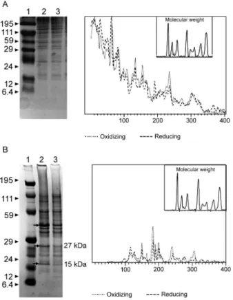
The electrophoresis profiles of the WP (Fig. 5A) showed relatively similar patterns when the conditions
were compared; however, the differences between the two conditions may be more clearly observed when examin-ing the electrophoresis profiles of the OMPs (Fig. 5B). When comparing the OMP extracts, some variability in the protein expression could reliably be detected. A pro-tein around 49 kDa was observed only under oxidising condition. Five proteins, near 60 kDa, 49 kDa, 39 kDa,
Fig. 4: total reducing sugar assay for the polysaccharide extracted from the Bacteroides fragilis ATCC 43859 strain grown under oxidizing and reducing conditions. The Student’s t test was used. The difference be-tween oxidizing and reducing was not significant (p > 0.05). The assay was performed in triplicate.
TABLE
Qualitative analysis of the surface antigens using monoclonal antibodies specific for the CPC of Bacteroides fragilis
Stranisb
ATCC 43859 ATCC 25285
Antibodiesa PH CE PH CE
Bf 5 + - +
-Bf 6 + - +
-Bf 7 + - +
-Bf 8 + - + -
Bf 12 + - + +
a: monoclonal antibodies specific for the CPC of B. fragilis were kindly provided by Dr Sheila Patrick and Dr. McCoy of the Queen’s University in Belfast, Ireland; b: no differences were found between the oxidizing and reducing conditions; CE: crude extract of the bacterial strains; PH: partial hydro-lysis of the polysaccharides; (+): recognition by the antibody; (-): no recognition by the antibody.
Fig. 5: evaluation of the interference of the redox potential in the ex-pression of whole proteins (WP) and outer membrane proteins (OMP) in Bacteroides fragilis ATCC 43859 strain. A: SDS-PAGE of the WP extracts; B: SDS-PAGE of the OMP extracts; A and B: Lane 1: mo-lecular weight standard (kDa); Lanes 2, 3: WP and OMP extracts un-der oxidizing and reducing conditions, respectively. Proteins that ap-peared or were more expressed under oxidizing conditions (arrows).
27 kDa and 15 kDa, were expressed more under oxidising than reducing conditions (Fig. 5B). The densitometry of the gel reflected these differences.
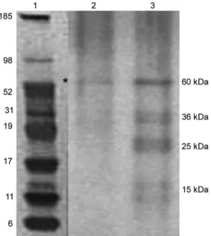
The immunoblotting analyses confirmed the involve-ment of the OMP in the adhesion to laminin (Fig. 6). We observed that, when cultured under high (oxidis-ing) Eh, some proteins capable of adhering to laminin were expressed more than when the bacterial cells were cultured under low (reducing) Eh. This result was con-firmed when the OMP extracts obtained from cultures kept under oxidising conditions were passed through an
affinity column and revealed bands of approximately 60 kDa, 36 kDa, 25 kDa and 15 kDa. For the reducing conditions, only a band of approximately 60 kDa was detected (Fig. 7).
DISCUSSION
Many events are involved in bacterial infections, and those events can compromise the functionality of a tis-sue, or result in its destruction. The adherence of micro-organisms to the host tissue is the first event and a crucial moment for colonisation. The whole adherence process involves many surface bacterial components, namely, adhesins that can recognise molecules in the host tissue or even components of the ECM, to colonise, invade and spread in the host (Patti et al. 1994). MSCRAMMS have been widely studied and represent an essential factor in the pathogenicity of some microorganisms. In patho-gens such as Pseudomonas aeruginosa (Plotkowski et al. 1996), Staphylococcus aureus (Peacock et al. 1999),
Fig. 7: SDS-PAGE of the outer membrane proteins (OMPs) of Bacte-roides fragilis ATCC 43859 strain passed through the affinity column with laminin. Lane 1: molecular weight standard; Lanes 2, 3: OMPs extracts of the ATCC 43859 strain under reducing and oxidizing condi-tions, respectively. Common band under both conditions (asterisk).
Yersinia pestis (Lähtennmaki et al. 1998) and Mycobac-terium leprae (Marques et al. 2001), MSCRAMMS that bind to laminin have already been identified. It was also reported that B. fragilis may recognise laminin, fibronec-tin, vitronectin and collagens (Nagy et al. 1994, Eiring et al. 1995). However, no data are available about the role played by such molecular recognition and binding in the infections caused by the species. Recently, Ferreira et al. (2006) have reported the ability of B. fragilis strains to adhere to laminin-1 and concluded that the molecules responsible for the recognition and binding to laminin were in the OMP extracts.
In the present study, to evaluate the interference of Eh in B. fragilis adherence to ECM components, we used a simple and rapid method, the latex agglutination as-say. The strain used in our study, ATCC 43859, revealed extensive adhesion to laminin when the bacterial cells tested were obtained from cultures kept under oxidising conditions. This strain was also capable of adhering to the other components tested. Some studies have demon-strated that pathogenic bacteria can adhere to molecules such as fibronectin and heparin sulfate to cause systemic infections (Henry-Stanley et al. 2005) and certainly these data related to B. fragilis should be better explored in further investigations.
Even though other authors have demonstrated the capacity of B. fragilis to adhere to laminin, this report shows the difference in adherence to this molecule when the strain is grown under oxidising and reducing con-ditions. The basal membrane, which is rich in laminin, is thin and has an intimate contact with the cytoplas-mic membrane of epithelial and endothelial cells (Inou 1989). Although there are at least 11 different isoforms of the laminin molecule, the best studied is the laminin type 1, isolated from EHS, used in our study. The basal membrane represents one of the barriers to B. fragilis
establishment and infection, following invasion of the peritoneal cavity and dissemination to other non-intes-tinal sites. Under normal circumstances, B. fragilis stays in harmony with the host and its growth is controlled. When lesions are formed, its microenvironment can be-come oxidising and B. fragilis turns into a pathogenic bacterium. Some studies have demonstrated that, when the Eh is altered, it can influence the behaviour of B. fragilis, which becomes more aggressive and invasive in HeLa cells (Goldner et al. 1993). ATCC strain 43859 has a peculiar characteristic; it was isolated from diarrhea and produces the B. fragilis toxin (BFT). This toxin can cause morphological and histological alterations, especially in polarised cells (Sears 2006). Cell polarisation derives from the signals that come from surface, cell-to-cell and cell-ECM activity, forming specialised membrane do-mains. The adhesion of proteins to the basal membrane and of integrins to the ECM, is essential to cell polarisa-tion (Gumbiner 1996). A subsequent role of BFT and an adhesion to laminin can be speculated and might emerge as an interesting field for further investigations.
conditions, but both conditions permitted the bacteria to reach the beginning of the log phase at the same time (af-ter about 6 h). Based on this fact, we decided to compare the behaviour of this strain at a time after 18 h, when the growth curves showed the greatest difference.
In fact, the most studied B. fragilis virulence factor is the CPC. This complex has a zwitterion nature (Stingele et al. 2004) and a critical involvement in abscess for-mation (Coyne et al. 2000). B. fragilis can modulate its surface antigenicity (phase dependent) and it has at least eight distinct polysaccharides (PSA to PSH) (Krinos et al. 2001). It is well documented that the CPC is involved in abscess formation (Stingele et al. 2004). This capsule modulation (Krinus et al. 2001) may be used as a strat-egy to help the bacteria evade the host immune system and at the same time to colonise the host. The TEM of ATCC strain 43859, using ruthenium red, revealed a very diverse population of cells, some with a thicker polysac-charide layer and others with none. This heterogeneity was verified in cultures kept in oxidising and reducing conditions. Patrick et al. (1999) have described that, in the same culture of B. fragilis, it is possible to find cells expressing at least three different kinds of capsule that could be visualised under TEM: thick, thin and an elec-tron dense layer (Patrick 1993). The qualitative analysis of the polysaccharides confirmed this result. The two strains tested, ATCC 43859 and ATCC 25285, were rec-ognised by antibodies after partial hydrolysis, and there was no difference between one condition and another. When the crude extract of ATCC strain 25285 was used, the Bf12 antibody recognised it, but still with no differ-ence between the two Eh conditions. Our results suggest that polysaccharide expression is neither influenced by the Eh nor involved in the recognition of the laminin.
SDS-PAGE of the WP of the ATCC strain 43859 did not show obvious differences between oxidising and re-ducing conditions. In contrast, electrophoretic profiles of the OMP showed differences between the extracts ob-tained from cells cultured in the two Eh conditions. Im-munoblotting with OMP extracts revealed a difference in the recognition of laminin when the proteins were ex-tracted from different culture conditions, oxidising and reducing. The laminin affinity column detected proteins of approximately 60 kDa, 36 kDa, 25 kDa and 15 kDa extracted from oxidised conditions. For the reduced con-ditions, only one band, at 60 kDa, was observed. Ferreira et al. (2006) have already demonstrated the involvement of the OMPs of B. fragilis in the adherence of laminin, and resident amino acid sequences inhibited this recog-nition. Our results suggest that structures present on the
B. fragilis surface can be influenced by Eh; moreover, the adhesins responsible for laminin recognition are manifested in the OMP extracts, and further investiga-tions are being done to confirm this.
ACKNOWLEDGEMENTS
To Prof. J. Smalley and Mr. A. Birss (University of Liv-erpool), for the use of an anaerobic chamber, to Dr. Sheila Patrick and L. McCoy (Queen’s University), for monoclonal antibodies, and Joaquim dos Santos, for technical support.
REFERENCES
Bölin I, Norlander L, Wolf-Watz H 1982. Temperature-inducible outer membrane protein of Yersinia pseudotuberculosis and Yersinia enterocolitica is associated with virulence plasmid. Infect Immun 17: 506-512.
Chaplin MF, Kennedy JK 1994. Carbohydrate analysis, a practical ap-proach, 2nd ed., Oxford University Press,United Kingdon, 350 pp.
Comstock LE, Coyne MJ, Tzianabos AO, Pantosti A, Onderdonk AB, Kasper DL 1999. Analysis of a capsular polysaccharide biosyn-thesis locus of Bacteroides fragilis. Infect Immun 67: 3525-3532.
Coyne MJ, Kalka-Mole W, Tzianabos AO, Kasper DL, Comstock LE 2000. Bacteroides fragilis NCTC 9343 produce at least three distinct capsular polysaccharides: cloning, characterization and reassignment of polysaccharide B and C biosynthesis loci. Infect Immun 69: 6176-6181.
Coyne MJ, Tzianabos AO, Mallory BC, Carey VJ, Kasper DL, Com-stock LE 2001. Polysaccharide biosynthesis locus required for virulence of Bacteroides fragilis. Infect Immun 69: 4342-4350.
Crago AM, Koronakis V 1999. Binding of extracellular matrix lamin-in to Escherichia coli expressing the Salmonella outer membrane proteins Rck and PagC. FEMS Microbiol Lett 176: 495-501.
Doyle SA 2005. Screening for the expression of soluble recombinant proteins in Escherichia coli. Methods Mol Biol 310: 115-121.
Eiring P, Manncke B, Gebracht K, Werner H 1995. Bacteroides fragilis adheres to laminin significantly stronger than Bacteroides thetaiotaomicron and other species of the genus. J Med Microbiol Virol Parasitol Infect Dis282: 279-286.
Ferreira EO, Lobo LA, Petrópolis DB, Avelar KES, Ferreira MC, Sil-va Filho FC, Domingues RMCP 2006. A Bacteroides fragilis sur-face glycoprotein mediates the interaction between the bacterium and the extracellular matrix component laminin-1. Res Microbiol 157: 960-966.
Finegold SM 1995. Overview of clinically important anaerobes. Clin Infect Dis 20: 205-207.
Goldner M, Coquis-Rondon M, Carlier JP 1993. Effect of growth of
Bacteroides fragilis at different redox levels on potential patho-genicity in a HeLa cells system: demonstration by confocal laser scanning microscopy. Zentralbl Bakteriol 278: 529-540.
Gumbiner BM 1996. Cell adhesion: the molecular basis of tissue ar-chitecture and morphogenesis. Cell 84: 345-357.
Henry-Stanley MJ, Hess DJ, Erlandsen SL, Wells CL 2005. Ability of the heparan sulfate proteoglycan syndecan-1 to participate in bac-terial translocation across the intestinal barrier. Shock 24: 571-576.
Inou ES 1989. Ultrastructure of basement membranes. Int Rev Citol 117: 57-98.
Jousiemies-Somier HR, Summanen P, Citron DM, Baron EJ, Wex-ler HM, Finegold FM 2002. Wadsworth anaerobes bacteriology manual, 6th ed., Star Publishing Company, Belmont, 152 pp.
Kern PA, Schotz MC 1987. An enzyme linked immunoassay for lipo-protein lipase. Anal Biochem 166: 27-35.
Krinos CM, Coyne MJ, Weinacht XG, Tzianabos AO, Kasper DL, Comstock LE 2001. Extensive surface diversity of a commensal microorganism by multiple DNA inversions. Nature 414: 555-558.
Laemmili UK 1970. Cleavage of structural proteins during the assem-bly of the head of bacteriophage T4. Nature 227: 680-685.
Ex-pression of plasminogen activator Pla of Yersinia pestis enhances bacterial attachment to the mammalian extracellular matrix. In-fect Immun 66: 5755-5762.
Ljungh A, Moran AP, Wadström T 1996. Interactions of bacterial ad-hesins with extracellular matrix and plasma proteins: pathogenic implications and therapeutic possibilities. FEMS Immunol Med Microbiol 16: 117-126.
Marques MAM, Mahapatra S, Sarno EN, Santos S, Spencer JS, Bren-nan PJ, Pessolani MCV 2001. Further biochemical characteriza-tion of Mycobacterium leprae laminin-binding proteins. Braz J Med Biol Res 34: 463-470.
Nagy E, Manncke B, Werner H 1994. Fibronectin and vitronectin binding of Bacteroides fragilis and eight other species of the ge-nus. Zentralbl Bakteriol 281: 235-239.
Naidu AS, Paulsson M, Wadström T 1988. Particle agglutination assays for rapid detection of fibronectin, fibrinogen and collagen recep-tors on Staphylococcus aureus. J Clin Microbiol 26: 1549-1554.
Patrick S 1993. The virulence of Bacteroides fragilis. Rev Med Mi-crobiol 4: 40-49.
Patrick S, Gilpin D, Stevenson L 1999. Detection of intra-strain an-tigenic variation of Bacteroides fragilis surface polysaccharides by monoclonal antibody labeling. Infect Immun 67: 4346-4351.
Patti JM, Allen BL, McGavin MJ, Höök M 1994. MSCRAMM - Me-diated adherence of microorganisms to host tissues. Ann Rev Mi-crobiol 48: 585-617.
Peacock SS, Foster TJ, Cameron BJ, Berendt AR 1999. Bacterial fi-bronectin binding proteins and endothelial cell surface fibronec-tin mediate adherence of Staphylococcus aureus to resting hu-man endothelial cells. Microbiology 145: 3477-3488.
Plotkowski MC, Tournier TM, Puchelle E 1996. Pseudomonas aeruginosa strains possess adhesins for laminin. Infect Immun 64: 600-605.
Sears CL 2006. The toxins of Bacteroides fragilis. Toxicon 39: 1737-1746.
Sijbrandi R, Den Blaauwen T, Tame JR, Oudega B, Luirink J, Otto BR 2005. Characterization of an iron-regulated alpha-enolase of
Bacteroides fragilis. Microbes Infect 7: 9-18.
Stingele F, Corthesy B, Kusy N, Porcelli SA, Kasper DL, Tzianabos AO 2004. Zwitterionic polysaccharides stimulate T cells with no
prefer-ential Vβ usage and promote anergy, resulting in protection against
experimental abscess formation. J Immunol 172: 1483-1490.