Mattia Esteves da Silva
Licenciado em Engenharia do Ambiente
Addressing the effects of short-term exposure to TiO2
nanoparticles in fish gills: An
ex-vivo
approach
Dissertação para obtenção do Grau de Mestre em
Engenharia do Ambiente
Orientador: Professora Doutora Marta Susana Silvestre
Gouveia Martins, Professora Auxiliar Convidada
do
DCEA,
Faculdade
de
Ciências
e
Tecnologias,
Universidade Nova de Lisboa.
Co-orientador: Doutor Pedro Manuel Broa Costa,
Investigador Auxiliar do DCEA, Faculdade de Ciências e
Tecnologias, Universidade Nova de Lisboa.
Juri:
Presidente: Professora Doutora Maria Helena Costa
Arguente: Doutora Cláudia Mieiro
Vogal: Professora Doutora Marta Martins
Addressing the effects of short-term exposure to TiO2 nanoparticles in fish gills: An ex-vivo
approach.
Mattia Esteves da Silva
Statement of copyright
A Faculdade de Ciências e Tecnologia e a Universidade Nova de Lisboa têm o direito, perpétuo e sem limites geográficos, de arquivar e publicar esta dissertação através de exemplares impressos reproduzidos em papel ou de forma digital, ou por qualquer outro meio conhecido, e de a divulgar através de repositórios científicos e de admitir a sua cópia e distribuição com objectivos educacionais ou de investigação, não comerciais, desde que seja dado crédito ao autor e editor.
Agradecimentos
Os meus primeiros agradecimentos são dirigidos à Professora Doutora Marta Martins pela possibilidade de realizar esta tese de mestrado sob a sua orientação. O rigor e qualidade, constante ao longo destes meses, a que fui submetido revelaram-se ferramentas fundamentais para a realização do melhor trabalho possível, que não seria possível sem a tua ajuda incansável. Agradeço o tempo despendido em mim e no meu trabalho e pela orientação, que muito contribuiu para a minha formação e enriquecimento profissional e que será importante no meu futuro e pela simpatia com que fui tratado.
Ao Doutor Pedro Costa, co-orientador deste trabalho, agradeço toda a disponibilidade e apoio demonstrados durante o desenvolvimento da tese. Tenho que agradecer a preocupação e atenção disponibilizada para o ensinamento de técnicas novas e pela partilha de novo conhecimento.
Ao Centro de Ciências do Mar e do Ambiente (MARE), Faculdade de Ciências e Tecnologia da Universidade NOVA de Lisboa, por ter aceite e oferecido os seus laboratórios e equipamento que possibilitaram a execução deste trabalho de investigação.
À Cátia, Carla, Nagore, Joana e Ana pelo apoio dado, esclarecimento de dúvidas, prontidão para ajudar e pelo carinho com que sempre fui tratado. Tornaram o trabalho muito mais fácil e acessível.
Aos meus amigos, um agradecimento especial pela preocupação, palavras de encorajamento, ajuda e distração durante a realização desta tese.
À minha família, um obrigado pelo apoio nesta etapa da minha vida. Um agradecimento especial aos meus pais, sem eles nada disto seria possível, como também ao meu irmão e aos meus tios, pela ajuda, compreensão, encaminhamento e apoio incansável que me deram ao longo desta etapa.
Por último, mas mais importante a ti Solange, que sempre foste o meu apoio para tudo o que desse e viesse. Não tenho palavras para dizer o quão grato estou por teres estado presente nesta fase da minha vida, pelo que fiz e pelo que ainda irei fazer. Permitiste que conseguisse lutar e alcançar os meus objectivos e não posso diminuir o teu contributo para que os atingisse com um simples obrigado. Espero que corresponda da melhor forma às expectativas que tens para mim e para nós, pois tu és o meu orgulho, o melhor que tenho.
Abstract
With the fast increase of world-wide consumption and lack of legislation of engineered nanomaterials (ENMs), it is expected an increase of artificial production and consequently, an increase of the number of nanoparticles (NPs) that are released into the environment. Most nanoparticles present in the aquatic environment, such as those of titanium oxide nTiO2, have effects on histopathological alterations on fish, creating several implications on their health, and, consequently influencing aquatic environment status.
The effects caused by the exposure to two realistic concentrations of nTiO2(20 and 200 μgL-1, plus controls) were evaluated in gills of fish (S. senegalensis), through a short-term ex vivo
approach, meaning that exposure was accomplished after dissection of gills from the animals. Alterations in gills were analysed, such as the formation of metal deposits and the specific alterations to gas-exchange epithelial and chloride cells. Standard histological techniques were coupled with fluorescent techniques to assess aforesaid alterations. Quantitative ad semi-quantitative approaches were employed.
Overall, the main alterations observed in gill exposed to nTiO2 treatments were epithelial lifting, chloride cells autolysis and goblet cell hypertrophy. Higher severity and dissemination of alterations was observed for gills exposed to the highest concentration (200 µg nTiO2 L-1). In accordance, the gill global histopathological condition indice (Ih) increased with the increase of nTiO2 concentration in water at T2 and T4. The number of CC (Chloride cells) and GC (Goblet cells) per interlamellar space also increased with the exposure to nTiO2, at T2, however without a clear relationship with the concentration. Metal deposits were found in gill macrophages, distributed consistently trough all treatments, failing to demonstrate any cause-effect relation between concentrations and time of exposure.
Overall, the present study indicates that under ecologically-relevant concentrations of nTiO2 caused moderately histopathological lesions in gills of S. senegalensis. Although, the alterations
in mucocytes indicated responses to the challenge, the exposure to TiO2 promoted osmotic imbalance. The present ex vivo study significantly contributed to define further procedures to
nanotoxicity studies.
Resumo
Com o rápido aumento do consumo a nível mundial e a falta de legislação de nanomaterias produzidos (ENM), espera-se um aumento da produção artificial e, consequentemente, um aumento da quantidade de nanopartículas (NPs) libertadas para o meio ambiente. A maioria das nanopartículas presentes no ambiente aquático, como as de óxido de titânio nTiO2, originam alterações histopatológicas nos peixes, criando várias implicações na saúde e, consequentemente, têm efeito sobre a saúde do meio aquático.
Os efeitos causados pela exposição a duas concentrações realistas e ambientalmente relevantes de nTiO2 (20 e 200 μgL-1, mais controlos) foram avaliados em brânquias de peixes (S. senegalensis), através de uma abordagem ex vivo de curto prazo, o que significa que a
exposição foi realizada após a dissecção de brânquias dos peixes. Foram analisadas alterações em brânquias, como a formação de depósitos metálicos e alterações de trocas gasosas em células epiteliais e de cloro. Técnicas histológicas padrão foram acopladas com técnicas fluorescentes para avaliar as alterações acima mencionadas. Foram utilizadas abordagens quantitativa e qualitativas.
Em geral, as principais alterações observadas nas brânquias expostas aos tratamentos foram o descolamento epitelial, autólise das células de cloro e hipertrofia das células de muco, onde
200 μg nTiO2 L-1 induziu alta severidade e disseminação. O índice de condição histopatológico global (Ih) aumentou a exposição às nTiO2 na água em T2 e T4. O número de CC (células de cloro) e GC (células de muco) por espaço interlamelar também aumentou após as brânquias terem sido expostas a nTiO2, porém sem uma relação clara com a concentração. Os depósitos de metal foram encontrados em macrófagos, distribuídos de forma consistente através de todos os tratamentos, não demonstrando qualquer relação causa-efeito entre as concentrações e o tempo de exposição.
Em geral, o presente estudo indica, que, sob concentraçõs ecológicamente relevantes de nTiO2, existem lesões histopatológicas moderadas em brânquias de S. senegalensis. Embora
Index
Agradecimentos... v
Abstract ... vii
Resumo ... viii
Figure Index ... xii
Table Index ... xiv
Abbreviation List ... ... xvi
1. Introduction ... 1
2. Objectives ... 5
3. Materials and methods ... 7
3.1. Test solutions ... 7
3.2. Bioassays ... 7
3.3. Histology ... 8
3.4. Gill Histopathology ... 9
3.5. Statistical analyses ... 10
4. Results ... 11
4.1. Gill histopathology ... 11
4.2. Quantitative histopathological measurements ... 13
4.3. Gill Histopathological condition indices ... 14
4.4. Correlation analyses ... 15
5. Discussion ... 17
5.1. Exposure to nTiO2 promote osmotic imbalance ... 17
5.2. Alterations in mucocytes reveal response to challenge ... 18
5.3. No clear evidence of nTiO2 uptake from standard histological methods ... 18
5.4. The ex vivo bioassays generated significant confounding factors ... 19
6. Conclusions ... 21
Figure Index
Figure 1 - Scheme of the two independent ex vivo bioassays performed with excised gills
of S. Senegalensis 8
Figure 2 - Gill histopathological sections of S.senegalensis stained with tetrachrome stain
and NFR 12
Figure 3 - Comparison of the mean number of chloride cell (CC), goblet cells (GC) and
metal deposits (MD) per interlamellar space (IS) 13
Figure 4 - Comparison of the average histopathological indice (Ih) 14
Figure 5 - Comparison of gill histopathological indices for each reaction pattern
Table Index
Table 1 - Histopathological alterations observed in the gills of S.senegalensis and their respective 10
Abbreviation List
CC Chloride cells
CCA Chloride cell autolysis
ENM Engineered nanomaterials
FL Gill filament
GC Goblet cells
HGC Hypertrophied goblet cells
IS Interlamellar space
Ih Histopathological indice
I1 Histopathological indice for the circulatory
disturbances/Inflammatory response response pattern
I2 Histopathological indice for the regressive response pattern
I3 Histopathological indice for the progressive response pattern
LM Lamellae
MMC Melanomacrophage
MD Metal deposits
NFR Nuclear Fast Red
NM Nanometer
NP Nanoparticle
nTiO2 Titanium dioxide nanoparticles
ROS Radical Oxygen Species
PC Pillar cells
PCB Polychlorinated biphenyl
TEM Transmission Electron Microscopy
1. Introduction
Nanoparticle (NPs), defined as particles with a size ranging between 1 and 100 nm on at least one dimension, have become rapidly introduced on the global economy, regarding nanotechnology products with a worth estimated at $26 billion, and expected to reach about $65 billion by 2019 (Winkler, 2016), overtaking the market at an increasing pace. Due to their nanoscale size, they have greater surface to volume ratio than bulk forms, which offers them unique physicochemical properties, namely larger reactivity and mobility (Rauscher et al., 2014), leading to numerous applications from biomedical to electronic science, cosmetic and pharmaceutical industries and environmental remediation.
Engineered nanomaterials (ENMs) are synthesized worldwide in various forms, shapes and sizes allowing adjustment to different functionalities but also increasing the scale by which entrance in the ecosystem occur. With the increase of world-wide consumption and lack of legislation of NPs, it is expected an increase of artificial production and consequently, an increase of the number of NPs that are released into the environment (Piccinno et al., 2012; Canesi et al., 2009). Their presence in the aquatic environment is likely to be non-uniform with higher concentrations in near-shore waters which are more impacted by run-off, wastewater discharge, and proximity to human populations (Gottschalk et al., 2011). When entering the aquatic environment, NPs will be subjected to several transformations, like dissolution, aggregation and sedimentation that will change their physico-chemical properties, which may influence their bioavailability and toxicity to aquatic organisms (Piccinno et al., 2012). Consequently, concerns about the safe use and environmental impacts of NPs in aquatic systems have been increasing and are essential for Environmental Risk Assessment in order to ensure the correct management of associated risk and, the safety of these manufactured materials, which are still poorly understood (Piperigkou et al., 2016; Toropova and Toropov, 2013).
The toxicity of nTiO2 to aquatic organisms is commonly attributed to three mechanisms: i) physicochemical stress in organs and tissues (cytotoxicity) caused by their size, shape and surface properties (Libralato et al., 2013; Vale et al., 2014); ii) chemical toxicity associated with NPs capacity to adsorb contaminants in the media (Pettibone et al., 2008; Cho et al., 2010); iii) phototoxicity associated with the formation of reactive oxygen species when nTiO2 are irradiated by UV light (Garvas et al., 2015). Some studies revealed several effects of nTiO2 in freshwater species such as: decreased immune response against pathogens (Blaise et al., 2008), increased bioaccumulation of contaminants associated with NPs and possible combined effects with other pollutants (Sun et al., 2009; Hu et al., 2011; Fan et al., 2012, Tan et al., 2012), immunotoxicity, cytotoxicity and oxidative stress as well as physiological and reproductive
alterations (Menard et al., 2011; Jovanović and Palić, 2012; Boyle et al., 2013; Diniz et al.,
2013; Vale et al., 2014). It was also reported that prolonged exposure of fish to nTiO2 induced biochemical and histopathological alterations in their gills, liver and intestines (Blaise et al., 2008; Boyle et al., 2013; Federici et al., 2007). Similar effects have been observed in invertebrate marine species (Blaise et al., 2008; Boyle et al., 2013; Federici et al., 2007).
Despite extensive research on freshwater species, few studies have been focusing on marine organisms. Despite their diversity and abundance, most studies have examined effects on a few representative species including Pseudomonas spp. (bacteria), Thalassiosira spp. (diatoms) and Mytilus spp. (mussels). For example, an in vivo study exposing M. galloprovincialis to nTiO2 revealed increased oxidative stress in digestive gland and, also effects on gene transcription (Barmo et al., 2013). Studies with marine fish are also scarce. Injection of Trachinotus carolinus
with nTiO2 led to genotoxicity and accumulation of nTiO2 in the kidney, gills, liver and muscle (Vignardi et al., 2015). Another study, reported sub-lethal adverse effects of nTiO2 on the early developmental stages of the brackish water species Oryzias latipes (Paterson et al., 2011). In fact, despite their relevance, estuarine and brackish water species, are seldom used in NP experiments. Indeed, these species may be more subjected to NPs toxicity, since the dynamic estuarine environment may increase the speciation of dissolved ions and the complexation of insoluble NPs, leading to sedimentation, and potential re-suspension after remobilization of sediments (Baker et al., 2014).
Among other marine organisms, fish is often chosen as model organism, due to their ecological and economical relevance. The gills are considered a target organ to assess the toxicity of several contaminants, because they are the main entry route of waterborne toxic substances and comprise important physiological functions, such as, gas exchange and ion transport (Stentiford et al., 2003; Riba et al., 2004, 2005; Costa et al. 2009, 2010, 2011). Thus, fish gill histopathology is regarded as an important tool in this research area being used in a growing number of studies, both in situ and ex situ (Stentiford et al., 2003; Riba et al., 2004, 2005; Costa
et al. 2009, 2010, 2011). However, mechanistic data on the etiology of gill histopathological lesions and alterations in fish exposed to ecologically-relevant concentrations of NPs is scarce.
In Environmental Toxicology, histopathological analyses has been tested and proposed as an efficient and sensitive biomarker to assess the health and environmental status of organisms exposed to environmental chemicals (Teh et al., 1997; Handy et al., 2002; Wester et al., 2002; Stentiford et al., 2003), mostly because they reflect organism health more realistically than biochemical biomarkers and can thus be better extrapolated to community- and ecosystem-level effects of toxicity (Au, 2004). Nevertheless, the establishment of cause-effect relationships between pathology and contamination is difficult through qualitative approaches. In order to fill this constrain, histopathological indices were developed providing numerical data based on a semi-quantitative approach, linking the qualitative and quantitative approaches. These indices are built taking the biological significance and, also the dissemination of histopathological changes, thus conferring also a wider biological significance (Costa et al., 2009; Vethaak and Wester, 1996). Still, fish histopathology is far from being standardized, having problems in establishing cause-effect relationships in higher vertebrates, as well as the lack of specificity of most biomarker candidates. Furthermore, there are yet few studies with fish exposed to environmentally realistic concentrations of TiO2 and even fewer concerning histopathology.
In vitro studies are considered the fastest and most convenient approach to assess the toxicity
of NPs (Park et al., 2009), due to minor ethical issues, easier logistics and decreased confounding effects compared to in vivo studies. These methodologies are well-established
2. Objectives
The present work aims to assess the histopathological effects caused by the exposure of flatfish (S. senegalensis) to realistic concentrations of nTiO2, through a short-term ex vivo approach, meaning that exposures were accomplished after dissection of gills from the fish.
Specifically, this Thesis intends to:
1. Identify histopathological lesions and alterations in the gills of S. senegalensis
after the exposure to n TiO2;
2. Assess time- and dose-responsiveness of effects from exposure; 3. Establish a link between potential and detectable n TiO2 in gills and the
effects observed;
4. Evaluate if ex vivo assays with fish gills are an appropriate method to assess
the effects of NPs.
3. Materials and methods
3.1. Test solutions
The nTiO2 nanoparticles (99.5%) were commercially obtained from aeroxide© P25. These NPs are pure titanium dioxide with high specific surface area and a mixture of anatase and rutile crystal structure. A stock solution with the concentration of 4 mgL-1 was prepared with seawater and then diluted with seawater to two target concentrations (20 and 200 µg nTiO2 L-1).
3.2. Bioassays
Two independent experiments were performed at University of Aveiro laboratory (Fig. 3.1). The bioassays consisted of ex vivo experiments using excised gills from S. senegalensis which were
exposed to the test solutions for 2h (T2) and 4h (T4), experiment A and B respectively. The fish were obtained from Aquacria aquaculture and acclimatized for 15 days.
Figure 3.1 –Scheme of the two-independent ex vivo bioassays, experiment A and B. Gill of S. senegalensis were exposed for 2 and 4h to two concentrations of nTiO2. For each experiment, gills were excised from fish (eight in total) and the branchial arches (eight) were randomly distributed among the three treatments (control, 20 and 200 µg nTiO2 L-1) to obtain two arches per treatment. The remaining two arches were fixed immediately after the excision and correspond to T0 gill samples.
3.3. Histology
Gill samples were prepared for histological analyses. In brief: the samples were dehydrated with a progressive series of ethanol (95% and 100% v/v respectively), intermediate impregnation with xylene and embedded in paraffin. Gills sections of 5µm thick, were obtained using a rotary microtome (Leica JUNG RM2035) and, at least 8 sections per slide were obtained. The slides were dewaxed and rehydrated with progressive series (30 seconds each) of Xylene, 100%, 95%, 70% v/v Ethanol and distilled water for 6 minutes and 3 distinct staining protocols were applied: i) Nuclear Fast Red, during 10 min, as contrastant, to detect nanomaterial deposits (Costa and Costa,2012); ii) Acridine Orange Fluorochrome (for 40 min), to identify mitochondria in chloride cells (Costa and Costa, 2012), and iii) Standard Tetrachromic procedure, combining
Alcian Blue (30 min), Weigert’s Haematoxylin (10 min) and Van Gieson’s dye (6 min), following Costa & Costa (2012) and Martins et al. (2016), for the detections of evident lesions and alterations and to reveal mucous substances and chloride cells respectively.
After each staining, the slides were cleared with Xylene and mounted with DPX resinous media (BHD, Pool, UK) and then rested for 24 hours. Four slides with (6-12 sections each) were prepares for each sample. A Leica DMLB microscope, equipped with a DFC480 digital camera, was used for the microscopic analysis. Image processing and analysis was performed with the software IrfanView, Image J (Schneider et al., 2012).
3.4. Gill Histopathology
Quantitative analyses were performed in gill samples by counting the chloride cells (CC), goblet cells (mucocytes) (GC) and metal deposits (MD) per interlamellar space (IS). Data were expressed as mean number of each gill cell metrics per interlamellar space.
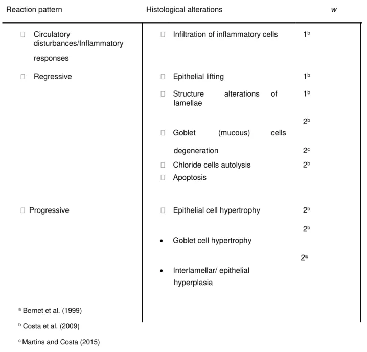
Semi-quantitative histopathological conditions indices were estimated for each individual, according to their biological significance of each surveyed alteration within the surveyed organ (score). The weight of alterations ranged between 1 (low severity) and 3 (high severity) and the score ranged from 0 (absent alteration) to 6 (diffuse alteration). The respective pathological alterations were classified into three reaction patterns: circulatory disturbance/inflammatory responses, regressive and progressive alterations. In table 3.1 are presented the histopathological alterations surveyed and their respective weights. The histopathological indices were estimated trough the following formula proposed by (Costa et al., 2013):
where wj is the weight of the jth histopathological alteration; ajh is the score attributed to the hth individual for the jth alteration and Mj is the maximum possible score, which normalizes Ih to a value between 0 and 1. As well as the global histopathological indice (Ih), partial indices including the different reaction patterns, I1 (circulatory disturbances/inflammatory responses), I2 (regressive changes) and I3 (progressive alterations) were also calculated.
Table 3.1 –Histopathological alterations observed in the gills ofS.senegalensisand their respective condition weights (w)
Reaction pattern Histological alterations w
Circulatory Infiltration of inflammatory cells 1b disturbances/Inflammatory
responses
Regressive Epithelial lifting 1b
Structure alterations of 1b lamellae
2b Goblet (mucous) cells
degeneration 2c
Chloride cells autolysis 2b Apoptosis
Progressive Epithelial cell hypertrophy 2b
2b • Goblet cell hypertrophy
2a • Interlamellar/ epithelial
hyperplasia
a Bernet et al. (1999)
b Costa et al. (2009)
c Martins and Costa (2015)
3.5. Statistical analyses
Shapiro-Wilks tests were performed to analyse the normality of the data obtained, followed by Levene test to analyse the homogeneity of variances. Due to the invalidation of at least one of the tests, the non-parametric Kruskal-Wallis one-way ANOVA followed by the Mann-Whitney U post hoc
test was employed for temporal comparisons as well as for gender and age comparisons.
Spearman’s rank statistic was applied to establish correlations. The statistical analysis was assessed
using Statistica 8.0 software (Starsoft, USA).
4. Results
4.1. Gill histopathology
Excised gills of S. Senegalensis (T0 gill samples) of each experiment (2h and 4 h of exposure) exhibited normal morphology (Fig. 4.1A), presenting well-defined lamellae attached to filaments, in accordance with previous descriptions for gills of juveniles of the species (Costa et al., 2009; Costa et al., 2010). The gill epithelium included pavement cells, goblet cells (mucocytes) and chloride cells, the latter type cells mostly located in the interlamellar space. The controls revealed some alterations of the epithelia relatively to T0 gills, namely, epithelial lifting and alterations of the epithelia structure (Fig. 4.1B).
Figure 4.1 –Gill histopathological sections ofS.senegalensis stained with tetrachrome stain (A,B,C,D,E) and NFR (F); (A) Overall aspect of the morphology of the gills obtained from T0 gill samples (gill excised and immediately fixed), exhibiting normal gill filament (fl), lamella (lm), pillar cells (pc), pavement cells (pv), chloride cells (cc) and goblet cells (gc); (B) Overall aspect of control samples, exhibiting epithelial lifting (white arrowhead) and structure alterations (black arrowhead); (C) Gill exposed to 200 µg nTiO2 L- 1 at T4 showing epithelial lifting (arrowhead) and chloride cell autolysis (cca) ; inset chloride cell autolysis (white arrowhead) and apoptosis (black arrowhead); (D and inset) hypertrophied goblet cells (arrows) and apoptosis (black arrowhead) present in gills exposed to 200 µg nTiO2 L-1; (E) interlamellar hyperplasia (arrows) observed in gills exposed to 20 µg nTiO2 L-1 treatment at T2; (F) metal deposits (white arrow), forming black deposits, macrophages (mmc) and interlamellar hyperplasia (black arrow) observed
in gills exposed to 20 µg nTiO2 L-1 treatment at T4.
4.2. Quantitative histopathological measurements
Significantly higher number of chloride cells per IS were obtained in gills exposed to nTiO2 treatments comparing to T0 gill samples and control treatment (Mann-Whitney U, p<0.01), at T2 (Fig. 4.2A). Conversely, no significant differences were observed between T0 gills and controls and between all treatments.
Figure 4.2 - Comparison of the mean number of chloride cell (A), goblet cells (B) and metal deposits (C) per interlamellar space (IS) between gills exposed to control, 20 and 200 µg nTiO2 L-1 treatments, for 2 (T2) and 4 h (T4) and T0 gill samples (gill excised and immediately fixed). * and **indicate significant differences between treatments and its respective control, p < 0.05 and p < 0.01, respectively (Mann Whitney U test). # and ## mean significant differences to its respective T0, p < 0.05 and p < 0.01 respectively (Mann Whitney U test). Error bars indicate standard deviation.
No significant differences were observed between T0 gill samples and treatments (control, 20 and
200 µg nTiO2 L-1) for both times of exposure (Fig 4.2C).
4.3. Gill Histopathological condition indices
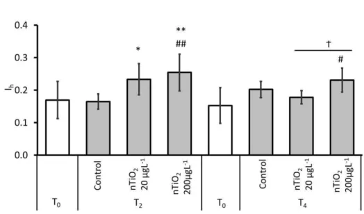
Gills exposed to nTiO2 for 2h revealed significantly higher global histopathological condition indice (Ih) compared to T0 and control gills (Fig.4.3). At this time point (T2) and considering exposures to nTiO2, the concentration of 200 µg nTiO2 L-1 yielded the highest Ih value, being significantly different from the respective T0 gill samples and from the controls (Mann-Whitney
U, p < 0.01). At T4, the highest Ihvalue was also registered for gills exposed to 200 µgL-1nTiO2, being significantly different to T0 gills and 20 µg nTiO2 L-1 treatment (Mann-Whitney U, p < 0.05).
Figure 4.3 - Comparison of the average histopathological indice (Ih) between gills exposed to control (C) and 20 and 200 µg nTiO2 L-1 treatments for 2 (T2) and 4 h (T4) and basal Ih from gills at the beginning of the experiment (T0 gills excised from fish and immediately fixed)..* and ** indicate significant differences between treatments and its respective control, p < 0.05 and p < 0.01, respectively (Mann Whitney U test). # and ## mean significant differences to its respective T0, p < 0.05 and p < 0.01 respectively (Mann Whitney U test). Ϯ indicate significant differences between nTiO2 treatments. Error bars indicate standard deviation.
In general, the average histopathological indices score obtained for the different reaction patterns exhibited similar variations as the global indices. Accordingly, gills exposed to nTiO2 treatments for 2h (T2) yielded higher of each individual indices (I1, I2, and I3) comparing to T0 and control gills (Fig. 4.4), however, the significant differences were only obtained for I2 and I3 indices (Fig. 4.4B, 4.4C), Mann-Whitney U, p<0.05 and p<0.01, respectively. No significant
differences were observed between nTiO2 treatments, however, the results show a tendency: the increase of indices for each reaction pattern with the increase of nTiO2 concentrations in water.At T4, the pattern was similar to T2, for I2 indice. Conversely, the progressive alterations reaction pattern indice (I3 were significantly lower for 20 µg nTiO2 L-1, relatively to T0 samples at both sampling times (Fig. 4.4C). Significant differences between nTiO2 treatments were only observed for I3, at T4.
significantly higher values for nTiO2 treatments relatively to T0 samples (p < 0.05). Conversely, the progressive alterations reaction pattern partial indice (I3) were significantly lower for 20 µg nTiO2 L-1, relatively to T0 samples (Fig. 4.4C). Significant differences between nTiO2 treatments were only observed for I3, at T4. No significant differences were observed regarding circulatory disturbances/inflammatory (I1), for both sampling times (Fig. 4.4A).
Figure 4.4 –Comparison of gill histopathological indices for each reaction pattern between gills exposed to control (C) and 20 and 200 µg nTiO2 L-1 treatments for 2 (T2) and 4 h (T4) and gills at the beginning of the experiment (T0 gills excised from fish and immediately fixed). Circulatory disturbances/Inflammatory response (I1); Regressive alterations (I2); Progressive alterations (I3). * and **indicate significant differences between treatments and its respective control, p < 0.05 and p < 0.01, respectively (Mann Whitney U test). # and ## mean significant differences to its respective T0, p < 0.05 and p < 0.01 respectively (MannWhitney U test). Ϯ indicate significant differences between nTiO2 treatments. Error bars indicate standard deviation.
4.4. Correlation analyses
H Spearman’s statistic showed the highest correlations between Ih, I3 (R=0.71, p<0.05) and I2 (R=0.79, p<0.05), demonstrating that progressive and regressive alterations were fundamental
to increase the histopathological indice. Regressive responses (I2) showed the highest correlations were obtained with apoptosis (R=0.67, p<0.05) and epithelial lifting (R=0.66, p<0.05). For the progressive responses (I3), goblet cell hypertrophy (R=0.79, p<0.05) was the
nor metal deposits mean value were highly correlated with any of the most significant alterations found trough the Spearman’s statistic.
5. Discussion
The present work is the first ex vivo experiment which used gills of S. senegalensis to assess
the toxicity of nTiO2. Few studies were done using ex vivo testing to assess TiO2, such as Valant & Drobne (2011) which assessed the biological reactivity of TiO2 by ex vivo testing, and
Brun et al. (2015), which assessed the titanium dioxide nanoparticle impact and translocation trough ex vivo gut epithelia.
The ex vivo test system employed in the present study provides evidence that gills were
moderately affected in the presence of nTiO2 in water. However, it is also relevant to note that some condition of the bioassays may also affect the normal functions of gills.
5.1. Exposure to nTiO2 promote osmotic imbalance
The presence of environmentally relevant concentrations of nTiO2 in the water yielded moderately histopathological lesions, mainly CC autolysis e GC hypertrophy. In fact, the nTiO2 increased the severity and dissemination of these alterations, as showed by the increment of the Ih relatively to T0 gill and control treatment, after 2 h of exposure. Representative correlations between the Ih and the respective responsive patterns were found, demonstrating that progressive and regressive alterations were fundamental to increase the histopathological indice (Van Dyk et al. 2009). Goblet cell hypertrophy and epithelial lifting showed high correlations when compared to histopathological indice. Progressive alterations, such as, hyperplasia and hypertrophy are considered protective measures against toxicants (Mallatt et al., 1985), possibly resulting in an unbalanced osmotic regulation, gas exchange and rapid mucous release.
In addition to the histopathological lesions, the number of chloride cells increased in gill exposed to nTiO2 comparing to control treatments, for 2h. It is well known that these cells are involved in ion transport in order to maintain the osmotic balance. As such, the present results may indicate that the immune system is reacting to the presence of nTiO2 in the water, producing more CC in gills. In fact, changes in number, size and distribution of CC in teleosts have been attributed to salinity (Karnaky et al., 1976) and also to metals (Giari et al., 2007). However, the results revealed that prolonged exposures, i.e. 4 hours of exposure (T4) does not imply the increase of CC, in ex vivo experiments. This may be attributed to the absence of a
systemic biological system when considering ex vivo, which may result in a deficit of osmoregulation.
5.2. Alterations in mucocytes reveal response to challenge
Mucous secretion (especially on the gill epithelium) is considered a defensive mechanism against toxicants (Handy et al., 1989). This common response of the mucus layer is excessive mucus production of mucosubstances, like glycoproteins and glycolipids, with the consequent swelling of epithelial cells followed by a change in the number of goblet cells (Bols et al., 2001). In the present study, the number of goblet cells per interlamellar space changed as a response to the exposure to nTiO2, however the pathway was time-dependent. Whereas the number of GC increased at T2, prolonged exposure (T4) decreased the number of this mucocytes. These results suggested a defensive mechanism toward the challenge, while a prolonged exposure to nTiO2, may overwhelmed the ability of immune system responses to cope with the injury, resulting in a decreasing number of goblet cells. In fact, changes in mucous cell function (number and size of goblet cells) have already been reported in studies when fish were exposed to distinct substances, like metals and organic toxicants (Costa et al., 2009; López-Galindo et al., 2010; Martins et al., 2015; Martins et al., 2016). In addition, excessive mucus production has also been documented in gills of rainbow trout after exposure to carbon based NPs (Smith et al., 2007). This response may be particularly critic in ex vivo systems, since they have not the
same intercellular interactions as in vivo systems, compromising the organism
immuno-response capability. The consequences of the reduction of mucus cells can enhance the infection potential, reduce lubrification of gill structures with consequent damage and hinder the excretion and regulation of metals and other ions.
5.3. No clear evidence of nTiO2 uptake from standard histological methods
Regarding metal deposits count, no significant differences were found between treatments or time of exposure. This result point to the problem commonly associated agglomeration of NPs in water. It is well known that NPs tend to agglomerate, especially in salt water, due to the increase of ionic strength which reduces the negativity of electrophoretic mobility of the particles (Batley et al., 2013). In addition, the bioavailability of NPs is influenced by the presence of
organic matter in water, changes in pH and ions (Baker et al., 2014). In fact, the Dynamic light scattering analysis indicated agglomeration of NPs, (data not shown). Probably this fact influenced the toxicity of the exposures since, generally, NPs agglomeration is associated with the increment
of NP’s size and the decrease of NP’s toxicity.
Some authors pointed that NP toxicity may be driven by the surface chemistry (reactivity) of the particles (Shaw and Handy, 2011). Parameters such as pH and light exposure (UV or natural) may modify the physiochemical characteristic of particles and make them more or less biologically active. It is pointed that nTiO2, when exposed to UV or natural light, due to their photocatalytic activity, can generate radical oxygen species (ROS), possibly inducing oxidative stress in cells (Xiong et al., 2011). In the present study, these parameters were monitored in both experiments and no differences were observed between control and nTiO2 treatments (data not shown).
Although metal deposits were found in gill macrophages, they were consistently distributed through all treatments. Generally, this response of the immune defence system is efficient in organisms and several NP aggregates will be expected in gill macrophage in in vivo experiments. Conversely, ex vivo systems may have limited this response. However, other methods, such as Transmission
Electron Microscopy (TEM), would added information about this issue.
5.4. The ex vivo bioassays generated significant confounding factors
The lesions observed in gills exposed to control treatment (sea water) showed severe epithelial lifting and, also, alterations of the structure of lamellae. The gills, because of their direct and permanent contact with water, are particularly sensitive to adverse environmental conditions (Thophon et al. 2003), including abrupt changes of salinity (Arjona et al., 2007). In addition, they are important organs since they perform vital functions such as gas exchange and ion osmoregulation. Through effective mechanisms of osmoregulation, euryhaline teleosts are able to cope with changes in salinity by the active transport of salts in the gills (Lin et al., 2004). In particular, S. Senegalensis, which is an estuarine species, is able to acclimate to different osmotic
conditions during short-term exposure (Arjona et al., 2007).
In fact, common alterations recorded in the present study, such as epithelial lifting, epithelial cell proliferation and lamellar fusion, have also been found in studies addressing osmoregulation in fish. Is the example of the in vivo work with Liza aurata fish after the exposure to acute increase in
This effect is a protective response to salinity stress since reduce gill surface area however, the efficiency of gas exchange will be severely affected. At the same time, changes in salinity also affect mitochondria-rich cells, i.e. chloride cells. The chloride cells of the gill secretory epithelium of euryhaline fish, such as S. senegalensis, adapt to the increment of salinity by stimulating chloride secretion, which basically pump salt from the blood to the outside medium and, in a second adaptation, by increasing the number and size of chloride cells (Arjona et al., 2007). In the present study, the gills were exposed to treatments immediately after excision and directly to sea water medium. Probably, these experimental conditions, i.e., ex vivo exposure in seawater, triggered
acute salinity stress, to which gills responded primarily with the alterations of the epithelial structure, however without significant chloride cell alterations, probably due to the short-time of the experiments (2 and 4 hours). In fact, the second adaptation mechanism takes place after days or weeks to the increment of salinity (Karnaky KJ JR, Kinter LB, Kinter WB, Stirling., 1976).
Ex vivo test reported in this study is well suited to the fast screening of the biological potential of
nanoparticles. Nonetheless, important factors should be assessed while preparing an ex vivo test,
such as the preparations of the solutions in the right environment, salinity and NPs characterization is important to avoid confounding factors.
Nonetheless, some improvements have to be implemented such as (i) the use of nanoparticulate TiO2, suspended in a physiological solution; (ii) the assess of the cell viability; (iii) the evaluation of the NP agglomeration using TEM (Transmission Electron Microscopy) and evaluation of their shape and size.
6. Conclusions
Overall, findings from this study indicate, that under ecologically relevant concentrations, nTiO2, can cause moderate effect on gills of S. Senegalensis in short-time exposures. However,
several parameters, such as pH of the exposure media or the presence of other molecules, may modify the physiochemical characteristic of particles and make them more or less biologically active than original or primary particle characteristics. It is, therefore, required to have a good understanding of the characteristics of particles to better understand their biological potential.
In conclusion, the ex vivo tests reported in this study are well suited to the fast screening of the
biological potential of nanoparticles, however, several issues have to be taken into account such as the medium for NPs and organ exposure. The data obtained from these experiments may significantly contribute to define further procedures to nanotoxicity studies.
7. References
Arjona, J.,2007. Acclimation of Solea senegalensis to different ambient temperatures: implications for thyroidal status and osmoregulation. 157(6): 1325–1335.
Andrade, T.A, 2016. Ex vivo model of human skin (hOSEC) as alternative to animal use for cosmetic tests. Procedia Engineering 110. 67 – 73.
Au, D.W.T., 2004. The application of histo-cytopathological biomarkers in marine pollution monitoring: a review. Mar. Pollut. Bull. 48, 817–834.
Baker, J.T., Tyler, R.C., Galloway, S.T., 2014. Impacts of metal and metal oxide nanoparticles on marine organisms. 186: 257-271.
Barmo, C., Ciacci, C., Canonico, B., Fabbri, R., Cortese, K., Balbi T., Marcomini, A., Pojana, G., Gallo, G., Canesi, L., 2013. In vivo effects of n-TiO2 on digestive gland and immune function of
the marine bivalve Mytilus galloprovincialis.15;132-133:9-18.
Bernardeschi, M., Guidi, P., Scarcelli, V., Lucchesi, P., Nigro, M., Frenzilli, G., 2010. Potential genotoxicity of nano-sized TiO2 particles in isolated bottle-nose dolphin (Tursiops truncates)
and human leukocytes.157, s14.
Blaise, C., Gagné, F., Férard, J.F., Eullaffroy, P., 2008. Ecotoxicity of selected nano- materials to aquatic organisms. Envir. Toxicol. 23, 591-598.
Batley, G.E., Kirby, J.K., McLaughlin, M.J., 2013. Fate and risks of nanomaterials in aquatic and terrestrial environments. Acc. Chem. Res. 46 (3), 854 - 862.
Boyle, D., Al-Bairuty, G.A., Ramsden, C.S., Sloman, K.A., Henry, T.B., Handy, R.D., 2013. Subtle
alterations in swimming speed distributions of rainbow trout exposed to titanium dioxide
nanoparticles are associated with gill rather than brain injury. Aquat. Toxicol. 126, 116e127.
Bols, N.C., Brubacher, J.L., Ganassin, R.C., Lee, L.E.J., 2001. Ecotoxicology and innate immunity in fish. Dev. Comp. Immunol. 25, 853–873.
Cabral, H., Costa,M.J.,1999. Differential use of nursery areas within the Tagus estuary by sympatric soles, Solea solea and Solea senegalensis. Environ. Biol. Fish. 56, 389–397.
within the nursery areas of the Tagus estuary, Portugal. J. Fish. Biol. 57, 1550–1562.
Canesi, L, Fabbri R, Gallo, G, Vallotto, D, Marcomini, A, Pojana, G., 2010. Biomarkers in Mytilus galloprovincialis exposed to suspensions of selected nanoparticles (nano-carbon black, C60 fullerene, nano-TiO2, nano-SiO2). Aquat Toxicol. 100:168–77.
Chen, X., Mao, S., 2007. Titanium dioxide nanomaterials: synthesis properties, modifications, and applications. Chem. Rev. 107, 2891–2959.
Cho, H.H., Wepasnick, K., Smith, B.A., Bangash, F.K., Fairbrother, D.H., Ball, W.P., 2010. Sorption of aqueous Zn II and Cd II by multiwall carbon nanotubes: the relative roles of oxygen-containing functional groups and graphenic garbon. Langmuir 26, 967–981.
Christoph Bartz, 2016. An ex vivo human cartilage repair model to evaluate the potency of a cartilage cell transplant. Journal of Translational Medicine. 14-317.
Costa, P.M., Lobo, J., Caeiro, S., Martins, M., Ferreira, A.M., Caetano, M., Vale, C., Del- Valls, T.A., Costa, M.H., 2008. Genotoxic damage in Solea senegalensis exposed to sediments from the Sado Estuary (Portugal): effects of metallic and organic contaminants. Mutat. Res. 654, 29–
37.
Costa, PM., Caeiro, S., Lobo, J., Martins, M., Ferreira, A.M., Caetano, M., Vale, C., DelValls, T.A., Costa, M.H., 2011. Estuarine ecological risk based on hepatic histopathological indices from laboratory and in situ tested fish. Mar Pollut Bull 62:55–65.
Costa, P.M., Lobo, J., Caeiro, S., Martins, M., Ferreira, A.M., Caetano, M., Vale, C., Del- Valls, T.A., Costa, M.H., 2008. Genotoxic damage in Solea senegalensis exposed to sediments from the Sado Estuary (Portugal): effects of metallic and organic contaminants. Mutat. Res. 654, 29–
37.
Costa, P. M., Diniz, M. S., Caeiro, S., Lobo, J., Martins, M., Ferreira, A. M., Costa, M. H. 2009. Histological biomarkers in liver and gills of juvenile Solea senegalensis exposed to contaminated estuarine sediments: A weighted indices approach. Aquatic Toxicology, 92(3), 202–212.
Costa, P.M., Caeiro, S., Diniz, M.S., Lobo, J., Martins, M., Ferreira, A.M., Costa, M.H., 2010. A description of chloride cell and kidney tubule alterations in the flatfish Solea senegalensis exposed to moderately contaminated sediments from the Sado estuary (Portugal). Journal of Sea Research, 64(4), 465–472.
Costa, P.M., Caeiro, S., Lobo, J., Martins, M., Ferreira, A.M., Caetano, M., Vale, C., DelValls, T.A., Costa, M.H., 2011. Estuarine ecological risk based on hepatic histopathological indices from laboratory and in situ tested fish. Mar Pollut Bull 62:55–65.
Costa, ,P.M., Costa, M.H, Costa., 2012. Development and aplicationof a novel histopathological multichrome techniqueon whole-body clam histopathology. J.Invertebr.Patho.110,411-414.
Costa, P.M., Caeiro, S., & Costa, M.H., 2013. Multi-organ histological observations on juvenile Senegalese soles exposed to low concentrations of waterborne cadmium. Fish Physiology and Biochemistry, 39(2), 143–158.
Diniz, S.M., Matos, P.A., Lourenço, A., Castro, L., Peres, l., Mendonça, E., Picado, A., 2013. Liver Alterations in Two Freshwater Fish Species (Carassius auratus and Danio rerio) Following Exposure to Different TiO2 Nanoparticle Concentrations.19, 1131-1140.
Emily, B., Frédérick, B., Giulia, V., Barbara, F., Stéphanie, S., Corinne, C., Christine, C., Thierry, R., Aloise, M., Nathalie, H., Marie, C., 2013. Titanium dioxide nanoparticle impact and translocation trough ex vivo, in vivo and in vitro gut epithelia. PLoS ONE. 9(5).
Fan, W., Cui, M., Shi, Z., Tan, C., Yang, X., 2012. Enhanced oxidative stress and physiological damage in Daphnia magna by copper in the presence of nano-TiO2. J. Nanomater. 10, 1687–
4110.
Farmen, E., Mikkelsen, H.N., Evensen, Ø., Einset, J., Heier, L.S., Rosseland, B.O., Salbu, B., Tollefsen, K.E., Oughton, D.H., 2012. Acute and sub-lethal effects in juvenile Atlantic salmon exposed to low μg/l concentration of Ag nanoparticles. Aquat. Toxicol. 108: 78-84.
Federici, G., Shaw, B.J., Handy, R.D., 2007. Toxicity of titanium dioxide nanoparticles to rainbow trout, (Oncorhynchus mykiss): gill injury, oxidative stress, and other physiological effects. Aquat. Toxicol 84:415–430.
Garvas, M., Testen, A., Umek, P., Gloter, A., Koklic, T., Strancar, J., 2015. Protein Corona Prevents TiO2 Phototoxicity. PLoS ONE. 10(6): e0129577.
Gottschalk, F., Sonderer, T., Scholz, R.W., Nowack, B., 2009. Modeled environmental concentrations of engeenered nanomaterials (TiO2, ZnO, Ag, CNT, Fellerenes) for different regions. Environ. Sci. Technol. 43, 9216–9222.
Gottschalk, F., Ort, C., Scholz, R.W., Nowack, B., 2011. Engineered nanomaterials in rivers exposure scenarios for Switzerland at high spatial and temporal resolution. Environ. Pollut. 159, 3439-3445.
Handy, R.D., Runnals, T., Russel, P.M., 2002. Histopathologic biomarkers in three spined sticklebacks, Gasterosteus aculeatus, from several rivers in southern Eng- land that meet the freshwater fisheries directive. Ecotoxicology 11, 467–479.
Handy RD, Henry TB, Scown TM, Johnston BD, Tyler CR (2008). Manufactured nanoparticles: their uptake and effects on fish—a mechanistic analysis. Ecotoxicology 17: 369–409.
Hu, X., Chen, Q., Jiang, L., Yu, Z., Jiang, D., Yin, D., 2011. Combined effects of titanium dioxide and humic acid on the bioaccumulation of cadmium in Zebrafish. Environ. Pollut. 159, 1151–
1158.
Jiménez-Tenorio, N., Morales-Caselles, C., Kalman, J., Salamanca, M.J., González de Canales, M.L., Sarasquete, C., DelValls, T.A., 2007. Determining sediment quality for regulatory proposes using fish chronic bioassays. Environ. Int. 33, 474–480.
Jovanović, B., & Palić, D., 2012. Immunotoxicology of non-functionalized engineered nanoparticles in aquatic organisms with special emphasis on fish-Review of current knowledge, gap identification, and call for further research. Aquatic Toxicology, 118–119, 141–151.
Karnaky, K.J. J.R., Kinter, L.B., Kinter, W.B., Stirling, C.E., 2016. Teleost chloride cell. II. Autoradiographic localization of gill Na,K-ATPase in killifish Fundulus heteroclitus adapted to low and high salinity environments. J Cell Biol 70:157—177.
Li, D., Cui, F., Zhao, Z., Liu, D., Xu, Y., Li, H., Yang, X., 2014. The impact of titanium dioxide nanoparticles on biological nitrogen removal from wastewater and bacterial community shifts in activated sludge. Biodegradation 25, 167-177.
Li, B., Huang,W., Zhang, C., Feng, S., Zhang, Z., Lei, Z., Sugiura, N., 2015. Effect of TiO2 nanoparticles on aerobic granulation of algal-bacterial symbiosis system and nutrients removal from synthetic wastewater. Bioresour. Technol. 187, 214-220.
Libralato, G., Minetto, D., Totaro, S., Micetic, I., Pigozzo, A., Sabbioni, E., Marcomini,A., Volpi Ghirardini, A., 2013. Mar. Environ. Res. 92, 71–78.
Lin, S.-H., Davidson, G.A., Secombies, C.J., Ellis, A.E., 1998. A morphological study of cells isolated from the perfused gill of dab and Atlantic salmon. J. Fish Biol. 53, 560-568.
M.J., 2010. Sublethal effects of the organic antifoulant Mexel®432 on osmorregulation and xenobiotic detoxification in the flatfish Solea Senegalensis. 79: 78-85.
Mallatt, J., 1985. Fish gill structural changes induced by toxicants and other irritants: a statistical review. Can J. Fish. Aquatic Sci. 42,630-648.
Martins, C., Alves de Matos, A.P., Costa, M.H., & Costa, P.M. 2015. Alterations in juvenile flatfish gill epithelia induced by sediment-bound toxicants: A comparative in situ and ex situ
study. Marine Environmental Research, 112, 122–130.
Martins, M., Santos, J., Costa M., Costa, P., 2016a. Apllying quantitative and semi-quantitative histopathology to adress the interaction between sediment-bound polycyclic aromatic hydrocarbons in fish gills. 79:266-275.
Menard, A., Drobne, D. & Jemec, A., 2011. Ecotoxicity of nanosized TiO2. Review of in vivo data. Environ Pollut 159:677-684.
Mu, L., Sprando, R., 2010. Application of nanotechnology in cosmetics. Pharm Res. 27:1746–
1749.
Mueller, N.C., Nowack, B., 2008. Exposure modeling of engineered nanoparticles in the environment. Environmental Science and Technology 42, 4447-4453.
Nel, A., Xia, T., Madler, L., Li, N., 2006. Toxic potential of materials at the nanolevel. Science 311, 622–627.
Park, M.V.D.Z., Lankveld, D.P.K., van Loveren, H., de Jong, W.H., 2009 The status of in vitro toxicity studies in the risk assessment of nanomaterials. Nanomedicine 4(6):669–685.
Paterson, G., Jamie, M.A., Ehsanul, H., Darcy, C. B., Chris, D. M., 2011. The toxicity of titanium dioxide nanopowder to early life stages of the Japanese medaka (Oryzias latipes).
82(7):1002-1009.
Pérez, S., Farré, M., Barceló, l.D., 2009. Analysis, behavior and ecotoxicity of carbon- based nanomaterials in the aquatic environment. TrAC Trends Anal. Chem. 28, 820–832.
Pettibone, J.M., Cwiertny, D.M., Scherer, M., Grassian, V.H., 2008. Adsorption of organic acids on TiO2 nanoparticles: effects of pH, nanoparticle size, and nanoparticle aggregation. Langmuir 24, 6659–6667.
Piccinno, F., Gottschalk, F., Seeger, S. & Nowack, B., 2012. Industrial production quantities and uses of ten engineered nanomaterials in Europe and the world. J Nanopart Res 14:1109-1120.
Rauscher, H., Roebben, G., Amenta, V., Sanfeliu, A.B., Calzolai, L., Emons, H.,Gaillard, C., Gibson, N., Linsinger, T., Mech, A., Pesudo, L.Q., Rasmunssen, K.,Sintes, J.R., Sokull-Klüttgen, B., Stamm, H., 2014. Towards a review of the ECrecommendation for a definition of the term nanomaterial. In: Report1831–9424. Luxembourg Publication Office of the European Union.
Riba, I., Casado-Martínez, M.C., Blasco, J., DelValls, T.A., 2004. Bioavailability of heavy metals bound to sediments affected by amining spill using Solea senegalensis and Scrobicularia plana.Mar. Environ. Res. 58, 395–399.
Riba, I., Blasco, J., Jiménez-Tenorio, N., González de Canales, M.L., DelValls, T.A., 2005. Heavy metal bioavailability and effects: II. Histopathology-bioaccumulation relationships caused by mining activities in the Gulf of Cádiz(SW, Spain).Chemo- sphere 58, 671–682
Shaw, B.J., Handy, R.D., 2011. Physiological effects of nanoparticles on fish: a comparison of nanometals versus metal ions. Environ. Int. 37: 1083-1097.
Schwaiger, J., 2001. Histopathological alterations and parasite infection in fish: indicators of multiple stress factors. J. Aquat. Ecosyst. Stress Recov. 8, 231–240.
Smith, C.J., Shaw, B.J., Handy, R.D., 2007. Toxicity of single walled carbon nanotubes to rainbow trout, (Oncorhynchus mykiss): Respiratory toxicity, organ pathologies, and other physiological effects. Aquat. Toxicol. 82, 94–109.
Stentiford, G.D., Longshaw, M., Lyons, B.P., Jones, G., Green, M., Feist, S.W., 2003. Histopathological biomarkers in estuarine fish species for the assessment of biological effects of contaminants. Mar. Environ. Res. 55, 137–159.
Sun, H., Zhang, X., Zhang, Z., Chen, Y., Crittenden, J., 2009. Influence of titanium dioxide nanoparticles on speciation and bioavailability of arsenite. Environ. Pollut. 157, 1165–1170.
Tan, C., Fan, W.,Wang, W., 2012. Role of titanium dioxide nanoparticles in the elevated uptake and retention of cadmium and zinc in Daphnia magna. Environ. Sci. Technol. 46, 469–476.
Teh, S.J., Adams, S.M., Hinton, D.E., 1997. Histopathologic biomarkers in feral fresh- water fish populations exposed to different types of contaminant stress. Aquat. Toxicol. 37, 51–70.
Thomas S., 2016. Human Ex-Vivo Liver Model for Acetaminophen-induced Liver Damage. 6: 316-319.
Thophon, S., Kruatrachue, M., Upatham, E., Pokethitiyook, P., Sahaphong, S., Jaritkhuan, S., 2003. Histopathological alterations of white seabass, Lates calcarifer, in acute and subcronic
cadium exposure. 121(3):307-320.
Toropov, A., Toropova, A., 2013. Optimal descriptor as a translator of ecletic information into the prediction of membrane damage by means if various TiO2 nanoparticles.93; 2650-2655.
Triebskorn, R., Telcean, I., Casper, H., Farkas, A., Sandu, C., Stan, G., Colãrescu, O.,Dori, T., Köhler, H.-R., 2008. Monitoring pollution in RiverMures¸ , Romania, part II: Metal accumulation and histopathology in fish. Environ. Monit. Assess. 141, 177–188.
Valant J., & Drobne D., 2011. Biological reactivity of TiO2 nanoparticles assessed by ex vivo
testing. 249: 835-842.
Vale, G., Franco, C., Diniz, M.S., Santos, M.M.C., Domingos, R.F., 2014. Ecotoxicol.Environ. Saf. 109, 161–168.
Van Dyk, J.C., Pieterse, G.M., Van Vuren, J.H.J., 2007. Histological changes in the liver of Oreochromis mossambicus (Cichlidae) after exposure to cadmium and zinc. Ecotoxicol. Environ. Safe. 66, 432–440.
Van Dyk, J.C., Marchand, M.J., Pieterse, G.M., Barnhoorn, I.E.J., Bornman, M.S., 2009. Histological changes in the gills of Clarias gariepinus (Teleostei: Clariidae) from a polluted South African urban aquatic system. Afr J Aqua Sci 34(3):283–291.
Vethaak, A.D., Wester, P.W., 1996. Diseases of flounder Platichthys flesus in Dutch coastal and estuarine waters, with particular reference to environmental stress factors. 26: 99-116.
Caroline, P.V., Fabio, M.H., Priscila, V.S., Caroline, M.C., Alex, S.D.M., Maria J.A.C.R. Passos., Thais. C.A.S., Juliana. M.N., Thiago, L.R.H., Li-Sei, W., Vicente, G., Ngan, V.P., 2015. Genotoxicity, potencial cytotoxicity and cell uptake of titanium dioxide nanoparticles in the marine fish Trachinotus carolinus (Linnaeus, 1766).
toxicology: opportunities for enhancement through histopathology. Environ. Toxicol. Pharmacol. 11, 289–295.
Xia, D., Jinyuan, C., Yanyuan, X., Meiron, Z., 2011. Effect of titanium dioxide nano-particles and some histological parameters of zebrafish (Danio rerio) after a long-term exposure.
101(3-4):493-499.
Xiong, D., Fang, T., Yu, L., Sima, X., Zhu, W., 2011. Effects of nano-scale TiO2, ZnO and their bulk counterparts on zebrafish: Acute toxicity, oxidative stress and oxidative damage. Science of the Total environment. 409:1444–1452.