Diogo Samuel Martins Almeida
Licenciado em Engenharia do Ambiente
Histopathological evaluation of two
Blennius
fishes exposed to microplastics
via feeding
Dissertação para obtenção do Grau de Mestre em Engenharia do Ambiente
Orientadora: Doutora Nagore Cuevas, investigadora do MARE-Centro de Ciências do Mar e do Ambiente.
Co-orientadora: Prof. Doutora Maria Helena Ferrão Ribeiro da Costa, Professora Associada com Agregação, Faculdade de Ciências e Tecnologia da Universidade Nova de Lisboa.
Júri:
Presidente: Prof. Doutora Paula Sobral Arguente: Prof. Doutora Paula Sobral
Vogais: Doutor Pedro Costa e Doutora Nagore Cuevas
ii Histopathological evaluation of two Blennius fishes exposed to microplastics via feeding
Diogo Samuel Martins Almeida
Statement of Copyright
iv
Agradecimentos
Em primeiro lugar gostaria de agradecer aos meus pais e irmão, por tudo aquilo que representam e porque sem eles dificilmente teria alcançado os meus objetivos.
Um obrigado a Professora Maria H. Costa pela oportunidade de realizar a tese no MARE.
A Nagore Cuevas, um obrigado especial pela constante partilha de conhecimento o que resultou num trabalho profícuo.
A todos os “MARES” (Ana Rodrigo, Carla Martins, Cátia Gonçalves, Joana Antunes, Marta Martins e Pedro Costa) um muito obrigado pela disponibilidade e apoio ao longo desta etapa, foram incansáveis.
A todos os professores com quem tive o prazer de me cruzar ao longo de todo o percurso académico e aqui refiro me desde os da Primaria até a Universidade, que de certa forma contribuíram para o meu desenvolvimento como pessoa e profissional.
vi
Abstract
The microplastic particles have dramatically increased in marine environments, emerging the concern of their potential adverse effects on marine biota, among others. These biological adverse effects of microplastics may result not only in physical harm such as internal abrasions and blockages, but also in an entrance vector of contaminants into marine organism. Therefore, the overall aim of this study was to assess the health status of two gobies (Blennius pholis and
Blennius galerita) exposed to microplastics, both coated and uncoated with antifouling paints
(i.e. common source of metals), via feeding for a month. For that purpose, multi-organ histopathological assessment (i.e. gills, liver, kidney and digestive tract) was carried out qualitatively and semi-quantitatively in gobies, as well as histochemical evaluation. The results showed no sign of microplastics ingestion, suggesting that these gobies were not able to ingest used microplastic spheres. Accordingly, no gross histological alterations were recorded in both fish species exposed to microplastics. Similarly, regardless the pathway of contaminants exposure (i.e. by ingestion or waterborne), animals exposed to contaminated microplastics presented similar histopathological levels than those treated with uncontaminated microplastics, or even than control. The affection degree of each target organ revealed gills and liver as the most affected organs following by kidney, digestive tract and spleen. Lamellar lifting, fat vacuolation of hepatocytes and melanomacrophage centers were the most prevalent alterations noticed in gills, liver and whole-body of fish, respectively. These findings may suggest that other factors, even natural occurrence, could be the cause of these mild histopathological alterations. Moreover, the use of Blennius gobies in multi-organ histopathology showed to be a suitable organism and tool for assessing the potential adverse effects caused by microplastics and their potential role as contaminants entrance. Therefore, smaller microplastic particles, both contaminated and uncontaminated, should be tested in gobies in order to clarify and evaluate their potential adverse effects via feeding.
Keywords: microplastic, gobies, histopathology, ingestion, contaminant release,
viii
Resumo
Os microplásticos têm aumentado drasticamente no meio marinho, ampliando a preocupação sobre os potenciais efeitos adversos na biodiversidade. Estes poderão ser causadores de danos físicos, como abrasões, bloqueios internos, ou atuar como vetores de entrada de contaminantes nos organismos marinhos. Por conseguinte, o objetivo geral deste trabalho foi avaliar o estado de saúde de dois cabozes (Blennius pholis e Blennius galerita) expostos a microplásticos com e sem revestimento de pintura anti-incrustante (i.e. fonte comum de metais no meio marinho) através da alimentação, durante um mês. Com esse propósito, foi feita, em cabozes, a avaliação qualitativa e semi-quantitativa histopatológica de vários órgãos (i.e. branquias, fígado, rim e trato digestivo). Os resultados, não mostraram sinais de ingestão de microplásticos, sugerindo os cabozes (B. pholis e B. galerita) como incapazes de ingerir microplásticos daquele tamanho. Consequentemente, não foram observadas quaisquer alterações significativas em ambas as espécies de peixes ao longo do ensaio laboratorial. Da mesma forma, independente emente da exposição aos contaminantes (i.e. por ingestão ou por via aquosa), os animais expostos a microplásticos contaminados apresentaram níveis histopatológicos similares aos expostos apenas a microplásticos ou mesmo aos do controlo. O grau de afetação de cada órgão, revelou branquias e fígado como os órgãos mais afetados seguindo-se rim, trato digestivo e baço. Deslocamento epitelial da lamela secundária, vacuolização dos hepatócitos e centros de melanomacrofagos, foram as alterações mais prevalentes observadas nas branquias, fígado e em todo o corpo do peixe, respetivamente. Em conclusão, as alterações histopatológicas registadas sugerem que a causa poderá ter sido resultado de outro fator ou de alterações naturais. No entanto, o uso de cabozes (Blennius) em histopatologia (avaliação de vários órgãos) revelou ser um método adequado para avaliar os potenciais efeitos adversos causados pelos microplásticos e seu papel como potencial entrada de contaminantes. Por esse motivo microplásticos de menores dimensões devem ser testados nos cabozes, a fim de esclarecer e avaliar os seus potenciais efeitos adversos através da alimentação.
x Contents
Figure index ... xii
Table index... xiv
Abbreviation list... xvi
1. Introduction ... 1
2. Objectives ... 4
3. Materials and methods ... 5
3.1. Experimental procedure ... 5
3.2. Histological procedure ... 6
3.3. Histopathological assessment ... 7
3.4. Statistical analyses... 8
4. Results ... 9
4.1. Biometric parameters ... 9
4.2. Signs of microplastics ingestion and copper deposits ... 9
4.3. Histopathological alterations and indices ... 10
4.3.1. Gills ... 10
4.3.2. Liver ... 12
4.3.3. Digestive tract ... 13
4.3.4. Kidney ... 14
4.3.5. Spleen ... 15
4.3.6. Global histopathological indices ... 15
4.3.7. Correlation analyses ... 16
5. Discussion ... 17
6. Conclusions ... 21
xii
Figure index
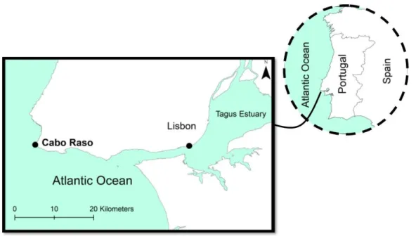
Figure 1. Location of sampling area (Cabo Raso, Portugal) where the animals were collected.
7
Figure 2. Experimental design showing five treatment diets carried out for each testing species and their respective replicate tanks.
9
Figure 3 Means ± SEM of biometric data (A: length; B: weight) of collected Blennius fish.
10
Figure 4. Diverse organs (i.e. liver and kidney) of gobies stained with Rubeanic Acid solution and counterstained with Nuclear Fast Red for observation of copper deposits.
11
Figure 5. Gills and liver sections of gobies collected in the experimental assay stained with Tetrachrome based on Alcian Blue, Weigert’s Hematoxylin and van Gieson’s.
12
Figure 6. Mean ± SEM of histopathological indices of liver, gills, kidney and digestive tract of collected Blennius fishes.
14
Figure 7. Digestive tract and kidney sections of gobies collected in the experimental assay stained with Tetrachrome based on Alcian Blue, Weigert’s Hematoxylin and van Gieson’s.
15
Figure 8. Spleen sections of gobies collected in the experimental assay stained with Tetrachrome based on Alcian Blue, Weigert’s Hematoxylin and van Gieson’s. 16
Figure 9. Mean ± SEM of global histopathological indices of collected Blennius
xiv
Table index
Table 1. Number of sampled individuals per treatment and species at the end of the
exposure period (i.e. after 30 days). 7
xvi
Abbreviation list
AF Antifouling paint
Cu Copper
Digestive tract HI Histopathological index of digestive tract
DPX Mounting medium fast (toluene base)
FCUL Faculty of Science at the University of Lisbon
Fe Iron
Gills HI Histopathological index of gills
Hg Mercury
Kidney HI Histopathological index of kidney
KW Kruskal Wallis – Multiple Comparisons
Liver HI Histopathological index of liver
MSFD Marine Strategy Framework Directive
PS Polystyrene
PVC Polyvinyl chloride
w/w Weigth/weigth
1
1.
Introduction
In modern society, plastic has achieved a pivotal status, with extensive commercial and industrial applications. In fact, the plastic has made a real contribution to meet targets for resource efficiency in many areas, including zero energy buildings, water saving, sustainable land use, extended shelf-life for products, diversified raw materials, greener mobility and renewable energies (PlasticsEurope, 2012). Hence, plastic production has dramatically increased from 1.5 million tonnes in the 1950 to approximately 322 million tonnes in 2015 (PlasticsEurope, 2016). This increasing plastic usage is provoking the uncontrollable accumulation of plastic wastes mainly in the terrestrial environment through accidental release and indiscriminate plastic discards (Barnes et al., 2009). Nevertheless, these plastic wastes also reach the marine coast by, wind and river-driven transport, becoming a prevalent widespread element of marine litter due to its lightweight durable nature (Moore, 2008; Thompson et al., 2009).
The first reports of plastic debris in the oceans were performed in early 1970s (Carpenter et al., 1972). From the following decades to the present, owing to the deepening knowledge on the marine ecological consequences of plastic wastes, the environmental problem of plastic debris accumulation has become of great concern both within scientific community and society ( e.g.
Cole et al., 2011; Fowler, 1987). In fact, a recent study finds that minute fragments of plastic
debris occur in aquatic environments worldwide, even in Antarctica (Barnes et al., 2009; Zarfl and Matthies, 2010). Likewise, as human population continues to increase, it is believed that the production of plastics and their wastes will also probably enhance, which may turn this social dilemma in a long-term environmental problem (Browne et al., 2011; Wright et al., 2013).
2 Thompson, 2009; Murray and Cowie, 2011). Considering the high relevance of this environmental problem, more knowledge on microplastics inputs, spatial and temporal distributions, transport dynamics, interactions with biota and adverse potential threat to marine biota and ecosystems is required. The assessment of microplastics in the marine environment, in fact, has recently been recognized by its inclusion as a priority descriptor in the Marine Strategy Framework Directive (MSFD, 2008/56/EC) as Descriptor 10 “Marine Litter”. More specifically, this descriptor demands more information related to types, sizes and composition of marine litter. By this way, this ecological data may help in the decision-making of scientific community, managers, policy makers and stakeholders (Boerger et al., 2010; De Stephanis et
al., 2013; Graham and Thompson, 2009; Lazar and Gračan, 2011; Murray and Cowie, 2011).
Nevertheless, a few scientific researches have been focused on the environmental problem caused by microplastics, up till now, especially with regard to potential adverse effects in marine organisms (Cole et al., 2011).
The microplastics bioaccumulation may result not only in physical harm such as internal abrasions and blockages, but also in a potential vehicle for the introduction of anthropogenic toxics by desorption processes (Andrady, 2011; Brennecke et al., 2016; Fossi et al., 2012,
2014; Koelmans et al., 2013; Wright et al., 2013). In fact, the capacity of microplastics to
increase bioavailability of contaminants from marine environment has already stated
(Brennecke et al., 2016). Among others, metal pollution is categorized of great concern, since
it is originated from multiple and common sources such as industrial wastes, fuel combustion and antifouling paints (Deheyn and Latz, 2006). The antifouling paints, in particular, are one of the major sources of heavy metals into the marine environment, through paint deterioration, metal release and subsequent dispersion (Berto et al., 2012; Tuner, 2010). The most modern marine antifouling paints contain a copper based biocidal pigment, which are applied to ship hulls and several other fixed marine structures (e.g. pilings, pontoons and buoys) to prevent the growth of fouling organisms (Almeida et al., 2007; Clode et al., 2011; Kiil et al., 2001; Valkirs
et al., 2003). The major environmental concern about the use of antifouling paints is currently
related to the passive leaching of Cu, among others, into waters (Katranitsas et al., 2003;
Warnken et al., 2004). Likewise, particles from antifouling paints may also be released into
3 One of the advantages of using biological effect techniques is that they indicate links between contaminant exposure and ecological endpoints (Au, 2004; Stentiford et al., 2003). Biomarkers are measurements of body fluids, cells or tissues at cellular, biochemical and molecular levels that indicate the presence of pollutants (exposure biomarkers) or the magnitude of the organism response (effects biomarkers) (NRC, 1987). Histopathology, in particular, is defined as the study of diseases and dysfunction of natural biological processes at tissue and cell levels (Chapman and Hollert, 2006) and thus measuring histopathological alterations in living organisms has been considered one of the most important approaches to assess pollution-driven adverse effects to organisms both in biomonitoring and laboratory studies (Costa et al., 2013;
Schultz et al., 2013; Stentiford et al., 2003). Moreover, histopathological techniques have
already been employed in studies related to microplastics and their respective adverse biological effects (Andrady, 2011; Cole et al., 2011; Fossi et al., 2012, 2014; Koelmans et al., 2013).
With the aim of addressing the assessment of adverse biological effects caused by microplastics, this study used two sympatric gobies, Blennius pholis and Blennius galerita, as potential test organisms. The B. pholis and B. galerita are common intertidal fishes living in exposed rocky shores in the marine littoral zones (Falcon et al., 2003), more specifically in the eastern Atlantic Ocean, the Mediterranean Sea and the Black Sea (Falcon et al., 2003; Zander,
1986). The Blennius fishes have a short generation time (12- 18 months) and can live in a
variety of substratum types during its life cycle, inhabiting in shallow pools when they are juveniles and in exposed rocky shores under stones and other protected microhabitats when they become adults (Faria et al., 2001).The Blennius fishes are believed to have limited mobility, hence organisms resilience to the inherently varying physico-chemical conditions is fundamental to their survival (McLusky and Elliott, 2007; Mitamura et al., 2005). Both fish species spawns from spring to summer in the Atlantic coast, although the reproductive cycle varies with latitude (Almada et al., 1996). In Portugal, in particular, the spawning season occurs in the cooler months, from October to May (Almada et al., 1990a; Faria et al., 2001). The main food items for this species consist of meiofauna and small macrofauna (Pihl, 1985; Salgado et
al., 2004). Owing to abovementioned characteristics, gobies have widely been used as suitable
4 2.
Objectives
The primary goal of this thesis is to assess histopathological alterations and lesions in whole body of two fish species of Blennius (i.e. B. pholis and B. galerita) exposed via feeding to several concentrations of microplastics (uncontaminated) for a month. As a secondary objective, heavy metal release from microplastics covered with an antifouling paint (contaminated) was assessed through the histopathological analyses in same testing species in order to detect whether microplastics may play a role as vector for heavy metal contamination and levels of these toxic compounds found in microplastics may cause potential adverse biological effects. For abovementioned purposes, histopathological disturbances were firstly evaluated qualitatively and then through the development of histopathological condition indices in several target organs (i.e. gills, gills, liver, kidney, digestive tract and spleen), as well as histochemical evaluation.
The present thesis attempts to address the following specific objectives:
• To identify histopathological alterations in multiple target organs (i.e. gills, liver, kidney, digestive tract and spleen) of two Blennius fishes after exposure (30 days) to uncontaminated microplastics via feeding.
• To determine and evaluate abovementioned histopathological effects in multiple target organs of gobies through qualitative and semi-quantitative histopathological assessment.
• To assess a potential role of microplastics covered with an antifouling paint (contaminated) as vector for entrance to heavy metal contamination in biota from marine environments.
• To investigate the potential adverse biological effects qualitatively and semi-quantitatively caused by heavy metal released contamination from microplastics in whole body of gobies.
5
3.
Materials and methods
3.1.Experimental procedure
The laboratory experiment was designed and carried out by the Faculty of Science at the University of Lisbon (FCUL). Two fish species of Blennius (B. pholis and B. galerita) were collected in July 2016 at Cabo Raso (Portugal) (Fig. 1), a negligibly disturbed area. Fish were transported to the laboratory in aerated seawater containers and 25 individuals were randomly placed into each aquarium for 10 days of acclimation. The animals were fed daily with 2.19 g of frozen grinded shrimp and maintained in continuous artificial sea water flow (20 ± 0.5ºC; 34ppm).
Fig. 1. Location of sampling area (Cabo Raso, Portugal) where the animals were collected.
6 marine and estuarine environments (Andrady, 2011). Specifically, 8 microplastic PS spheres were added to daily feed in 0.1% (w/w) experimental groups, while in 0.2 % (w/w) treatments 16 microplastic PS spheres were provided. To sum up, as Figure 2 illustrates, this experimental procedure presented five experimental groups, treatment C as control, treatments 0.1 and 0.2 with uncontaminated PS microplastics and, treatments 0.1mp and 0.2mp with microplastics covered with AF (contaminated). Three replicate tanks were assigned to each treatment diet and species.
Fig. 2. Experimental design showing five treatment diets [(C) control; (0.1) 0.1% w/w; (0.2) 0.2% w/w; (0.1mp)
0.1% w/w with AF; and (0.2mp) 0.2% w/w with AF] carried out for each testing species and their respective replicate tanks. PS: Polystyrene; AF: antifouling paint; n: number of microplastic spheres provided daily.
The number of the individuals sampled per treatment and species at the end of the exposure period (i.e. after 30 days) and processed for histological process is illustrated in Table 1. During the experiment, fish were monitored for any possible signs of impaired health status.
Table 1. Number of sampled individuals per treatment and species at the end of the exposure period (i.e. after 30
days). n.d.: no data.
C 0.1 0.2 0.1mp 0.2mp
B. pholis 6 6 n.d. 6 2
B. galerita 6 6 6 6 5
3.2.Histological procedure
Collected fish samples were immersed in Bouin-Hollande’s (10% v/v formaldehyde and 7% v/v acetic acid to which picric acid was added till saturation) or Davidson (formalin- ethanol- acetic acid) fixative for 24 - 36 h at 4°C. Afterwards, whole body samples were dehydrated in a graded progressive series of propanol (70, 96, 100%) followed by an embedding in paraffin. Embedded tissue samples of whole fish were cut (5 µm - 7 µm) using a rotary microtome (Jung
(c)'' d (c)' d (c) (0.1)'' d (0.1)' d (0.1) (PS) (n8) (0.2)'' d (0.2)' d (0.1mp)'' d (0.1mp)' d (0.2mp)'' d (0.2mp)' d (0.2) (PS) (n16) (0.1mp)
(PS) + (AF)
(n8)
(0.2mp)
(PS) + (AF)
7 RM2035, Leica Microsystems). At least four slides from each sample were obtained, each containing 4 - 6 sections. Tissue samples were deparaffinated and rehydrated before staining. Diverse dyes were employed for histochemical and histopathological evaluation: (1) Haematoxylin and Eosin (H&E) for structural and morphological screening; (2) Rubeanic Acid solution counterstained with Nuclear Fast Red in order to analyze and evaluate copper deposits and (3) tetrachrome stain based on Alcian Blue, Weigert’s Hematoxylin and van Gieson’s to the differentiation of distinct cell types, though enhanced chromatic segregation of tissues and cells within tissues (Costa and Costa, 2012 with few modifications). Afterwards, all slides were dried and mounted with glycerine solution (50 - 75%). Unlike common histopathological procedure, isopropanol and glycerine were employed instead ethanol and DPX, respectively, to avoid microplastics degradation (Gonçalves et al., 2017).
3.3.Histopathological assessment
8 Where Ih is the histopathological condition indices for the individual h; wj the weight of the jth histopathological alteration; ajh the score attributed to the hth individual for the jth alteration and Mj is the maximum attributable value for the jth alteration, i.e., weight × maximum score.
The equation’s denominator normalizes Ih to a value between 0 and 1, thus permitting comparisons between distinct situations (such as different organs and species).
A list of all observed pathologies and respective condition weigh, considered for the estimation of histopathological indices are illustrated in Table 2. A blind review of slides was performed to ensure the accuracy of histopathological evaluation.
Table 2. Histopathological alterations recorded in liver, gills, kidney and stomach of Blennius and their respective
biological significance (w). MMCs: melanomacrophage centers; HP: hyperplasia.
3.4.Statistical analyses
The non-parametric test Kruskal-Wallis Multiple Comparison test (KW) was employed in quantitative and semi-quantitative parameters after invalidation of at least one of the assumption of parametric tests (homocedasticity by Levene’s test and normality by Kolmogoroff-Smirnoff’s test). Spearman rank correlations were also performed in order to associate analized variables (i.e. biometric data and histopathological indices). A significance level α was set at 0.05 for all analyses. The statistical analysis was conducted with the Statistica 8.0 software (Statsoft, USA).
Liver w Gills w Kidney w Stomach w
Circulatory disturbances
Hyperaemia 1
Haemorrhage 1
Inflammation
response MMCs 1 MMCs 1 MMCs 1 MMCs 1
Regressive changes
Lamellar Lifting 2
Progressive changes
Fat vacuolation
9 4. Results
4.1.Biometric parameters
The length and weight of gobies ranged between 2.0 - 6.5 mm and 0.02 - 3.0 g, respectively. Mean values of both biometric variables were illustrated in Figure 3, grouped according to the fish species and experimental treatment. Significant differences in the length and weight were observed between individuals from the control and treatment 0.1mp for the B. pholis fish species (KW, p< 0.05). On the contrary, no significant differences in biometric parameters were obtained among treatments for the B. galerita fish species (KW, p< 0.05). Moreover, biometric parameters in both fish species were similar in all experimental treatments (KW, p< 0.05).
Fig. 3. Means ± SEM of biometric data (A: length; B: weight) of collected Blennius fishes. Statistical differences
among experimental treatments in B. pholis fish species were reflected by letters. C: control; 0.1: 0.1% w/w; 0.2:
0.2% w/w; 0.1mp: 0.1% w/w with AF; 0.2mp: 0.2% w/w with AF; n.d.: no data.
4.2.Signs of microplastics ingestion and copper deposits
10 Fig. 4. Liver and Kidney stained of Blennius fishes with Rubeanic Acid solution and counterstained with Nuclear
Fast Red for observation of copper deposits: (A) Liver section showing a blood vessel surrounded by small copper deposits (circle); (B) Kidney section presenting copper deposits into a renal tubule (circle). Scale bar: 35 µm.
4.3.Histopathological alterations and indices
No gross histopathological lesions were observed in any of the organs analysed (i.e. gills, liver, kidney, digestive tract and spleen) for both species in all experimental groups. The main histopathological alterations registered in target organs were illustrated in abovementioned Table 2 (see material and methods section). The biological importance (weigh), according to the histopathological index applied, ranged between 1 and 3, although in the present work the maximum weigh registered was of 2, in the case of lamellar lifting and hyperplasia of goblet cells, both lesions in gills (Table 2). Likewise, the dissemination degree (score) of recorded histopathological alterations was relatively low, being values between 2 and 4 the most common within the multi-organ histopathological assessment in both fish species.
4.3.1. Gills
Normal gills revealed the typical structure of lamellae (i.e. primary and secondary), in which the single-cell thick lamellar epithelia contained goblet and chloride cells supported by a complex system of blood vessels (Fig. 5A).
11 in the gills for both fish species. B. pholis, in particular, displayed a prevalence ranged between 0 - 33%, while B.galerita demonstrated less frequency (0 - 20%). This latter fish species also presented one case of progressive changes in gills, more specifically hyperplasia of goblet cells. No circulatory disturbances were observed in both fish species within all experimental groups.
Fig.5 Gills and liver sections of Blennius fishes stained with Tetrachrome based on Alcian Blue, Weigert’s
Hematoxylin and van Gieson’s: (A) Normal gills with well differentiated primary (l) and secondary lamellae (sl), gill filament support cartilage (ct), goblet cell (gc) and chloride cell (cc); (B) Gills affect by lamellar lifting (arrows and inset); (C) Normal liver presenting hepatocytes (h) with a homogenous cytoplasm and a central spherical and well defined nucleus (hn). (D) Liver tissue affected entirely by fat vacuolation (v) of hepatocytes and hyperaemia (hy). bv: blood vessel. Scale bar: 35 µm, inset 20 µm.
12 No significant statistical differences were found among the treatments in each fish species. However, significant statistical differences (KW, p < 0.05) were observed between species for the treatments 0.1, 0.1mp and 0.2mp (Fig. 7).
4.3.2. Liver
The normal liver structure of gobies was characterized by hepatocytes presenting a homogenous cytoplasm and a central spherical nucleus, aligned with sinusoids (Fig. 5C). In this fish species, pancreatic tissue was comprised within the liver forming a single organ known as hepatopancreas.
The occurrence of lesions in the liver of collected fish was relatively low, being fat vacuolation of hepatocytes the most frequent alteration in both fish species. In those cases, nuclei of hepatocytes were remarkably displaced to the periphery of the cells becoming difficult to distinguish the normal hepatic lobular architecture (Fig. 5D). B. pholis presented prevalence ranged between 60 - 100%, showing the lowest value in animals from the control group. In the case of B. galerita, fat vacuolation was shown in frequencies of 0 - 80%. Noted that B. galerita fish collected from the treatment 0.2mp did not demonstrated any fat vacuolation of hepatocytes. Inflammatory responses, which include presence of melanomacrophage centers (MMCs), were the second most common reaction pattern, observing that B. pholis presented lower prevalence (0 - 50%) than B. galerita (40 - 100%). It should be emphasized that both species presented fat vacuolation as the second highest prevalence (40% B.pholis and 83% B.
galerita) in individuals from the control group. Circulatory disturbances such as haemorrhage
and hyperaemia in individuals of B. pholis presented prevalence ranged between 0 - 50% and 0 - 60%, respectively. Nevertheless, no signs of these circulatory alterations were shown in fish from the control for the B. galerita species, while in individuals from the rest of treatments presented prevalence between 0 - 20% for haemorrhage and 0 - 100% for hyperaemia. According to B. pholis results, hyperaemia was not shown in individuals from the control.
B. galerita presented the lowest histopathological index of liver (HI Liver) in the treatment
13 Significant statistical differences (KW, p< 0.05) were found for the species B. galerita (Fig. 6) between control and the treatments 0.2 and 0.2mp. Differences were also observed between species for the treatment 0.2mp (KW, p < 0.05) (Fig. 6).
Fig. 6. Mean histopathological indices of liver, gills, kidney and digestive tract of two fishes collected in the experimental assay. Letters indicate statistically significant differences among the treatments for the B. galerita
fish species and (*) between the species. Error bars indicate the standard error (SEM). C: control; 0.1: 0.1% w/w; 0.2: 0.2% w/w; 0.1mp: 0.1% w/w with AF; 0.2mp: 0.2% w/w with AF. n.d.: no data
4.3.3.Digestive tract
14 Fig. 7. Sections of digestive tract and kidney of Blennius fishes stained with Tetrachrome based on Alcian Blue,
Weigert’s Hematoxylin and van Gieson’s. (A) Normal structure of digestive tract composed by: gastric gland (gg) and gastric pit (gp); mucous cell (mc); muscularis mucosae (m) and lumen (lu). (B) Digestive tract with MMCs (circle). (C) Normal structure of kidney composed wit renal tubule (t) lined with a normal epithelial cell (ec); (D) Kidney tissue with MMCs (dashed circle and inset). Scale bar: 35 µm; inset 20 µm.
4.3.4. Kidney
The kidney normal structure was composed of renal tubules surrounded by hematopoietic interstitial tissue containing mainly erythrocytes and blast (Fig 7C). Within this study, presence of MMCs was only observed, which is characterized as inflammatory change. This latter alteration was recorded in individuals of B. pholis from the treatment 0.2mp with a prevalence of 50%, while in B. galerita was registered in all treatments, except in the treatment 0.1mp with prevalence between 33 and 67% (Fig. 7D).
15 animals of B. pholis this alteration was only observed in the treatment 0.2mp (Fig. 6). Nevertheless, when the statistical evaluations were possible, no significant statistical differences were observed between the species and among the treatments (Fig. 6).
4.3.5.Spleen
The spleen structure was composed mainly of blood vessels, red pulp and white pulp. The red pulp, which occupied the majority of the organ, consisted of a cellular reticulum with hematopoietic tissue and blood sinuses. The white pulp, on the contrary, was often scarce (Fig. 8).
In this organ, only inflammation responses were observed in both fish species from all experimental groups, more specifically the presence of MMCs (Fig. 8). B. pholis showed a prevalence of 100% in the control group and treatment 0.1. B. galerita presented prevalence ranging between 0 - 100%, being the control the most affected group, while the treatment 0.2 and 0.1mp did not showed any inflammation responses. Note that the sample size of this organ obtained in individuals from different experimental group was rather low and thus estimation of histopathological indices and subsequent statistical analyses were not performed and plotted.
Fig. 8. Section of spleen of Blennius fish stained with Tetrachrome based on Alcian Blue, Weigert’s Hematoxylin
and van Gieson’s. Spleen section affected by MMCs (circles). rp: red pulp; wp: white pulp; bv: blood vessels. Scale bar 35 µm.
4.3.6. Global histopathological indices
16 of tested individuals (Fig. 9). Considering the different affection degree in each target organ, gills and liver demonstrated to be the most affected organs in this laboratory experiment, while kidney and digestive tract, together with spleen, were less impacted (gills > liver > kidney > digestive tract). Thus, both liver and gills presented more influence in the estimation of global histopathological indices (Fig. 6, 9).
In general, B. pholis registered higher Global HI than B. galerita. There were no signs of tendencies among different treatments in the histopathological indices for both species (Fig. 9). Global HI, conversely, revealed significant statistical differences (KW, p< 0.05) between species in the treatments 0.1 and 0.1mp (Fig. 9).
Fig. 9. Means of global histopathological indices, which are the sum of histopathological indices of the different
target organs (i.e. liver, gills, kidney and digestive tract), registered in two Blennius fishes. (*) indicates
statistically significant differences between the species. Error bars indicate the standard error (SEM). C: control; 0.1: 0.1% w/w; 0.2: 0.2% w/w; 0.1mp: 0.1% w/w with AF; 0.2mp: 0.2% w/w with AF. n.d.: no data.
4.3.7.Correlation analyses
17 5. Discussion
In the present work, Blennius fishes, B. pholis and B. galerita, were exposed to two concentrations of microplastics (both contaminated and uncontaminated) during a month. Even so, the findings did not reveal signs of microplastics ingestion, which may suggest that these gobies were not able to ingest this kind of microplastic spheres. Therefore, negligible adverse biological effects were shown in the multi-organ histopathological assessment (i.e. liver, gills, kidney, digestive tract, and spleen) in gobies exposed to uncontaminated microplastics. Accordingly, regardless the pathway of contaminants (by ingestion or waterborne), exposure to contaminated microplastics did not cause gross histopathological adverse effects in gobies. Nevertheless, this study did not clarify whether microplastics may play a role as vector for entrance to heavy metal contamination in biota from marine environments.
According to the findings, the size of microplastics, selection of appropriate fish species (e.g. feeding behaviour) and its respective biometric parameters were relevant factors to be considered within experimental design, when the ingestion of these microplastic particles was expected. In comparison with the basis of this experimental design (Pedá et al., 2016), the diameter of microplastic PS particles employed within this laboratory assay were almost three times larger (0.7 - 0.9 mm > 0.3 mm), while the size of fishes was considerably lower (3 g < 140 g). For instance, Oliveira et al., (2013) reported the adverse effects of remarkably smaller microplastics (1 - 5µm diameter) ingestion in Pomatoschistus microps gobies, which feature similar size than Blennius species. Similarly, Mota (2017) presents a study with Diplodus
Sargus, larger fish than Blennius fishes,based on the same bioassay conditions as in the present
18 In view of that ingestion of microplastics may not occur, the potential role of these particles as entrance vector of metals from antifouling paints by ingestion pathway was not evidenced within the present study. The release of metals from antifouling paints may be held directly in seawater, being these contaminants diluted in the medium. Therefore, the levels of metals in waterborne and its bioavailability, specifically of copper, did not cause any relevant bioaccumulation and histopathological alterations in diverse target tissues of both fish species. The punctual cooper deposits observed into the target organs may be caused from natural occurrence, since metals such Cu, Fe and Zn are essential metals and play important role in biological metabolic processes (Arulkumar et al., 2017). Conversely, Sivaperumal et al.,
(2007) and Velusamy et al. (2014) stated that essential metals can also produce toxic effects at
high residual concentrations. Indeed, according to Brennecke et al., (2016) and Soroldoni et
al., (2017), antifouling paints can be a source of metals to be desorbed through microplastics
into the seawater and thus be bioavailable and bioaccumulated in marine living organisms.
Among the most obvious adverse effects caused by microplastics ingestion in fish species are the physical blockage of the digestive tract and its influence in feeding (Sá et al., 2015; Jovanovic, 2017). Some authors, conversely, have reported more severe histopathological alterations caused by microplastics such as widening of the lamina propria and hypertrophy of goblet cells in diverse fish species after exposure of 30 days (Pedá et al., 2016). Thus, contrary to the expectations, digestive tract was the less impacted target organ, showing only mild inflammatory responses. As reported in other studies, microplastics can pass through the digestive tract and be expelled from the body or be retained in the gastrointestinal tract, causing internal abrasion and inflammatory responses (Moos et al. 2012). Regarding other target organs, differences in histopathological affection degree were observed, being listed from most impacted to less as gills, liver, kidney and spleen. As it has been already reported, this different affection degree was probably owing to the specific biological function and sensitivity of each target organ (Bernet et al., 1999; Costa et al., 2009, 2013). Nevertheless, assessed organs within this work have already stated as suitable target organs to evaluate adverse biological effects in fish (Bernet et al., 1999; Costa et al., 2009; Cuevas et al. 2016).
19 contaminants resulting in adverse biological effects (Evans et al., 2005; Movahedinia et al., 2009). Unlike in other studies in which liver is reported as the most affected organ after contamination exposure (Costa et al., 2009; Cuevas et al., 2016; Karami et al., 2016), gills presented the highest histopathological indices in this work. This may be explained due to the abovementioned fairly slight waterborne metal release from microplastic particles, which may suggest gills as the only organ in direct contact with potential metal pollutants. On the other hand, it should be noted that animals collected from natural sites, often present a baseline level of non-specific gill lesions supporting the statement of the natural occurrence (Costa el al., 2009; Cuevas et al., 2015).
In the case of liver, kidney and spleen, they were employed within several works as target organs for the assessment of adverse biological effects (Bernet 1999; Costa et al., 2009, 2013; Cuevas et al., 2016). Liver, in particular, presents a relevant detoxification role, which regulates metabolism, allows the digestive system to extract more nutrients and remove wastes from blood, to aid in recycling old blood cells, any damage to the liver could impair circulatory lipid levels in the body (Begriche et al., 2011). Liver presented vacuolation of hepatocytes as the most common alteration in both fish species. Some authors defend that the condition of hepatocytes is highly dependent on the reproductive stage and the availability of an adequate supply (Davies and Vethaak, 2012). On the contrary, this histopathological disturbance has also been related to exposure of mixtures of metal and organic contaminants (Van Dyk et al., 2007; Triebskorn et al., 2008). Kidney, in turn, operates as an important organ related to electrolyte and water balance and the maintenance of a stable internal environment (Bernet et al., 1999; Costa et al., 2010). Melanomacrophage centers (MMCs) were the only alteration registered in this organ, although this alteration was observed in whole body of gobies. This frequent alteration in fish has specific functions such as deposition sites for intracellular bacteria, retention of iron, presentation to immune cells and collection of products of cellular degradation (Agius et al, 2003; Evans and Nowak, 2016; Wolke, 1992). However, according to some authors the number and dissemination of MMCs can be influenced by dietary supplementation, as well as exposure to infectious agents (Hur et al., 2006; Manrique et al.,
2014; Pronina et al. 2014). Spleen, generally, is located close to the stomach and it mainly acts
20 According to the results, B. pholis and B. galerita were not suitable species to evaluate potential adverse effects of the selected PS microplastic spheres owing to their large size for this fish species. However, gobies were reported as suitable test organisms for assessing adverse effect of both microplastic ingestion and potential pollutants (Sá et al., 2015; Cuevas
et al., 2016; Oliveira et al., 2013). Likewise, this work presented a setback related to the low
sample size of treatments 0.2 and 0.2mp in B. pholis fish, which hindered the comparison between species. Nevertheless, these sympatric fish species presented similar biological adverse effect within this study, reflecting that both fish species may be used indistinctly, which may facilitate upcoming field and laboratory works.
21 6. Conclusions
22
7.
References
Agius, C., Roberts, R.J., 2003. Melano-macrophage centres and their role in fish pathology. J. Fish Dis. 26, 499–509. https://doi:10.1046/j.1365-2761.2003.00485.
Almada, V.C., Barata, E.N., Gonçalves, E.J., Oliveira, R.F. 1990a. On the Breeding season of Lipophrys polis (Pisces: Blennidae) at Arrábida, Portugal. J. Mar. Biol. Assoc. U. K. 70, 913–916. https://doi.org/10.1017/S0025315400059142.
Almada, V.C., Carreiro, H., Faria, C., Gonçalves, E.J., 1996. The breeding season of Coryphoblennius galerita in Portuguese waters. J. Fish Biol. 48, 295–297. https://doi:10.1111/j.1095-8649.1996.tb01121. Almeida, E., Diamantino, T.C., de Sousa, O., 2007. Marine paints: the particular case of antifouling paints. Prog. Org. Coatings 59, 2–20. https://doi:10.1016/j.porgcoat.2007.01.017.
Andrady, A.L., 2011. Microplastics in the marine environment. Mar. Pollut. Bull. 62, 1596–1605.
https://doi:10.1016/j.marpolbul.2011.05.030.
Arulkumar, A., Paramasivam, S., Rajaram, R., 2017. Toxic heavy metals in commercially important food fishes collected from Palk Bay, Southeastern India. Mar. Pollut. Bull. 119, 454–459.
https://doi:10.1016/j.marpolbul.2017.03.045.
Au, D.W.T., 2004. The application of histo-cytopathological biomarkers in marine pollution monitoring: A review. Mar. Pollut. Bull. 48, 817–834. https://doi:10.1016/j.marpolbul.2004.02.032.
Barnes, D.K.A., Galgani, F., Thompson, R.C., Barlaz, M., 2009. Accumulation and fragmentation of plastic debris in global environments. Philos. Trans. R. Soc. B Biol. Sci. 364, 1985–1998.
https://doi:10.1098/rstb.2008.0205.
Begriche, K., Marston, O.J., Rossi, J., Burke, L.K., Mcdonald, P., Heisler, L.K., Butler, A.A., 2012. Melanocortin-3 receptors are involved in adaptation to restricted feeding. Genes Brain Behav. 11, 291–
302. https://doi:10.1111/j.1601-183X.2012.00766.x.
Bernet, D., Schmidt, H., Meier, W., Burkhardt-Holm, P., Wahli, T., 1999. Histopathology in fish: Proposal for a protocol to assess aquatic pollution. J. Fish Dis. 22, 25–34. https://doi:10.1046/j.1365-2761.1999.00134.x.
Berto F., Cacciatore, F., Covelli, S., Rampazzo, D., Boscolo, R., Giovanardi, O., Giani, M., 2012. Tin free antifouling paints as potential contamination source of metals in sediments and gastropods of the southern Venice lagoon. Cont. Shelf Res. 45, 34–41. https://doi:10.1016/j.csr.2012.05.017.
Boerger, C.M., Lattin, G.L., Moore, S.L., Moore, C.J., 2010. Plastic ingestion by planktivorous fishes in the North Pacific Central Gyre. Mar. Pollut. Bull. 60, 2275–2278.
https://doi:10.1016/j.marpolbul.2010.08.007.
Brennecke, D., Duarte, B., Paiva, F., Caçador, I., Canning-Clode, J., 2016. Microplastics as vector for heavy metal contamination from the marine environment. Estuar. Coast. Shelf Sci. 178, 189–195.
23 Browne, M.A., Dissanayake, A., Galloway, T.S., Lowe, D.M., Thompson, R.C., 2008. Ingested microscopic plastic translocates to the circulatory system of the mussel, Mytilus edulis (L.). Environ. Sci. Technol 42, 5026–5031. https://doi:10.1021/es800249a CCC: $40.75.
Browne, M.A., Crump, P., Niven, S.J., Teuten, E., Tonkin, A., Galloway, T., Thompson, R., 2011. Accumulation of microplastic on shorelines woldwide: sources and sinks. Environ. Sci. Technol. 45, 9175–9179. https://doi:10.1021/es201811s.
Canning-Clode, J., Fofonoff, P., Riedel, G.F., Torchin, M., Ruiz, G.M., 2011. The effects of copper pollution on fouling assemblage diversity: a tropical-temperate comparison. PLoS One. 6, e18026
https://doi:10.1371/journal.pone.0018026.
Carpenter, E.J., Smith, K.L., Science, S., Series, N., Mar, N., 1972. Plastics on the Sargasso Sea surface. Science 175, 1240–1241. https://doi.org/10.1126/science.175.4027.1240.
Chapman, P.M., Hollert, H., 2006. Should the sediment quality triad become a tetrad, a pentad, or possibly even a hexad? J. Soils Sediments 6, 4–8. https://doi:10.1065/jss2006.01.152.
Cole, M., Lindeque, P., Halsband, C., Galloway, T.S., 2011. Microplastics as contaminants in the marine environment: a review. Mar. Pollut. Bull. 62, 2588–2597.
https://doi:10.1016/j.marpolbul.2011.09.025.
Comber, S.D.W., Gardner, M.J., Boxall, A.B.A., 2002. Survey of four marine antifoulant constituents (copper, zinc, diuron and Irgarol 1051) in two UK estuaries. J. Environ. Monit. 4, 417–425.
https://doi:10.1039/b202019j.
Costa, P.M., Diniz, M.S., Caeiro, S., Lobo, J., Martins, M., Ferreira, A.M., Caetano, M., Vale, C., DelValls, T.Á., Costa, M.H., 2009. Histological biomarkers in liver and gills of juvenile Solea senegalensis exposed to contaminated estuarine sediments: a weighted indices approach. Aquat. Toxicol. 92, 202–212. https://doi:10.1016/j.aquatox.20
Costa, P.M., Caeiro, S., Diniz, M.S., Lobo, J., Martins, M., Ferreira, A.M., Caetano, M., Vale, C., DelValls, T.Á., Costa, M.H., 2010. A description of chloride cell and kidney tubule alterations in the flatfish Solea senegalensis exposed to moderately contaminated sediments from the Sado estuary (Portugal). J. Sea Res. 64, 465–472. https://doi:10.1016/j.seares.2010.01.005
Costa, P.M., Costa, M.H., 2012. Development and application of a novel histological multichrome technique for clam histopathology. J. Invertebr. Pathol. 110, 411–414.
https://doi:10.1016/j.jip.2012.04.013.
Costa, P.M., Carreira, S., Costa, M.H., Caeiro, S., 2013. Development of histopathological indices in a commercial marine bivalve (Ruditapes decussatus) to determine environmental quality. Aquat. Toxicol. 126, 442–454. https://doi:10.1016/j.aquatox.2012.08.013.
24 Cuevas, N., Zorita, I., Franco, J., Costa, P.M., Larreta, J., 2016. Multi-organ histopathology in gobies for estuarine environmental risk assessment: a case study in the Ibaizabal estuary (SE Bay of Biscay). Estuar. Coast. Shelf Sci. 179, 145–154. http:// doi:10.1016/j.ecss.2015.11.023.
Davies, I. M. and Vethaak, A. D. 2012. Integrated marine environmental monitoring of chemicals and their effects. ICES Cooperative Research Report No. 315. 277.
http://dx.doi.org/10.1016/j.marenvres.2016.05.014
De Stephanis, R., Giménez, J., Carpinelli, E., Gutierrez-Exposito, C., Cañadas, A., 2013. As main meal for sperm whales: Plastics debris. Mar. Pollut. Bull. 69, 206–214.
http://doi:10.1016/j.marpolbul.2013.01.033.
Deheyn, D.D., Latz, M.I., 2006. Bioavailability of metals along a contamination gradient in San Diego Bay (California, USA). Chemosphere 63, 818–834. http://doi:10.1016/j.chemosphere.2005.07.066.
Evans, D.H., 2005. The multifunctional fish gill: dominant site of gas exchange, osmoregulation, acid-base regulation, and excretion of nitrogenous waste. Physiol. Rev. 85, 97–177.
http://doi:10.1152/physrev.00050.2003.
Evans, D., Nowak, B., 2016. Effect of ranching time on melanomacrophage centres in anterior kidney and spleen of Southern bluefin tuna, Thunnus maccoyii. Fish Shellfish Immunol. 59, 358–364.
http://doi:10.1016/j.fsi.2016.11.014.
Faria, C., 2001. Microhabitat segregation in three rocky intertidal fish species in Portugal: does it reflect interspecific competition? J. Fish Biol. 58, 145–159. http://doi:10.1006/jfbi.2000.1434.
Fossi, M.C., Coppola, D., Baini, M., Giannetti, M., Guerranti, C., Marsili, L., Panti, C., de Sabata, E., Clò, S., 2014. Large filter feeding marine organisms as indicators of microplastic in the pelagic environment: the case studies of the Mediterranean basking shark (Cetorhinus maximus) and fin whale (Balaenoptera physalus). Mar. Environ. Res. 100, 17–24. http://doi:10.1016/j.marenvres.2014.02.002.
Fossi, M.C., Panti, C., Guerranti, C., Coppola, D., Giannetti, M., Marsili, L., Minutoli, R., 2012. Are baleen whales exposed to the threat of microplastics? A case study of the Mediterranean fin whale
(Balaenoptera physalus). Mar. Pollut. Bull. 64, 2374–2379.
http://doi:10.1016/j.marpolbul.2012.08.013.
Fowler, C.W., 1987. Marine debris and northern fur seals: a case study. Mar. Pollut. Bull. 18, 326–335.
http://doi:10.1016/S0025-326X(87)80020-6.
Galgani, Oosterbaan, L., Poitou, I., Hanke, G., Thompson, R., Amato, E., Janssen, C., Galgani, F., Fleet, D., Franeker, J. Van, Katsanevakis, S., Maes, T., 2010. Marine Strategy Framework Directive: Task Group 10 Report Marine Litter. http://doi:10.2788/86941.
25 Grabowska, J., Grabowski, M., Kostecka, A., 2009. Diet and feeding habits of monkey goby (Neogobius fluviatilis) in a newly invaded area. Biol. Invasions 11, 2161–2170. http://doi:10.1007/s10530-009-9499-z.
Graham, E.R., Thompson, J.T., 2009. Deposit- and suspension-feeding sea cucumbers (Echinodermata) ingest plastic fragments. J. Exp. Mar. Bio. Ecol. 368, 22–29. http://doi:10.1016/j.jembe.2008.09.007.
Graham, E.R., Thompson, J.T., 2009. Journal of experimental marine biology and ecology deposit- and suspension-feeding sea cucumbers (Echinodermata) ingest plastic fragments. J. Exp. Mar. Bio. Ecol. 368, 22–29. http://doi:10.1016/j.jembe.2008.09.007.
Hur, J.W., Woo, S.R., Jo, J.H., Park, I.S., 2006. Effects of starvation on kidney melano-macrophage centre in olive flounder, Paralichthys olivaceus (Temminck and Schlegel). Aquac. Res. 37, 821–825.
http://doi:10.1111/j.1365-2109.2006.01498.x.
Falcon, A.B. and G.G., 2003. Peces de la laguna de Khnifiss (Sahata, NW de Africa y de los sectores costeros proximos). Revista de la Academia Canaria de Ciencias 152, 139–152.
http://www.mdc.ulpgc.es/cdm/ref/collection/racc/id/256.
Jovanovi, B., 2017. Ingestion of microplastics by fish and its potential consequences from a physical perspective. Integr. Environ. Asess. Manag. 13, 510–515. http://doi:10.1002/ieam.1913.
Karami, A., Romano, N., Galloway, T., Hamzah, H., 2016. Virgin microplastics cause toxicity and modulate the impacts of phenanthrene on biomarker responses in African catfish (Clarias gariepinus). Environ. Res. 151, 58–70. http://doi:10.1016/j.envres.2016.07.024.
Katranitsas, A., Castritsi-Catharios, J., Persoone, G., 2003. The effects of a copper-based antifouling paint on mortality and enzymatic activity of a non-target marine organism. Mar. Pollut. Bull. 46, 1491– 1494. http://doi:10.1016/S0025-326X(03)00253-4.
Kiil, S., Weinell, C.E., Pedersen, M.S., Dam-Johansen, K., 2001. Analysis of self-polishing antifouling paints using rotary experiments and mathematical modeling. Ind. Eng. Chem. Res. 40, 3906–3920.
http://doi:10.1021/ie010242n.
Koelmans, A.A., Besseling, E., Wegner, A., Foekema, E.M., 2013. Plastic as a carrier of POPs to aquatic organisms: a model analysis. Environ. Sci. Technol. 47, 7812–7820.
http://doi:10.1021/es401169n.
Lazar, B., Gračan, R., 2011. Ingestion of marine debris by loggerhead sea turtles, Caretta caretta, in the Adriatic Sea. Mar. Pollut. Bull. 62, 43–47. http://doi:10.1016/j.marpolbul.2010.09.013.
Lim, L.-S., Lai, S.-K.J., Yong, A.S.-K., Shapawi, R., Kawamura, G., 2017. Feeding response of marble goby (Oxyeleotris marmorata ) to organic acids, amino acids, sugars and some classical taste substances. Appl. Anim. Behav. Sci. 196, 113–118. http://doi:10.1016/j.applanim.2017.06.014.
26 efficiency, and possible in fl uence of developmental conditions. Environ. Pollut. 196, 41–44.
http://doi:10.1016/j.envpol.2014.10.026.
Manrique, W.G., da Silva Claudiano, G., Petrillo, T.R., Pardi de Castro, M., Pereira Figueiredo, M.A., de Andrade Belo, M.A., Engracia de Moraes, J.R., de Moraes, F.R., 2014. Response of splenic melanomacrophage centers of Oreochromis niloticus (Linnaeus, 1758) to inflammatory stimuli by BCG and foreign bodies. J. Appl. Ichthyol. 30, 1001–1006. http://doi:10.1111/jai.12445.
McLusky, D.S., Elliott, M., 2007. Transitional waters: A new approach, semantics or just muddying the waters? Estuar. Coast. Shelf Sci. 71, 359–363. http://doi:10.1016/j.ecss.2006.08.025.
Mitamura, H., Arai, N., Sakamoto, W., Mitsunaga, Y., Tanaka, H., Mukai, Y., Nakamura, K., Sasaki, M., Yoneda, Y., 2005. Role of olfaction and vision in homing behaviour of black rockfish Sebastes inermis. J. Exp. Mar. Bio. Ecol. 322, 123–134. http://doi:10.1016/j.jembe.2005.02.010.
Moore, C.J., 2008. Synthetic polymers in the marine environment: a rapidly increasing, long-term threat. Environ. Res. 108, 131–139. http://doi:10.1016/j.envres.2008.07.025.
Moos, N., Burkhardt-Holm, P., Köhler, A., 2012. Uptake and effects of microplastics on cells and tissue of the blue mussel Mytilus edulis L. after an experimental exposure. Env. Sci Technol 46, 1–5. http://doi:
10.1021/es302332w.
Movahedinia A., Abtahi B. and Bahmani M., 2012. Gill histopathological lesions of the Sturgeons. Asian J. Anim. Vet. Adv.7, 710-717. http://doi: 10.3923/ajava.2012.710.717.
Mota, A., 2017 The potential of microplastic pellets as a vector to metal contamination in two sympatric marine species, Master Thesis, FCT UNL.
Murray, F., Rhys, P., Sea, C., 2011. Plastic contamination in the decapod crustacean Nephrops
norvegicus (Linnaeus, 1758). Mar. Pollut. Bull. 62, 1207–1217.
http://doi:10.1016/j.marpolbul.2011.03.032.
NRC: Committee on Biological Markers of the National Research Council, 1987. Biological markers in environmental health research. Environ. Health Perspect. 74, 3–9.
Oliveira, M., Ribeiro, A., Hylland, K., Guilhermino, L., 2013. Single and combined effects of microplastics and pyrene on juveniles (0 + group) of the common goby Pomatoschistus microps (Teleostei , Gobiidae ). Ecol. Indic. 34, 641–647. http://doi:10.1016/j.ecolind.2013.06.019.
Parks, R., Donnier-Marechal, M., Frickers, P.E., Turner, A., Readman, J.W., 2010. Antifouling biocides in discarded marine paint particles. Mar. Pollut. Bull. 60, 1226–1230.
http://doi:10.1016/j.marpolbul.2010.03.022.
Pedà, C., Caccamo, L., Fossi, M.C., Gai, F., Andaloro, F., Genovese, L., Perdichizzi, A., Romeo, T., Maricchiolo, G., 2016. Intestinal alterations in European sea bass Dicentrarchus labrax (Linnaeus, 1758) exposed to microplastics: Preliminary results. Environ. Pollut. 212, 251–256.
http://doi:10.1016/j.envpol.2016.01.083.
27 Plastics Europe, 2012. An analysis of European plastics production, demand and waste data http://doi:10.1016/j.marpolbul.2012.01.015.
PlasticsEurope, 2016. An analysis of European plastics production, demand and waste data. http://doi:10.1016/j.marpolbul.2015.01.018.
Pronina, S. V., Batueva, M.D.-D., Pronin, N.M., 2014. Characteristics of melanomacrophage centers in the liver and spleen of the roach Rutilus rutilus (Cypriniformes: Cyprinidae) in Lake Kotokel during the Haff disease outbreak. J. Ichthyol. 54, 104–110. http://doi:10.1134/S003294521401010X.
Richardson, B.J., Velusamy, A., Kumar, P.S., Ram, A., Chinnadurai, S., 2014. Bioaccumulation of heavy metals in commercially important marine fishes from Mumbai Harbor, India. Mar. Pollut. Bull. 81, 218–224. http://doi:10.1016/j.marpolbul.2014.01.049.
Salgado, J.P., Cabral, H.N., Costa, M.J., 2004. Feeding ecology of the gobies Pomatoschistus minutus (Pallas , 1770 ) and Pomatoschistus microps (Krøyer , 1838) in the upper Tagus estuary , Portugal. Sci. Mar. 68, 425–434. http://doi:10.3989/scimar.2004.68n3425.
Schultz, M.M., Minarik, T.A., Martinovic-Weigelt, D., Curran, E.M., Bartell, S.E., Schoenfuss, H.L., 2013. Environmental estrogens in an urban aquatic ecosystem: II. Biological effects. Environ. Int. 61, 138–149. http://doi:10.1016/j.envint.2013.08.006.
Secombes, C., Manning, M., 1980. Comparative studies on the immune-system of fishes and amphibians - Antigen localization in the Carp Cyprinus-Carpio L. J. Fish Dis. 3, 399–412. http://doi:
10.1111/j.1365-2761.1980.tb00424.
Secombes, D.J., Manning, M.J., 1982. Localization of immune complexes and heat-aggregated immunoglobulin in the carp Cyprinus carpio L. Immunology 47, 101–105. http://doi:
10.1111/j.1365-2761.1980.tb00424.
Sivaperumal, P., Sankar, T. V, Nair, P.G.V., 2007. Food Chemistry Heavy metal concentrations in fish, shellfish and fish products from internal markets of India vis-a-vis international standards 102, 612–
620. http://doi:10.1016/j.foodchem.2006.05.041.
Soroldoni, S., Abreu, F., Castro, Í.B., Duarte, F.A., Pinho, G.L.L., 2017. Are antifouling paint particles a continuous source of toxic chemicals to the marine environment? J. Hazard. Mater. 330, 76–82. http://doi:10.1016/j.jhazmat.2017.02.001.
Stentiford, G.D., Longshaw, M., Lyons, B.P., Jones, G., Green, M., Feist, S.W., 2003. Histopathological biomarkers in estuarine fish species for the assessment of biological effects of contaminants. Mar. Environ. Res. 55, 137–159. http://doi:10.1016/S0141-1136(02)00212.
Triebskorn, R., Telcean, I., Casper, H., Farkas, A., Sandu, C., Stan, G., Colârescu, O., Dori, T., Köhler, H.R., 2008. Monitoring pollution in River Mureş, Romania, part II: metal accumulation and histopathology in fish. Environ. Monit. Assess. 141, 177–188. http://doi:10.1007/s10661-007-9886-9.
28 Turner, A., 2010. Marine pollution from antifouling paint particles. Mar. Pollut. Bull. 60, 159–171.
http://doi:10.1016/j.marpolbul.2009.12.004.
Valkirs, A.O., Seligman, P.F., Haslbeck, E., Caso, J.S., 2003. Measurement of copper release rates from antifouling paint under laboratory and in situ conditions: Implications for loading estimation to marine water bodies. Mar. Pollut. Bull. 46, 763–779. http://doi:10.1016/S0025-326X(03)00044-4.
van Dyk, J.C., Pieterse, G.M., van Vuren, J.H.J., 2007. Histological changes in the liver of Oreochromis mossambicus (Cichlidae) after exposure to cadmium and zinc. Ecotoxicol. Environ. Saf. 66, 432–440.
http://doi:10.1016/j.ecoenv.2005.10.012.
Warnken, J., Dunn, R.J.K., Teasdale, P.R., 2004. Investigation of recreational boats as a source of copper at anchorage sites using time-integrated diffusive gradients in thin film and sediment measurements. Mar. Pollut. Bull. 49, 833–843. http://doi:10.1016/j.marpolbul.2004.06.012.
Wolke, R.E., 1992. Piscine macrophage aggregates: A review. Annu. Rev. Fish Dis. 2, 91–108.
http://doi:10.1016/0959-8030(92)90058-6.
Wright, S.L., Thompson, R.C., Galloway, T.S., 2013. The physical impacts of microplastics on marine organisms: A review. Environ. Pollut. 178, 483–492.http://doi:10.1016/j.envpol.2013.02.031.
Zander, C.D., 1986. Blenniidae. In: Whitehead, P., Bauchot, M.L., Hureau, J.C., Nielsen, J., Tortonese, E. (Eds). Fishes of the North-eastern Atlantic and the Mediterranean. UNESCO, Paris. 1096–1112.
Zarfl, C., Matthies, M., 2010. Are marine plastic particles transport vectors for organic pollutants to the Arctic? Mar. Pollut. Bull. 60, 1810–1814. http://doi:10.1016/j.marpolbul.2010.05.026.
![Fig. 2. Experimental design showing five treatment diets [(C) control; (0.1) 0.1% w/w; (0.2) 0.2% w/w; (0.1mp) 0.1% w/w with AF; and (0.2mp) 0.2% w/w with AF] carried out for each testing species and their respective replicate tanks](https://thumb-eu.123doks.com/thumbv2/123dok_br/16660059.742208/24.892.174.720.349.557/experimental-showing-treatment-control-carried-testing-respective-replicate.webp)


