Universidade de Lisboa
Faculdade de Medicina Veterinária
Computed Tomography of Clinical and Subclinical Middle Ear Disease in
Domestic Rabbits (Oryctolagus cuniculus)
Ricardo Emanuel Castanheira de Matos
Constituição do Júri Orientador
Doutora Ana Mafalda Gonçalves Xavier Félix Lourenço Dr. Margret Thompson Doutor António José de Almeida Ferreira
Co-Orientador
Universidade de Lisboa
Faculdade de Medicina Veterinária
Computed Tomography of Clinical and Subclinical Middle Ear Disease in
Domestic Rabbits (Oryctolagus cuniculus)
Ricardo Emanuel Castanheira de Matos
Dissertação de Mestrado Integrado em Medicina Veterinária
Constituição do Júri Orientador
Doutora Ana Mafalda Gonçalves Xavier Félix Lourenço Dr. Margret Thompson Doutor António José de Almeida Ferreira
Co-Orientador
Acknowledgments
First and foremost, thank you to my lovely wife, Dr. Marta Castelhano, for the continuous support and encouragement provided during all these years away from home. I would not be where I am today and who I am today without her.
I would also like to thank my parents for the financial support for my several trips to Cornell University as a student, which opened the door for the many opportunities following graduation. I am extremely grateful for all the help and support provided by the collaborators in the study: Dr. Meg Thompson, Associate Clinical Professor, Section Chief and project advisor; Dr. Ruth Van Hatten, Lecturer, Section of Veterinary Radiology, Department of Clinical Sciences; and Dr. Jennifer Ruby, Class of 2014, College of Veterinary Medicine, Cornell University. I am also grateful to all clients and referring veterinarians for answering the many follow up questions needed to complete the study. To Lyn Park, thank you for preparing the CT image composites, and for assisting with editing and formatting of the document.
A special thanks to Professor António Ferreira for serving as a co-advisor in the project.
Last but not least, I am very thankful for the continuous mentorship and friendship of Professor Constança Pomba.
Abstract
Otitis media is commonly reported in rabbits. Proposed predisposing factors include upper respiratory infection and otitis externa/ear position. Otitis media can be difficult to diagnose, as affected rabbits are often asymptomatic unless it is associated with otitis externa or interna. Computed tomography (CT) is considered the “gold standard” for evaluation of the middle ear in dogs and cats. Medical records were searched for rabbits that had a head CT scan performed. Eighty eight cases met the inclusion criteria and were assigned to 2 groups based on reason for head CT (group 1: ear related clinical signs; group 2: non-ear related clinical signs). The prevalence of clinical and subclinical middle ear disease was 57% and 27% respectively as defined by increased attenuation within the tympanic cavity. Lysis of the tympanic bulla was associated with clinical disease and weakly associated with clinical progression of subclinical middle ear disease to clinical disease. Upon follow up of rabbits with subclinical middle ear disease, most cases remained subclinical after CT exam. A strong correlation was found between otitis media and lop-ear position and otitis externa. No correlation was found between middle ear disease and upper respiratory disease.
Key words: rabbit, computed tomography, otitis media, otitis externa, otitis interna, vestibular disease
Resumo
A otite média ocorre frequentemente em coelhos domésticos. Os factores de risco para a ocorrência da doença em coelhos inclui doença do trato respiratório superior e otite externa/posição da orelha. A otite média nos coelhos é normalmente assintomática e difícil de diagnosticar, excepto quando associada com otite externa ou interna. A tomografia computorizada (TC) é considerada a modalidade de imagiologia de referência para o diagnóstico de otite média em animais domésticos. Para o estudo retrospectivo, foram utilizados registos clínicos de coelhos com história clínica compatível com otite média ou outros problemas na cabeça mas não relacionados com o ouvido e que incluíam TC da cabeça. Os animais foram distribuídos em dois grupos consoante a indicação clínica para realização de TC da cabeça (grupo 1: problemas que sugerem presença de otite; grupo 2: problemas na região da cabeça mas não relacionados com o ouvido). A prevalência de otite média clínica no grupo 1 foi de 57% e de otite média sub-clínica no grupo 2 foi de 27%. O sinal mais comum na TC do ouvido médio foi aumento da densidade na cavidade tímpânica . Nos casos de otite média clínica ocorreu com mais frequência o sinal de lesão lítica da bolha timpânica comparativamente com os casos de otite média sub-clínica. Nos animais do grupo 2 com alterações do ouvido médio na TC, foi realizado acompanhamento clínico para determinar a progressão da otite média sub-clínica. Na maioria dos casos, o animal não desenvolveu sinais clínicos sugestivos de otite média. Os resultados do estudo confirmaram que existe uma relação significativa entre otite média e otite externa e posição da orelha para baixo, mas não entre otite média e doença do trato respiratório superior.
Palavras chave: Coelho, tomografia computorizada, otite média, otite externa, otite interna, doença vestibular.
Table of Contents
Acknowledgments ... i Abstract ... iii Resumo ... iv Index- Figures ... vi Index-Tables ... viiIndex- Charts ... viii
Abbreviations ... ix
1 – Introduction ... 1
1.1 - Anatomy of the rabbit ear ... 1
1.2 - Pathophysiology of Otitis Media in Rabbits ... 6
1.3 - Clinical Signs of Otitis Media ... 8
1.4 – Diagnosis ... 8
1.5 - Specific Aims of the Study ... 16
2 - Materials and Methods ... 18
2.1 - Case selection ... 18 2.2 – Imaging ... 18 2.3 - Image evaluation ... 20 2.4 - Follow up ... 21 2.5 - Data analysis ... 21 3 – Results ... 22 3.1 - Case Selection ... 22
3.2- Prevalence of middle ear CT changes ... 28
3.3- Characterization of CT abnormalities of the middle ear ... 29
3.4 - Predisposing factors of middle ear disease ... 37
4- Discussion ... 39
Index- Figures
Figure 1 – Rabbit ear anatomy (adapted from (Barone & Simoens, 2010)). ... 2
Figure 2 – Tympanic bulla of the rabbit (adapted from Popesko, Rajtova and Horak, 1992) ... 2
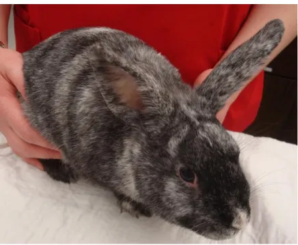
Figure 3 – Upright eared rabbit. ... 3
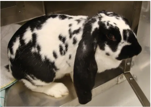
Figure 4 – Lop-eared rabbit. ... 4
Figure 5 – Diagram of the mammalian ear (adapted from McLaughlin & Chiasson, 1979). ... 5
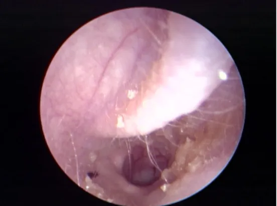
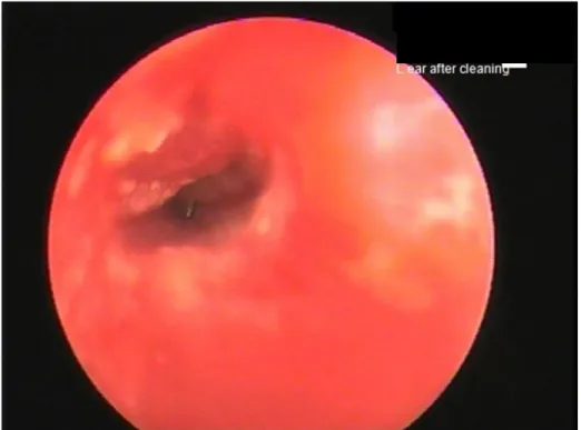
Figure 6 – Normal ear canal of a rabbit... 9
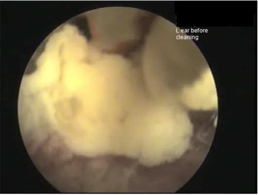
Figure 7 – Ear canal of a rabbit with otitis externa with accumulation of purulent material within the canal. ... 10
Figure 8 – Ear canal of a rabbit with otitis externa. ... 11
Figure 9 – Right side facial nerve paralysis in a rabbit with otitis media. ... 12
Figure 10 – Contracture of muscles of the left side of the face in a rabbit with chronic left facial nerve paralysis secondary to otitis media. ... 13
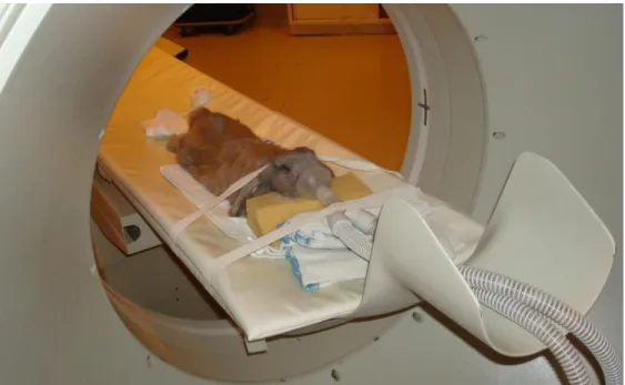
Figure 11 – General anesthesia for head CT in a rabbit positioned in ventral recumbence. ... 19
Figure 12 - Single slice axial CT unit Picker PQS. ... 19
Figure 13 - Sixteen slice helical CT unit Toshiba Acquilion LB. ... 20
Figure 14- CT images of a rabbit with dental disease and otitis externa ... 24
Figure 15 - CT images of a rabbit with Abscess/dental disease without ear lesions ... 25
Figure 16 – CT images of a rabbit with upper respiratory disease and otitis externa. ... 26
Figure 17 – CT images of a rabbit with a soft tissue mass head region and otitis externa ... 27
Figure 18- CT images of a rabbit with otitis externa and media ... 30
Figure 19 - CT images of a rabbit with otitis externa and media ... 31
Figure 20 – CT images of a rabbit with otitis externa/media and an otolith ... 32
Figure 21- CT images of a rabbit with progressive otitis media ... 34
Index-Tables
Table 1 – Prevalence of TB attenuation, increased thickness and lysis for rabbits in group 1 and group 2 ... 33
Table 2 - Prevalence of middle, external and inner ear CT changes in rabbits in group 1 and group 2. ... 36
Index- Charts
Chart 1- Total number and percentage of rabbits presented for evaluation of ear disease and/or signs that suggest presence of ear disease (group 1) and in rabbits presented for evaluation of other conditions not related to or suggestive of ear disease (group 2). ... 23
Chart 2- Distribution of rabbits in group 2 by reason for head CT. ... 23
Abbreviations
CT- Computed Tomography
CUHA- Cornell University Hospital for Animals MRI- Magnetic Resonance Imaging
TB- tympanic bulla SD- standard deviation
1 - Introduction
Domestic rabbits (Oryctolagus cuniculus) belong to the order Lagomorpha, family Leporidae (Vella & Donnelly, 2012). Although rabbits are traditionally used in biomedical research and as a source of meat or fur, the species is becoming increasingly popular as pets (Shepherd, 2008). As a result, rabbits are often presented to veterinarians with owners seeking and requesting advanced diagnostic and treatment options.
1.1 - Anatomy of the rabbit ear
The ear consists of 3 distinct anatomical and functional components: the external, the middle and the inner ear. The external ear consists of the pinna and the external acoustic meatus (Barone & Simoens, 2010). Rabbits’ pinna is well developed and is supported by the large auricular cartilage (Figure 1). The pinna is used for thermoregulation and visual communication, in addition to sound collection (Chitty & Raftery, 2013; King, Hall, Cranfield, & Sullivan, 2007). The external acoustic meatus receives and concentrates air vibrations, transmitting them to the middle ear. The meatus is vertically oriented in rabbits, with no horizontal component. It is supported by a large and complex annular cartilage and the acoustic meatus cartilage (Figure 1). A smaller, dorsally placed scutiform cartilage is also present. The acoustic meatus cartilage interlocks with the vertical bony acoustic duct arising from the tympanic bulla (TB) (Barone & Simoens, 2010; Chitty & Raftery, 2013). The duct is well-developed in rabbits, as opposed to the short bone canal of the dog and the bone ring of the cat. The bony tube mean length is 0.76 cm+/-0.04 and width is 0.65 cm +/-0.06 cm, and extends caudo-dorsally from the lateral aspect of the TB (Figure 2) (King et al., 2007; King, Cranfield, Hall, Hammond, & Sullivan, 2010a; King, Cranfield, Hall, Hammond, & Sullivan, 2010b).
Figure 1 – Rabbit ear anatomy (adapted from Barone & Simoens, 2010).
Figure 2 – Tympanic bulla of the rabbit: 1- external acoustic porus, 2-
external acoustic meatus, 3 tympanic bulla, 9- mastoid process (adapted
from Popesko, Rajtova and Horak, 1992).
The external acoustic meatus is delineated by an irregularly shaped tragus (Figure 1) (Chitty & Raftery, 2013). In upright eared rabbits (Figure 3), the external ear cartilages interdigitate and form a ridged structure and vertical ear canal. Lop-eared rabbits (Figure 4) have a 3-5 mm separation between the cartilage of the acoustic meatus and the tragus, which results in folding of the ear and closure of the external acoustic meatus (Chitty & Raftery, 2013). The pendulous ear seen in certain dog breeds and one cat breed (Scottish Fold) results from folding of the external ear at the level of the distal pinna/auricular cartilage (Evans & de Lahunta, 2013; Heine, 2004; Hudson, 2010). The closure of the meatus in lop-eared rabbits can cause excessive accumulation of cerumen proximal to the fold (Chitty & Raftery, 2013), similar to what is described in dogs and cats with obstructive ear disease (Glaze, 2013).
Figure 4 – Lop-eared rabbit.
The middle ear is formed by an air-filled cavity within the temporal bone, connected to the nasopharynx via the auditory tube and closed to the external acoustic meatus by the tympanic membrane (Barone & Simoens, 2010). The rabbit tympanic membrane is elliptical with a near vertical long axis (5.75 mm) and a near horizontal minor axis (5 mm) (Mayer, 2011). The membrane is relatively thin when compared with the tympanic membrane of dogs and cats, and has a translucent blue coloration on examination (Chitty & Raftery, 2013).
The basic anatomy of the rabbit tympanic bulla is similar to the dog and cat (King et al., 2007; King et al., 2010a; King et al., 2010b). The rabbit TB is located on the ventral aspect of the temporal bone (Figure 2). It presents a smooth convex outer bone surface, with mean dimensions of 1.13+/- 0.06 cm long, 0.9+/-0.04 cm wide and 0.96+/-0.08 cm deep, which results in a relatively rounder appearance (King et al., 2007). The TB bone wall is thinnest ventrally and thickest laterally, the latter due to the presence of a smooth internal bone rim between the external acoustic meatus and the TB wall (present in dogs and cats) and a roughened external bone rim (exclusive to rabbits) (King et al., 2007). The rabbit TB wall lacks the bone ridge present between the TB wall and the petrous temporal bone found in dogs (King et al., 2007; King et al., 2010b). The mastoid and paracondylar or jugular processes, well developed in the
rabbit, extend ventral to the caudolateral aspect of the TB (King et al., 2007; King et al., 2010a; King et al., 2010b).
Three auditory ossicles (malleus, incus and stapes) are present within the cavity (Figure 5). These ossicles connect the tympanic membrane to the oval window and are responsible for transmitting the air vibrations from the external to the inner ear (Barone & Simoens, 2010). The facial nerve emerges from the stylomastoid foramen and runs along the lateral aspect of the TB (Chitty & Raftery, 2013).
Figure 5 – Diagram of the mammalian ear (adapted from McLaughlin &
Chiasson, 1979).
The rabbit middle ear is lined by ciliated columnar epithelium, with simple squamous epithelium over the tympanic membrane and auditory ossicles (Flatt, Deyoung, & Hogle, 1977).
The rabbit inner ear structure and function is similar to other domestic mammals. It comprises the semicircular canals and the cochlea, lodged within the temporal bone and with two membranous surfaces at the base (oval window and round window- Figure 5). The inner ear is responsible for hearing and balance (Barone & Simoens, 2010).
1.2 -
Pathophysiology of Otitis Media in Rabbits
Otitis media, or inflammatory disease of the middle ear, is commonly reported in rabbits (Chitty & Raftery, 2013; Flatt et al., 1977; Oglesbee, 2006b; Snyder, Fox, & Soave, 1973). Most infections are caused by Pasteurella multocida, with other organisms such as Staphylococcus
aureus, Streptococcus species, Pseudomonas aeruginosa, Bordetella bronchiseptica, Escherichia coli and Proteus mirabilis less frequently reported (Deeb, DiGiacomo, Bernard, & Silbernagel,
1990; DiGiacomo, Garlinghouse, & Van Hoosier, 1983; Fisher, 2012; Flatt et al., 1977; Fox, Norberg, & Myers, 1971; Kunstyr & Naumann, 1985; Smith & Webster, 1925; Snyder et al., 1973; Snyder, Fox, Campbell, & Soave, 1976).
The primary infection is believed to result from bacteria within the nasopharynx reaching the middle ear via the auditory tube (Flatt et al., 1977; Harcourt-Brown, 2002b; Smith & Webster, 1925). This pathogenesis is supported by the fact that most infections are caused by Pasteurella
multocida, a respiratory pathogen (Deeb et al., 1990; DiGiacomo et al., 1983; Fisher, 2012; Flatt
et al., 1977; Fox et al., 1971; Kunstyr & Naumann, 1985; Smith & Webster, 1925; Snyder et al., 1973). A high prevalence of middle ear disease is reported in rabbits with concurrent upper respiratory infections (78% (Smith & Webster, 1925) and 85% (Deeb et al., 1990)).
Disease etiology in rabbits, as for other species, is likely to be multifactorial. Factors such as pathogenicity of the organisms involved, concurrent disease and/or infection, interaction between the microrganisms and the host immune system and anatomical variations may all play a role in the development of otitis media in rabbits. Viral upper respiratory tract infections in humans appear to predispose for development of otitis media. Inflammation of the mucosa associated with the infection creates conditions that promote bacterial colonization and adhesion to the cells. The normal Eustachian tube function and pressure regulation mechanisms are affected, which allows invasion of the middle ear (Qureishi, Lee, Belfield, Birchall, & Daniel, 2014; Sismanis, 1991). The predisposing effect of viral infections in the development of bacterial otitis media has been shown in chinchilla middle ear disease model studies (Brockson et al., 2012; Giebink, Ripley, & Wright, 1987). The type and/or pathogenicity of the organism involved may also affect the outcome of infection. In an experimental otitis media study in rabbits,
translocate from the nasopharynx to the middle ear and cause disease (Usviatsov & Dolgov, 2009).
Similarly, chronic inflammation associated with allergies is considered as a common cause of otitis media in children (Sismanis, 1991). This association has also been reported in dogs (Glaze, 2013). Chronic inflammation associated with allergic rhinitis/sinusitis in rabbits could initiate and maintain the hyper-secretory state of the middle ear mucosa and resulting chronic otitis media (Ovesen, Paaske, Blegvad, & Elbrond, 1992; Schousboe, Rasmussen, & Ovesen, 2001). The microflora of the oropharynx would be not only important in the development of infection but also for maintenance of disease within the middle ear (Ovesen et al., 1992).
Craniofacial malformations of the skull in humans have been associated with otitis media by direct effect of Eustachian tube function or indirectly via dysfunction of the tensor veli palatine muscle (Sismanis, 1991). This association is also well described in brachycephalic dogs (Owen, Lamb, Lu, & Targett, 2004). Suppurative otitis media can be experimentally induced in rabbits by altering the auditory tube function without the inoculation of bacteria (Ovesen et al., 1992; Schousboe et al., 2001). The altered conformation of the skull described in some pet rabbit breeds, reported to predispose those rabbits to dental and nasolacrimal duct problems (Meredith, 2006) may also result in altered function of the auditory tube and predisposition to otitis media. Otitis media also develops secondarily to bacterial or parasitic otitis externa (Bjotvedt & Geib, 1981; Flatt et al., 1977; Kumar, Rajashekar, & Rao, 2000; Oglesbee, 2006a). Otitis externa is an important predisposing factor for development of otitis media in dogs, reported to occur in 50-80% of dogs with chronic otitis externa (Cole, Kwochka, Kowalski, & Hillier, 1998). The incidence of otitis media in rabbits with otitis externa is unknown. Otitis externa occurs more commonly in lop-eared rabbits, possibly due to abnormal conformation of the external ear canal described above (Oglesbee, 2006a). The development of otitis externa in lop-eared rabbits could be due to cerumen build-up as seen in dogs and cats with otitis externa secondary to obstructive ear disease, and/or excessive moisture within the ear canal, as for otitis externa in pendulous-eared dogs.
Other causes of middle ear disease in dogs and cats (polyps, neoplasia, cholesteatomas) have not been reported in rabbits (King et al., 2007).
1.3 - Clinical Signs of Otitis Media
Otitis media can be difficult to diagnose in live rabbits, as affected rabbits are often asymptomatic unless disease is associated with otitis externa or interna (Chow, 2011; Eatwell et al., 2013; Flatt et al., 1977; Harcourt-Brown, 2002a; Smith & Webster, 1925). Rabbits with otitis externa may be subclinical or present signs similar to other animals with this condition, including head shaking, ear scratching, malodorous ears and/or holding the affected ear(s) down for rabbits with upright ears (Oglesbee, 2006a).
Middle ear infection can spread to the inner ear causing vestibular disease due to labyrinthitis, or through the internal acoustic meatus and along the vestibulocochlear nerve to the brain, resulting in severe neurologic signs due to encephalomyelitis (Fisher, 2012; Fox et al., 1971; Gruber, Pakozdy, Weissenbock, Csokai, & Kunzel, 2009; Harcourt-Brown, 2002b; Kunstyr & Naumann, 1985; Smith & Webster, 1925). Otogenic intracranial infection is reported to be rare in humans, (Kangsanarak, Navacharoen, Fooanant, & Ruckphaopunt, 1995), dogs and cats (Sturges et al., 2006). However, it is reported to be relatively common in rabbits, affecting as much as 50% of rabbits with otitis media (Gruber et al., 2009; Kunstyr & Naumann, 1985).
1.4 - Diagnosis
Examination of the ears should be routinely performed in rabbits presented for health examination and for evaluation of clinical signs that may suggest ear disease. The exam should start with evaluating the pinna for asymmetry, skin lesions, masses or evidence of discharge. The base of the ears should be palpated for asymmetry or evidence of pain upon manipulation of the area. The ear canal should be examined for presence of purulent discharge, excessive ear wax, mites, inflammation, stenosis or any other abnormalities (Figures 6 and 7) (Chitty & Raftery, 2013; Oglesbee, 2006a). The deep location of the tympanic membrane (ventro-medial to the interdigitation of the cartilagenous and bony external acoustic meatus) makes visualization difficult during conscious routine ear exam (Chitty & Raftery, 2013). Sedation or general anesthesia is often required for complete otoscopic and/or endoscopic examination of the ear canal and visualization of the tympanic membrane. Rabbits with otitis media may present a bulging and discolored (white) tympanic membrane due to the accumulation of purulent material
within the tympanic bulla (Capello, 2004). The presence of a ruptured tympanic membrane supports the diagnosis of otitis media. If the tympanic membrane is intact and visible and otitis media is suspected, or when discoloration and/or bulging of the membrane are/is present, a myringotomy may be performed for sample collection from the middle ear for cytology and microbiology. This procedure will also allow drainage of the middle ear (Cole, 2004).
Figure 6 – Normal ear canal of a rabbit; note the minimal ear wax (bottom
right).
Figure 7 – Ear canal of a rabbit with otitis externa with accumulation of
purulent material within the canal (photo courtesy of Dr. Mike Carey).
Although examination of the external ear canal and tympanic membrane has been proposed for diagnosis of subclinical otitis media, neither the presence of a normal tympanic membrane nor a normal external ear canal excludes otitis media (Flatt et al., 1977; Oglesbee, 2006a; Smith & Webster, 1925). As many as 72% of dogs with otitis media present intact tympanic membranes on otoscopic examination (Cole, 2004). In addition, examination of the tympanic membrane can be challenging for anatomical reasons, species variation (lop-eared rabbits) and when otitis externa is present (Capello, 2004; Chow, 2011).
Exudate within the ear canal should be collected and evaluated cytologically for presence of inflammatory cells, bacteria and/or yeast, and samples collected for bacterial and/or fungal culture if indicated. When the tympanic membrane is ruptured, it may be difficult to determine if the purulent sample within the ear canal is representative of disease process of the middle ear or external ear, i.e., if the otitis media is the primary disease or secondary to progressive otitis externa (Chow, 2011).
Aggressive ear cleaning to facilitate examination of the tympanic membrane can result in iatrogenic (further) irritation of the ear canal and/or rupture of the tympanic membrane (Figure 8).
Figure 8 – Ear canal of a rabbit with otitis externa; note the swelling and
irritation of the ear canal secondary to aggressive flushing and cleaning
(photo courtesy of Dr. Mike Carey).
Electrophysiological tests such as the brainstem auditory evoked response (BAER) can be used to assess to earing and assist in the diagnosis of ear disease in dogs (Scheifele & Clark, 2012). This test has been used experimentally in rabbits (Pettigrew & Morey, 1987), but its use for assisting in the diagnosis of middle ear disease in this species has not been investigated.
For rabbits presenting with vestibular disease, a neurologic exam should be performed to determine if disease is peripherally or centrally located. Rabbits with otitis interna present with clinical signs of peripheral vestibular disease, including head tilt, nystagmus, ataxia and/or
side infection (Kunstyr & Naumann, 1985). Facial nerve paralysis and Horner’s syndrome can also occur with otitis media and interna in rabbits (Figure 9) (Oglesbee, 2006b). With chronic facial nerve paralysis, fibrosis of the denervated muscles results in contracture of the affected side of the face (Figure 10) (Eatwell et al., 2013; Oglesbee, 2006b). Concurrent head tremors, proprioceptive deficits and hypermetria suggest central vestibular disease (Keeble, 2006b). The main differential diagnosis for rabbits presenting with vestibular signs is Encephalitozoon
cuniculi, a microsporidium parasite that can cause encephalitis (Fisher, 2012; Harcourt-Brown,
2002b). Otitis media/interna is commonly associated with signs of peripheral vestibular disease, while Encephalitozoon cuniculi causes central vestibular disease (Fisher, 2012). However, in cases of otogenic intracranial infection, signs of central vestibular disease may be preceded by peripheral vestibular disease signs, making distinction difficult (Harcourt-Brown, 2002b; Sturges et al., 2006). Even though serologic tests are available for Pasteurella multocida and
Encephalitozoon cuniculi that could assist in differentiating peripheral from central vestibular
disease, a positive titer reflects exposure and does not correlate with active disease for either organism (Fisher, 2012; Harcourt-Brown, 2002b).
Figure 9 – Right side facial nerve paralysis in a rabbit with otitis media.
Figure 10 – Contracture of muscles of the left side of the face in a rabbit with
chronic left facial nerve paralysis secondary to otitis media.
Due to the difficulties in diagnosing otitis media on physical exam and differentiating peripheral from central vestibular disease, and the relatively high prevalence of otogenic intracranial infection in rabbits, diagnostic imaging is important for evaluation of the middle ear and early identification of disease.
Radiography is most commonly used since it is readily available (Harcourt-Brown, 2002b; Keeble, 2006a). The normal radiographic anatomy of the rabbit has been described (King et al., 2010a; King et al., 2010b). As for dogs and cats, a minimum of 3 to 4 projections (dorsoventral view and 2 to 3 oblique views) are needed to evaluate the rabbit’s middle ear (Hammond, Sullivan, Posthumus, & King, 2010). The open mouth rostrocaudal projection cannot be used in rabbits since the limited angle of jaw opening results in superimposition of the tympanic bulla with the dental arcades (Hammond et al., 2010). Radiographic changes associated with otitis
Schwarz, 2003; Harcourt-Brown, 2002b; Remedios, Fowler, & Pharr, 1991). Since significant radiographic changes may not be visible for several weeks, false-negative results can occur with acute otitis media (Harcourt-Brown, 2002b; Rohleder et al., 2006). In most cases, otitis interna cannot be diagnosed on routine radiography. However, radiographic evidence of otitis media may support diagnosis of otitis interna in animals with signs of peripheral vestibular disease (Shepherd, 2008).
Routine radiography in dogs is considered highly specific but moderately sensitive for the diagnosis of middle ear disease, as 25% of the patients with otitis media in surgery presented no radiographic changes (Remedios et al., 1991). Although the accuracy of radiography for detection of fluid in the TB of rabbit cadavers was high (77-80%) (Hammond et al., 2010), there are no studies investigating sensitivity and specific of radiography for diagnosis of otitis media in clinical cases. In addition, the obliquity required for evaluation of the middle ear in rabbit cadavers may be difficult to achieve in clinical patients, with false-positive results possible due to superimposition of osseous structures on radiographic images (Hammond et al., 2010).
The use of contrast radiography has been suggested for evaluation of the integrity of the tympanic membrane and outline of the TB. However, the presence of thick exudate or pus in the ear canal and/or narrowing of the lumen seen in cases of otitis externa may cause false negative results (Chitty & Raftery, 2013).
Ultrasonography can be used to evaluate the middle ear. This imaging modality is readily available, is well tolerated, does not require sedation or anesthesia. In addition, it is both sensitive and specific in detecting presence of fluid within the TB, a common feature of acute otitis media (Griffiths, Sullivan, O'Neill, & Reid, 2003). The technique and normal ultrasonographic appearance of the rabbit TB has been described and its efficacy for detection of fluid within the tympanic membrane investigated in a rabbit cadavers (King et al., 2007; King, Posthumus, Hammond, & Sullivan, 2012). However, the use of this technique for imaging the middle ear is limited by acoustic shadowing and reverberation artifact effect. In addition, there are currently no reports of use of ultrasound for evaluation of clinical cases of otitis media in rabbits.
Computed tomography (CT) is considered the “gold standard” for evaluation of the middle ear in dogs and cats (Bischoff & Kneller, 2004; Doust et al., 2007; Garosi et al., 2003). Although radiography continues to be more accessible, CT units are becoming more readily accessible and
not exclusive to Universities, with many referral specialty hospitals offering this imaging modality. This imaging modality allows evaluation of the external, middle and inner portions of the ear, in addition to other clinically relevant structures of the rabbit head, including teeth and upper respiratory tract (Keeble, 2006a; Knipe, 2007; Van Caelenberg et al., 2010). Because of the reduced superimposition of bony structures and ability to window and level images to emphasize different tissues, abnormalities of the middle ear are more apparent with this modality when compared with radiography, allowing early diagnosis of subtle changes (Bischoff & Kneller, 2004) and more accurate prognosis and treatment planning (Garosi et al., 2003; Stieve-Caldwell, Morandi, Souza, & Adams, 2009). CT is considered superior to magnetic resonance imaging (MRI) for evaluating the bone structures of the middle and inner ear (Garosi et al., 2003). CT was reported to be more sensitive than and as specific as radiography for diagnosing and characterizing the severity of middle ear disease in dogs (Rohleder et al., 2006). The normal CT appearance of the rabbit ear has been described (Van Caelenberg et al., 2010). In a rabbit cadaver study, single slice CT was more accurate than ultrasound and radiography in detecting the presence of fluid within the tympanic bulla (King et al., 2012).
CT findings in dogs and cats with otitis media include thickening, irregularity, proliferation and/or lysis of the TB wall. In addition, a fluid/soft tissue density may be seen within the lumen of the bulla (Bischoff & Kneller, 2004; Garosi et al., 2003; Shanaman, Seiler, & Holt, 2012). The use of thin slices, bone algorithm and viewing at a window width greater than 2,000 CT numbers is recommended to prevent bulla thickening artifact created by the presence of fluid within the tympanic bulla (Barthez, Koblik, Hornof, Wisner, & Seibert, 1996). CT changes associated with otitis externa include mineralization of the ear canal, narrowing of the lumen of the external ear canal, and soft tissue density material in the lumen of the ear canal. Otitis interna is difficult to diagnose using CT unless it is associated with severe destruction of the inner ear bony structures (Bischoff & Kneller, 2004).
The use of CT for diagnosis of otitis media in rabbits has been described in the review articles (Chow, 2011; Keeble, 2006a) and book chapters (Fisher, 2012; Veraa & Schoemaker, 2013), with limited references in clinical cases (Eatwell et al., 2013; Stieve-Caldwell et al., 2009). Magnetic resonance imaging is considered superior to CT in evaluation of the soft tissues of the
Lucas, & Schmitz, 2000; Bischoff & Kneller, 2004; Dvir, Kirberger, & Terblanche, 2000; Garosi et al., 2003; Van Caelenberg et al., 2011). MRI findings in cases of otitis media include medium signal intensity material in the TB on T-1 weighted images and hyperintense on T2-weighted images. Enhancement of the inner margin of the TB can be seen on T1-weighted images with contrast agent administration. Narrowing of the external ear canal, increased signal intensity and thickening of the canal wall on T1- and T2-weighted images can be seen on MRI images in cases of otitis externa. The lack of signal intensity from the intralabyrinth fluid on T2-weighted images can be seen with otitis interna, although this can also be seen in normal ears. Administration of gadolinium-DTPA contrast material results in meningeal enhancement on T1-weighted images in cases of otitis interna (Bischoff & Kneller, 2004). Low field MRI appearance of the normal rabbit ear has been described (Van Caelenberg et al., 2011).
The prolonged anesthesia time required for examination, the limited availability of equipment and financial constraints still limit use of MRI routinely in rabbits (Van Caelenberg et al., 2011). The use of MRI for evaluation of the inner ear and central nervous system in rabbits with vestibular disease has not been reported.
Other causes of middle ear disease in dogs and cats (polyps, neoplasia, cholesteatomas) for which MRI would be useful/better than CT have not been reported in rabbits (King et al., 2007).
1.5 - Specific Aims of the Study
Postmortem studies investigating subclinical otitis media in rabbits report the prevalence ranging from 11.5% (Deeb et al., 1990) to 32% (Flatt et al., 1977; Smith & Webster, 1925). Different studies report a variation in the prevalence of otitis media in rabbits with vestibular disease: 5.5% in a postmortem study of rabbits with neurologic disease (Gruber et al., 2009), 24% in a
Encephalitozoon cuniculi seroprevalence study (Jeklova et al., 2010), and 63% in a postmortem
study of rabbits with vestibular disease (Kunstyr & Naumann, 1985). Most of these studies report prevalence of subclinical and clinical middle ear disease at necropsy and in meat rabbits or research rabbits, not antemortem or in domestic rabbits. Therefore, the first aim of this study was to determine the prevalence of clinical and subclinical middle ear disease in a group of domestic rabbits that underwent head CT.
Due to the often subclinical presentation of otitis media, the difficulty in differentiating peripheral from central vestibular disease, and the relatively high prevalence of otogenic intracranial infection in rabbits when compared to other species, diagnostic imaging is important for evaluation of the middle ear and early identification of disease. Computed tomography allows cross-sectional evaluation of the entire head, including teeth and upper respiratory tract, without superimposition of structures (Bischoff & Kneller, 2004; Doust et al., 2007; Garosi et al., 2003; Keeble, 2006a; Knipe, 2007; Van Caelenberg et al., 2010). The use of CT for diagnosis of otitis media in rabbits has been described in the literature (Chow, 2011; Eatwell et al., 2013; Fisher, 2012; Keeble, 2006a; Stieve-Caldwell et al., 2009; Veraa & Schoemaker, 2013). However, the information is descriptive and limited to CT changes in cases of clinical otitis media. Therefore, the second aim of this study is to describe and compare CT abnormalities of the middle ear in rabbits with clinical and subclinical middle ear disease.
Although predisposing factors (upper respiratory disease, otitis externa and lop-ear position) and mechanisms of infection are described in the rabbit literature, the relative importance of each for development of otitis media in rabbits is unknown. Because there are multiple possible causes of otitis media in the rabbit that are not fully understood, the third aim of this study was to evaluate the frequency of proposed predisposing factors of otitis media (upper respiratory disease, otitis externa and lop-ear position) in rabbits with subclinical and clinical otitis media, and in rabbits with and without otitis media.
To address these three study aims, multiple hypothesis were investigated. First, it was hypothesized that CT signs of otitis media is a common finding in rabbits that undergo head CT for reasons other than ear disease. Second, it was hypothesized that CT findings of middle ear disease would not be different in rabbits with clinical and subclinical otitis media. Third, it was hypothesized that the frequency of predisposing factors to otitis media would occur more commonly in rabbits with clinical and subclinical otitis media than in rabbits without otitis media.
2 - Materials and Methods
2.1 - Case selection
The medical records for rabbits that were presented to Cornell University Hospital for Animals (CUHA) from June 2007 to February 2014 were searched for patients that had a head CT scan performed. Information collected from the medical record included age, weight, gender, ear position, past medical history, reason for visit, and physical exam findings. Rabbits were assigned to one of two groups based on the reason for visit and head CT. Group 1 included rabbits that were presented for evaluation of clinical otitis externa (head shaking, pinnae pruritus, malodorous ears and/or holding the affected ear(s) down for rabbits with upright ears) or for evaluation of signs that suggested the presence of middle ear disease (signs of vestibular disease, facial nerve paralysis). Group 2 included rabbits that were presented for evaluation of conditions not related to ear disease. These conditions included dental disease, upper respiratory disease, and other conditions of the head region, such as ocular/periocular disease, and/or presence of soft tissue or bone mass/masses.
2.2 - Imaging
All head CT examinations were performed under sedation (ketamine 10 mg/kg and diazepam 0.5 mg/kg IV) and/or general anesthesia (isoflurane, via face mask; Figure 11). CT scans were obtained using one of two units: a single slice CT unit (Picker PQS Picker International, Cleveland, OH, USA; Figure 12) for cases examined from August 2008 to July 2009, or a 16 slice helical CT unit (Toshiba Acquilion LB, Toshiba America Medical Systems Inc., Tustin, CA, USA; Figure 13) for cases examined from August 2009 to March 2014. Scans were acquired in 1 or 2 mm contiguous transverse slices with kVp of 120 and automatic MaS, with rabbits positioned in ventral recumbency. Most scans were performed using a soft tissue and bone algorithm (in 72 of the 88 exams performed), with a few performed using a bone algorithm only (15 out of 88) or a soft tissue algorithm only (1 out of 88). Images were displayed in 1-2mm contiguous slices in bone and soft tissue windows.
Figure 11 – General anesthesia for head CT in a rabbit positioned in ventral
recumbence.
Figure 13 - Sixteen slice helical CT unit Toshiba Acquilion LB.
2.3 - Image evaluation
The external, middle and inner portions of the ear were evaluated for each case by two radiologists. Image evaluation was performed in chronologic order of CT exam. Reviewers were blinded to the group assignment, reason for head CT exam, and to the other reviewer’s interpretation of ear CT abnormalities. Once the image review process was completed, inter-reviewer discrepancies were addressed with collaborative image evaluation during which both reviewers agreed on a single diagnosis. Studies were interpreted on a commercial picture archiving and communication system (Carestream Vue PACS, Carestream Health Inc., Rochester, NY, USA) with multiple planes and windows available. The external ear was evaluated for increased attenuation within the canal, thickness of the wall of the external ear canal, mineralization of the auricular cartilages and presence of a mass or masses within or associated with the external ear canal. The middle ear was evaluated for the presence of
attenuating material within the tympanic cavity and/or increased thickness, irregularity, expansion or lysis of the tympanic cavity wall. The inner ear was evaluated for sclerosis or lysis of the petrous temporal bone and/or loss of cochlear semicircular canal definition. CT lesions of the external, middle and inner ear were categorized as absent or present. In addition, the presence of abnormalities in the upper respiratory tract, teeth and other significant CT findings within the head region were recorded.
2.4 - Follow up
Information regarding the progression of clinical and subclinical cases of middle ear disease was obtained by follow up visits at CUHA and/or telephone or electronic mail contact with the owners and/or referring veterinarians. When available, follow up CT exam ear findings were also characterized as described above and compared with the initial CT findings.
2.5 - Data analysis
Descriptive statistics methods were used for data analysis and calculated using a spread sheet software for numerical data (Excel 2013, Microsoft Corporation, Redmond, WA, USA)and an online data analysis program for categorical data (http://graphpad.com/quickcalcs/contingency1/; Graph Pad Software, La Jolla, CA, USA).Numerical data (age, weight) was described using mean and standard deviation. Categorical data (presence/absence of CT changes of the external, middle or inner ear; unilateral/bilateral disease; type of lesion present in the middle ear, ear position, and upper respiratory disease) was described as the frequency of occurrence of each category by group. The Χ2 or Fisher exact tests, depending on the sample size, was used to determine the association between the type of CT middle ear lesion and reason for visit (group 1 and 2) and within group 2, between rabbits with that remained subclinical and rabbits that eventually develop clinical middle ear disease. These same tests were used to determine the association between middle ear disease (clinical/subclinical and presence/absence) and the proposed middle ear predisposing factors (lop ear position, otitis externa, upper respiratory
3 - Results
3.1 - Case Selection
A total of 88 of 784 rabbits met the inclusion criteria established. Twenty one rabbits (24%) were presented for evaluation of ear disease and/or signs that suggest presence of ear disease (group 1; Chart 1). Presenting complaints included clinical otitis externa (6), facial nerve paralysis (3), and signs of vestibular disease (20). The group consisted of 9 males (43%) and 12 females (57%). These rabbits had a mean SD age of 4.4 2.6 years (range, 0.1 to 8 years) and mean weight of 1.9 ± 0.92 kg (range, 0.6 to 4.9 kg). Sixty seven rabbits included in the study (76%) were presented for evaluation of other conditions not related to or suggestive of ear disease (group 2; Chart 1 and 2), including dental disease (33; Figure 14 and 15), upper respiratory disease (18; Figure 16) and other conditions involving the skull/head (16; Figure 17). The group consisted of 47 males (70%) and 20 females (30%). Rabbits in group 2 had a mean age of 4.9 ± 2.51 years (range, 0.2 to 11.6 years) and mean weight of 2.26 ± 0.9 kg (range, 0.7 to 5.3 kg).
Chart 1- Total number and percentage of rabbits presented for evaluation of
ear disease and/or signs that suggest presence of ear disease (group 1) and in
rabbits presented for evaluation of other conditions not related to or
suggestive of ear disease (group 2).
Chart 2- Distribution of rabbits in group 2 by reason for head CT (total
number and percentage).
Figure 14- CT images of a rabbit with dental disease and otitis externa.
Sagittal (A) and transverse (B, C) CT images of a 4 year old female spayed lop-ear rabbit with chronic dental disease. The images are displayed using a bone algorithm/window and in 1 mm contiguous slices. All cheek teeth contain variable lucencies and are moderate-to-severely malaligned with an irregular occlusion surface (Figure A). The right lower fourth premolar contains a chronic fracture with the root displaced rostral and lateral to the lower third premolar (Figure A). In addition, moderate points on the lingual surface of the lower cheek teeth and buccal surface of the upper cheek teeth can be seen (Figure B). There is an increased attenuation within both external ear canals (Figure C).
Figure 15 - CT images of a rabbit with Abscess/dental disease without ear
lesions.
Sagittal (A), dorsal (B) and transverse (C, D) CT images of a 2 year old male intact rabbit with a rostral facial mass and difficulty breathing. The images are displayed using a bone algorithm/window and in 1 mm contiguous slices. The surface of the nose is severely swollen dorsally. The swelling extends deep into the right nasal cavity as a very large, locally extensive, soft tissue attenuating mass with severe destruction of the nasal and facial bones. Many poorly defined mineral opacities are interspersed within the soft tissue mass (Figure A-C). The right upper first incisor alveolus is severely expanded and the tooth root is surrounded by a thick rim of soft tissue and a thin incomplete rim of bone. The right lower incisor is missing (Figure C). The external, middle and inner ears are normal (Figure A and D).
Figure 16 – CT images of a rabbit with upper respiratory disease and otitis
externa.
Dorsal (A) and transverse (B, C) CT images of a 1 year old female spayed lop-ear rabbit with active left-sided nasal discharge. The images are displayed using a bone algorithm/window and in 1 mm contiguous slices. The left side of the nasal cavity contains a severe amount of soft tissue attenuating material that extends from the upper incisors to the cribiform plate and extends into the left nasopharynx (Figure A). At the level of the left upper first premolar there is a large mass effect that disrupts and extends through the left maxilla causing a bulge on the left muzzle. The mass causes destruction of the nasal conchae with no deviation of the nasal septum (Figure B). The left external ear canal contains soft tissue attenuating material, and the right contains minimal material (Figure C).
Figure 17 – CT images of a rabbit with a soft tissue mass head region and
otitis externa.
Transverse (A, B, D) and dorsal (C) CT images of a 1 year old male intact rabbit with a mass on the left side of the face. The images are displayed using a bone (A, D) or soft tissue (B, C) algorithm/window and in 1 mm contiguous slices. In left cheek, from the level of the orbit to the ear, there is a large, soft tissue attenuating mass (Figure A-C). The left ear contains a mild amount of soft tissue attenuating material (Figure D).
3.2- Prevalence of middle ear CT changes
Twelve of the 21 rabbits in group 1 (57%) had CT abnormalities of the middle ear (Chart 3). Among the 20 rabbits with signs of vestibular disease in group 1, 11 (55%) demonstrated CT changes consistent with middle ear disease. The other 9 (45%) showed no CT changes in the external, middle or inner ears and were diagnosed with presumptive central vestibular disease. Eighteen of the 67 rabbits in group 2 (27%) showed CT abnormalities of the middle ear (Chart 3).
Chart 3- Prevalence of CT changes
of the middle ear disease in rabbits
presented for evaluation of ear disease and/or signs that suggest presence of
ear disease (group 1) and in rabbits presented for evaluation of other
conditions not related to or suggestive of ear disease (group 2).
3.3- Characterization of CT abnormalities of the middle ear
For group 1 rabbits with CT changes of the middle ear, an equal number of rabbits (6) showed unilateral and bilateral changes, for a total of 18 affected ears. All affected ears displayed increased attenuation within the tympanic cavity; of the 18 ears, 10 had lysis of the bulla (prevalence, 56%) and 3 had thick tympanic bulla (prevalence, 17%) (Figure 18-20).
The 3 rabbits in group 1 with facial nerve paralysis had CT changes of the middle ear. One rabbit had middle ear changes ipsilateral to facial nerve paralysis and no vestibular signs. The other two rabbits had bilateral middle ear changes as well as head tilt and facial nerve paralysis ipsilateral to inner ear CT changes. The type and severity of middle ear CT changes in rabbits with facial nerve paralysis were similar to the other vestibular rabbits in group 1 without facial nerve paralysis.
Five of the vestibular rabbits with CT changes of the middle ear had unilateral disease and ipsilateral vestibular signs. The remaining six vestibular rabbits showed bilateral middle ear disease. Vestibular signs were ipsilateral to the side with more severe middle ear CT lesions (2 cases) or to the side with inner ear CT changes (2 cases). The two remaining rabbits with vestibular signs had bilateral middle and inner ear disease. Despite similar CT abnormalities and severity of disease for both middle and inner ears, these rabbits demonstrated unilateral vestibular signs.
For group 2, unilateral middle ear disease was identified in 10/18 rabbits (55%) and bilateral disease by 8/18 rabbits (45%), for a total of 26 affected ears. As for group 1, all affected middle ears of rabbits in group 2 had increased attenuation within the tympanic cavity. The prevalence of bulla lysis was 19% (5/26) and of increased bulla thickness was 19% (5/26) (Table 1). When comparing the prevalence of middle ear changes between the two groups, the higher prevalence of bulla lysis in rabbits with clinical middle ear disease (group 1) was statistically significant (p<0.001).
Figure 18- CT images of a rabbit with otitis externa and media.
Transverse (A,B) and dorsal (C) CT images at the level of the tympanic bulla of a 5 year old male castrated Velveteen Lop rabbit with left facial nerve paralysis and no head tilt. The images are displayed using a bone (A, C) or soft tissue (B) algorithm/window and in 1 mm contiguous slices. There is an increased attenuation within both external ear canals and the left ear canal is mineralized with a mild mass effect and thickened wall (Figure A). The left tympanic bulla is subjectively thickened along the ventral aspect and is occupied by a fluid/soft tissue attenuating material. There is lysis present on the lateral aspect of the left tympanic bulla. The right tympanic bulla has no abnormalities. The inner ears appear intact (Figure A-C).
Figure 19 - CT images of a rabbit with otitis externa and media.
Transverse (A,B) and dorsal (C) CT images at the level of the tympanic bulla of a 7 year old female spayed lop-eared rabbit with an acute right head tilt and mild right facial nerve paralysis. The images are displayed using a bone (A, C) or soft tissue (B) algorithm/window and in 1 mm contiguous slices. Both external ear canals are expanded with a soft tissue attenuating material (Figure A and B). The left external ear canal is replaced by a large soft tissue attenuating mass expanding from the left middle ear causing local disruption and lysis of the left tympanic bulla and left inner ear. The right tympanic bulla is filled with soft tissue attenuating material but the normal structure of the bulla is maintained and lysis of the petrous temporal bone is not detected (Figure A-C).
Figure 20 – CT images of a rabbit with otitis externa/media and an otolith
.Transverse (A,B) and dorsal (C) CT images at the level of the tympanic bulla of a 6 year old male intact Miniature Lop rabbit with a left head tilt (no facial nerve paralysis). The images are displayed using a bone (A, C) or soft tissue (B) algorithm/window and in 1 mm contiguous slices. There is an increased attenuation within both external ear canals and both ear canals appear thickened with mineralized cartilages (Figure A). Bilateral otitis media is evident with both bulla occupied by a fluid/soft tissue attenuating material, and lysis is present at the lateral aspect of the left tympanic bulla. In the right tympanic bulla an otolith is noted. The inner ears are unaffected (Figure A-C).
Table 1 – Prevalence of TB attenuation, increased thickness and lysis for
rabbits in group 1 and group 2.
Group 1 Group 2
Tympanic bulla attenuation 100% (18/18) 100% (26/26)
Tympanic bulla thickening 3% (3/18) 19% (5/26)
Tympanic bulla lysis 56% (10/18) 19% (5/26)
One rabbit with subclinical middle ear CT changes was euthanized during the CT visit due to advanced dental disease. Follow up information was available for all other rabbits with subclinical middle ear disease, with post-CT monitoring time median of 23 months (range 2 months-5 years). Five out of 17 (29%) of group 2 rabbits with CT abnormalities of the middle ear eventually developed temporary (2/5) or persistent (3/5) signs of peripheral vestibular disease, on average 10 months after the CT exam (range 3 days-2 years). CT scan of the head was repeated in 2 of the rabbits that developed persistent vestibular signs and images confirmed the progression of middle ear disease in comparison with the initial CT exam (Figure 21).
The remaining 13/17 rabbits (71%) with subclinical middle ear disease did not develop signs suggestive of ear disease for a period ranging from 2 months to 5 years after CT examination (median 9 months). For two of the rabbits that remained subclinical, CT examination of the head was repeated for evaluation of chronic upper respiratory disease and no progression of middle ear lesions was reported (Figure 22).
Figure 21- CT images of a rabbit with progressive otitis media.
Transverse CT images at the level of the tympanic bulla of a 2 year old male intact Miniature Rex rabbit with a chronic upper respiratory infection. The images are displayed using a bone algorithm/window and were obtained in 1 mm contiguous slices. Bilateral otitis media is evident with both bullae occupied by a soft tissue attenuating material, and lysis present at the lateral aspect of the right tympanic bulla. The inner ears are unaffected (Figure A). Figure B is a transverse CT image at the level of the tympanic bulla of the same rabbit in Figure A 10 months later when the rabbit presented with a right head tilt and worsening upper respiratory infection. The bilateral otitis media has advanced to include expansile lytic lesions along both tympanic bullae, with the right being more severe than the left. The inner ears remain unaffected. Figure C: transverse CT image at the level of the tympanic bulla of the same rabbit in Figure A and B 13 months after the first CT scan when the rabbit developed neurologic clinical signs after discontinuing medications. The bilateral otitis media remains severe as before. There is a new development of lysis of the left inner ear; however the right inner ear remains unaffected.
Figure 22 - CT images of a rabbit with non-progressive otitis media.
Transverse CT images at the level of the tympanic bulla of a 2 year old male castrated Miniature Holland Lop rabbit with chronic upper respiratory disease and mild left facial nerve paralysis. The images are displayed using a bone algorithm/window and were obtained in 1 mm contiguous slices. In Figure A, both tympanic bullae are occupied by a soft tissue attenuating material, and both inner ears appear intact. Additional images in the CT scan display a severe, destructive rhinopathy on the right (image not shown). Figure B displays a transverse CT image at the level of the tympanic bulla of the same rabbit in Figure A 2 years later to evaluate the chronic upper respiratory disease. The bilateral otitis externa and media are unchanged and the destructive rhinopathy is progressive (image not shown).
When comparing middle ear CT lesions between group 2 rabbits that remained subclinical and rabbits that eventually develop clinical signs, the prevalence of attenuation and thickening of the tympanic bulla was the similar. However, rabbits that eventually developed clinical signs had a higher prevalence of bulla lysis when compared with rabbits that remained subclinical (3/5, 60% vs. 1/10, 9%). Nevertheless, the difference of prevalence of lytic changes of the bulla between these two subgroups was not statistically significant (p=0.262).
(euthanized due to progression of dental disease). The other rabbit developed vestibular signs ipsilateral to the affected inner ear 3 days after the CT exam. These signs resolved within 2 days, during antibiotic therapy for an upper respiratory infection. Two years later, the rabbit continues to show no recurrence of vestibular signs.
Table 2 - Prevalence of middle, external and inner ear CT changes in rabbits
in group 1 and group 2. Data for group 2 is also presented by reason for head
CT.
Group 1 Group 2
All Upper resp. disease Dental disease Other
Total number 21/88 67/88 18/67 33/67 16/67 Middle ear changes -Total -Unilateral -Bilateral 12/21 57% 50% 50% 18/67 27% 55% 45% 7/18 39% 0 100% 8/33 24% 88% 12% 3/16 19% 100% 0 External ear changes - Total - Unilateral - Bilateral 10/21 48% 20% 80% 32/67 48% 9% 91% 11/18 61% 0 100 15/33 45% 7% 93% 6/16 38% 33% 67% Inner ear changes
- Total - Unilateral - Bilateral 4/21 19% 50% 50% 2/67 3% 100% 0% 1/18 6% 100% 0% 1/33 3% 100% 0% 0/16
3.4 - Predisposing factors of middle ear disease
Lop eared rabbits accounted for 43% of the rabbits in group 1. Of the 12 rabbits in group 1 with middle ear changes on CT, 9 (75%) were lop-eared and 3 (25%) were upright-eared. All rabbits with presumptive central vestibular disease had upright ears. For group 2, the distribution of lop-eared rabbits was similar to group 1 (28/67; 42%). In regards to ear position for rabbits with CT changes of the middle ear in this group, 12 rabbits (67%) had lop ears and 6 rabbits (33%) had upright ears. When combining rabbits with subclinical and clinical middle ear disease (30 cases), 21 rabbits (70%) had lop ears and 9 rabbits (30%) had upright ears. For rabbits with no middle ear changes on CT (58), 16 rabbits (28%) had lop ears and 42 rabbits (72%) had upright ears. Although there was no correlation between ear position and clinical or subclinical ear disease, there was a statistically significant correlation between lop ears and presence of CT changes of the middle ear (p<0.001). The study results have also shown a statistically significant correlation between upright ears and presumptive central vestibular disease.
Ten out of the twelve rabbits (83%) in group 1 with CT changes of the middle ear also showed CT abnormalities of the external ear, with 2 (17%) displaying only middle ear changes. No rabbits in this group had CT changes limited to the external ear. For group 2, 15 out of the 18 rabbits (83%) with CT middle ear lesions demonstrated CT abnormalities of the external ear, with 3 (17%) displaying only middle ear changes. In contrast, 17/32 (53%) of rabbits with external ear CT lesions showed no CT lesions in the middle ears. None of these rabbits developed signs of otitis externa, media or interna following CT examination. When combining rabbits with subclinical and clinical middle ear disease (30 cases), 25 rabbits (83%) had changes in the external ear and 5 rabbits (17%) had normal external ears on CT. For rabbits with no middle ear changes on CT (58), 17 (29%) rabbits had changes in the external ear and 41 (71%) had normal external ears on CT. Although the prevalence of otitis externa in group 1 and 2 was the same, there was a statistically significant (p<0.001) correlation between otitis externa and presence of CT changes of the middle ear.
Only 3 out of 12 rabbits (25%) in group 1 with CT changes of the middle ear had a history of upper respiratory disease (2 rabbits) or showed CT changes of the upper respiratory tract (1
Table 1 lists the prevalence of middle ear disease by reason for presentation for head CT (group 2). Rabbits presenting for evaluation of signs of upper respiratory disease had higher prevalence of CT changes of the middle ear, with all rabbits exhibiting bilateral disease. However, the difference of prevalence of CT changes of the middle ear between the different reasons for head CT in group 2 rabbits was not statistically significant. When combining rabbits with subclinical and clinical middle ear disease (30 cases), 10 rabbits (33%) had a history of and/or CT changes consistent with upper respiratory disease, and 20 rabbits (67%) had neither history of nor CT changes within the upper respiratory tract. For rabbits with no middle ear changes on CT (58), 20 rabbits (34%) had a history of and/or CT changes consistent with upper respiratory disease rabbits, and 38 rabbits (66%) had neither history of nor CT changes within the upper respiratory tract. There was no statistically significant correlation between upper respiratory disease and presence of middle ear CT changes (p=0.88).
When comparing rabbits in group 2 that had middle ear CT changes and remained subclinical (n=12) with rabbits that were subclinical and eventually developed vestibular signs (5), no statistically significant correlation existed between progression of disease and ear position, presence of external ear disease or reason for head CT.
4- Discussion
This study describes the prevalence and characteristics of CT changes of the middle ear in rabbits presenting with signs consistent or suggestive of ear disease (clinical) and with no signs of ear disease (subclinical). The prevalence of clinical and subclinical middle ear disease was 57% and 27% respectively. Lysis of the tympanic bulla was more prevalent in clinical cases of middle ear disease and, although not statistically significant, was associated with progression of subclinical middle ear disease to vestibular disease. Otitis externa and ear position were shown to be important predisposing factors for the development of middle ear disease in rabbits. No strong correlation was found between the presence of middle ear disease and upper respiratory tract disease, with other factors likely to play a role in development of otitis media in rabbits.
The overall prevalence of clinical middle ear disease in this retrospective study was 57%. When considering rabbits with vestibular signs, the prevalence was 55%. Previous studies report significantly different prevalence values (5.5% (Gruber et al., 2009), 24% (Jeklova et al., 2010) and 63% (Kunstyr & Naumann, 1985)). These discrepancies are likely attributed to differences in rabbit populations, inclusion criteria, and/or method of diagnosis of otitis media. For the study describing a prevalence of 24%, diagnosis was based on otoscopic examination and radiographic abnormalities (Jeklova et al., 2010). The higher prevalence of middle ear disease reported in the current retrospective study could be due to the increased sensitivity and accuracy of CT in identifying changes of the middle ear when compared with routine radiography, as previously reported (Hammond et al., 2010; King et al., 2012; Rohleder et al., 2006). One limitation to the current and Jeklova et al. studies is that the imaging abnormalities of the middle ear were not confirmed with surgery, histopathology or necropsy. The higher prevalence (63%) of otitis media in the retrospective postmortem study of rabbits with vestibular disease suggests the occurrence of false-negative results with radiography and CT, as reported in dogs with middle ear disease (Rohleder et al., 2006).
The prevalence of middle ear CT changes in group 2 was 27%. Although this value is within the reported range of 11.5% to 32%, all previous studies reporting the incidence of subclinical middle ear disease are based on postmortem findings of meat rabbits (Deeb et al., 1990; Flatt et