Universidade de Aveiro
2016 Departamento de Biologia
Rodrigo Dinis
Crespo
Dano cromossómico em peixes (Anguilla
anguilla L.) induzido por exposição ao
fungicida Mancozan
®e em período de
pós-exposição
Chromosomal
damage
in
fish
(Anguilla
anguilla
L.)
induced
by
the
fungicide
Mancozan
®upon exposure and post-exposure
periods
DECLARAÇÃO
Declaro que este trabalho académico foi integralmente elaborado por mim. Todas as fontes e obras consultadas estão devidamente referenciadas à semelhança das citações dessas obras que aparecem inequivocamente identificadas. Deste modo, não existe qualquer tipo de plágio de textos publicados (em fontes impressas, não impressas ou na internet) ou de outros trabalhos académicos.
Universidade de Aveiro
2016
Departamento de Biologia
Rodrigo Dinis
Crespo
Dano cromossómico em peixes (Anguilla
anguilla L.) induzido por exposição ao
fungicida Mancozan
®e em período de
pós-exposição
Chromosomal
damage
in
fish
(Anguilla
anguilla
L.)
induced
by
the
fungicide
Mancozan
®upon exposure and post-exposure
periods
Dissertação apresentada à Universidade de Aveiro para cumprimento dos requisitos necessários à obtenção do grau de Mestre em Biologia Aplicada, realizada sob a orientação científica da Doutora Sofia Isabel Antunes Gomes Guilherme, Investigadora em Pós-doutoramento do Departamento de Biologia da Universidade de Aveiro, e do Professor Doutor Mário Guilherme Garcês Pacheco, Professor Auxiliar com Agregação do Departamento de Biologia da Universidade de Aveiro
o júri
presidente
arguente
orientador
Prof. Doutor João António de Almeida Serôdio
Professor Auxiliar com Agregação do Departamento de Biologia da Universidade de Aveiro
Prof. Doutora Isabel O’Neill de Mascarenhas Gaivão
Professora Auxiliar do Departamento de Genética e Biotecnologia da Universidade de Trás-os- Montes e Alto Douro
Doutora Sofia Isabel Antunes Gomes Guilherme
agradecimentos À Sofia, pela amizade, companheirismo e apoio incondicional
demonstrados durante todo o percurso da tese. Pela motivação, disponibilidade e paciência infindáveis, por todo o conhecimento transmitido e por toda a confiança depositada, o meu muito obrigado. Ao professor Mário, por todo o auxílio, confiança e amizade demonstrados. Obrigado por todo o apoio prestado e por todo o conhecimento que me cedeu.
À Ana, ao Ricardo Costa, à Olívia, ao Guilherme, à Párástu e restantes colegas de laboratório por todo o auxílio e amizade proporcionados durante a elaboração deste trabalho. Obrigado por todos os momentos de diversão, por toda a ajuda cedida e pela vossa presença constante.
Aos meus colegas de curso, Márcio, Bruno, Sara, Liliana e Cláudia por todos os momentos de diversão, por todo o apoio interminável e acima de tudo pela vossa amizade.
À minha família por sempre me terem apoiado em todas as minhas escolhas. Obrigado por tudo o que sou hoje, pela educação e auxílio dados no passado e por tudo o que certamente me proporcionarão no futuro. À Luciane pela presença e motivação constantes durante toda esta caminhada. Obrigado.
Ao João e a todas as pessoas que de alguma forma contribuíram para a elaboração desta tese. Obrigado por terem acompanhado e auxiliado durante este percurso.
Palavras-chave
Resumo
Contaminação aquática, Mancozan®, Anguilla anguilla, dano cromossomal, índice de maturidade eritrocítica (IME).
Mancozan® é um fungicida da família dos ditiocarbamatos amplamente utilizado em todo o mundo, tendo o seu consumo vindo a aumentar de ano para ano. Como consequência do seu uso intensivo, é possível encontrar os seus metabolitos nos ecossistemas aquáticos, representando assim um potencial perigo para organismos não-alvo. Vários estudos realizados com o seu princípio ativo, mancozeb, têm reportado um impacto negativo para peixes. Contudo, os potenciais efeitos tóxicos, e particularmente a genotoxicidade da formulação comercial Mancozan®, são ainda pouco conhecidos. Assim, e com o intuito de melhorar este conhecimento, o presente estudo pretendeu avaliar o potencial genotóxico da formulação comercial Mancozan® para células sanguíneas de enguia europeia (Anguilla anguilla), após uma exposição de curta duração (3 dias) a concentrações ambientalmente realistas (2.8 e 28 μg.L-1
). No sentido de avaliar a progressão do dano após a cessação da exposição, foi incluído um período de pós-exposição (1, 7 e 14 dias) onde os peixes foram transferidos para água sem pesticida. O dano genético foi avaliado através do teste das ANE (anormalidades nucleares eritrocíticas). Em simultâneo, determinou-se a frequência de eritrócitos imaturos (EI), com o intuito de fornecer informação indirecta acerca do catabolismo dos eritrócitos e taxa de eritropoiese. No sentido de melhor discriminar o estado de maturidade dos eritrócitos, adoptou-se o índice de maturidade eritrocítica (IME).
Os resultados deste estudo demonstraram o potencial genotóxico do Mancozan®, em particular para a concentração testada mais elevada, apresentando uma indução de dano cromossomal, após 3 dias de exposição, assim como uma rápida recuperação no período de pós-exposição. Por outro lado, a concentração mais baixa induziu dano cromossomal apenas 14 dias após o fim do período de exposição, já no período de pós-exposição, sugerindo uma degradação progressiva e o colapso das defesas eritrocíticas das enguias. Ao mesmo tempo foi observado, através da frequência de eritrócitos imaturos (EI) em conjunto com o IME, que o balanço entre a eritropoiese, a eliminação de eritrócitos e o ritmo de maturação celular não foram afetados pela exposição ao fungicida, não existindo uma influência no aparecimento de ANE. Deste modo, estes resultados reforçam a constatação dos perigos associados aos pesticidas para organismos não-alvo, dando um especial enfoque à ocorrência de uma destabilização de curto-termo do genoma dos peixes, como um efeito nefasto provocado pelas aplicações sazonais de pesticidas.
keywords
abstract
Aquatic contamination, Mancozan®, Anguilla anguilla, chromosomal damage, erithrocyte maturity index (EMI).
Mancozan® is a dithiocarbamate fungicide used worldwide and its consumption has been increasing year after year. Due to its extensive use, their metabolites can be easily found in aquatic ecosystems around the world, representing thus a potential hazard to non-target organisms. Many studies performed with its active ingredient, mancozeb, have demonstrated its negative impact to fish. However, the toxic effects, and particularly the genotoxic potential of the commercial formulation Mancozan® are still poorly understood. Thus, and in order to improve the knowledge concerning this thematic, the present study aimed to evaluate the genotoxic potential of the fungicide Mancozan® to blood cells of the European eel (Anguilla anguilla) following a short-term (3 days) exposure to environmental realistic concentrations (2.8 and 28 μg.L-1
). With the intuit of investigate the damage progression after the contamination source cessation, a post-exposure period (1, 7 and 14 days) was included, where fish were transferred to fungicide-free water. In order to evaluate the genetic damage, the ENA (erythrocytic nuclear abnormalities) assay was performed. Additionally, the frequency of immature erythrocytes (IE) was scored in order to provide indirect information on the erythrocyte catabolism and erythropoiesis rate. As a complement, the EMI (erythrocyte maturity index) was adopted in order to better discriminate the stage of erythrocytic immaturity.
The obtained results demonstrated the potential of the highest concentration of Mancozan® to induce chromosomal damage, following a 3 days exposure, as well as a rapid recover in the post-exposure period. On the other hand, the lowest concentration of the commercial formulation was able to induce chromosomal damage only 14 days after the end of the exposure period, suggesting a progressive degradation and the collapse of eel erythrocytes defences. At the same time it was observed, through the IE assay and the calculation of the EMI, that the balance between the erythropoiesis, erythrocytes elimination and the cellular maturation rate was not affected by the exposure to the fungicide and consequently had no influence in the appearance of ENA. Globally, these results reinforce the idea concerning the pesticide risks to non-target organisms, highlighting the occurrence of a short-term genome-destabilizing in fish, as a result of occasional pesticide applications.
Table of contents
1. Introduction 17
1.1 The importance of the use of pesticides 17
1.1.1 Pesticides fate and risk to the aquatic environment 18
1.1.2 Fish as non-target organisms 19 1.1.3 The impact of fungicides 20
1.1.3.1 The particular case of Mancozan®, a mancozeb-based fungicide 21
1.2 The importance of DNA integrity 22
1.2.1 How can fungicides affect the DNA molecule? 23
1.2.2 The erythrocytic nuclear abnormalities (ENA) assay as a genotoxicity biomarker 25 1.2.3 Nuclear volume and cellular dynamics – a pathway to assess cell maturity 26 1.2.3.1 Immature erythrocytes (IE) frequency and erythrocyte maturity index (EMI) –
what can they tell us? 27
1.3 Thesis goals 28 2 Material and Methods 29
2.1 Chemicals 29
2.2 Test animals and experimental design 29
2.3 ENA assay 30
2.4 IE frequency 31
2.5 Erythrocyte Maturity Index (EMI) 31
2.6 Statistical analysis 32
3 Results 33
3.1 ENA assay 33
3.2 Cell maturity as a proxy for evaluation of erythrocyte population dynamics 35 4 Discussion
4.1 Genotoxic risk of Mancozan®
4.2 Contribution to the clarification of hematological dynamics 4.3 Implications for the definition of biomonitoring strategies
37 37 40 41 5 Final Remarks 42 References 43
17
1.
Introduction
1.1 The importance of the use of pesticides
Pesticides are defined by US EPA (2015) as any substance or mixture of substances intended for preventing, destroying, repelling, or mitigating any pest. Pesticides can be grouped according their target organisms, as insecticides, herbicides, rodenticides, fungicides, acaricides, algaecide, bactericida, mulluscicides, piscicide, nematicide and others (Fishel, 2014; Taylor et al., 2007). Moreover, pesticides can be grouped according to their chemistry properties in carbamates, neonicotinoids, organochlorine,
organophosphate, pyrethroid and others (US EPA, 2015a).
The first intentional use of pesticides remounts to Sumerians period (4500 years ago) that rubbed sulfur compounds on their bodies with the view to avoid insects and mites (Taylor et al., 2007). Nevertheless, the history of synthetic pesticides only begins in the early 1940s with the introduction of dichlorodiphenyltrichloroethane (DDT). Due their effectiveness, pesticides like DDT were largely applied in agro-industries and, consequently, embedded in the environment. Nevertheless, it was only in 1962 that the world begins to consider the potential long-term adverse effects of pesticides when Rachel Carson publishes the book “Silent Spring”, highlighting the potential risks of pesticides for the environment (Fishel, 2013). Consequently, US EPA was created in 1970 with the aim to protect the environment and the health of humans and other animals (Fishel, 2013).
The pesticide use increases crop yields and quality as well as decreases the prices of foods. However, they entails health risks to people who apply agrochemicals, contaminating crop products with harmful chemical residues, develop pest resistance and
contaminate soils and water resources (Hannaway and Shuler, 2016). In 2013, were sold
in Europe more than 356 thousand tonnes of pesticides. Fungicides and bactericides represent 42%, while 36% are herbicides, haulm destructors and moss killers, 13% are other plant protection products, insecticides and acaricides correspond to 5%, while plant grow regulators and mulluscicides represent 3% and 1%, respectively (Eurostat, 2016). As a direct consequence, concentrations of pesticides in groundwater exceeding the quality standards have been reported in several countries of Europe. Moreover, excessive levels
18
for one or more pesticides were detected in about 7 % of the groundwater stations (Eurostat, 2013).
1.1.1 Pesticides fate and risk to the aquatic environment
During pesticide preparation and their subsequent application, large chemical amounts do not reach the target. Fortunately, these losses might be mitigated by abiotic degradation, as hydrolysis or photolysis, or biologically by microorganisms. However, after the application, chemicals can migrate off-site due to aerial drift, surface runoff and/or leaching to water bodies (Eurostat, 2013; Wightwick and Allinson, 2007) reaching the aquatic environment. According to Eurostat (2013), the pesticide fate, and consequently the environmental risk, is managed by vapor pressure, sorption characteristics, solubility in water, and environmental persistence which will determine agrochemicals concentration in the aquatic environment. Even so, agro-environmental factors as soil type, pH, rainfall, light length, topography or agricultural management practices plays a crucial role in the fate of pesticides in aquatic ecosystem (Wightwick et al., 2010).
The risk of pesticides to aquatic organisms largely depends of their toxicity, exposure time, dose rate, and persistence in the environment. Once in the water, the effects induced in non-target organisms are a result from this conjunction of factors. The exposure to a pesticide is also dependent of its bio-availability, bioconcentration, biomagnification, and persistence in the environment, which expresses the length of time
that the pesticide remains in the environment, being also known as “half-life”. According
to their persistence pesticides can be grouped in nonpersistent (half-life less than 30 days), moderately persistent (half-life between 30 and 100 days) and persistent (half-life greater than 100 days) (Rao et al., 2012). The most persistent pesticides such as DDT, chlordane, and dieldrin can be detected in sediments and biota decades after the prohibition of their use in agriculture (USGS, 2014). As a consequence, higher the persistence, higher the possibility to be available in the environment (Helfrich et al., 2009). On the other hand, the persistence of pesticides in the water is deeply dependent with their susceptibility to be attacked by micro-organisms, enzymes and light (Eurostat,
19
2013). Notwithstanding, pesticides can be leached from soil or biota to the water. For this reason, the ideal pesticide must be quickly degraded.
1.1.2 Fish as non-target organisms
Once in the aquatic environment, pesticides can directly affect fish by three different ways: (i) by direct absorption through the skin, (ii) by direct uptake through the gills during ventilation process and (iii) by drinking pesticide-contaminated water or feeding on pesticide-contaminated prey (Helfrich et al., 2009). The easiness for pesticides of being
incorporated by fish allowed Belenguer et al., (2014) to detect and quantify 3 different
pesticides (chlorpyriphos, ethion and diazinon) in six species of fish (largemouth bass, northern pike, iberian gudgeon, eastern iberian barbell, pumpkinseed and european eel) in a Spanish river. As a consequence, the main pesticide-caused effects reported in fish are changes in feeding habits, attack ability, avoidance and reproductive behaviours, histologic and hematologic parameters, anti-oxidant defense system, nutrient profile,
hormonal or enzymatic alterations, oxygen consumption, and DNA damage ( Sana and
Zorriehzahra, 2015).
Several studies performed with various groups of pesticides demonstrate their ability to affect fish. Exposures to herbicides such as 2,4-dichlorophenol and paraquat induced
alterations in antioxidant responses (Parvez and Raisuddin, 2006; Zhang et al., 2004)
while Roundup®, glyphosate and Garlon® demonstrate their genotoxic potential
(Guilherme et al., 2010, 2012a, 2012b). On the other hand, exposures to insecticides like deltametrin, endosulfan, carbaryl and methyl parathion are linked to the induction of oxidative stress, lipid peroxidation, alterations in antioxidant enzymes, histopatological
alterations and inhibition of AChE in fish (Capkin et al., 2006; Guiloski et al., 2013; Sayeed
et al., 2003). Moreover, fish exposures to fungicides such as azoxystrobin, propiconazole,
trifloxystrobin, kresoxim-methyl or captan induced oxidative stress, histopatological
alterations, developmental toxicology, genotoxicity and alterations in
acetylcholinesterase enzyme (Boran et al., 2012; Han et al., 2016; Liu et al., 2013; Mu et al., 2013; Srivastava and Singh, 2014b; Tabassum et al., 2016).
20
Water inputs of pesticides are usually discontinuous due the periodic or seasonal application of the agrochemicals as a consequence to the crop seasonality. Moreover, the fish mobility and avoidance behaviour may also turn the fish exposure to these contaminants intermittent (time-scale of days), being thus likely a following period in non-contaminated environments. For this reason, the clarification concerning the progression of the toxicity during a post-exposure period appears to be of the highest importance in toxicological studies with fish.
1.1.3 The impact of fungicides
Fungicides are widely adopted in horticultural production in order to control fungal diseases such as downy mildew (Plasmopara viticola), grey mould (Botrytis cinerea), and black spot (Diplocarpon rosae). However, fungal diseases are difficult to eradicate since outbreaks can arise across several growing seasons, as a consequence of spore germination after the winter (Wightwick et al., 2010). For this reason, the regular application of fungicides throughout the growing season (whether or not a fungal infection is present) is strongly advised by preventative agrochemical spray programs (McConnell et al., 2003).
Although the consumption of fungicides (and fungicide active ingredients) has been decreasing in Portugal from 12.820 tonnes in 2008 to 7.201,6 tonnes in 2013 (Eurostat, 2015), it continues to be the most sold pesticide group, whereby the questions about their risk to aquatic organisms remain. Several studies demonstrated the effective risk of fungicides to aquatic organisms, namely to fish. Fungicides as captan and propiconazole promote histopathological alterations in Oncorhynchus mykiss and Channa punctate, respectively (Boran et al., 2012; Tabassum et al., 2016), while thifluzamide and difenoconazole showed to be teratogenic to Danio rerio (Mu et al., 2013; Yang et al., 2016), being the latter also a growth inhibitor (Mu et al., 2013). Bouhafs et al. (2015) and Srivastava and Singh (2014b) demonstrated the ability of Artea® 330EC and propiconazole to induce alterations in acetylcholinesterase enzyme in Gambusia affinis and Clarias
batrachus, respectively. Moreover, exposure to trifloxystrobin, azoxystrobin and
21
idella (Liu et al., 2013) and Danio rerio (Han et al., 2016). Other hand, propiconazole had
demonstrated the same ability in Channa punctata (Tabassum et al., 2016) and
Oncorhynchus mykiss (Zhi-Hua et al., 2013) while difenoconazole and Tattto® affect antioxidant responses in Danio rerio (Mu et al., 2013), and Carassius auratus (Atamaniuk et al., 2013). In addition, trifloxystrobin, azoxystrobin, kresoxim-methyl demonstrated to be able to induce gene expression alterations in Ctenopharyngodon idella (Liu et al., 2013). Furthermore, azoxystrobin showed also to be able to induce DNA strand-breaks in
Danio rerio (Han et al., 2016).
1.1.3.1 The particular case of Mancozan®, a mancozeb-based fungicide
Mancozan® is a mancozeb-based fungicide which has been extensively used in agro-industry. This commercial formulation has mancozeb (containing zinc and manganese as
co-metals) as its active ingredient (80%), which belongs to the
ethylenebisdithiocarbamates (EBDC) class, and presents methanamine (3%) as surfactant. On the other hand, mancozeb (Figure 1), is a broad spectrum fungicide used in agriculture, professional turf management and horticulture (US EPA, 2005). It has a reduced half-life in water (less than 2 days) (Atreya and Sitaula, 2011; Williams, 2007; Xu, 2000), originating several breakdown products as ethylenediamine (EDA), ethylenethiourea (ETU), ethyleneurea (EU) and ethylenebis(isothiocyanate) sulphide (EBIS) (Atreya and Sitaula, 2011; Lindsay, 2001; Srivastava and Singh, 2013a). As respecting to the mode of action, mancozeb is classified by the Fungicide Resistance Action Committee (FRAC) in mode of action (MOA) group M3. In other words, mancozeb is a multi-site inhibitor and it is considered to belong to a low risk group without any signs of resistance development (Frac, 2016; Gullino et al., 2010). Mancozeb can be considered a pro-fungicide, since once in water it breaks down into EBIS that, in presence of UV light, can be converted in ethylenebis (isothiocyanate) (EBI). In turn, it is believed that EBIS and EBI are active toxicants and interfere with enzymes containing sulphydryl groups (Gullino et al., 2010). Consequently, there are some evidences that the disruption of core enzymatic processes inhibits or interferes with at least six different biochemical processes
22
inside the fungal cell cytoplasm and mitochondria, including the inhibition of spore germination (Gullino et al., 2010).
Figure 1 - Chemical structure of mancozeb (Source: Montesano and Wang, 2011).
Considering the potential toxicity of the commercial formulation Mancozan® to aquatic organisms, the information available is very scarce, though it is disclosed by its own manufacturer (Bayer CropScience). In addition, Marques et al. (2016) demonstrated its ability to induce chromosomal damage in the fish Anguilla anguilla.
Similarly to Mancozan®, the toxicity of its active ingredient mancozeb to fish was also considered high or very high (US EPA, 2006). Moreover, Figueiredo-Fernandes et al. (2006) demonstrates the mancozeb ability to induce hepatocelular changes in the fish
Oreochromis niloticus, while Shahi and Singh (2014) and Srivastava and Singh (2013b)
reveal its ability to create chromosomal abnormalities in the fish Clarias batrachus. In what concerns to the surfactant methanamine, it can be hydrolyzed in a few days to ammonia and formaldehyde under neutral pH conditions, followed by a complete biological degradation (European Comission, 2007), being considered as no toxic to aquatic organisms (European Comission, 2007).
1.2 The importance of DNA integrity
A DNA molecule consists in two long complementary polynucleotide chains, linked by hydrogen bonds, carrying each of them the information for all the proteins that every cells of the organism will ever synthesize. DNA integrity is crucial for the cell. In this sense, DNA is the repository of genetic information in cells being responsible for the normal organism operation, from birth to the death. Moreover, genome is also responsible for the transmission of information to the next generation, ensuring the specie perpetuation (Alberts et al., 2002).
23
DNA integrity can be affected spontaneously as a consequence of normal cell metabolism, such DNA replication during cell division. DNA polymerase is really the major factor in determining the spontaneous mutation rate in an organism adding wrong nucleotides during DNA replication (Suzanne, 2008). On the other hand, DNA molecule is subjected to environmental attacks, being liable to be damaged. When genotoxic damage is detected, cell activates a complex signalling networks that will determine cell fate (Roos et al., 2016). Despite the cell have DNA repair mechanisms which can replace DNA integrity with a subsequent cell survival, their action is not entirely effective. If DNA damage cannot be repaired the mutation induced is preserved, probably bringing consequences for the cell and organism or even leads to the cell death (Suzanne, 2008). Moreover, when it's about of gametes the integrity of genome is essential for reproduction and healthy offspring. According to Wynand et al. (2016), the future of DNA damage cell is determined by all the factors involved in DNA damage recognition, as DNA repair and damage tolerance, activation of apoptosis, necrosis, autophagy and senescence. Thus, the fate of a genome damage cell depends on the agreement of all this factors.
1.2.1 How can fungicides affect the DNA molecule?
Several DNA lesions like DNA adducts, DNA single- and double-strand breaks or DNA oxidation can be induced as a consequence of exposure to fungicides (Calviello et al., 2006; Capriglione et al., 2011; Han et al., 2016). Moreover, chromosomal aberrations and micronuclei can also be induced after exposure to fungicides (Capriglione et al., 2011; Goldoni and da Silva, 2012; Marques et al., 2016; Shahi and Singh, 2014; Srivastava and Singh, 2013b). DNA adducts can be induced in genome, as a consequence of a direct attack to DNA, creating a covalent binding with the molecule (Tretyakova et al., 2012). They characterize a precocious event generated by exposure to a genotoxic molecule being then possible to be removed by DNA repair mechanisms. Moreover, DNA integrity is permanently perturbed by endogenous and spontaneous molecules/ions, like reactive oxygen species (ROS), which may occur naturally, as consequence of the basal cell metabolism. Additionally, also environmental genotoxicants, such as pesticides, have the
24
capability of enhance ROS formation as a consequence of pro-oxidants/antioxidants balance destabilization (Agrawal and Sharma, 2010). Whatever their origin, ROS may damage DNA (and other biomolecules, such as lipids and proteins) which may disturb nucleotide bases of DNA or induce base modifications or even strand-breaks. Among DNA strand-breaks, single strand-breaks are easily repaired, while double strand-breaks generate greater concern as it constitute effective breaks in chromosomes and, in case of not being repaired, can originate nuclear abnormalities, cancer cells or even cell dead (Weaver, 2014). Nuclear abnormalities can be triggered by genotoxicans (or spontaneously) and can be distinguish according changes in the number of chromosomes (numerical) or in their structure (structural) (Russell, 2002). Clastogenicity (chromosome aberration or breakage) and aneugenicity (loss or gain of small numbers of chromosomes as well as dysfunction at the mitotic spindle level) are the main responsible for nuclear abnormalities (including micronuclei) (Fenech, 2000). Micronuclei can be induced as a consequence of interaction with a genotoxic molecule. Micronuclei are constituted by fragments or an entire chromosome as a consequence of a deficient segregation during anaphase (Fenech, 2000). Moreover, micronuclei may result from direct double-strand DNA breakage, conversion of single-strand breaks in double-strand breaks after cell replication, or inhibition of DNA synthesis (Mateuca et al., 2006). In this way, several studies performed with fungicides had demonstrated their ability to induce micronuclei and other nuclear abnormalities.
Capriglione et al. (2011) demonstrated micronuclei and chromosome aberrations induction in Podarcis sicula after exposure to thiophanate-methyl. In the same direction, an acute exposure to mancozeb allowed Goldoni and da Silva(2012) to detected micronuclei and chromosome aberrations induction in Astyanax Jacuhiensis, while Shahi and Singh (2014) reported micronuclei generation in Clarias batracus. In turn, Falfushynska et al. (2013) described an increase in micronuclei and nuclear abnormalities in Anodonta anatina after an exposure to Tattoo®.
25
1.2.2 The erythrocytic nuclear abnormalities (ENA) assay as a genotoxicity biomarker
Genetic biomarkers emerge as valuable tools for the early assessment of environmental pollution exposures (Andrade et al., 2004; Coronas et al., 2009), namely in exposures to low concentrations of genotoxicants (Scalon et al., 2010).
The ENA assay depends on whether genotoxins can induce micronuclei and other nuclear abnormalities in nucleated mature erythrocytes (Pacheco and Santos, 1997). In this way, fish erythrocytes are commonly used to assess the genotoxic potential of pollutants or other adverse environmental factors, due to their sensitivity to xenobiotics (Witeska, 2013). The first description of nuclear abnormalities was performed by Carrasco et al. (1990). In order to better determine their frequencies, the nuclear abnormalities were divided in the following categories: kidney shaped nuclei (K), lobed nuclei (L), binucleate or segmented nuclei (S), vacuolated nuclei (V) and micronuclei (MN). These categories are indicators of cleavage (clastogenicity) or in extreme cases, of the complete loss of a chromosome, as well as dysfunction at the mitotic spindle level (aneugenicity) (Fenech, 2000; Guilherme et al., 2010; Stoiber et al., 2004). Therefore, chromosomal damage represents lesions hardly to be repaired, being considered less transient alterations, displaying thus a later appearance. Due to the simplicity of this technique and the robust results obtained, ENA assay showed to be a suitable marker for the evaluation of genotoxic damage of fungicides in fish (Goldoni and da Silva, 2012; Shahi and Singh, 2014; Srivastava and Singh, 2013b).
26 Figure 2 - Immature (A- right) and mature (A - left) erythrocytes of A. anguilla with nuclear normal shape. Mature erythrocytes with nuclear abnormalities: kidney shaped (B), lobed (C and D), segmented (E), vacuolated (F) and micronuclei (G). Giemsa stain. Bar = 10µm. Source: Marques et al., 2014.
1.2.3 Nuclear volume and cellular dynamics – a pathway to assess cell maturity The ratio between nucleus and cytoplasm volumes may vary during cell cycle, being also crucial for cell integrity (Webster et al., 2009). The nucleus and cytoplasm volumes are reciprocally related and this relation is referred as karyoplasmic ratio (Gregory, 2005). Moreover, nuclear shape is important for cell function in certain specialized types of cells and can be related with chromatin reorganization and thereby regulate gene expression (Webster et al., 2009). In the particular case of fish erythrocytes, they have the particularity of being morphologically different according to their mature stage. Mature erythrocytes vary from oval to ellipsoidal with an abundant pale eosinophil cytoplasm and contain a central oval to ellipsoidal nucleus, while immature erythrocytes are round cells with a high (and less condensed) nucleus centrally positioned, containing less cytoplasm comparatively with mature erythrocytes (Campbell, 2015). In short, during maturation process cell and nuclear volume decreases, chromatin condenses, RNA is degraded and hemoglobin content increases in erythrocytes (Campbell, 2015; Vacca et al., 2014). Moreover, during this process rarefied or pale-staining areas and vacuoles can emerge as consequence of degradation of organelles like mitochondria (Campbell, 2015).
27
1.2.3.1 Immature erythrocytes (IE) frequency and erythrocyte maturity index (EMI) – what can they tell us?
Immature erythrocyte (IE) frequency is based on the hematological dynamics with the view to obtain indirect information of erythrocyte catabolism (hemocatheresis), as well as the erythropoiesis rate. The use of this parameter is relevant in toxicological studies, particularly jointly with the ENA assay, allowing to clarify if a genetic damage recover is either due to a removal of damaged cells or due to a dilution effect by production of new erythrocytes.
As a complement to the information given by the IE parameter, the erythrocyte maturity index (EMI) was developed in this study, on the basis of the procedure described by Maceda-Veiga et al. (2010). The EMI continuously distributes the erythrocytes in accordance with their maturity stadium, taking into account the ratio between the minor nucleus radius and major cytoplasm radius. Since the maturity process is characterized by a nucleus condensation (as a consequence of chromatin condensation) and cytoplasm elongation the ratio between the minor axis of nucleus and the major axis of cytoplasm decreases as the cell becomes more mature. Thus, lower the EMI, more mature will be the erythrocyte. Thus, the EMI allows the understanding of the progressive route to cell maturity and also to distinguish a continuum of classes, which are between mature and immature status, representing a substantial improvement in relation to the IE frequency evaluation (that artificially consider only two maturity status). Moreover, EMI is particularly important in genotoxicological studies with longer duration (days to weeks) since it makes possible to monitor in detail the maturity state of erythrocytes across the time and not only the input of new (immature) cells detected by the IE frequency. In this way, EMI emerges as a complement of IE frequency assessment.
28
1.3 Thesis goals
Considering the context of the fungicides genotoxicity to fish, the present study had as main goals:
(i) to assess the genotoxic potential, in terms of chromosomal damage, of environmental realistic concentrations of the formulation Mancozan®;
(ii) to assess the persistence of effects induced during the exposure after the cessation of the exposure to the fungicide;
(iii) to improve the knowledge concerning the changes in erythrocytes population dynamic caused by the fungicide genotoxicity induction (measured as ENA) along exposure and post-exposure periods;
(iv) to provide scientific data able to improve agro-industry managing practices, with the view to mitigate the agrochemical effects in aquatic environment, namely in fish.
29
2.
Material and Methods
2.1 Chemicals
The commercial formulation tested – Mancozan® - was obtained from Bayer CropScience Portugal, containing 80% (p/p) of mancozeb as active ingredient and the surfactant methanamide (3%). All the other chemicals used were obtained from the Sigma–Aldrich Chemical Company (Madrid, Spain).
2.2 Test animals and experimental design
Specimens of juvenile European eel (Anguilla anguilla) with an average weight of 0.25 ± 0.02 g (glass eel stage) were captured from Minho river mouth, Caminha, Portugal. Animals were acclimated to the laboratory for 15 days, maintained in 60-L aquaria under a natural photoperiod, in aerated, filtered, dechlorinated and recirculating tap water, with the following physicochemical conditions: salinity 0, temperature 20 ± 1 ºC, pH 7.3 ± 0.2, nitrate 25 ± 0.5 mg.L-1, nitrite 0.05 ± 0.01 mg.L-1, ammonia 0.1 ± 0.01 mg.L-1, dissolved oxygen 8.1 ± 0.5 mg.L-1. During this period, fish were daily fed with fish roe. The experiments were carried out in 1-L aquaria, in a semi-static mode, under the conditions described for the acclimation period.
Fish were exposed to two environmentally realistic concentrations of Mancozan®, 2.8 µg.L-1 (M1) and 28 µg.L-1 (M2), (equivalent to 2.25 and 22.5 µg.L-1 of the active ingredient mancozeb, respectively) during 3 days (exposure period). Another set of fish, previously exposed for 3 days to the mentioned Mancozan® concentrations, was then transferred to fungicide-free water and sampled after 1, 7 and 14 days (post-exposure period). In parallel, during the exposure and post-exposure periods, the same number of fish (two groups) were maintained in fungicide-free water (negative control groups) and sampled accordingly.
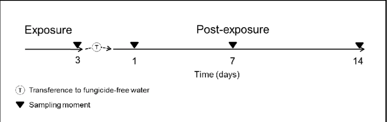
The schematic experimental design of experience, including exposure period and the transference to the post-exposure period, as well as the sampling moments is depicted below in the figure 3. The experiment (exposure and post-exposure periods) was carried out using aquaria duplicates (n = 2) where each aquarium had a group of 3 or 4 fish (7 fish per each condition/time). Water medium, in both exposure and post-exposure periods,
30
was daily renewed (100%). Fish were not fed during the exposure period. In the post-exposure period, fish were daily fed with fish roe, except at the day immediately before samplings. Animals were sacrificed by cervical transaction at the post-opercular region and blood collected from the heart using heparinized capillary tubes. Blood smears were immediately prepared for ENA, IE and EMI assays.
Figure 3 - Schematic representation of the experimental design, elucidating the sampling moments at 3 days (Exposure period) and the subsequent transference to fungicide-free water followed by sampling moments at 1, 7 and 14 days (Post-exposure period).
2.3 ENA assay
The ENA assay was performed in mature peripheral erythrocytes of Anguilla anguilla, as described by (Pacheco and Santos, 1997). For that, one blood smear per animal was fixed with methanol during 10 min and stained with Giemsa (5%) during 30 min. Then, slides were coded and scored blind. From each smear, 1000 erythrocytes were considered, under a 1000x magnification (microscope Olympus BX50), to evaluate the relative frequency of the following nuclear lesions: kidney shaped nuclei (K), lobed nuclei (L), binucleate or segmented nuclei (S), vacuolated nuclei (V) and micronuclei (MN) (Pacheco and Santos, 1997) (Fig. 1). Final results were expressed as the mean value (‰) of the sum for all the lesions observed (K + L + S + V + MN).
31
2.4 IE frequency
Immature erythrocytes were scored (under a 1000x magnification - microscope Olympus BX50) on 1000 erythrocytes (mature and immature) per fish. Results were presented as a frequency, according to the expression:
IE frequency (‰) =ME+IEIE × 1000;
where ME represents mature and IE the immature erythrocytes. Mature erythrocytes can be distinguished from immature since the latter present a rounder and larger nucleus and a bluish-gray cytoplasm, as established by Takashi Hibiya et al. (1982) and Smith (1990)(Fig. 2).
2.5 Erythrocyte maturity index (EMI)
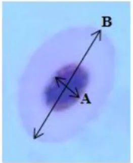
The EMI parameter was improved from Maceda-Veiga et al. (2010) in order to complement the IE frequency information and consisted in dividing the minor axis of the nucleus (Fig. 4, axis A) by the major cell axis length (Fig. 4, axis B) to obtain a ratio. Ten fields per sample/slide were examined at 1000× magnification and photographed. Erythrocytes measurements were performed using the ImageJ® software. EMI was calculated on 25 random cells of each field (250 in total).
Figure 4 - Schematic representation of the axes used for EMI calculation: A - minor axis of the nucleus; B - major cell axis.
After the calculation, EMI values were grouped considering class intervals of 0.1 units (]0-0.1] (class 1) up to ]0.6-0.7] (class7)), where the highest ratio corresponds to erythrocytes in a maximum immaturity status. For each fish, maturity classes were expressed as a percentage of the total number of cells analysed, in order to simplify the
32
analysis of EMI data. The use of this index intended to improve the interpretation of erythropoiesis-associated processes.
2.6 Statistical analysis
Statistica 8.0 software (StatSoft, Inc., OK, USA) was used for statistical analysis. All data were first tested for normality (Shapiro - Wilk test) and homogeneity of variance (Levene’s test) to meet statistical demands. For each sampling moment, one-way Analyses of Variance (ANOVA) was applied, followed by a post hoc Tukey's significance test for all pairwise comparisons. When the assumptions for parametric statistics failed (even after the data transformation), the Kruskal–Wallis ANOVA by Ranks (non-parametric) was performed.
For all the analyses, differences between means were considered significant when p < 0.05 (Zar, 1996).
33
3.
Results
3.1 ENA assay
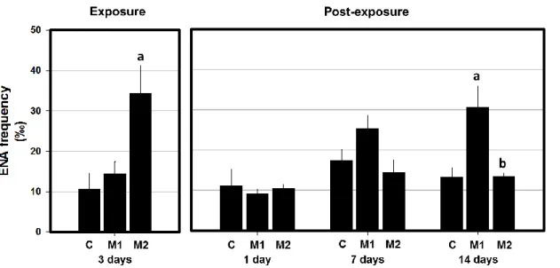
During the exposure period of 3 days, fish that have been exposed to the highest Mancozan® concentration (28 μg.L−1 - M2 group) demonstrated a significant increase of ENA frequency, when compared with the respective control (Fig. 5). Likewise, the results in terms of individual analysis of each nuclear lesion category (Table 1), considering the M2 group, showed a significant increase in S (segmented) category and in sub-total (K+L+S+V), when compared with the respective control. Moreover, the K (kidney Shaped) category was identified as the most prevalent considering all the experimental groups (control and treated), tough not showing statistically significant differences.
Figure 5 - Mean frequency (‰) of erythrocytic nuclear abnormalities (ENA) in peripheral erythrocytes of A. anguilla exposed to 2.8 and 28 μg.L−1 Mancozan® (M1, M2, respectively), during 3 days (Exposure period), and allowed to recover in fungicide-free water during 1, 7 and 14 days (Post-exposure period). Bars represent the standard error. Statistically significant differences (p<0.05), within the same exposure time, are: (a) in relation to control group (C); (b) between treatments.
In what concerns to the post-exposure period, no significant differences were observed on the first day in ENA frequency, either as overall values or as frequencies of individual categories (Table 1). Similarly, no significant differences were observed in the 7th day of post-exposure, with the exception of the K category, which was significantly
34
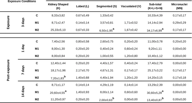
Kidney Shaped
(K) Lobed (L) Segmented (S) Vacuolated (V)
Sub-total (K+L+S+N) Micronuclei (MN) C 8,33±3,82 0,67±0,49 1,33±0,42 10,33±4,39 0,17±0,17 M1 8,71±2,47 0,14±0,14 3,57±0,81 1,71±0,52 14,14±2,94 0,29±0,29 M2 25,33±5,10 0,67±0,33 6,50±1,95a 1,67±0,42 34,17±6,99a 0,17±0,17 C 7,40±2,56 0,80±0,58 2,60±0,75 0,20±0,20 11,00±3,78 0,20±0,20 M1 8,00±1,30 0,20±0,20 0,40±0,24 0,60±0,24 9,20±1,11 0,00±0,00 M2 8,00±0,84 0,20±0,20 1,00±0,55 1,20±0,80 10,40±1,12 0,00±0,00 C 12,40±1,44 0,20±0,20 4,40±1,57 0,40±0,24 17,40±2,79 0,00±0,00 M1 18,17±1,96 2,17±0,70 4,67±1,31 0,17±0,17 25,17±3,22 0,17±0,17 M2 7,20±1,83b 1,40±0,68 4,40±1,94 1,20±1,20 14,20±3,15 0,17±0,18 C 8,71±1,17 0,14±0,14 4,29±1,19 0,14±0,14 13,29±2,39 0,00±0,00 M1 20,60±4,55a 1,40±0,93 8,00±1,14 0,60±0,60 30,60±5,28a 0,00±0,00 M2 11,20±0,97 0,20±0,20 2,00±0,63b 0,00±0,00 13,40±0,87b 0,00±0,00 1 4 d ay s
Nuclear Abnormalities Categories
3 d ay s 1 d ay 7 d ay s Exposure Conditions Ex p o su re Po st -e xp o su re
lower in M2 condition when compared with M1 (Table 1). The 14 day of post exposure reflected a significant increase of ENA frequency in the M1 group (2.8 μg.L−1 Mancozan®) when compared simultaneously with control and M2 groups (Fig. 5). The 14th day in fungicide-free water displayed also a significant increase in the frequency of K category for M1 group, in comparison to control. Moreover, M2 group displayed a significant decrease in relation to the lowest concentration (M1) concerning the S category. Sub-total values presented by M1 group was significantly higher than both control and M2 groups. During the whole post-exposure period, K category was found as the most abundant category, considering either the control or treated groups (Table 1).
Table 1 - Mean frequency (‰) of each nuclear abnormality category (± standard error) in peripheral erythrocytes of A. anguilla exposed to 2.8 and 28 μg.L−1 Mancozan® (M1, M2, respectively), during 3 days (Exposure) and submitted to a period in fungicide-free water during 1, 7 and 14 days (Post-exposure). Statistically significant differences (p<0.05) are: (a) in relation to control (C), within the same exposure time; (b) between treatments, within the same exposure time.
35
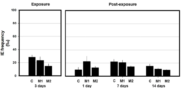
3.2 Cell maturity as a proxy for evaluation of erythrocyte population dynamics No statistically significant differences were found concerning the IE frequency, neither during the exposure nor the post-exposure period (Fig. 6).
Figure 6 - Mean frequency (‰) of immature erythrocytes (IE) in peripheral erythrocytes of A. anguilla exposed to 2.8 and 28 μg.L−1 Mancozan® (M1, M2, respectively), during 3 days (Exposure period), and allowed to recover in fungicide-free water during 1, 7 and 14 days (Post-exposure period). Bars represent the standard error. Statistically significant differences (p<0.05), within the same exposure time, are: (a) in relation to control group (C); (b) between treatments.
Likewise IE frequency, no significant differences were detected concerning the defined class intervals of EMI along the whole experiment (Fig. 7). Looking to the relative abundance of each class during the exposure period, the class 3 (orange) was found as the most frequent, considering either the control or treated groups. Similarly, in the post-exposure period the class 3 (orange) emerged as the most frequent with the exception for control group of fourteenth day. Nevertheless, only M1 group presented a similar pattern in abundance distribution between classes along the whole experiment (Fig. 7).
36 Fi gu re 7 - Me an f requen cy (%) o f EMI c las ses in pe ri ph er al er yth ro cytes o f A . a n g u ill a exp o sed to 2 .8 an d 28 μ g. L −1 Ma nc o zan ® (M1 , M2 , r es pec ti ve ly ), du ri ng 3 da ys ( Ex po sur e) an d sub m itt ed to a per io d in f un gi ci de -f re e wa ter dur ing 1 , 7 a nd 1 4 da ys ( P o st -exp o su re) . Sta n da rd e rr o r om itt ed .
37
4.
Discussion
Due to their frequent use in agro-industry, fungicides and their subproducts were detected in aquatic environment worldwide (Belenguer et al., 2014; Geissen et al., 2010; Melgar et al., 2008; Wightwick et al., 2012). Considering this, their presence is often associated with adverse effects in non-target organisms like fish. Histopathological alterations (Boran et al., 2012; Tabassum et al., 2016; Zhi-Hua et al., 2013), growth inhibition (Mu et al., 2013), alterations in antioxidant responses (Han et al., 2016; Liu et al., 2013; Tabassum et al., 2016; Zhi-Hua et al., 2013), inhibitions of acetylcholinesterase activity (Bouhafs, et al., 2015; Srivastava and Singh, 2014b) or gene expression (Liu et al., 2013) are some of them. However, the effects at the DNA level have a particular interest in toxicology, since a single alteration in DNA molecule can trigger harmful effects on the normal cell metabolism and fate. In addition, several fungicides have proven their genotoxic potential to fish (Han et al., 2016; Liu et al., 2013; Shahi and Singh, 2014; Srivastava and Singh, 2013b). However, a single fish study is available concerning the genotoxicity of Mancozan® (Marques et al., 2016), a widely used dithiocarbamate fungicide formulation.
In this way, the present work aimed to shed a light on the genotoxic risk associated to occasional exposures of non-target organisms to fungicides. Notwithstanding, the persistence of fungicide effects were assessed after the cessation of the exposure, in a post-exposure period, where fish were transferred to fungicide-free water. Moreover, this study was also intended to contribute to the establishment of biomonitoring tools, aiming an efficient assessment of the risk to non-target organisms (fish), associated to the input of fungicides in the aquatic environment.
4.1 Genotoxic risk of Mancozan®
Mancozan® hazard to aquatic environment is assumed by its manufacturer and recently Marques et al. (2016) proved its genotoxicity to fish. Whereby there still is a lack of knowledge concerning its pernicious genotoxic effects that, jointly with its utilization worldwide, increases the interest of this study.
38
The exposure to the higher tested concentration of Mancozan (M2 – 28 µg.L ) presented clastogenic and/or aneugenic effects in A. anguilla erythrocytes, after a 3 days of exposure, attesting the genotoxic potential of this commercial formulation. Accordingly to a previous study performed by Srivastava and Singh (2013b), which detected the appearance of chromosomal abnormalities in the fish Clarius batrachus after a 3 days exposure to mancozeb, the observed genotoxicity may be linked to an eventual action of this active principle. However, the use of extremely high (sub-lethal) doses by these authors, when compared to the environmental relevant concentrations presently used, may compromise the connection.
Another purpose of the present study was to evaluate the A. anguilla ability to recover from the genetic damage induced by a short-term exposure to the fungicide upon the exposure cessation. It is well-known that fish can move within and between habitats, in different spacial scales, determined by a variety of purposes such as to escape from predators, prey capture, migrations, spawning, and avoidance of local poor conditions. Consequently, fish can move, intentionally or unintentionally, from contaminated
towards non-contaminated sites. Thus, in order to improve the knowledge concerning the
progression of the chromosomal damage following the cessation of exposure, as a realistic natural situation, the DNA integrity was assessed after transference of previously exposed fish to fungicide-free water. In this regard, a post-exposure period was performed in order to follow up the previously observed genetic damage. Bear this in mind, in the day immediately after the transference to the fungicide-free medium, fish presented a reduction of the ENA frequency, since M2 group returned to control levels, that were maintained, at least, for more thirteen days. Nevertheless, the 14th day of post-exposure revealed an interesting finding. The M1 group (2.8 µg.L-1, the lowest concentration of Mancozan®), which has always presented ENA levels similar to control, revealed, for the first time, a significant increase of abnormal nuclei, highlighting a worrying long-lasting damage effect. Despite the maintenance of the chromosomal integrity on the seventh day of post-exposure, in both treated conditions, the lowest concentration (M1) presented an increasing tendency (statistically non-significant) in the ENA frequency. Even so, a significant increasing in the chromosomal damage was only
39
detected in the M1 condition 7 days later, in the 14th day of post-exposure. In this direction and taking into account the mancozeb toxicity reported to fish (US EPA, 2006), it is expectable that this active principle and/or its metabolites play a relevant role in the late appearance of cytogenetic damage in M1 condition (in the 14th day of the post-exposure period). In addition, and considering the already stated pro-oxidant potential of mancozeb (Calviello et al., 2006; Domico et al., 2007), it is important to consider that ROS generated can react with macromolecules, like lipids, inducing lipid peroxidation (LPO). Even so, the increase of LPO levels detected by Kubrak et al., (2012) and Atamaniuk et al., (2013) in brain, gills and kidney, after an acute exposure to a mancozeb-based fungicide (Tattoo®) in same extent supports this idea. Since lipid peroxidation is a degenerative process which affects phospholipids of membranes under oxidative conditions, it can be responsible for the loss of cell integrity and consequently, this fragility makes cells (and DNA) more susceptible to be attacked (Sevgiler et al., 2004). Bearing this in mind and taking into account the pro-oxidant potential demonstrated by mancozeb (Calviello et al., 2006; Srivastava et al., 2012; Srivastava and Singh, 2014a), the obtained results lead to suppose that the exposure to 2.8 µg.L-1 of Mancozan® induce a progressive loss of antioxidant defences in erythrocytes allowing the development of chromosomal damages (significantly higher) 14 days after exposure. At the same time, may be proposed that higher concentrations of fungicide (M2) induced the activation of the erythrocyte antioxidant defenses, as a response to the ROS over-generation (Srivastava et al., 2012). Initially, it is possible that during the exposure period, the oxidant potential of the agrochemical (28 µg.L-1 Mancozan® - M2) was so strong that antioxidants defenses were not able to effectively control it (even by a short time). However, after the removal of fungicide (post-exposure period) antioxidant defenses in fish exposed to 28 µg.L-1 Mancozan® were keep high enabling the recover and preservation of the chromosomal integrity until the end of the experiment.
Despite all these facts, the presence of methanamine in the commercial formulation Mancozan® should not be disregarded. As an attempt to clarify the possibility of an oxidative cause on the genetic damage induced by Mancozan®, further studies considering the commercial formulation and the active ingredient, in parallel, should be
40
performed. Thus, the analysis of the cell antioxidant responses emerges, undoubtedly, as a useful tool. Moreover, the use of the comet assay improved with DNA lesion-specific repair enzymes, which highlight specifically oxidized DNA bases, is strongly recommended. Moreover, the jointly use of the comet assay and ENA test would improve the knowledge concerning the magnitude of DNA damage, since these tests appear as complementary.
4.2 Contribution to the clarification of hematological dynamics
In order to improve the knowledge concerning the hematological dynamics and taking into account that erythropoiesis (synthesis of erythrocytes) can influence the ENA frequency by the existence of a dilution effect, the frequency of immature erythrocytes (IE) was scored. Moreover, and since the interpretation of IE frequency data only gives a momentary perspective of the erythropoiesis rate, the erythrocyte maturity index (EMI) was adopted to clarify the cell population dynamics during the whole experiment, allowing thus the interpretation of the evolution of the immature erythrocytes (represented in the EMI figure by dark blue, blue and green areas, Fig. 7). In what concerns to the simple analysis of the IE occurrence, the erythrocyte population dynamics does not seem to have been affected during the exposure period. Thus, and considering the EMI data, it was observed that after a 3-days exposure, it was possible to found a decrease tendency corresponding to the class 4 (yellow area) offset by a class 2 increase (red area) in treated groups (more evident in M1 condition), when compared with control. This tendency of replace cells with a lower maturation degree (class 4) by cells with a higher mature degree (class 2) can be interpreted as an aging of erythrocyte exposed populations. These results point to a reduction of erythropoiesis rate in exposed fish but the link between the exposure to the fungicide and the erythropoiesis inhibition cannot be stablished at this time.
Regarding to the first day of post-exposure period, when the hematological dynamics was analysed, EMI and IE parameters were consistent. No differences were observed. Thereby, EMI (dark blue, blue and green areas, class 7, 6 and 5, respectively) and IE
41
frequency clearly indicate that the synthesis of new erythrocytes was not the mechanism responsible both for maintenance of chromosomal integrity of M1 group, in first day of post-exposure, as for the recovery presented by M2 group. Thus, it can be stated that chromosomal integrity, in the M2 group, was recovered mainly due the erythrophagia of damage erythrocytes in spleen (simultaneously with the production ), as already stated by (Pacheco and Santos, 2002). Even so, the simultaneous production of erythrocytes with normal nucleus denoting the loss of genotoxic action and/or the increase of the antigenotoxic processes effectiveness.
Globally, the IE frequency has been stated as a useful tool in the analysis of hematological dynamics, essentially when coupled with ENA assay (Pacheco and Santos, 2002). In turn, EMI showed be a promising technique. Although no significant changes have been detected during the experiment, this parameter allowed a broader view of the maturity pathway of erythrocytes population.
4.3 Implications for the definition of biomonitoring strategies
The usefulness of biomonitoring strategies depends on the capacity to reflect the magnitude and persistence of an environmental risk. Thus, the present study strengthened the utility of ENA assay in the detection of genotoxic effects in fish exposed to fungicide-contaminated waters. Since fungicides are seasonally applied, their input in the aquatic environment is considered intermittent, increasing the difficulty to detect its impact in biomonitoring programs, as time-scaling can be limiting. Since fish are not always sampled under the action of the pesticide (and their biological effects not always are instantaneous), it is very important to find specific biomarkers with the view to assess key biological responses to correctly correspond to the delay between the exposure and sampling time. ENA assay was able to detect DNA damage induced by Mancozan® 14 days after the cessation of the contamination pressure, which highlights the importance of this endpoint as a biomarker of exposure.
It is also remarkable that the late reappearance of genetic damage presented in this study highlights the occurrence of a long-term genome-destabilizing action (e.g. carcinogenic and reproductive impairments) in fish, as a result of occasional pesticide
42
applications. Whereupon, the present findings emerge as a relevant source of knowledge that should be integrated in the definition of biomonitoring strategies as well as on the improvement of agro-industrial practices.
5. Final Remarks
The Mancozan® genotoxic potential was demonstrated in erythrocytes of Anguilla
anguilla exposed to environmental realistic concentrations of this fungicide. This
commercial formulation has demonstrated its capacity to induce cytogenetic effects, as chromosome or chromatid breaks or loss (measured by ENA assay), in both tested concentrations.
The synthesis of erythrocytes (inferred by IE frequency) appeared to not be affected by exposure to the pesticide. Moreover, the use of EMI increased the knowledge concerning the information given by IE frequency, showing the magnitude of erythrocytes maturity, and also reinforcing the occurrence of no changes in erythrocyte population dynamics.
Globally, the genotoxic effects of Mancozan® (with special attention to those revealed after 14 days in the post-exposure period) highlight the risk for fish populations in what concerns to long-term adverse effects (e.g. carcinogenic and reproductive deficiencies).
43
References
Agrawal, A., Sharma, B. (2010). Pesticides induced oxidative stress in mammalian systems. International Journal of Biological and Medical Research, 3(3), 90–104.
Alberts, B., Johnson, A., Lewis, J., Raff, M., Roberts, K., Walter, P. (2002). The Structure and Function of DNA (4th ed.). New York: Garland Science.
Andrade, V., Silva, J., Silva, F., Heuser, V., Dias, J., Lu, M. (2004). Fish as Bioindicators to Assess the Effects of Pollution in Two Southern Brazilian Rivers Using the Comet Assay and Micronucleus Test. Environ. Mol. Mutagen., 468, 459–468.
Atamaniuk, T. M., Kubrak, O. I., Husak, V. V., Storey, K. B., Lushchak, V. I. (2013). The Mancozeb-Containing Carbamate Fungicide Tattoo Induces Mild Oxidative Stress in Goldfish Brain, Liver, and Kidney. Environmental Toxicology, 1227–1235. http://doi.org/10.1002/tox
Atreya, K., Sitaula, B. K. (2011). Mancozeb: growing risk for agricultural communities? Himalayan Journal of Sciences, 6(8), 9–10. http://doi.org/10.3126/hjs.v6i8.1794
Belenguer, V., Martinez-Capel, F., Masiá, A., Picó, Y. (2014). Patterns of presence and concentration of pesticides in fish and waters of the Júcar River (Eastern Spain). Journal of Hazardous Materials, 265, 271–279. http://doi.org/10.1016/j.jhazmat.2013.11.016
Boran, H., Capkin, E., Altinok, I., Terzi, E. (2012). Assessment of acute toxicity and histopathology of the fungicide captan in rainbow trout. Experimental and Toxicologic Pathology, 64(3), 175–179. http://doi.org/10.1016/j.etp.2010.08.003
Bouhafs, N. B., Djebbar, M. R., Berebbeh, H. (2015). Elevation of Glutathione-S-Transfer a se and Inhibition of Cholinesterase Activity As biomarkers of Fungicide Toxicity in Mosquito fish Gambusia affinis, 9(July), 191–197.
Calviello, G., Piccioni, E., Boninsegna, A., Tedesco, B., Maggiano, N., Serini, S., Wolf, F. I., Palozza, P. (2006). DNA damage and apoptosis induction by the pesticide Mancozeb in rat cells: Involvement of the oxidative mechanism. Toxicology and Applied Pharmacology, 211(2), 87– 96. http://doi.org/10.1016/j.taap.2005.06.001
Campbell, T. W. (2015). Exotic Animal Hematology and Cytology (4th ed.). Wiley Blackwell. Retrieved from https://books.google.com/books?id=zAZ9BgAAQBAJ&pgis=1
Capkin, E., Altinok, I., Karahan, S. (2006). Water quality and fish size affect toxicity of endosulfan, an organochlorine pesticide, to rainbow trout. Chemosphere, 64(10), 1793–1800. http://doi.org/10.1016/j.chemosphere.2005.12.050
44
Capriglione, T., De Iorio, S., Gay, F., Capaldo, A., Vaccaro, M. C., Morescalchi, M. A., Laforgia, V. (2011). Genotoxic effects of the fungicide thiophanate-methyl on Podarcis sicula assessed by micronucleus test, comet assay and chromosome analysis. Ecotoxicology, 20(4), 885–891. http://doi.org/10.1007/s10646-011-0655-8
Carrasco, K. R., Tilbury, K. L., Myers, M. S. (1990). Assessment of the Piscine Micronucleus Test as an in situ Biological indicator of Chemical Contaminant Effects. Canadian Journal of Fisheries and Aquatic Sciences, 47(11), 2123–2136.
Caspers, H. (1982). An Atlas of Fish Histology. Normal and Pathological Features. (H. Takashi, Ed.)Internationale Revue der gesamten Hydrobiologie und Hydrographie (Vol. 69). http://doi.org/10.1002/iroh.19840690307
Comission, E. (2007). SIDS INITIAL ASSESSMENT PROFILE DE / eu.
Coronas, M. V., Pereira, T. S., Rocha, J. A. V, Lemos, A. T., Fachel, J. M. G., Salvadori, D. M. F., Vargas, V. M. F. (2009). Genetic biomonitoring of an urban population exposed to mutagenic
airborne pollutants. Environment International, 35(7), 1023–1029.
http://doi.org/10.1016/j.envint.2009.05.001
Domico, L. M., Cooper, K. R., Bernard, L. P., Zeevalk, G. D. (2007). Reactive oxygen species generation by the ethylene-bis-dithiocarbamate (EBDC) fungicide mancozeb and its contribution to neuronal toxicity in mesencephalic cells. NeuroToxicology, 28(6), 1079–1091. http://doi.org/10.1016/j.neuro.2007.04.008
Eurostat. (2013). Agri-environmental indicator - pesticide pollution of water. Retrieved June 9, 2016, from http://ec.europa.eu/eurostat/statistics-explained/index.php/Agri-environmental_indicator_-_pesticide_pollution_of_water
Eurostat. (2015). Agri-environmental indicator - consumption of pesticides - Statistics Explained.
Retrieved April 20, 2016, from
http://ec.europa.eu/eurostat/statistics- explained/index.php/Agri-environmental_indicator_-_consumption_of_pesticides#Measurements
Eurostat. (2016). Pesticide sales statistics - Statistics Explained. Retrieved May 27, 2016, from http://ec.europa.eu/eurostat/statistics-explained/index.php/Pesticide_sales_statistics Falfushynska, H. I., Gnatyshyna, L. L., Stoliar, O. B. (2013). In situ exposure history modulates the
molecular responses to carbamate fungicide Tattoo in bivalve mollusk. Ecotoxicology, 22(3), 433–445. http://doi.org/10.1007/s10646-012-1037-6
45
Figueiredo-Fernandes, a., Fontaínhas-Fernandes, a., Monteiro, R., Reis-Henriques, M. a., Rocha, E. (2006). Effects of the fungicide mancozeb on liver structure of Nile tilapia, Oreochromis niloticus: Assessment and quantification of induced cytological changes using qualitative histopathology and the stereological point-sampled intercept method. Bulletin of Environmental Contamination and Toxicology, 76(2), 249–255. http://doi.org/10.1007/s00128-006-0914-1
Fishel, F. M. (2013). Pest Management and Pesticides : A Historical Early Uses of Pesticides and
Other Insect-Vectored Diseases, (February), 2–6. Retrieved from
https://edis.ifas.ufl.edu/pdffiles/PI/PI21900.pdf Fishel, F. M. (2014). How are Pesticides Classified ? 1, 1–2.
Frac. (2016). FRAC Code List © * 2016: Fungicides sorted by mode of action (including FRAC Code
numbering), 1–10. Retrieved from
http://www.frac.info/docs/default-source/publications/frac-code-list/frac-code-list-2015-finalC2AD7AA36764.pdf?sfvrsn=4 Geissen, V., Ramos, F. Q., Bastidas-Bastidas, P. D. J., Díaz-González, G., Bello-Mendoza, R.,
Huerta-Lwanga, E., Ruiz-Suárez, L. E. (2010). Soil and water pollution in a banana production region in tropical Mexico. Bulletin of Environmental Contamination and Toxicology, 85(4), 407–413. http://doi.org/10.1007/s00128-010-0077-y
Goldoni, a, da Silva, L. B. (2012). Mutagenic Potential of the Fungicide Mancozeb in Astyanax Jacuhiensis (Teleostei: Characidae). Bioscience Journal, 28(2), 297–301. Retrieved from <Go to ISI>://WOS:000305333400018
Gregory, T. R. (2005). CHAPTER 1 – Genome Size Evolution in Animals. In The Evolution of the Genome (pp. 3–87). http://doi.org/10.1016/B978-012301463-4/50003-6
Guilherme, S., Gaivão, I., Santos, M. a., Pacheco, M. (2010). European eel (Anguilla anguilla) genotoxic and pro-oxidant responses following short-term exposure to Roundup® - A
glyphosate-based herbicide. Mutagenesis, 25(5), 523–530.
http://doi.org/10.1093/mutage/geq038
Guilherme, S., Gaivão, I., Santos, M. A., Pacheco, M. (2012a). DNA damage in fish (Anguilla anguilla) exposed to a glyphosate-based herbicide – Elucidation of organ-specificity and the
role of oxidative stress. Mutat. Res.-Gen. Tox. En., 743, 1–9.