Participatio n o f nitric o xide in
the nucle us isthm i in CO
2
-drive
to bre athing in to ads
Departamentos de Fisiologia, Faculdade de O dontologia de Ribeirão Preto and Faculdade de Medicina de Ribeirão Preto, Universidade de São Paulo, Ribeirão Preto, SP, Brasil
L.H. Gargaglioni and L.G.S. Branco
Abstract
The nucleus isthmi (NI) is a mesencephalic structure of the amphibian brain. It has been reported that NI plays an important role in integration of CO2 chemoreceptor information and glutamate is probably
in-volved in this function. However, very little is known about the mechanisms involved. Recently, it has been shown that nitric oxide synthase (NOS) is expressed in the brain of the frog. Thus the gas nitric oxide (NO) may be involved in different functions in the brain of amphibians and may act as a neurotransmitter or neuromodulator. We tested the hypothesis that NO plays a role in CO2-drive to breathing,
specifically in the NI comparing pulmonary ventilation, breathing frequency and tidal volume, after microinjecting 100 nmol/0.5 µl of L-NAME (a nonselective NO synthase inhibitor) into the NI of toads (Bufo paracnemis) exposed to normocapnia and hypercapnia. Control animals received microinjections of vehicle of the same volume. Under normocapnia no significant changes were observed between control and L-NAME-treated toads. Hypercapnia caused a significant (P<0.01) increase in ventilation only after intracerebral microinjection of L-NAME. Exposure to hypercapnia caused a significant increase in breathing frequency both in control and L-NAME-treated toads (P<0.01 for the control group and P<0.001 for the L-NAME group). The tidal volume of the L-NAME group tended to be higher than in the control group under hypercapnia, but the increase was not statistically signifi-cant. The data indicate that NO in the NI has an inhibitory effect only when the respiratory drive is high (hypercapnia), probably acting on tidal volume. The observations reported in the present investigation, together with other studies on the presence of NOS in amphibians, indicate a considerable degree of phylogenetic conservation of the NO pathway amongst vertebrates.
Co rre spo nde nce
L.G.S. Branco
Departamento de Fisiologia Faculdade de O dontologia de Ribeirão Preto, USP 14040-904 Ribeirão Preto, SP Brasil
Fax: + 55-16-633-0999 E-mail: branco@ forp.usp.br Presented at the Meeting “NO Brazil, Basic and Clinical Aspects of Nitric O xide”, Foz do Iguaçu, PR, Brazil, March 10-13, 1999.
Research supported by FAPESP and CNPq. L.H. Gargaglioni was the recipient of a Doctoral FAPESP fellowship.
Received June 17, 1999 Accepted July 24, 1999
Ke y wo rds
·Nitric oxide ·Amphibian ·Hypercapnia ·Ventilation ·Nucleus isthmi
·Toad
·CO2
·L-NAME
Intro ductio n
The breathing pattern of most fish, birds and mammals is continuous, whereas that of most air-breathing fish, amphibia, and rep-tiles is not. These animals exhibit one of two
basic types of intermittent breathing in which lung ventilation occurs in single events or is grouped into episodes of many breaths sepa-rated by non-ventilatory (apneic) periods of variable duration (1). The emergence of CO2
meta-morphosis may be an important factor in the production of breathing episodes (2).
In bullfrogs, the area where brainstem transections affected the breathing pattern included the nucleus isthmi (NI) (2). This nucleus is a mesencephalic structure of the amphibian brain located between the roof of the midbrain and the cerebellum (3). NI goes through substantial cellular arrangement and differentiation during amphibian metamor-phosis, a period also associated with the onset of episodic lung ventilation in bull-frogs (4). The NI also plays an important role in CO2 chemodetection or, more likely, in
the integration of CO2 chemoreceptor
infor-mation (2). NI is an important site for the control of breathing and glutamate is prob-ably involved in this function (2).
Glutamate released from one presynaptic terminal acts on both N-methyl-D-aspartate (NMDA) and non-NMDA classes of gluta-mate receptors. It has been shown that acti-vation of NMDA receptors results in an in-crease in intracellular calcium which can stimulate constitutive nitric oxide synthase (NOS), an enzyme that generates nitric ox-ide (NO) from the amino acid L-arginine (5,6). The NO formed diffuses to the presyn-aptic terminal where it stimulates guanylate cyclase and elevates cyclic GMP concentra-tion (7), leading to a further increase in the release of glutamate and a greater augmenta-tion of synaptic transmission (6). A number of recent studies have shown that arginine analogs such as Nw-nitro-L-arginine methyl ester (L-NAME) inhibit NO synthesis (6).
More recently, it has been shown that NOS is expressed in a subpopulation of neu-rons throughout the brain of the frog (8). The distribution pattern reveals certain similari-ties to that of other species. Expression of NOS is not limited to a particular system. Therefore, NO may be involved in different functions in the brain of amphibians.
It has been recently shown that the NI plays an important role in respiratory control and in CO2 chemodetection in bullfrogs (2).
It is possible that the NO pathway is in-volved in this control. The aim of the present study was to assess the role of the NO path-way in CO2-drive to breathing specifically in
the NI by microinjecting a nonselective NOS blocker (L-NAME) into the NI of toads (Bufo paracnemis) under conditions of normocap-nia and hypercapnormocap-nia.
Mate rial and Me tho ds
Bufo paracnemis toads (mass 126.8 ± 2.4 g, N = 16) were collected in the vicinity of Ribeirão Preto, SP, Brazil, during the rainy summer months. The toads were maintained in containers with free access to water and basking area. Food was withheld for two weeks before the experiment.
Surgical m e tho ds
Animals were anesthetized by submer-gence in 0.3% MS-222 and a silastic tube segment, 1.5 mm in outer diameter, was introduced into the right femoral artery. The animals head was then fixed to a stereotaxic apparatus and the skin covering the skull was removed with the aid of a bone scraper. An opening was made in the skull above the midbrain region using a small drill. A guide cannula prepared from a hypodermic needle segment 14 mm in length and 0.6 mm in outer diameter was attached to the tower of the stereotaxic apparatus and placed in con-tact with the exposed surface of the midbrain inside the NI region. The orifice around the cannula was filled with a paste consisting of a mixture of equal parts of paraffin and glycerine. The cannula was fixed to the skull with acrylic cement. The experiments were initiated 48 h after brain surgery.
Me asure m e nts o f blo o d pre ssure , he art rate
and ve ntilatio n
pressure transducer (Narco, P-1000B) and the signals from the transducer were recorded on paper (Narcotrace 80). Heart rate (HR) was determined by counting pressure pulses. Pulmonary ventilation (VI) was
meas-ured directly using a pneumotachographic method (9) based on the Poiseuille principle that the laminar flow of a gas is proportional to the pressure gradient across a tube. A lightweight transparent face mask provided an air-tight connection between the nostrils and a Fleisch tube. Inspiratory and expira-tory gas flows were monitored by means of a differential pressure transducer (Validyne 451871) connected to the same physiograph.
Expe rim e ntal pro to co ls
Two days after surgery, the animals were housed individually in a plastic chamber kept at the experimental temperature of 25oC.
The animal chamber was continuously flushed with humidified room air. In one group (N = 8), experimental animals re-ceived one microinjection of 100 nmol/0.5 µl L-NAME (Sigma Chemical Co., St. Louis, MO, USA) dissolved in mock cerebrospinal fluid (mCSF). The basic composition of the mCSF solutions in mEq/l was: 83.6 Na+, 2.7
K+
, 0.9 Ca2+
, 0.45 Mg2+
and 0.45 SO4
2-. Bicarbonate and chloride concentrations were adjusted in order to attain the desired pH values for the individual perfusate solu-tions. Control animals (N = 8) were treated with intracerebral microinjections of vehicle in the same volume. Intracerebral injections were performed with a thin dental needle introduced until its tip was 2 mm below the cannula end. A volume of 0.5 µl was injected over a period of 30 s using a Hamilton mi-crosyringe. The movement of an air bubble inside the PE 10 polyethylene tubing con-necting the microsyringe to a dental needle confirmed drug flow. Doses and methods of administration were chosen on the basis of a previous study (10).
Once conditions were stable in the
nor-mocapnic condition, BP and VI were
re-corded. A hypercapnic gas mixture of 3% CO2 (AGA, Sertãozinho, SP, Brazil) was
then applied for 30 min.
Histo lo gy
At the end of experiments the animals were sacrificed with ether and perfused through the heart with Ringer followed by 10% formalin. A dental needle was inserted through the guide cannula and a 0.5 µl mi-croinjection of Evans blue was performed. The heads of the animals were removed and fixed in 10% formalin. The brains were re-moved from the skull, immersed in paraffin, sectioned on a microtome, and stained with hematoxylin-eosin for light microscopy de-termination of the region reached by the microinjection needle.
Calculatio ns and statistical analysis
All values are reported as means ± SEM. BP, HR and VI were calculated on the basis
of 10-min recording periods. BF was quanti-fied by analyzing the number of respiratory events (lung breaths) per minute. Tidal vol-ume (VT) was obtained from the integrated
area of the inspired flow signal. VI (VI = VT
x BF) was expressed as ml BTPS (body temperature and pressure, saturated with water vapor) kg-1 min-1. The effects of
hyper-capnia were evaluated by analysis of vari-ance (ANOVA) and the difference between means was assessed by Tukeys test. A P value of less than 0.05 was considered sig-nificant.
Re sults
Effe cts o f L-NAME micro inje ctio n into the
nucle us isthmi o n ve ntilato ry re spo nse to
hype rcapnia
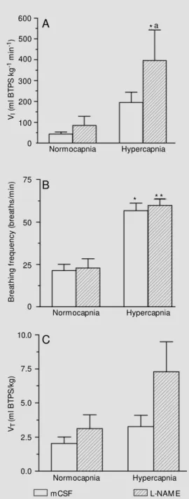
exposure to hypercapnia caused a signifi-cant increase in BF (P<0.01 for the control group and P<0.001 for the L-NAME group). Figure 1C shows that during hypercapnia, the VT of L-NAME group tended to be higher
than in the control group, but the increase was not statistically significant.
Figure 2 shows the pulmonary ventila-tion recordings obtained after mCSF or L-NAME microinjections during normocapnia and hypercapnia.
Effe cts o f L-NAME micro inje ctio n into the
nucle us isthmi o n blo o d pre ssure and he art
rate re spo nse to hype rcapnia
Table 1 shows the effects of treatments on BP and HR. None of the experimental conditions had any significant effect on BP and HR.
D iscussio n
The present study provides evidence that the gas NO plays a role in NI neurotransmis-sion involved in CO2-drive to breathing since
intracerebral microinjection of a nonselec-tive NOS blocker (L-NAME) increased the ventilatory CO2 response.
Despite recent advances, the mechanisms of neurorespiratory control in amphibians are not fully understood. Most early studies on pulmonary ventilation in amphibians fo-cused on the mechanics of pulmonary venti-lation, the mechanism behind the positive pressure inflation of the lungs (11,12). Addi-tionally, the periodic pattern of ventilation was described as breath-holding alternating with bursts of pulmonary ventilation, which in some anuran amphibians are initiated by stepwise pulmonary deflation followed by lung reinflation (13,14). In anurans, studies have evaluated the chemical drive to breathe and the receptors involved. The ventilatory responses to hypoxia were assessed in Bufo paracnemis (15), and the other studies char-acterized the arterial O2 receptors (16,17), as VI
(
m
l
B
T
P
S
k
g
-1 m
in
-1)
600
500
400
300
200
100
0
*a
Normocapnia Hypercapnia
B
re
a
th
in
g
f
re
q
u
e
n
c
y
(
b
re
a
th
s
/m
in
) 75
50
25
0
Normocapnia Hypercapnia
VT
(m
l
B
T
P
S
/k
g
)
10.0
7.5
5.0
2.5
0.0
Normocapnia Hypercapnia
* * *
A
B
C
mCSF L-NAM E
Figure 1 - Effect of microinjec-tion of L-NAM E (100 nmol/0.5 µl) or mCSF (0.5 µl) into the nucleus isthmi on VI (A), BF (B) and VT (C), in animals exposed to normocapnia and hypercapnia (3% CO2). * P<0.01, * * P<0.001 indicate a significant difference in mean values before and after 30 min of 3% CO2. aP<0.01 indi-cates a significant difference be-tw een control and the L-NAM E group (Tukey’s test).
no significant changes in VI were observed
between control and L-NAME-treated toads. Hypercapnia caused a significant (P<0.01) increase in VI only after L-NAME treatment.
well as the central acid-base receptors of
Bufo paracnemis (18,19). The respiratory control of ectotherms resembles the mam-malian control system; the rhythmogenic and pattern-forming elements in each are adapt-ed to meet the demands determinadapt-ed by the environment, behavior, metabolic needs, and breathing mechanisms. However, studies about neurorespiratory control in ectotherm vertebrates are scarce (20,21). Recently, Kinkead et al. (2) found that NI plays an important role in respiratory control by main-taining eucapnic motor output and allowing full expression of the CO2 response, since
bilateral lesions of NI in bullfrogs by micro-injection of kainic acid caused a reduction of eucapnic ventilation and of CO2
chemosen-sivity. However, it has been previously shown that kainic acid causes damage at the site of injection together with seizures and loss of cells some distance from the injection (22). Thus, it is often difficult to attribute the changes observed in this experimental prepa-ration to damage to the brain area of interest. Additionally, the experiments were per-formed approximately 1.5 h after the kainic acid injections, despite the fact that this neu-rotoxin has a strong and long-lasting excita-tory effect (23).
The experimental approach used in the present study differs considerably from that followed by Kinkead et al. (2). They per-formed this study in decerebrate, paralyzed and unidirectionally ventilated bullfrogs. These differences preclude a detailed com-parison between their study and the present data although both investigations provide support for a participation of the NI in inte-gration of CO2-drive to breathing.
Glutamate is widely distributed in the central nervous system and is supposed to act as a neurotransmitter in the NI for the control of breathing (2). NO is linked to glutamatergic neurotransmission in the cen-tral nervous system (24,25) and activation of glutamate receptors in the nucleus tractus solitarius (NTS) and paragigantocellular
nucleus is necessary to maintain normal lev-els of blood pressure and ventilation and also for a normal ventilatory CO2 response
to occur (26). It has been demonstrated that NO enhances the excitability and spontane-ous discharge rates of neurons in the NTS (27) and may act as a retrograde messenger in an L-glutamate-releasing positive feed-back system, also in the NTS, involved in the increase of ventilation during hypoxia (24). NO has an excitatory effect on the discharge rates of neurons in the pontine respiratory group, whereas L-NNA produces a disrup-tion of the pneumotaxic mechanism (28).
A number of recent studies have shown that NO accounts for a large part of the biological actions of endothelin-derived
re-Table 1 - Effects of L-NAM E microinjection into the nucleus isthmi on the blood pressure and heart rate response of Bufo paracnemis to hypercapnia.
Values are reported as means ± SEM . mCSF, M ock cerebrospinal fluid.
Treatment Inspired CO2 Blood pressure Heart rate
(% ) (mmHg) (min-1)
mCSF (0.5 µl) 0 36.7 ± 3.8 41.2 ± 4
(N = 8) 3 34.9 ± 2.3 46.0 ± 7.2
L-NAM E (100 nmol/0.5 µl) 0 30.3 ± 4.1 47.6 ± 5
(N = 8) 3 27.7 ± 1.6 48.6 ± 6.7
20 ml/s
10 s
A
B
Air 3% CO2
laxing factor (29). The importance of NO can be demonstrated by inhibition of the effects of NO (30) using L-arginine analogs such as L-NAME. In the present study, we have chosen L-NAME because it is a nonse-lective inhibitor of NOS and acts on both the constitutive and inducible isoforms of the enzymes.
The microinjection of L-NAME into the NI altered the CO2-induced hyperventilation
(Figure 1). When the animals were exposed to hypercapnia after receiving L-NAME, a significant increase in ventilatory CO2
re-sponse was observed. Probably, this might be due to the fact that NO in the NI may have an inhibitory influence on the integration of the CO2-drive to breathing. Conversely, a
number of studies indicate NO as an excita-tory neurotransmitter (7,6). In agreement with our study, a previous study, Iadecolaet al. (31) reported that intravenous administra-tion of L-NAME to rats leads to a partial inhibition, i.e., ~50% inhibition of brain NOS catalytic activity. NO, besides increasing cGMP, can also inhibit NMDA-induced in-tracellular calcium entry and NOS activity, exercising a local negative feedback that may be an important intracellular mechanism
in the regulation of NO synthesis because NO has been shown to be potentially cyto-toxic (32). Such a role for NO may compli-cate the interpretation of data obtained by application of L-NAME. It seems that the role of NO is more complex than previously thought, and may vary according to the site under study.
NO acts as a physiological messenger molecule that may serve as a neurotransmit-ter in the central nervous system (29). In amphibians, NO has been suggested to be a neurotransmitter in the gut (33). We have demonstrated that NO in the NI has an inhib-itory influence only when the respiratory drive is high (hypercapnia), acting on VT.
Conversely, the present observations, to-gether with other studies on the presence of NO synthase in amphibians (8,33,34), indi-cate a considerable degree of phylogenetic conservation of the NO pathway amongst vertebrates.
Ackno wle dgm e nts
We thank Mr. Rubens F. Melo for excel-lent technical assistance.
Re fe re nce s
1. M ilsom WK (1991). Intermittent breath-ing in vertebrates. Annual Review of Phys-iology,53: 87-105.
2. Kinkead R, Harris M B & M ilson W K (1997). The role of the nucleus isthmi in respiratory pattern formation in bullfrogs.
Journal of Experimental Biology, 200: 1781-1793.
3. Senn DG (1972). Development of tegmen-tal and rhombencephalic structures in a frog (Rana temporaria). Acta Anatomica, 82: 525-548.
4. Burggren WW & Infantino RL (1994). The respiratory transition from w ater to air breathing during amphibian metamorpho-sis. American Zoology, 34: 238-246. 5. Know les RG, Palacios M , Palmer RM J &
M oncada S (1989). Formation of nitric ox-ide from L-arginine in the central nervous system: A transduction mechanism of
stimulation of the soluble guanylate cy-clase. Proceedings of the National Acade-my of Sciences, USA,86: 5159-5162. 6. M oncada S & Higgs EA (1993). The
L-arginine-nitric oxide. New England Jour-nal of M edicine, 329: 2002-2012. 7. Bredt DS & Snyder SH (1992). Nitric
ox-ide, a novel neuronal messenger. Neuron, 18: 3-11.
8. Brüning G & M ayer B (1996). Localization of nitric oxide synthase in the brain of the frog, Xenopus laevis. Brain Research, 741: 331-343.
9. Glass M L, Wood SC & Johansen K (1978). The application of pneumotachography on small unrestrained animals. Comparative Biochemistry and Physiology, 59A: 425-427.
10. Guimarães FS, Aguiar JC, Del Bel EA & Ballejo G (1994). Anxiolytic effect of nitric
oxide synthase inhibitors microinjected into the dorsal central grey. NeuroReport, 5: 1929-1932.
11. Dejong HJ & Gans C (1969). On the mech-anism of respiration in bullfrog, Rana ca-tesbeiana: a reassessment. Journal of M orphology, 127: 259-290.
12. Jones RM (1982). How toads breathe: control of air flow to and from the lung by the nares in Bufo marinus. Respiration Physiology, 49: 251-265.
13. Boutilier RG (1984). Characterization of the intermittent breathing in Xenopus laevis. Journal of Experimental Biology, 110: 291-309.
14. Shelton G & Boutilier RG (1982). Apnoea in amphibians and reptiles. Journal of Ex-perimental Biology,100: 245-273. 15. Kruhøf f er M , Glass M L, Abe AS &
in an amphibian Bufo paracnemis: effects of temperature and hypoxia. Respiration Physiology,69: 267-275.
16. Ishii K, Ishii K & Husakabe T (1985). Chemo- and baroreceptor innervation of the aortic trunk of the toad Bufo vulgaris. Respiration Physiology,60: 365-375. 17. Van Vliet BN & West NH (1992).
Func-tional characteristics of arterial chemore-ceptors in an amphibian (Bufo marinus).
Respiration Physiology, 88: 113-127. 18. Branco LGS, Glass M L & Hoffmann A
(1992). Central chemoreceptor drive to breathing in unanesthetized toads, Bufo paracmenis. Respiration Physiology, 87: 195-204.
19. Branco LGS, Glass M L, W ang T & Hoffmann A (1993). Temperature and cen-tral chemoreceptor drive to ventilation in toad (Bufo paracnemis).Respiration Phys-iology, 93: 337-346.
20. Shelton G, Jones R & M ilsom WK (1986). Control of breathing in ectotherm verte-brates. In: Fishman AP, Cherniack NS, Widdicombe JG & Geiger SR (Editors),
Handbook of Physiology. Section 3,The Respiratory System. Control of Breathing. Vol. 2. Part 2. American Physiological So-ciety, Bethesda, M D, 857-909.
21. M ilsom WK (1995). The role of CO2/pH chemoreceptors in ventilatory control. Brazilian Journal of M edical and Biological Research, 28: 1147-1160.
22. Jarrard LE (1991). Use of ibotenic acid to
selectively lesion brain structures. In: Conn M P (Editor), Lesions and Transplan-tations. M ethods in Neuroscience. Vol. 7. Academic Press, New York, 58-70. 23. Watanabe K, Tanaka T & Yonemasu Y
(1987). Ibotenic acid-induced limbic sei-zures and neuronal degeneration. Brain and Nerve, 39: 505-508.
24. Ogaw a H, M izysaw a A, Kikuchi Y, Hida W, M iki H & Shirato K (1995). Nitric oxide as retrograde messenger in the nucleus tractus solitarii of rats during hypoxia.
Journal of Physiology, 486: 494-504. 25. Teppema L, Berkenbosch A & Olievier C
(1997). Effect of Nw-nitro-L-arginine on ventilatory response to hypercapnia in anesthetized cats. Journal of Applied Physiology,82: 292-297.
26. Nattie EE & Li AH (1995). Rat retrotrap-ezoid nucleus iono- and metabotropic glu-t am aglu-t e recepglu-t ors and glu-t he conglu-t rol of breathing. Journal of Applied Physiology, 78: 153-163.
27. M a S, Abboud FM & Felder RB (1995). Effects of L-arginine-derived nitric oxide synthesis on neuronal activity in nucleus tractus solitarius. American Journal of Physiology, 268: 487-491.
28. Ling L, Karius DR, Fiscus RR & Speck DF (1992). Endogenous nitric oxide required for an integrative respiratory function in the cat brain. Journal of Neurophysiology, 68: 1910-1912.
29. M oncada S, Palmer RM J & Higgs EA
(1991). Nitric oxide: physiology, patho-physiology and pharmacology. Pharmaco-logical Review s, 43: 109-142.
30. Rees DD, Palmer R, Schultz R, Hodson H & M oncada S (1990). Characterization of three inhibitors of endothelial nitric oxide synthase in vitro and in vivo. British Jour-nal of Pharmacology, 101: 746-752. 31. Iadecola C, Xu X, Zhang F, Hu J &
el-Fakahany EE (1994). Prolonged inhibition of nitric oxide synthase by short-term sys-temic administration of nitro-L-arginine methyl ester. Neurochemical Research, 19: 501-505.
32. Cappendijk SLT, Vries R & Dzoljic M R (1993). Inhibitory effect of nitric oxide (NO) synthase inhibitors on naloxone-pre-cipitated w ithdraw al syndrome in mor-phine-dependent mice. Neuroscience Let-ters, 162: 97-100.
33. Li ZS, Furnes JB, Young HM & Campbell G (1992). Nitric oxide synthase immunore-activity and NADPH diaphorase enzyme activity in neurons of the gastrointestinal tract of the toad, Bufo marinus. Archives of Histology and Cytology,55: 333-350. 34. Bodegas M E, Villaro AC, M ontuenga LM ,