UNIVERSIDADE DE LISBOA FACULDADE DE MEDICINA DENTÁRIA
“THE EFFECT OF AN ADDICIONAL ADHESIVE LAYER ON
DENTIN BOND STRENGTH: COMPARISON WITH
MANUFACTURE PROTOCOL”
Ana Soraia Pinheiro Andrade
Dissertação
Mestrado Integrado em Medicina Dentária
UNIVERSIDADE DE LISBOA FACULDADE DE MEDICINA DENTÁRIA
“THE EFFECT OF AN ADDICIONAL ADHESIVE LAYER ON
DENTIN BOND STRENGTH: COMPARISON WITH
MANUFACTURE PROTOCOL”
Ana Soraia Pinheiro Andrade
Orientador:
Dr. Bernardo Romão de Sousa
Co-orientadora:
Prof.ª Doutora Manuela Lopes
Mestrado Integrado em Medicina Dentária
Any piece of knowledge I acquire today has a value at this moment exactly proportioned to my skill to deal with it. Tomorrow, when I know more,
I recall that piece of knowledge and use it better.
AGRADECIMENTOS
Ao Dr. Bernardo Romão de Sousa, pela constante disponibilidade, pelo incentivo, pela persistência e exigência que durante a realização deste trabalho me fizeram ser mais e melhor. Foi um privilégio tê-lo como meu orientador.
Ao Prof. Doutor Alexandre Cavalheiro, por ter tornado possível a realização deste projecto, pela indispensável ajuda na análise estatística e por toda a disponibilidade e partilha de conhecimentos.
À Prof.ª Doutora Manuela Lopes, por toda a ajuda disponibilizada no decorrer deste trabalho.
À Dr.ª Ana Pequeno, Dr.ª Catarina Coito, Tatiana, Joana e Mafalda pela preciosa ajuda durante todo o procedimento experimental.
À Carolina Peneque, por todo o tempo e ajuda disponibilizados.
À Catarina Marques, à Catarina Pinto, à Madalena, Marialice, Inês e Sara Pestana pela amizade e apoio no decorrer desta e de tantas outras etapas ao longo deste últimos 5 anos.
Ao meu irmão, pelo amor, constante incentivo e reconhecimento. A ele, por tudo o que representa.
À Soraya, pela insubstituível presença, pela irmandade e pelo companheirismo. No trabalho e na vida.
Ao João Lagrange, pela amizade e por ser fonte de inspiração. Um sentido e merecido agracedimento.
Aos meus pais e irmãs, pelo amor e todo o apoio.
Aos meus avós, pelo constante cuidado e amor incondicional. Obrigada por tornarem tudo isto possível.
THE EFFECT OF AN ADDITIONAL ADHESIVE LAYER ON DENTIN BOND STRENGTH:
COMPARISON WITH MANUFACTURE PROTOCOL
v GENERAL INDEX
Abstract Resumo
I - Literature review
1. Adhesives System Classification a. Total-etch (etch&rinse b. Self-etch /etch&dry 2. Universal Adhesives II – Purpose
III – Materials and Methods 1. Type of study 2. Design of the study 3. Teeth preparation
4. Distribution and treatment of the crown segments 5. Specimens preparation for the microtensile tests 6. Microtensile Tests
7. Statistical Analysis IV – Results
V – Discussion VI – Conclusion
VII – Literature References VIII – Anexx vii viii 1 2 2 3 4 6 7 7 7 7 8 10 11 12 14 17 22 23 I
THE EFFECT OF AN ADDITIONAL ADHESIVE LAYER ON DENTIN BOND STRENGTH:
COMPARISON WITH MANUFACTURE PROTOCOL
v TABLES INDEX
Table 1 – Composition, manufacturer, lot and validity of Scotchbond
Universal Adhesive 12
Table 2 – Composition, manufacturer, lot and validity of Scotchbond
Universal Etchant, Adper Scotchbond Multi-purpose and Composite
ENAMEL plus HRi3 13
Table 3 – Number of sticks (N); Micro-tensile (μTBS) mean values;
Standard deviation (Std. Deviation) and Standard Error Mean (Std. Error Mean) 14
Table 4 and 5 – Failure mode: A- adhesive failure; CC- Composite cohesive
failure; CD- dentine cohesive failure; M – mixed failure 16
GRAPHICS INDEX
Graphic 1 and 2 – Tests of Normality for the SBU TE D and
SBU+A TE D group 15
Graphic 3 – Box-whisker plot of the μTBS for SBU TE D and
SBU+A TE D 15
Graphic 4 – Mean values of adhesive strength (by type of failure
and the test group). A- adhesive failure; CC- Composite cohesive
failure; CD- dentine cohesive failure; M – mixed failure 16
FIGURES INDEX
Figure 1 – Scotchbond Universal Adhesive 9
THE EFFECT OF AN ADDITIONAL ADHESIVE LAYER ON DENTIN BOND STRENGTH:
COMPARISON WITH MANUFACTURE PROTOCOL
vi ABREVIATIONS
% - per cent
10-MDP - 10-Methacryloyloxydecyl dihydrogen phosphate
Bis-GMA - bisphenol A diglycidyl methacrylate
cm – Centimeters
Et al. – Et alli
ER – Etch & Rinse
SE – Self - etch
HEMA - 2-hydroxyethyl methacrylate
mm – Millimeters
mm/min – Millimeter per minute
mm2- Square millimeter
MPa - MegaPascal
nm - Nanometer
p – Significance value
Phenyl-P – 2-(methacryloyloxyethyl)phenyl hydrogenphosphate
SBU - Scotchbond Universal
SBU TE D – Scotchbond Universal Adhesive applied in total-etch mode (3M ESPE, St. Paul, MN, USA)
SBU+A TE D – Scotchbond Universal Adhesive applied in total-etch mode with an additional layer of hydrophobic adhesive resin (Adper Scotchbond Multi-purpose, 3M ESPE, Seefeld, Germany)
SPSS - Statistical Package for Social Sciences
μm – Micrometer
μTBS – Microtensile bond strength
mW/cm2 - milliWatt per square centimeter
THE EFFECT OF AN ADDITIONAL ADHESIVE LAYER ON DENTIN BOND STRENGTH:
COMPARISON WITH MANUFACTURE PROTOCOL
vii ABSTRACT
The aesthetic requirements of today's society led to the development of a new concept of adhesive dentistry, in which manufacturers are challenged to design the simplest, user-friendly and least technique-sensitive-adhesive. Thus, a newly developed class of dental adhesives has appeared on the market – the universal adhesives systems.
Purpose: To evaluate the micro-tensile bond strength to dentine of a universal
adhesive system (Scotchbond Universal Adhesive, 3M ESPE, St Paul, MN) in total-etch mode comparing two different approaches: as per manufacturer instructions (SBU TE D) and with the application of an additional layer of hydrophobic adhesive resin (SBU+A TE D) (Adper Scotchbond Multi-purpose, 3M ESPE, Seefeld, Germany)
Materials and Methods: Six human teeth were used to obtain crown segments
by exposing middle dentine and then randomly distributing them into two groups (n=6): Group 1 - SBU TE D and group 2 – SBU+A TE D. Resin composite restorations were placed over the prepared dentinal surface, and then sectioned longitudinally in both ‘x’ and ‘y’ directions with a low speed diamond disk. Sticks with 1mm2
of cross sectional area were obtained and stored in distilled water for 24hours at 37ºC. The specimens were tested to assess dentin bond strength by using micro-tensile tests (μTBS) and the statistical analysis of the results was performed by independent samples t-test when the assumption of normality was valid.
Results: SBU TE D showed lower μTBS mean (33,32±13,5 MPa) than SBU+A
TE D (36.92±20,1 MPa). However, no statistical significant differences were found between groups (p > 0,05).
Conclusions: The use of an additional hydrophobic resin layer coating does not
improve dentin bond strength of a new universal adhesive when used in the total-etch strategy.
Keywords: universal adhesives; total-etch mode; dentin; micro-tensile bond
THE EFFECT OF AN ADDITIONAL ADHESIVE LAYER ON DENTIN BOND STRENGTH:
COMPARISON WITH MANUFACTURE PROTOCOL
viii RESUMO
A dentisteria minimamente invasiva tem evoluído de forma a corresponder às exigências estéticas da sociedade actual. Os sistemas adesivos utilizados nas restaurações estéticas com resinas compostas assumem assim um papel cada vez mais importante na área da Medicina Dentária.
Há duas técnicas distintas através das quais se pode fazer a adesão ao substrato dentário: a técnica “etch&rinse” (ER) baseia-se no condicionamento simultâneo do esmalte e dentina, o qual, após lavagem, desmineraliza o esmalte e a superfície da dentina e remove a totalidade da “smear layer” (SL). A técnica “self-etch” (SE) envolve o uso de monómeros acídicos, que desmineralizam e infiltram o esmalte e a dentina e dissolvem parcialmente a SL, incorporando-a, juntamente com os cristais de hidroxiapatite, no processo de adesão.
'Multi-mode', 'multi-purpose' ou 'adesivos universais' são os nomes de uma nova classe de adesivos dentários em que os fabricantes têm investido, alegando que um único producto pode ser usado em diferentes estratégias (ER e SE) sem comprometimento da eficácia da adesão. Este novo conceito versátil preconiza a utilização da estratégia mais simples para cada situação e dando ao clínico várias possibilidades de acordo com a sua preferência pessoal.
Um desses novos adesivos é o Scotchbond Universal (3M ESPE, St Paul, MN, EUA) que contém na sua constituiçção moléculas de 10-MDP e um copolímero ácido polialquenóico que desempenham um papel fulcral no processo de adesão. O 10-MDP é um monómero funcional com capacidade de formar ligações iónicas com o cálcio da hidroxiapatite. O ácido polialquenóico também tem a capacidade de estabelecer ligações químicas com o cálcio da hidroxiapatite, podendo competir com o 10-MDP para o estabelecimento dessas mesmas ligações.
A utilização destes novos adesivos amelo-dentinários sobre a dentina aparenta não ser ainda isenta de dificuldades. Existindo de um modo geral na formação destes adesivos, uma maior quantidade de solventes e monómeros hidrofílicos, verifica-se que a camada híbrida por eles criada apresenta uma maior quantidade de solvente residual aprisionado. A hidrofilia do adesivo aplicado sobre dentinas faz com que este se comporte como uma membrana semipermeável, o que leva à passagem de fluido através
THE EFFECT OF AN ADDITIONAL ADHESIVE LAYER ON DENTIN BOND STRENGTH:
COMPARISON WITH MANUFACTURE PROTOCOL
ix da interface resina-dentina, sendo uma possível causa de sensibilidade pós-operatória e
levando posteriormente à degradação da camada adesiva.
A aplicação de uma camada adicional de uma resina hidrofóbica sobre o adesivo pré-polimerizado pretende contornar estas desvantagens.
Até à data poucos estudos foram realizados procurando estudar o desempenho deste novo tipo de adesivos.
Objectivo: Através de testes de microtração, avaliar as forças de adesão à
dentina do adesivo universal Scotchbond Universal (3M ESPE, St. Paul, MN – SBU TE D), aplicado segundo a técnica total-etch: 1) segundo as instruções do fabricante e 2) utilizando uma camada de resina adesiva adicional (SBU+A TE D) (Adper Scotchbond Multi-purpose, 3M ESPE, Seefeld, Germany). A hipótese nula testada neste estudo foi de que não há diferença nas forças de união à dentina entre o adesivo Scotchbond Universal usado no modo total-etch, segundo as instruções do fabricante e com uma camada adicional de resina adesiva hidrofóbica (Adper Scotchbond Multi-purpose).
Materiais e Métodos: Um total de seis terceiros molares extraídos
recentemente, intactos e isentos de cárie ou restaurações foram armazenados em Cloramina T 0,5% (Sigma Chemical Co., St Louis, MO, USA) a 4ºC durante uma semana e posteriormente deixados em água destilada a 4ºC por um período inferior a três meses.
A partir de cada dente, obtiveram-se segmentos de coroa através de dois cortes paralelos à face oclusal: 1) 1-2 mm abaixo da junção amelocementária para remover as raízes; 2) remoção do esmalte oclusal e exposição da dentina média. Para o efeito, foi utilizado um disco diamantado a baixa velocidade (Diamond Wafering Blade - 10,2cmx0,3mm - Series 15HC, Buehler Ltd., Lake Bluff, IL, USA) sob irrigação com água destilada, num micrómetro de tecidos duros (IsometTM 1000 Precision Saw, Buehler Ltd. Ltd., Lake Buff, IL, USA). Seguidamente, para criação de smear-layer de forma padronizada, foi realizado polimento da superfície dentinária com tira de lixa de sílica-carboneto grão 600 (Ultra-Prep, Buehler Ltd., Lake Bluff, IL, USA) durante 60 segundos.
Os segmentos da coroa foram distribuídos aleatoriamente em dois grupos (n=6) para aplicação do sistema adesivo: Grupo 1 - SBU TE D e grupo 2 - SBU + A TE D.
THE EFFECT OF AN ADDITIONAL ADHESIVE LAYER ON DENTIN BOND STRENGTH:
COMPARISON WITH MANUFACTURE PROTOCOL
x Seguidamente, sobre a superfície preparada, foi aplicada a resina composta ENAMEL
plus HRi (Micerium S.p.A. Avegno (GE), Italy), cor UD4. O compósito foi polimerizado em incrementos de 2 mm até um total de 6 mm, tendo sido efectuada uma polimerização adicional de 10 segundos em cada uma das faces mesial, distal, vestibular e lingual.
Através de secções longitudinais segundo os eixos ‘x’ e ‘y’, executadas com um disco de diamante a baixa velocidade, obtiveram-se palitos com uma área de cerca de 1mm2 que foram armazenados em água destilada por 24horas a 37ºC.
Numa máquina de teste universal (Instron® 4502 Series, Serial no. H3307, Instron Corporation, Canton, MA, USA) e utilizando testes de microtração (μTBS), as amostras foram testadas para avaliar as forças de adesão (MPa) à dentina. Com uma craveira digital foram medidas as faces dos palitos para calcular a área de adesão (em mm2) e calculadas as forças de adesão.
Utilizando um estereomicroscópio (Nikon, Japan) com ampliação de 10x, foi analisado o tipo de fractura de cada amostra, tendo as mesmas sido classificadas de acordo com a zona onde ocorreu fractura: 1) adesivas (na interface adesivo/compósito); 2) coesiva de compósito ou de dentina (fractura ocorre exclusivamente no compósito ou na dentina, respectivamente) ou 3) mista (fractura envolve a dentina e o compósito). O tipo de fractura foi analisado pelo mesmo observador e os dados foram tratados recorrendo ao teste paramétrico de amostras independentes (t-test), após ser verificada a existência de uma distribuição normal.
Resultados: Um total de 166 (cento e sessenta e seis) palitos foi testado: 77
(setenta e sete) pertencentes ao grupo do SBU TE D e 89 (oitenta e nove) do grupo do SBU+A TE D. A análise estatística inferencial recorreu ao t-test para amostras independentes. Testes de Kolmogorov-Smirnov e Shapiro-Wilk foram realizados para verificar a normalidade da totalidade das amostras. Para avaliar a homogeneidade das variâncias foi executado o teste de Levene. Ainda que o grupo SBU TE D apresente um valor médio de forças de adesão à dentina (33,32± 13,5 MPa) superior ao SBU+A TE D (36.92±20,1MPa), a análise estatística não revelou diferenças estatisticamente significativas entre ambos os grupos, para p≥0,05 (p = 0,186).
THE EFFECT OF AN ADDITIONAL ADHESIVE LAYER ON DENTIN BOND STRENGTH:
COMPARISON WITH MANUFACTURE PROTOCOL
xi
Conclusões: Uma vez que não se detectou diferença estatisticamente
significativa quando os valores de resistência adesiva SBU TE D foram comparados com SBU + A TE D, a hipótese nula foi aceite. Assim, não obstante às limitações deste estudo, a utilização de um revestimento adicional de resina hidrofóbica parece não contribuir para melhorar as forças de adesão do adesivo Scotchbond Universal, quando usado com a estratégia de TE. Existe a necessidade de mais estudos laboratoriais e clínicos para avaliar o desempenho a longo prazo desta nova categoria de adesivos.
THE EFFECT OF AN ADDITIONAL ADHESIVE LAYER ON DENTIN BOND STRENGTH:
COMPARISON WITH MANUFACTURE PROTOCOL
1 I - LITERATURE REVIEW
Minimally invasive dentistry has evolved to meet the aesthetic requirements of today's society. The adhesive systems used in aesthetic restorations with resin composites assume an increasingly important role in the field of operative dentistry (Summitt et al. 2006).
Adhesive dentistry began in the mid-1960s with Dr. Michael Buonocore on the benefits of acid etching the tooth substrate before placing an aesthetic restoration (Breschi et al. 2008, Perdigao et al. 2014, Van Meerbeek B et al. 1998).
With the development of clinical etching technique, a new concept of adhesive dentistry has started, in which manufacturers are challenged to design the simplest, user-friendly and least technique-sensitive adhesive (Peumans et al. 2005).
The main objective of Dental adhesives is to provide our restorations with retention and resistance to dislodging mechanical forces. They should also allow a proper sealing of the restoration margins to avoid leakage that leads to discoloration of the restoration margins, and eventually loss of retention (Van Landuyt et al. 2007).
Currently, the effectiveness and durability of adhesion to enamel is a proven medical procedure. However, and despite all the progress achieved in both clinical and laboratory conditions, adhesion to dentin is still seen as unpredictable (Van Meerbeek B et al. 2003).
Adhesion to the tooth substrate is based on an exchange process, where the inorganic components of the tooth are replaced by synthetic resin. Initially, this process involves the removal of calcium phosphates, exposing the microporosity enamel and dentin. Subsequently, a second phase called hybridization or the formation of the ‘hybrid layer’, involves infiltration and subsequent in situ polymerization of the resin within these same microporosities. This whole process results in a micromechanical retention, this being a requirement to achieve effective adhesion (De Munck et al. 2005, Van Landuyt et al. 2007, Van Meerbeek et al. 2011).
Along with this concept, in order to improve bond stability through time, the potential benefits of a supplementary chemical interaction between tooth structure and
THE EFFECT OF AN ADDITIONAL ADHESIVE LAYER ON DENTIN BOND STRENGTH:
COMPARISON WITH MANUFACTURE PROTOCOL
2 functional monomers present in the adhesives is being studied for the last few years
(Van Meerbeek B et al. 2003, Pashley et al. 2011).
1. ADHESIVES SYSTEM CLASSIFICATION
There are two distinct techniques through which one can promote adhesion to tooth substrate: The "etch & rinse" (E&R) technique can be used in three or two-step protocols and the “self-etch” technique in a one or two-step protocol.
In 1995, Buonocore, using techniques of industrial bonding, postulated that acids could be used as a surface treatment before application of the resins and suggested that it was the formation of resin tags that caused the principal adhesion of the resins to acid-etched enamel (Buonocore 1995).
A) Total - Etch (etch & rinse)
The "etch & rinse" ( ER ) technique is based on the simultaneous etching with phosphoric acid (35-37%) of enamel and dentin, demineralizing enamel and the dentin surface as well as removing the " smear layer " (Van Meerbeek B et al. 2003, Pashley et al. 2011).
In three steps systems, these adhesives are applied with acid etching, rinsed with water, followed by the application of primer and adhesive separately; in two steps systems, the primer and adhesive are mixed into a single solution (Van Meerbeek B et al. 2003, Muñoz M et al. 2013).
The idea that etching of enamel increases the area, energy surface (Muñoz et al. 2013) and micro-retentions (in hydroxyapatite rich substrates where the resin can penetrate) results in a micromechanical bond, is well-accepted today (Van Meerbeek et al. 2010).
In total-etch adhesives, an initial etching step with phosphoric acid (35-37%) is required, etching enamel and dentin at the same time (Pashley et al. 2011). Acid etching dentin causes complete demineralization in a depth of 5-8μm in intertubular dentin surface, exposing the collagen fibrillar matrix almost without hydroxyapatite (Meerbeek et al. 2005, Pashley et al. 2011). The main mechanism of adhesion to dentin of these adhesives is based on diffusion and depends on the hybridization or infiltration
THE EFFECT OF AN ADDITIONAL ADHESIVE LAYER ON DENTIN BOND STRENGTH:
COMPARISON WITH MANUFACTURE PROTOCOL
3 of the resin in the opened dentinal tubules and exposed collagen matrix, forming a
hybrid layer (Van Meerbeek B et al. 2003).
The application of a primer, which contains specific resin monomers, such as 2-Hydroxy ethyl methacrylate (HEMA) dissolved in a solvent (water, acetone or ethanol), is the next and the most sensitive step of the ER technique. The responsible for transforming the hydrophilic dentine surface into a hydrophobic surface is HEMA (monomer with simultaneously hydrophobic and hydrophilic properties) (Van Meerbeek et al. 2003). The penetration of monomers into the collagen matrix and removal of the remaining water from the dentine surface depends on the solvent (Van Landuyt KL et al. 2007).
The micromechanical retention is achieved with the last step of the E&R technique: adhesive resin application. The adhesive penetrates into the exposed collagen matrix and the dentine tubules (Cardoso MV et al. 2001, Meerbeek et al. 2005).
B) Self – Etch (etch & dry)
These adhesives may have a one - or two - step protocol, depending on whether the self-etching primer and adhesive are presented separately or together in a single solution (Van Meerbeek et al. 2011). They do not require a separate acid etching step because the "self- etch" (SE) technique involves the use of acidic monomers which infiltrate and demineralize the enamel (in a limited way) and dentin and partially dissolve the smear layer (SL), incorporating it together with the hydroxyapatite crystals in the adhesion process (Van Meerbeek et al. 1998, Yoshida et al. 2004, Van Meerbeek et al. 2011).
Depending on its pH, self-etch adhesives can be classified as "strong" (pH <1), "intermediately strong" (pH between 1 and 2), "mild" (pH ≈ 2) and "ultra-mild" (pH> 2.5) (Van Meerbeek et al. 2011).
Tooth demineralization and infiltration of the resin occur simultaneously, thus leading to the complete penetration of the adhesive, (Carvalho et al. 2005) as the tooth tissue is beeing altered.
The reduction in the effectiveness of the adhesion to enamel is a disadvantage of the SE systems, since the increase in the surface area of intact enamel is lower than that obtained with phosphoric acid and is pH dependent (Kanemura et al. 1999,
THE EFFECT OF AN ADDITIONAL ADHESIVE LAYER ON DENTIN BOND STRENGTH:
COMPARISON WITH MANUFACTURE PROTOCOL
4 Pashley&Tay 2001). The fact that enamel cannot be conditioned to the same depth (as
with phosphoric acid) results in lower adhesion forces and frequent occurrence of marginal discrepancies (Perdigao et al. 2005).
The literature indicates that self-etch two-step adhesive systems have shown to be clinically reliable and predictable with good clinical performance, unlike the self-etch one step protocol, demonstrating a clinically ineffective performance (Peumans et al. 2005).
2. UNIVERSAL ADHESIVES
‘Multi-mode’, ‘multi-purpose’ or ‘universal’ adhesives are the names of a new class of dental adhesives which manufacturers have been developing (Muñoz M et al. 2013) claiming that a single solution can be used in different strategies: etch & rinse and self-etch (Hanabusa M et al. 2012, Muñoz M et al. 2013) without compromise of the effectiveness of adhesion. Manufacturers also recommended an alternative “selective enamel etching” technique (Perdigão J&D. 2014). This versatile new concept advocates the use of the simplest strategy in each situation (Muñoz M et al. 2013).
One of these new universal adhesives is Scotchbond Universal Adhesive (SBU) (3M ESPE, St Paul, MN, USA), which contains 10-MDP and a polyalkenoic acid copolymer in its composition (Muñoz M et al. 2013, Perdigao J et al. 2014). These two components can compete as both of them bond chemically to hydroxyapatite’s calcium (Muñoz MA et al. 2014). The bonding capacity of SBU can be a result of two chemical bonding mechanisms, due to the presence of 10-MDP monomer and polyalkenoic acid copolymer (Perdigão J et al. 2012) and mechanical interlocking at the dentine surface (Yoshida Y et al. 2012). As mentioned before, etching dentin with phosphoric acid causes demineralization in intertubular dentin surface, exposing the collagen fibrillar matrix. It is not clear yet if and how MDP-containing adhesives are able to bond ionically to calcium-deprived etched dentin (Perdigão J&D. 2014). Enamel etching with phosphoric acid is still recommended in this type of adhesives, because is has been shown that bond strengths are higher and a deep enamel etching pattern is only achieved when enamel is separately etched with phosphoric acid (Perdigão J&D. 2014).
THE EFFECT OF AN ADDITIONAL ADHESIVE LAYER ON DENTIN BOND STRENGTH:
COMPARISON WITH MANUFACTURE PROTOCOL
5 This kind of adhesive systems showed similar values regarding immediate
micro-tensile bond strength of SBU used as a etch-and-rinse or etch-and-dry adhesive (Muñoz MA et al. 2014). However, many studies have been performed to study the performance of these new adhesives in the long term. The application technique, often performed without regards to manufacturer's instructions and not taking into account the composition of the adhesive and the substrate, has been studied in order to prevent failure of the resin restorations.
The increased amount of residual solvents entrapped in the adhesive layer is caused by a greater amount of solvents and hydrophilic monomers in the adhesive formulations (Yiu CK et al. 2005). As one-step SE universal adhesives, most two-step ER adhesives also contain water and organic solvents to help infiltrate demineralized dentin. The hydrophilicity of this adhesives makes them behave as semi-permeable membranes, which leads to fluid transudation across the resin-dentin interface, and consequently to bond degradation (Perdigão J&D. 2014). So, the formation of a high cross-linking polymer may be hampered by accumulation of hydrophilic monomers and especially residual solvents, (Paul SJ et al. 1999, Ye Q et al. 2007) leading to a decrease in the degree of conversion (Cadenaro M et al. 2005) which may reduce resin–dentine bond strengths, (Hass V et al. 2011) and increase the permeability of the adhesive layer after polymerization (Muñoz MA et al. 2014).
The application of an additional layer of a hydrophobic resin coating over the unpolymerized adhesive appears to be an alternative solution to overcome this drawbacks. This additional coating resin aims at increasing the thickness and uniformity of the adhesive layer, (King NM et al. 2005) as well as decreasing the amount of hydrophilic monomers, the flow of fluid through the adhesive interface and consequently, the degradation pattern in vitro and in vivo (Reis A et al. 2008, Reis A et al. 2009).
THE EFFECT OF AN ADDITIONAL ADHESIVE LAYER ON DENTIN BOND STRENGTH:
COMPARISON WITH MANUFACTURE PROTOCOL
6 PURPOSE
In vitro experimental protocol, evaluating microtensile dentin bond strength of a universal adhesive (Scotchbond Universal Adhesive, 3M ESPE, Seefeld, Germany) used in total-etch mode (SBU TE D) in two different strategies:
As per manufacturer instructions
With an additional layer of hydrophobic adhesive resin (SBU+A TE D) (Adper Scotchbond Multi-purpose, 3M ESPE, Seefeld, Germany)
The following null hypothesis was tested in this study:
1. There is no difference in bond strength to dentin between the universal adhesive Scotchbond Universal in total-etch mode (per manufacturer’s instructions) and with an additional layer of hydrophobic adhesive resin (Adper Scotchbond Multi-purpose), using micro-tensile tests.
THE EFFECT OF AN ADDITIONAL ADHESIVE LAYER ON DENTIN BOND STRENGTH:
COMPARISON WITH MANUFACTURE PROTOCOL
7 III - MATERIALS AND METHODS
1. Type of study
This was an experimental in vitro study with the purpose of evaluating microtensile dentin bond strength of an universal adhesive used in total-etch mode: 1) as per manufacturer instructions, or 2) with an additional layer of hydrophobic adhesive resin.
2. Design of the study
A convenient sample of six recently extracted third molars, intact and without macroscopic evidence of caries or restorations, was used in this study. Before preparation, the teeth were randomly selected from a group of teeth, firstly stored in a 0,5% chloramine T solution (Sigma Chemical Co., St Louis, MO, USA) at 4ºC for one week and then, left in distilled water at 4ºC, according to the ISO TR 11405 standard, for no more than three months.
3. Teeth preparation
From each tooth, a crown segment was obtained exposing middle dentin by sectioning the crowns with two cuts, a few millimeters apart, parallel to the occlusal surface, with a precision diamond disk at low speed (Diamond Wafering Blade -10,2cmx0,3mm - Series 15HC, Buehler Ltd., Lake Bluff, IL, USA) on a hard tissue microtome (IsometTM 1000, Buehler Ltd., Lake Buff, IL, EUA) under distilled water irrigation, in the following way:
1. The teeth were attached to an acrylic holder with sticky wax, perpendicular to the long axis of the tooth.
2. The first cut was made parallel to the occlusal surface 1-2 mm below the cementoenamel junction to remove the roots.
3. The second cut, parallell to the first, was made within 1-2 mm from the pulp horns.
4. The pulpal tissues were removed from the pulp chamber with a curette and then filled with cyanoacrylate glue (737 Black Magic
THE EFFECT OF AN ADDITIONAL ADHESIVE LAYER ON DENTIN BOND STRENGTH:
COMPARISON WITH MANUFACTURE PROTOCOL
8 Toughened adhesive, Permabond, Hampshire, UK). The crown segments
were then glued with cyanoacrylate glue (737 Black Magic Toughened adhesive, Permabond, Hampshire, UK) to the acrylic holders, by the pulpal side.
5. Dentin surface was polished with 600-grit silica-carbide (SiC) sandpaper (Ultra-Prep, Buehler Ltd., Lake Bluff, IL, EUA) on a mechanical grinder (Ecomet 3, Buehler Ltd., Lake Buff, IL, EUA) during 60 seconds under water irrigation by the same operator (Pashley et al., 1988).
4. Distribution and treatment of the crown segments
The crown segments were kept in distilled water until the moment of treatment. The six crown segments were randomly assigned to one of the two adhesive groups. The order in which the crown segments were treated was random, to avoid a possible bias due to any particular sequence of treatment. Every treatment procedures were performed by the same operator in the way described above:
Group A – Scotchbond Universal as per manufacturer’s instructions – total-etch (etch-and-rinse) technique on dentin (SBU TE D):
1. The occlusal surface was rinsed with water. The excess of water was removed from the dentin surface using a moist cotton pellet, so that the surface remained shiny and visibly moist.
2. The adhesive1 was applied with a disposable microbrush, to the entire dentine surface, gently scru bbing it for 20 seconds.
3. The surface was then gently air-dried until it ceased to show any movement and the solvent was completely evaporated, forming a thin, homogenous and shiny film. Beginning with a soft blow of air 1 - Scotchbond Universal Adhesive – See on table 1: Composition, manufacturer, lot and validity of used
THE EFFECT OF AN ADDITIONAL ADHESIVE LAYER ON DENTIN BOND STRENGTH:
COMPARISON WITH MANUFACTURE PROTOCOL
9 from a distance of approximately 10 cm, the air pressure was increased
while decreasing distance, finishing at a distance of approximately 1-2 cm from the surface at maximum air pressure.
4. Finally, the surface was polymerized for 10 seconds at a distance of 1-2mm and a light intensity of 600 mW/cm2 with a curing light Elipar S10 (3M ESPE Seefeld, Germany).
Figure 1: Scotchbond Universal Adhesive.
Group B – Scotchbond Universal + Scotchbond multipurpose adhesive – total-etch (total-etch-and-rinse) technique on dentin (SBU+A TE D)*:
1. The occlusal surface was rinsed with water. The excess of water was removed from the dentin surface using a moist cotton pellet, so that the surface remained shiny and visibly moist.
2. The adhesive was applied with a disposable microbrush, to the entire dentine surface, gently scru bbing it for 20 seconds.
3. The surface was then gently air-dried until it ceased to show any movement and the solvent was completely evaporated, forming a thin, homogenous and shiny film. Beginning with a soft blow of air from a distance of approximately 10 cm, the air pressure was increased while decreasing distance, finishing at a distance of approximately 1-2 cm from the surface at maximum air pressure.
THE EFFECT OF AN ADDITIONAL ADHESIVE LAYER ON DENTIN BOND STRENGTH:
COMPARISON WITH MANUFACTURE PROTOCOL
10 4. A layer of Adper Scotchbond Multipurpose 2 adhesive was
applied using a disposable microbrush, leaving a uniform, thin and even adhesive layer, removing the excess of adhesive with the same microbrush, as needed.
5. Finally, the surface was polymerized for 10 seconds at a distance of 1-2mm and a light intensity of 600 mW/cm2 with a curing light Elipar S10 (3M ESPE Seefeld, Germany).
Resin composite build-ups were performed using ENAMEL plus HRi, shade UD4 (Micerium S.p.A. Avegno (GE) Italy) 3, in three increments of 2 mm each.
Each increment was light cured for 20 seconds, according to manufacturer's instructions, until a total height of 6 mm. Additional light polymerization was performed on facial, lingual, mesial and distal surfaces of the composite build-up for 10 seconds each.
All light curing was performed with a light intensity of 600 mW/cm2 using a LED light-activation unit (ELIPAR S10, 3M ESPE Seefeld, Germany), with the 13 mm light guide held 1-2 mm from the treatment surface. The output of the curing light was periodically verified at >600 mW/cm2 with a radiometer (Curing Radiometer P/N 10503, USA) throughout the procedure.
5. Specimens preparation for the microtensile tests
All teeth were painted with waterproof ink in different colors. The exterior surface of the resin composite was also painted, in order to identify, and then, exclude from the study the sticks in which the adhesion was performed on enamel.
Afterwards, the teeth were stored in distilled water in an incubator for 24 hours at 37°C. Date and time of the restoration were registered.
The teeth were longitudinally sectioned in both “x” and “y” directions with a slow-speed diamond disk (Diamond Wafering Blade -10,2cmx0,3mm- Series 15HC,
2 - Adper Scotchbond Multipurpose – See on table 1: Composition, manufacturer, lot and validity of used
materials.
THE EFFECT OF AN ADDITIONAL ADHESIVE LAYER ON DENTIN BOND STRENGTH:
COMPARISON WITH MANUFACTURE PROTOCOL
11 Buehler Ltd., Lake Bluff, IL, USA) under water irrigation, using a microtome
(IsometTM, Buehler Ltd. Ltd., Lake Buff, IL, EUA), to obtain sticks with a cross-sectional area of approximately 1 mm2.
A final cut was made at the base of the root, perpendicular to the long axis of the tooth, to separate the sticks from the acrylic support.
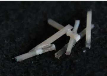
Figure 2: Sticks.
Debonded or lost sticks were all registered. Debonded sticks were those separated at the adhesive interface during the cutting procedure. Lost sticks were those which were lost or fractured during test preparation.
The remaining sticks were kept in distilled water for a maximum of 24 hours, until microtensile tests were performed.
6. Microtensile Tests
The specimens were individually attached to a stainless-steel grooved Geraldelli´s jig with cyanoacrylate glue (737 Black Magic Toughened adhesive, Permabond, H0ampshire, UK) and then submitted to a tension load using a universal testing machine (Instron® 4502 Series, Serial no. H3307, Instron Corporation, Canton, MA, USA), at a crosshead speed of 1mm/min until fracture occurred, with the stress to failure expressed in MPa.
THE EFFECT OF AN ADDITIONAL ADHESIVE LAYER ON DENTIN BOND STRENGTH:
COMPARISON WITH MANUFACTURE PROTOCOL
12 A digital caliper (Ficher Darex®, France) was used to measure the sides of the
bonding interface and calculate the bonding area in mm2. The μTBS (MPa) values were calculated by dividing the load (N) at failure by the area (mm2) of each stick.
The failure modes were analyzed under a stereomicroscope (Nikon, Japan) at 10x magnification to determine the mode of failure: failure occurring at the dentin-adhesive interface (A); cohesive when the failure occurred in dentin (CD) or in composite (CC); and mixed (M) , failure with composite and dentin at the interface. Observations of mode of fracture were performed by the same operator.
7. Statistical Analysis
The statistical analysis of the results was performed through descriptive and inference methods. An independent sample t-test was performed when the assumption of normality was valid.
Pre-testing failures that occurred during specimen preparation were treated as left-censored data and assigned a bond strength value of 1 MPa (Armstrong et al. 2010).
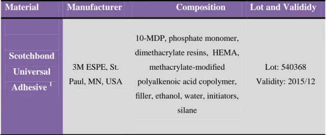
Table 1 – Composition, manufacturer, lot and validity of Scotchbond universal Adhesive.
Material Manufacturer Composition Lot and Valididy
Scotchbond Universal Adhesive 1 3M ESPE, St. Paul, MN, USA 10-MDP, phosphate monomer, dimethacrylate resins, HEMA,
methacrylate-modified polyalkenoic acid copolymer, filler, ethanol, water, initiators,
silane
Lot: 540368 Validity: 2015/12
THE EFFECT OF AN ADDITIONAL ADHESIVE LAYER ON DENTIN BOND STRENGTH:
COMPARISON WITH MANUFACTURE PROTOCOL
13 Table 2 – Composition, manufacturer, lot and validity of Scotchbond Universal Etchant, Adper
Scotchbond Multi-purpose and Composite ENAMEL plus HRi3.
Material Manufacturer Composition Lot and Valididy
3M ESPE, St. Paul, MN, USA
Scotchbond Universal
Etchant
32% phosphoric acid, water, synthetic amorphous silica, polyethylene, aluminum oxide
Lot: 537103 Valididy: 2015/11 Adper Scotchbond Multi-purpose 2 Component 1(etchant): 35% H3PO4 Primer: 3.3 Component 2: (Scotchbond
Multi-Purpose primer), HEMA, polyalkenoic acid polymer,
water Bonding: 8.2
Component 3: (Scotchbond
Multi-Purpose adhesive) Bis-GMA, HEMA,tertiary mines (both for light-cure and self-cure initiators), photo-initiator
Lot: N421442 Valididy: 2015/08 Composite ENAMEL plus HRi 3 Micerium S.p.A. Avegno (GE) Italy
Dimethacrylate, barium glass, ytterbium trifluoride, mixed oxides, prepolymers, additives, stabilizers, catalysts, pigments.
Lot: 2012000921 Lot: 2013008624 Valididy: 2018/06
THE EFFECT OF AN ADDITIONAL ADHESIVE LAYER ON DENTIN BOND STRENGTH:
COMPARISON WITH MANUFACTURE PROTOCOL
14 IV - RESULTS
A total of 166 (one hundred and sixty six) sticks were tested: 77 (seventy seven) using Scotchbond Universal Adhesive in total-etch mode as per manufacturer’s instructions (SBU TE D, n=77) and 89 (eighty nine) using Scotchbond Universal Adhesive with an extra hydrophobic coat of Scotchbond multipurpose adhesive (SBU+A TE D, n=89).
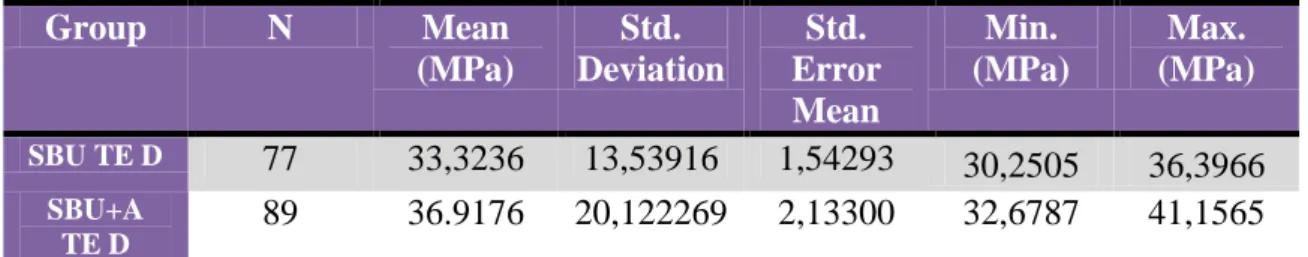
Micro-tensile bond strength (μTBS) mean values in MPa, standard deviations, and numbers of sticks tested per group are described in table 2.
Group N Mean (MPa) Std. Deviation Std. Error Mean Min. (MPa) Max. (MPa) SBU TE D 77 33,3236 13,53916 1,54293 30,2505 36,3966 SBU+A TE D 89 36.9176 20,122269 2,13300 32,6787 41,1565
Table 3 – Number of sticks (N); Micro-tensile (μTBS) mean values; Standard deviation
(Std. Deviation) and Standard Error Mean (Std. Error Mean).
Statistical analysis was performed using the SPSS 19.0 software for Windows (SPSS Inc, Chicago IL, USA). The t-test for independent samples was used for a confidence level of 95%. Kolmogorov-Smirnov and Shapiro-Wilk tests were used to verify the assumption of normality of the sample data. The homogeneity of variance was confirmed by Levene's test. Since the significance value (p) was superior to 0,05, the variances were assumed as equal.
THE EFFECT OF AN ADDITIONAL ADHESIVE LAYER ON DENTIN BOND STRENGTH:
COMPARISON WITH MANUFACTURE PROTOCOL
15 Graphic 1 and 2: Tests of Normality for the SBU TE D and SBU+A TE D group.
The distribution of μTBS is shown in graphic 3, where the median μTBS is represented by the central line of the box.
THE EFFECT OF AN ADDITIONAL ADHESIVE LAYER ON DENTIN BOND STRENGTH:
COMPARISON WITH MANUFACTURE PROTOCOL
16 The type of failures distribution per group is displayed on table 3 and 4. In group
1 (SBU TE D) most failures were mixed (M); in group 2 (SBU+A TE D) most failures were adhesive (A).
Group Failure Mode Number of fractures SBU TE D
A 19 (24.6%)
CC 13 (16.9%)
CD 13 (16.9%)
M 32 (41.6%)
Group Failure Mode Number of fractures SBU+A TE D
A 37 (41.6%)
CC 15 (16.8%)
CD 7 (7.8%)
M 30 (33.8%)
Table 4 and 5 - Failure mode: A - adhesive failure; CC - Composite cohesive failure;
CD - dentine cohesive failure; M - mixed failure.
In graphic 4, are showed the mean values of adhesive strength by type of failure and the test group.
Graphic 4 - Mean values of adhesive strength (by type of failure and the test group).
A- adhesive failure; CC- Composite cohesive failure; CD- dentine cohesive failure; M – mixed failure. 0 5 10 15 20 25 30 35 40 45 50 SBU TE D SBU+A TE D A CC CD M
THE EFFECT OF AN ADDITIONAL ADHESIVE LAYER ON DENTIN BOND STRENGTH:
COMPARISON WITH MANUFACTURE PROTOCOL
17 V - DISCUSSION
Adhesive systems available on the market can be classified into two categories: etch-and-rinse and self-etch strategies, in versions of three (only ER), two or one application step. Given the differences in professional judgment for the selection of the adhesive strategy and number of steps, some manufacturers have released more versatile adhesive systems that include etch-and-rinse (two step) and self-etch (one or two step) options: Universal adhesive systems. Scotchbond Universal Adhesive is one of these universal adhesives. However, it should be noted that despite the over-simplification of procedures reported by manufacturers (that clinicians tend to like because it saves them a considerable amount of time) the advantages of regular clinical use, still requires clinical and experimental evidence.
This experimental study evaluated the bond strength to dentine of a new universal adhesive system in two different groups: SBU TE D as group 1 used in total-etch mode as per manufacturer’s instructions and SBU+A TE D as group 2, used in total-etch mode with an additional adhesive layer.
As required from the ISO TR 11405 (Standardization. 2003) standard the teeth selected were stored in 0.5% chloramine T (Sigma Chemical Co., St. Louis, MO, USA) at 4°C for one week and then left in distilled water at 4°C for less than three months.
When we address the study of adhesion forces to tooth substrate, we have to take into consideration a number of different parameters that can affect the final outcome. Dentin preparation before bonding procedures is one of them. Depending on the type of surface preparation, we can create different variations of smear-layer (in size and structure’s) which play an important role in adhesion (Van Meerbeek et al. 2011). The use of sandpaper to create smear-layer was tested by Tao et. al (1987), who found only a small difference in bond strength when the smear layer was created with dental burs or sandpaper. In this study, a standardized and uniform smear-layer was created on each tooth, with the purpose of creating similar conditions to those occurring in a clinic situation.
Also, all of the adhesive procedures for both groups (SBU TE and SBU+A TE D) were performed by the same operator in a random sequence, thus eliminating any possible bias by repeating the same procedures.
THE EFFECT OF AN ADDITIONAL ADHESIVE LAYER ON DENTIN BOND STRENGTH:
COMPARISON WITH MANUFACTURE PROTOCOL
18 According to the manufacturer’s instructions, the ENAMEL plus HRi composite
used in restorative procedures was polymerized for 20 seconds. However, in order to avoid composite cohesive failures, an additional light polymerization was performed on mesial, distal, facial and lingual surfaces, for 10 seconds each.
Another parameter that may influence strongly the bond strength results is the cross-sectional area of the sticks. It is important that the cuts performed over the tooth substrate and resin composite restoration are as standardized as possible. In our study, teeth were longitudinally sectioned to obtain sticks with approximately 1mm2 of cross sectional area.
Micro-tensile test was used to assess the dentine bond strength of the resin-dentine interface. According with the available literature, many scientific papers use the micro-tensile bond strength approach (Van Meerbeek et al. 2010). It’s important to mention that bond strengths values cannot be considered as a specific material property (De Munck et al. 2005) because the results depend largely on experimental factors such as the type of resin composite, stress rate, sample size and geometry, and the actual test method (Phrukkanon S et al. 1998, Sudsangiam S&R 1999). For this reason, the absolute test values cannot be used to draw conclusions from, or be compared with, data recorded in other studies. One can only interpret the relative study outcome because they are a valid basis for further interpretation/comparison of the results (De Munck et al. 2005).
During the preparation, debonded and lost sticks for the micro-tensile tests were registered and considered as pre-testing failures. As in other studies, the pre-testing failures were excluded from statistical analysis (Perdigão J et al. 2014, Taschner M et al. 2014). Since pre-testing sticks had a certain (unknown) bond strength, including these pre-testing sticks in the total sample with a given bond values of 0 MPa, would severely penalize the final outcome of adhesive performance. On the other hand, this approach may overestimate the bond strength values (Van Meerbeek et al. 2010).
In this study, SBU TE D showed lowest μTBS mean values (33,32±13,5 MPa) than SBU+A TE D (36,92±20,1 MPa). However, since p > 0,05, there is no statistical differences in dentine μTBS between the two groups tested. These results may suggest that SBU TE D has similar performance when compared to SBU+A TE D regarding to μTBS. Consequently, the null hypothesis was accepted.
THE EFFECT OF AN ADDITIONAL ADHESIVE LAYER ON DENTIN BOND STRENGTH:
COMPARISON WITH MANUFACTURE PROTOCOL
19 Only one study, performed by Muñoz et al. (2014), compares μTBS values of
Scotchbond Universal Adhesive applied in ER mode and Scotchbond Universal Adhesive applied in ER mode with an additional layer of hydrophobic adhesive resin (Adper Scotchbond Multi-purpose, 3M ESPE, Seefeld, Germany). The results of µTBS testing using SBU in ER approach and SBU+A TE were similar to those achieved in our study.
In that study, the authors tested the μTBS to dentine of several universal adhesive systems: Scotchbond Universal Adhesive (SBU, 3M ESPE, St. Paul, MN, USa) , All-Bond Universal (ABU, Bisco Inc., Schaumburg, IL, USA) and G-Bond Plus [GBP, GC Corporation Tokyo, Japan ). In each different adhesive system, the universal adhesives were applied in both SE and ER techniques and an additional layer of hydrophobic resin coating was applied to all of them (HE, Heliobond, Ivoclar Vivadent, Schaan, Liechtenstein). The aim was to compare the immediate resin dentine microtensile bond strengths, nanoleakage and in situ degree of conversion of three universal adhesives with or without an additional hydrophobic resin coating. As in our study, the universal adhesive (SBU) was applied following the manufacturer’s instructions and with an extra layer of adhesive resin. The authors reported lower μTBS mean values with SBU TE D (32,3±3,7) than SBU+A TE (34,6±4,1) but no statistical significant differences between groups (p > 0,05).
Muñoz et al (2014) reported that most failures were adhesive for both groups: SBU TE D (48) vs SBU + A TE D (45). The majority of SBU TE D failures showed in our study were mixed (32) followed by adhesive failures (19) while SBU + A TE D showed more adhesive failures (37).
Although no statistical differences in dentin bond strength between the two adhesive systems were found, the addition of an extra hydrophobic adhesive layer leads to the creation of a larger interface between the substrate and the composite resin, and this may partially explain why most failures observed in the SBU+A TE D group were of the adhesive type. It can be speculated that the placement of this additional layer of hydrophobic resin over the unpolymerized universal adhesive will necessarily create a different hybrid layer, thus also partially explaining the higher incidence of adhesive failures.
Cohesive failures are frequently related with higher bond strength values (Perdigão J et al. 2006). In a study performed by Muñoz et al. (2013), the authors
THE EFFECT OF AN ADDITIONAL ADHESIVE LAYER ON DENTIN BOND STRENGTH:
COMPARISON WITH MANUFACTURE PROTOCOL
20 compared the immediate microtensile bond strengths, nanoleakage and in situ degree of
conversion of three universal adhesives applied to dentine according to the ER and the SE strategies. As control materials, the 2-step ER, Adper Single Bond 2; and the 2-step SE, Clearfil SE Bond were used. The following three universal adhesive systems were tested: Peak Universal Adhesive System, applied as a 2-step ER and 2-step SE; Scotchbond Universal Adhesive applied as a 2-step ER and 1-step SE; and All Bond Universal applied as a 2-step ER and 1-step SE.
In both studies (Muñoz M et al. 2013, Muñoz MA et al. 2014), the SBU used by ER technique showed no composite cohesive failures. However, each increment was light cured for 40s using a LED light-curing unit set at 1200 mW/cm2. Thus, although we performed 4 additional 10 seconds polymerization cycles on mesial, distal, lingual and buccal surfaces in our study, the number of composite cohesive failures may be explained by insufficient polymerization (Silva A 2008) and the use of a darker shade of resin composite, which reduces light transmission.
Failure mode analyses were performed by the same operator with the help of a stereomicroscope at 10x magnification. However, for a more reliable examination, it is desirable to perform this analysis with the help of scanning electron microscopy (SEM) (Armstrong SR. et al. 1998). In such manner, classification of mode of fracture is likely to be less operator-dependent, more reliable and accurate.
SBU+A TE D showed higher μTBS mean values than SBU TE D, although statistical significance was found. Unsolvated hydrophobic monomers are added to the adhesive interface by the extra layer of hydrophobic resin, which decreases the relative concentration of retained solvents and unreacted monomers in the adhesive layer (Breschi et al. 2008). Consequently, this increases the ultimate tensile strength of the adhesive interface, due to the formation of a more densely packed hybrid layer, making it more resistant to the tensile forces during μTBS testing and less prone to degradation effects over time (Reis A et al. 2007, Reis A et al. 2008, Muñoz MA et al. 2014).
As previously mentioned, the presence of 10-MDP molecule and polyalkenoic acid copolymer (first used in resin-modified glass-ionomer cement VitrebondTM), in the Scotchbond Universal formulation, appears to contribute to its good performance regarding μTBS, due to the two mechanisms of chemical bond present. Furthermore, the fact that it was used according to the total etch technique, whose performance is well documented, was in itself a good starting point. On the other hand, it should be noted
THE EFFECT OF AN ADDITIONAL ADHESIVE LAYER ON DENTIN BOND STRENGTH:
COMPARISON WITH MANUFACTURE PROTOCOL
21 that the results could have been influenced by the size of the tested sample in our study.
One can speculate that a more powerful sample comparing both adhesive protocols could reveal statistically significant differences.
With respect to clinical implications, taking into account the results obtained in our study, there seems to be no advantage in adding an additional layer of hydrophobic adhesive to the universal adhesive and by doing so, alter the manufacter´s protocol. This extra clinical step implies a higher consumption of material and time without any apparent benefit.
There are very few studies on this new type of UA. In the future, more clinical and laboratory studies are needed so that they can be compared with "gold standard" adhesives and thus assess their behavior in the short and long term.
THE EFFECT OF AN ADDITIONAL ADHESIVE LAYER ON DENTIN BOND STRENGTH:
COMPARISON WITH MANUFACTURE PROTOCOL
22 VI - CONCLUSION
There were no significantly statistical differences when μTBS values of SBU TE D were compared with SBU+A TE D. The null hypothesis is therefore accepted.
Hence, despite the limitations of this study, adding an additional hydrophobic resin coating layer does not seem to improve the adhesive performance of resin-dentine bond strengths of Scotchbond Universal when used in the TE strategy.
THE EFFECT OF AN ADDITIONAL ADHESIVE LAYER ON DENTIN BOND STRENGTH:
COMPARISON WITH MANUFACTURE PROTOCOL
23
VII - LITERATURE REFERENCES
Armstrong, S., S. Geraldeli, R. Maia, L. H. Raposo, C. J. Soares and J. Yamagawa (2010). "Adhesion to tooth structure: a critical review of "micro" bond strength test methods." Dent Mater 26(2): e50-62.
Armstrong SR., Boyer DB. and K. JC. (1998). "Microtensile bond strength testing and failure analysis of two dentin adhesives." Dental materials 14: 44-20.
Breschi, L., A. Mazzoni, A. Ruggeri, M. Cadenaro, R. Di Lenarda and E. De Stefano Dorigo (2008). "Dental adhesion review: aging and stability of the bonded interface." Dent Mater 24(1): 90-101.
Buonocore, M. G. A. (1995). " A simple method of increase the adhesion of acrylic filling materials to enamel surfaces." J. Dent. Res., Alexandria 34(6): 849-853.
Cadenaro M, Antoniolli F, Sauro S, Tay FR, Di Lenarda R and e. a. Prati C (2005). "Degree of conversion and permeability of dental adhesives." European Journal of Oral Sciences 113: 525-530.
Cardoso MV, Neves AA, Mine A, Coutinho E, Van Landuyt K, De Munck J and V. M. B. (2001). "Current aspects on bonding effectiveness and stability in adhesive dentistry." Australian Dental Journa 56: 31-44.
Carvalho, R. M., S. Chersoni, R. Frankenberger, D. H. Pashley, C. Prati and F. R. Tay (2005). "A challenge to the conventional wisdom that simultaneous etching and resin infiltration always occurs in self-etch adhesives." Biomaterials 26(9): 1035-1042.
De Munck, J., K. Van Landuyt, M. Peumans, A. Poitevin, P. Lambrechts, M. Braem and B. Van Meerbeek (2005). "A Critical Review of the Durability of Adhesion to Tooth Tissue: Methods and Results." Journal of Dental Research 84(2): 118-132.
Hanabusa M, Mine A, Kuboki T, Momoi Y, Van Ende A, Van Meerbeek B and D. M. J (2012). "Bonding effectiveness of a new ‘multi-mode’ adhesive to enamel and dentine." Journal of Dentistry 40: 475-484.
Hass V, Folkuenig MS, Reis A and L. AD. (2011). "Influence of adhesive properties on resin-dentin bond strength of onestep self-etching adhesives." Journal of Adhesive Dentistry 13: 417-424.
Kanemura, N., H. Sano and J. Tagami (1999). "Tensile bond strength to and SEM evaluation of ground and intact enamel surfaces." J Dent 27(7): 523-530.
King NM, Tay FR, Pashley DH, Hashimoto M, B. Ito S and e. a. WW (2005). "Conversion of one-step to two-step self-etch adhesives for improved efficacy and extended application." American Journal of Dentistry 18: 126-134.
THE EFFECT OF AN ADDITIONAL ADHESIVE LAYER ON DENTIN BOND STRENGTH:
COMPARISON WITH MANUFACTURE PROTOCOL
24 Meerbeek, V., V. Landuyt, J. Munck, M. Hashimoto, M. Peumans, P. Lambrechts, Y.
Yoshida and S. Inoue (2005). "Technique-Sensitivity of Contemporary Adhesives." Dent Mater Journal 24(1): 1-13.
Muñoz M, Luque I, Hass V, Reis A, Loguercio AD and B. NH (2013). "Immediate bonding properties of universal adhesives to dentine." Journal of Dentistry 41(5): 404– 411.
Muñoz MA, Sezinando A, Luque-Martinez I, Szesz AL, Reis A, Loguercio AD, Bombarda NH and P. J (2014). "Influence of a hydrophobic resin coating on the bonding efficacy of three universal adhesives." Journal of Dentistry 42: 595-602.
Pashley, D. H. and F. R. Tay (2001). "Aggressiveness of contemporary self-etching adhesives. Part II: etching effects on unground enamel." Dent Mater 17(5): 430-444. Pashley, D. H., F. R. Tay, L. Breschi, L. Tjaderhane, R. M. Carvalho, M. Carrilho and A. Tezvergil-Mutluay (2011). "State of the art etch-and-rinse adhesives." Dent Mater
27(1): 1-16.
Paul SJ, Leach M, Rueggeberg FA and P. DH. (1999). " Effect of water content on the physical properties of model dentine primer and bonding resins." Journal of Dentistry
27: 209-214.
Perdigão J and L. A. D. (2014). "Universal or Multi-mode Adhesives: Why and How?" Journal of Adhesive Dentistry 6(2): 193-194.
Perdigão J, Gomes G, Gondo R and Fundingsland JW (2006). "In Vitro Bonding Performance of All-in-one Adhesives. Part I - Microtensile Bond Strensths." Journal of Adhesive Dentistry 8: 367-373.
Perdigao J, Munoz M, S. A, Martinez L, S. R, R. A and L. AD (2014). "Immediate Adhesive Properties to Dentin and Enamel of a Universal Adhesive Associated With a Hydrophobic Resin Coat." Operative Dentistry 39(2): 11.
Perdigão J, Muñoz MA, Sezinando A, Luque-Martinez IV, Staichak R, Reis S and Loguercio AD (2014). "Immediate Adhesive Properties to Dentin and Enamel of a Universal Adhesive Associated With a Hydrophobic Resin Coat." Operative Dentistry
39(2).
Perdigão J, Sezinando A and Monteiro PC (2012). "Laboratory bonding ability of a multi-purpose dentin adhesive." American Journal of Dentistry 25: 153-158.
Perdigao, J., A. R. Carmo, C. Anauate-Netto, R. Amore, H. R. Lewgoy, H. J. Cordeiro, M. Dutra-Correa and N. Castilhos (2005). "Clinical performance of a self-etching adhesive at 18 months." Am J Dent 18(2): 135-140.
Perdigao, J., M. Munoz, A. Sezinando, I. Luque-Martinez, R. Staichak, A. Reis and A. Loguercio (2014). "Immediate Adhesive Properties to Dentin and Enamel of a
THE EFFECT OF AN ADDITIONAL ADHESIVE LAYER ON DENTIN BOND STRENGTH:
COMPARISON WITH MANUFACTURE PROTOCOL
25 Universal Adhesive Associated With a Hydrophobic Resin Coat." Operative Dentistry
39(2): 11.
Peumans, M., P. Kanumilli, J. De Munck, K. Van Landuyt, P. Lambrechts and B. Van Meerbeek (2005). "Clinical effectiveness of contemporary adhesives: a systematic review of current clinical trials." Dent Mater 21(9): 864-881.
Phrukkanon S, Burrow MF and T. M. (1998). "The influence of cross-sectional shape and surface area on the microtensile bond test. ." Dent Mater 14: 212-221.
Reis A, Albuquerque M, Pegoraro M, Mattei G, Bauer JR, Grande RH, Klein-Junior CA, Baumhardt-Neto R and L. AD (2008). "Can the durability of one self-etch adhesives be improved by double application or by an extra layer of hidrofobic resin? ." J Dent 36: 309-315.
Reis A, Carvalho Cardoso P, Vieira LC, Baratieri LN, Grande RH and L. AD. (2008). " Effect of prolonged application times on the durability of resin-dentin bonds." Dent Mater 24: 639-644.
Reis A, Grande RH, Oliveira GM, Lopes GC, L. AD. and (2007). "A 2-year evaluation of moisture on microtensile bond strength and nanoleakage." Dent Mater 23: 862-870. Reis A, Leite TM, Matte K, Michaels R, Amaral RC, Geraldeli S and L. AD (2009). "Improving clinical retention of one-step self-etching adhesive systems with ann additonal hydrophobic adhesive layer." J Am Dent Assoc 140: 877-885.
Silva A (2008). Efeito do tempo de polimerização nas forças de adesão entre a dentina e as resinas compostas (dissertação), Instituto Superior de Ciências da Saúde – Egas Moniz.
Standardization., I. O. f. (2003). SO/TR 11405 Dental Materials - Testing of adhesion to tooth structure. . Geneva, Switzerland, WHO: 1-16.
Sudsangiam S and V. N. R (1999). "Do dentin bond strength tests serve a useful purpose?" J Adhes Dent 1: 57-67.
Summitt, J., J. Robbins, T. Hilton, R. Schwartz and (2006). Fundamentals of Operative Dentistry.
Taschner M, Kümmerling M, Lohbauer U, Breschi L, Petschelt A and Frankenberger R (2014). "Effect of Double-layer Application on Dentin Bond Durability of One-step Self-etch Adhesives." Operative Dentistry 39(2).
Van Landuyt KL, Snauwaert J, De Munck J, Peumans M, Yoshida Y, Poitevin A, Coutinho E, Suzuki K, Lambrechts P and Van Meerbeek B (2007). "Systematic review of the chemical composition of contemporary dental adhesives." Biomaterials 28: 3757-3785.
THE EFFECT OF AN ADDITIONAL ADHESIVE LAYER ON DENTIN BOND STRENGTH:
COMPARISON WITH MANUFACTURE PROTOCOL
26 Van Landuyt, K. L., J. Snauwaert, J. De Munck, M. Peumans, Y. Yoshida, A. Poitevin,
E. Coutinho, K. Suzuki, P. Lambrechts and B. Van Meerbeek (2007). "Systematic review of the chemical composition of contemporary dental adhesives." Biomaterials
28(26): 3757-3785.
Van Meerbeek B, De Munck J, Yoshida Y, Inoue S, Vargas M, Vijay P, Van Landuyt K, L. P and V. G (2003). "Adhesion to Enamel and Dentin: Current Status and Future Challenges." Operative Dentistry 28(3): 215-235.
Van Meerbeek B, Perdigão J, Lambrechts P and V. G. ( 1998). "The clinical performance of adhesives." J Dent: 26: 21-20.
Van Meerbeek, B., J. De Munck, Y. Yoshida, S. Inoue, M. Vargas, P. Vijay, K. Van Landuyt, P. Lambrechts and G. Vanherle (2003). "Buonocore memorial lecture. Adhesion to enamel and dentin: current status and future challenges." Opererative Dentistry 28(3): 215-235.
Van Meerbeek, B., J. Perdigao, P. Lambrechts and G. Vanherle (1998). "The clinical performance of adhesives." J Dent 26(1): 1-20.
Van Meerbeek, B., M. Peumans, A. Poitevin, A. Mine, A. Van Ende, A. Neves and J. De Munck (2010). "Relationship between bond-strength tests and clinical outcomes." Dent Mater 26(2): e100-121.
Van Meerbeek, B., K. Yoshihara, Y. Yoshida, A. Mine, J. De Munck and K. L. Van Landuyt (2011). "State of the art of self-etch adhesives." Dent Mater 27(1): 17-28. Ye Q, Spencer P, Wang Y, M. A. and R. P. A (2007). "Relationship of solvent to the photopolymerization process, properties, and structure in model dentin adhesives." Journal of Biomedical Materials 80: 342-350.
Yiu CK, Pashley EL, Hiraishi N, King NM, Goracci C and e. a. Ferrari M (2005). "Solvent and water retention in dental adhesive blends after evaporation. ." Biomaterials
26(68): 63-72.
Yoshida Y, Yoshihara K, Nagaoka N, Hayakawa S, Torii Y, Ogawa T, Osaka A and Van Meerbeek B (2012). "Self-assembled Nano-layering at the Adhesive Interface." Journal of Dental Research 91(4): 379-381.
Yoshida, Y., K. Nagakane, R. Fukuda, Y. Nakayama, M. Okazaki, H. Shintani, S. Inoue, Y. Tagawa, K. Suzuki, J. De Munck and B. Van Meerbeek (2004). "Comparative study on adhesive performance of functional monomers." J Dent Res 83(6): 454-458.
I
VI - Anexx
II
ANEXX 1
III
A) MANUFACTURER’S INSTRUCTIONS
Scotchbond Universal (3M ESPE, St. Paul, MN) – Total-etch Strategy
1. Apply a commonly used phosphoric acid etching gel (about 35%), e.g. Scotchbond Universal Etchant, to the prepared and unprepared (if present) tooth structure (enamel and dentin) and allow to react for 15 sec.
2. Rinse thoroughly with water and dry with water-free and oil-free air or with cotton pellets; do not overdry.
3. Use the disposable applicator to apply the adhesive to the entire tooth structure and rub it in for 20 sec. Avoid contact between the adhesive and the oral mucosa.
4. The surface was then gently air-dried until it ceased to show any movement and the solvent was completely evaporated, forming a thin, homogenous and shiny film.
5. Light cure the adhesive with any commonly used curing light for 10 sec.