UNIVERSIDADE DE LISBOA
FACULDADE DE CIÊNCIAS
DEPARTAMENTO DE QUÍMICA E BIOQUÍMICA
THE INTERPLAY BETWEEN HIV-1 gp41,
MEMBRANES AND PRE-FUSION ANTIVIRAL
AGENTS
Ana Salomé Rocha do Nascimento Veiga
DOUTORAMENTO EM BIOQUÍMICA (Especialidade de Biofísica Molecular)
UNIVERSIDADE DE LISBOA
FACULDADE DE CIÊNCIAS
DEPARTAMENTO DE QUÍMICA E BIOQUÍMICA
THE INTERPLAY BETWEEN HIV-1 gp41,
MEMBRANES AND PRE-FUSION ANTIVIRAL
AGENTS
Ana Salomé Rocha do Nascimento Veiga
Tese orientada pelo Prof. Doutor Miguel Augusto Rico Botas Castanho
DOUTORAMENTO EM BIOQUÍMICA (Especialidade de Biofísica Molecular)
Prefácio
iii
PREFÁCIO
Os estudos que resultaram nesta tese foram objecto dos seguintes artigos já publicados ou submetidos para publicação:
Veiga S, Henriques S, Santos NC, and Castanho M (2004) Putative role of membranes in the HIV fusion inhibitor enfuvirtide mode of action at the molecular level. Biochem. J. 377, 107-110.
Veiga AS, Santos NC, Loura LMS, Fedorov A, and Castanho MARB (2004) HIV fusion inhibitor peptide T-1249 is able to insert or adsorb to lipidic bilayers. Putative correlation with improved efficiency. J. Am. Chem. Soc. 126, 14758-14763.
Veiga S, Yuan Y, Li X, Santos NC, Liu G, and Castanho MARB (2006) Why are HIV-1 fusion inhibitors not effective against SARS-CoV? Biophysical evaluation of molecular interactions. Biochim. Biophys. Acta 1760, 55-61.
Veiga AS, Santos NC, and Castanho MARB (2006) An insight on the leading HIV entry inhibitors. Recent Patents on Anti-Infective Drug Discovery 1, 67-73.
Veiga AS and Castanho MARB (2006) The membranes’ role in the HIV-1 neutralizing monoclonal antibody 2F5 mode of action needs re-evaluation. Antiviral Res. 71, 69-72.
Veiga AS and Castanho MARB (2007) The influence of cholesterol on the interaction of HIV gp41 membrane proximal domain derived peptides with lipid bilayers. FEBS J. 274, 5096-5104. Veiga AS, Pattenden LK, Fletcher JM, Castanho MARB, and Aguilar M (2007) HIV-1 Monoclonal Antibodies 2F5 and 4E10 membranes interaction in the presence and absence of its epitopes. Submetido.
Um outro estudo envolvendo também proteínas virais, mas não directamente relacionado com o estudo aqui apresentado, é apresentado no anexo II:
Pérez-Berná AJ, Veiga AS, Castanho MARB, and Villalaín J. (2007) Hepatitis C virus core protein binding to lipid membranes: the role of domains 1 and 2. J. Viral Hepatitis, in press.
Prefácio
iv
Gostaria de agradecer a todos os que de algum modo contribuiram para que a realização desta tese fosse possível.
À Fundação para a Ciência e a Tecnologia, pelo apoio financeiro concedido (bolsa SFRH/BD/14336/2003).
Ao Centro de Química e Bioquímica da Faculdade de Ciências da Universidade de Lisboa pelas condições de trabalho necessárias para a realização deste trabalho.
Ao meu orientador de trabalho, Professor Miguel Castanho, por me aceitar no seu laboratório e tornar possíveis as condições necessárias à realização dos trabalhos da tese. Agradeço ainda por toda a dedicação e apoio ao meu trabalho, mas principalmente pela amizade.
Aos meus colegas do grupo de Biofísica Molecular: Henri Franquelim, Marta Ribeiro e Manuel Melo pela amizade, companheirismo e boa disposição no dia-a-dia do laboratório. Um agradecimento especial à Sónia Henriques, companheira de licenciatura e doutoramento, pelo companherismo e ajuda, por todos os momentos partilhados, pelas úteis discussões científicas, e não só, e principalmente pela amizade.
Um agradecimento especial ao Professor Manuel Prieto pelo constante encorajamento e discussões científicas, disponibilidade para a realização de alguns trabalhos experimentais no seu laboratório, e também pela sua amizade. Ao professor Luís Loura por toda a ajuda e disponibilidade, em especial na realização dos estudos de espectroscopia de fluorescência resolvida no tempo do inibidor de fusão T-1249. Aos restantes membros do grupo de Biofísica Molecular do Centro de Química-Física Molecular do Instituto Superior Técnico, por toda a ajuda e disponibilidade. Ao Doutor Alexander Fedorov, pela contribuição essencial para este trabalho através da realização das medidas de fluorescência resolvidas no tempo.
Ao Professor Nuno Santos, por toda a ajuda concedida e disponibilidade demonstrada ao longo de todo o doutoramento, mas também pela amizade.
À Professora Marie-Isabel Aguilar por me ter recebido no seu laboratório para os estudos de Surface Plasmon Ressonance dos anticorpos 2F5 e 4E10, e por todo o apoio concedido. Ao Leonard Pattenden pelo seu apoio durante a realização do trabalho experimental, e pelo seu entusiasmo pelo trabalho. Ao Jordan Fletcher que tornou possível a síntese do péptido necessário à realização do trabalho experimental. Um agradecimento especial à Sharon Unabia por toda a assitência técnica, ajuda e
Prefácio
v disponibilidade e por todos os momentos de diversão. Um agradecimento também aos restantes elementos do grupo por todas a ajuda e disponibilidade demonstradas.
À Roche (Palo Alto, CA, EUA) por ter facultado os inibidores de fusão T20 e T-1249.
Ao Professor Cláudio Soares pela ajuda nos modelos moleculares do T20 e T-1249. Ao Professor João Pessoa, Instituto Superior Técnico, por me ter facultado a utilização do aparelho de dicroísmo circular. Um agradecimento especial à Susana por toda a incasável ajuda na realização das medidas.
Ao Professor José Villalaín por me ter recebido no seu laboratório para a realização dos estudos de FTIR e por todo o apoio concedido. À Ana Joaquina Pérez-Berná pelo seu apoio durante a realização do trabalho experimental, e pela sua amizade. À Ana Isabel Goméz por toda a assitência técnica, ajuda, disponibilidade e pela amizade. Um agradecimento também aos restantes elementos do grupo por todas a ajuda e disponibilidade demonstradas.
À Virginia, companheira de licenciatura, por toda a amizade, partilha de muitos bons momentos, e pelo encorajamento constante no trabalho.
E finalmente gostava de agradecer aos meus pais pelo apoio e encorajamento sem os quais esta tese não seria possível.
Resumo
vii
RESUMO
O vírus da imunodeficiência humana (VIH) é o agente causador do síndrome da imunodeficiência adquirida (SIDA) e o seu modo de actuação consiste, sobretudo, em infectar e destruir células T CD4+ do organismo hospedeiro. Existem dois tipos do vírus, VIH-1 e VIH-2, mas é o VIH-1 o mais virulento e o responsável pela maior parte dos casos de SIDA no mundo.
O envelope do vírus consiste numa bicamada lipídica, derivada da célula hospedeira, que possui um complexo glicoproteíco que confere ao vírus a capacidade para entrar nas células alvo. Este complexo é formado pelas subunidades gp120 (subunidade superfície) e gp41 (subunidade transmembranar), que se mantêm associadas de modo não covalente e oligomerizam sob a forma de trímeros na superfície do vírus. A gp41 é uma proteína transmembranar constituída por um domínio extracelular (ectodomínio), um domínio transmembranar e um domínio intracelular (endodomínio). O ectodomínio da gp41 contém várias regiões funcionais importantes para o processo de fusão viral. O péptido de fusão, localizado no terminal N da proteína, é uma região hidrófoba e rica em resíduos de glicina. Este domínio é essencial para o processo de fusão, nomeadamente para a penetração inicial da membrana da célula alvo. No seguimento do péptido de fusão encontram-se duas regiões que têm tendência para formar superenrolamentos de hélice α. A região adjacente ao péptido de fusão é chamada NHR e a região CHR é a que precede o segmento transmembranar. Os péptidos derivados destas regiões são chamados péptidos N- e C-, respectivamente. No terminal C da proteína, entre a região CHR e o domínio transmembranar, situa-se a chamada região próxima da membrana (MPR – membrane proximal region). Esta região é rica em resíduos de triptofano e parece ter também um papel chave na fusão viral.
O primeiro passo no processo de infecção consiste na ligação da gp120 ao receptor (CD4) e co-receptores (CXCR4 ou CCR5) na superfície das células alvo. Esta interacção induz alterações conformacionais na gp120 e modula as interacções gp120/gp41. A gp41 sofre então uma alteração conformacional e a sua estrutura nativa transita para uma estrutura intermediária de pré-hairpin. Estas variações na gp41 incluem a exposição do péptido de fusão e a sua inserção na membrana alvo. Posteriormente dá-se uma associação entre as regiões NHR e CHR levando à formação
Resumo
viii
da estrutura em hairpin (trímero em dímeros de hélices antiparalelas), activa para a fusão, o que leva à aproximação das membranas celular e viral. Na estrutura em hairpin a região NHR forma três hélices centrais arranjadas num superenrolamento trimérico, enquanto que a região CHR forma três hélices exteriores que encaixam de um modo antiparalelo em cavidades hidrófobas na superfície do superenrolamento. O que ocorre desde a aproximação das membranas até à fusão completa não está clarificado mas parece envolver a formação de agregados dos trímeros da proteína do envelope para formar poros de fusão.
O tratamento mais comum para os doentes com SIDA baseia-se na aplicação de combinações de drogas anti-VIH, nomeadamente inibidores do transcriptase reversa e do protease, dois enzimas virais com importância ao nível da replicação do vírus. Apesar dos benefícios do uso destas drogas, a sua utilização é limitada devido os seus efeitos tóxicos e aparecimento de estirpes resistentes. Assim, tornou-se óbvia a necessidade de desenvolvimento de drogas mais eficientes, menos tóxicas e que permitam eliminar os efeitos adversos associados à acção das drogas no interior das células.
Do trabalho científico desenvolvido nos últimos anos, resultaram fármacos que têm como objectivo actuar ao nível da entrada do vírus nas células, nomeadamente, na ligação da gp120 ao CD4, na ligação da gp120 aos co-receptores e na fusão do envelope do vírus com a membrana alvo. Apesar da enorme quantidade de compostos em desenvolvimento e até em fase de testes clínicos, o T20 (também chamado Enfuvirtide ou pelo nome comercial da marca Fuzeon), um inibidor de fusão, é o único já aprovado para aplicação clínica. No entanto, apesar da sua comprovada eficácia clínica, não há consenso sobre o seu modo de actuação ao nível molecular. O mecanismo de acção mais aceite envolve a interacção do T20 com a região NHR da gp41, na fase de pré-hairpin, impedindo a formação da estrutura em hairpin e deste modo a fusão viral com a membrana celular.
As membranas biológicas são entidades complexas, constituídas principalmente por lípidos e proteínas, que limitam as células, constituindo uma barreira entre o meio exterior e interior. A base estrutural das biomembranas é a bicamada lipídica. No entanto, a distribuição lipídica nas biomembranas pode ser assimétrica levando a que as duas camadas tenham uma composição lipídica diferente. Além disso, apesar de grande parte dos lipídos na bicamada formarem uma mistura uniforme e homogénea na
Resumo
ix chamada fase fluida, podem ocorrer heterogeneidades laterais, ou domínios, com composições lipídicas distintas. Os rafts lipídicos, são domínios encontrados nas membranas plasmáticas que são enriquecidos em colesterol e esfingolípidos. Estes domínios existem numa fase de líquido ordenado que é mais ordenada e compacta que o resto da bicamada, em fase fluida. Tem sido proposto que os rafts podem funcionar como plataformas para a reunião de complexos macromoleculares associados a membranas com importância ao nível de vários processos celulares, entrada de vírus como o VIH-1 para as células (uma vez que receptores e co-receptores se encontram provavelmente associados a rafts) e a formação de vírus a partir da célula infectada. De resto, a membrana deste vírus é muito enriquecida em colesterol e esfingolípidos, relativamente aos níveis encontrados nas membranas das células alvo, sendo sugerido que isso se deve ao facto de a formação dos novos vírus se fazer através de rafts lipídicos. O colesterol possui um papel central uma vez que mantém os rafts num estado funcional.
O estudo da interacção entre péptidos e/ou proteínas e biomembranas é muitas vezes necessário de modo a clarificar alguns processos em que ambos estejam envolvidos. No entanto, as membranas biológicas são sistemas demasiado complexos para serem usadas como tal neste tipo de estudo. Uma alternativa viável é o estudo destas interacções em sistemas modelo de membranas, nomeadamente vesículas lipídicas unilamelares. Podendo ser preparadas em diferentes tamanhos, foram no presente trabalho utilizados na sua maioria vesículas unilamelares grandes com diâmetro aproximado de 100 nm. Estes sistemas miméticos de membranas biológicas permitem uma base de estudo simplificada em simultâneo com a manutenção das principais propriedades das biomembranas.
Os estudos realizados ao longo deste trabalho foram essencialmente biofísicos, baseados em técnicas de espectroscopia de fluorescência, dado o facto de os péptidos e proteínas estudados serem intrinsecamente fluorescentes sem necessidade de acoplamento de sondas.
O T20, um péptido sintético que corresponde a uma sequência linear da gp41 do VIH-1 (entre o terminal C da região CHR e o terminal N da MPR), é um potente inibidor da fusão deste vírus. Apesar de estar aprovado para uso clínico, não existe ainda consenso acerca da sua acção ao nível molecular. Investigou-se a possível existência de um papel para as membranas lipídicas no modo de acção do T20, sendo que este envolvimento é sugerido tanto pela estrutura secundária do péptido, como por
Resumo
x
estudos de hidrofobicidade teórica e por trabalhos já publicados. Os resultados obtidos permitem inferir um modelo para descrever a acção do T20. O T20 insere-se na membrana principalmente nas zonas mais fluidas, ao nível da camada externa da bicamada lipídica. Devido ao elevado conteúdo em colesterol do envelope do vírus, o T20 não tem tendência a ligar-se a este. Assim, o T20 concentra-se na membrana celular podendo esta funcionar como um reservatório. No entanto, a concentração do T20 no ambiente aquoso pode ficar suficientemente elevada de modo a permitir ligação à gp41, inibindo deste modo o processo de fusão pelo modelo de acção mais aceite. Por outro lado, a ligação do T20 à membrana pode possibilitar a sua ligação a outras regiões da gp41, diferentes da ligação que ocorre em solução aquosa, e que também permitem o bloqueio da fusão.
O T-1249 é um péptido inibidor de fusão do VIH-1 de segunda geração, mais potente do que o T20 e activo em estirpes que apresentam resistência à inibição pelo T20. Este péptido foi obtido através de técnicas de design de drogas, sendo composto por sequências do VIH-1, VIH-2 e do vírus da imunodeficiência símio (VIS). Não havendo também consenso sobre o seu mecanismo de acção e tendo em conta os resultados obtidos com o T20, a interacção deste pétido com membranas lipídicas foi também investigada. Tal como o T20, o T-1249 insere-se na camada externa da bicamada lipídica, nas zonas fluídas da membrana. No entanto, ao contrário do T20, o T-1249 tem a capacidade de adsorver a domínios ricos em colesterol. Esta capacidade aumenta a concentração local de T-1249 quer nas zonas da célula onde ocorre a fusão, os rafts lipídicos, quer na membrana do vírus, também rica em colesterol. Assim, tanto a membrana da célula como a membrana do vírus podem funcionar como um reservatório de péptido. O aumento da eficácia clínica do T-1249 em relação ao T20 pode estar relacionada com as diferenças da interacção de ambos com as membranas, descritas para os dois inibidores.
O SARS-CoV é um novo coronavírus identificado como o agente causador do síndrome respiratório agudo. A glicoproteína S do envelope do vírus é responsável, à semelhança da gp120/gp41, pela entrada do vírus nas células alvo. Alguns estudos apontam para um mecanismo de entrada na célula semelhante ao usado pelo VIH-1. Sendo um novo vírus não estão ainda disponíveis medicamentos específicos para o inibir. Dadas as semelhanças entre os dois vírus e com base numa análise estrutural da gp41 e da proteína S2, foi proposto que o T20 poderia inibir a fusão do SARS-CoV, através de um mecanismo semelhante ao mais aceite, ou seja, a inibição da formação da
Resumo
xi estrutura em hairpin. Num estudo efectuado no âmbito desta tese foi investigado o potencial do T20 e T-1249 para inibirem o SARS-CoV. Os resultados obtidos permitem concluir que apesar de ocorrer a interacção entre o T20 e o T-1249 e a região NHR do vírus, que pode levar à inibição do SARS-CoV, esta ligação pode não ser suficientemente forte para conseguir uma inibição efectiva.
A MPR no ectodomínio da gp41 do VIH-1 tem um papel chave no processo de fusão viral. Tem-se mostrado que esta região tem a capacidade de interactuar com membranas e provocar a sua destabilização, num processo semelhante à acção do péptido de fusão na membrana alvo. Dada a proximidade entre esta região e a membrana do vírus o efeito deve ser exercido sobre esta. Além disso, esta região contém uma pequena sequência normalmente definida como um domínio de ligação preferencial ao colesterol. Investigou-se a influência do colesterol na interacção com membranas de péptidos correspondentes à MPR como um todo e sem a sequência de ligação específica ao colesterol, e de um péptido correspondente apenas ao domínio de ligação ao colesterol. Propõe-se que a MPR pode ter uma função muito específica no processo de fusão viral de membranas, através da acção concertada de domínios de ligação ao colesterol e domínios que não ligam ao colesterol.
O 2F5 e o 4E10 são dois anticorpos com uma acção potente contra o VIH-1, cujos epitopos se encontram localizados na MPR no ectodomínio da gp41. Tem sido sugerido que as membranas lipídicas, mais concretamente a membrana do vírus, podem ter um papel no mecanismo de acção destes anticorpos, que ainda não é conhecido. Alguns estudos propuseram que estes anticorpos podem interactuar com a membrana viral através de uma superfíce hidrófoba presente numa das cadeias pesadas. Estudou-se a interacção do 2F5 e do 4E10 com modelos de biomembranas, na presença e na ausência de um péptido que contempla o epitopo de ambos, de modo a investigar o possível papel das membranas no seu modo de acção. Tanto o 2F5 como o 4E10 têm a capacidade de interactuar com membranas, interacção mais fraca no caso do 2F5, e com o seu epitopo quando este está presente nas mesmas. Esta capacidade pode ser uma adaptação dos anticorpos para se ligarem a um epitopo que está tão próximo da membrana ou devido à existência de uma dependência da membrana na conformação do epitopo óptima para a ligação.
Summary
xiii
SUMMARY
The HIV-1 envelope glycoprotein complex plays an important role in viral entry. Its surface subunit gp120 is responsible for virus binding to cellular receptors, and the transmembrane subunit gp41 mediates fusion of the virus with the target cell. Different functional regions can be identified in gp41, each one with importance in the fusion process.
T20 and T-1249 are two HIV-1 fusion inhibitor peptides, being T20 already approved for clinical application in AIDS treatment. The work here presented describes research on the role of biomembranes on their mode of action. A model for the role of biomembranes was proposed.
Given the similarities between the HIV-1 and SARS-CoV, and the urgency to obtain licensed drugs to treat SARS, the possibility that T20 and T-1249 can work as SARS-CoV inhibitors was investigated. The results obtained show that despite the hypothesis that T20 could inhibit SARS-CoV fusion is correct, the effect may not be strong enough for practical application.
A small sequence inside the HIV-1 gp41 ectodomain membrane proximal region (MPR) is commonly referred to as a cholesterol binding domain. As the MPR plays a key role in the HIV-1 fusion process and the viral envelope is rich in cholesterol, the influence of cholesterol on the interaction of the MPR derived peptides with membrane model systems was studied.
2F5 and 4E10 are two potent and broadly neutralizing antibodies against HIV-1, whose epitopes are localized in the gp41 MPR. It was proposed that these antibodies can interact with the viral membrane trough a hydrophobic surface present on their third complementarity-determining region of the heavy chain. The 2F5 and 4E10 interactions with biomembranes models, in the absence and presence of its epitopes, were investigated in order to reveal the membranes role in the antibodies mechanism of action.
Contents xv
CONTENTS
Prefácio iii Resumo vii Summary xiii Contents xvAbbreviations and Symbols List xvii
I – INTRODUCTION 1
1. Human immunodeficiency virus (HIV) 1
1.1 HIV replication cycle and antiviral strategies 1
1.2 HIV entry and its inhibition 4
1.3 HIV fusion inhibitors 8
1.4 Anti-HIV antibodies 9
2. Biological membranes 11
2.1 Biological membranes lipid composition 11
2.2 Biological membranes asymmetry and heterogeneity 13
2.3 Lipid rafts 14
2.4 Cholesterol and rafts role on HIV infection 15
2.5 Membrane model systems 16
3. Peptides 18
3.1 Peptides as models 18
3.2 Peptide-membrane interactions 19
II – MEMBRANES ROLE ON THE MODE OF ACTION OF
THE HIV-1 FUSION INHIBITORS T20 AND T-1249 21
1. Introduction 21
2. Putative role of membranes in the HIV fusion inhibitor enfuvirtide
mode of action at the molecular level 25
3. HIV fusion inhibitor peptide T-1249 is able to insert or adsorb to
Contents
xvi
III – PUTATIVE INHIBITION OF SARS-CoV BY THE HIV-1
FUSION INHIBITORS T20 AND T-1249 49
1. Introduction 49
2. Why are HIV-1 fusion inhibitors not effective against SARS-CoV?
Biophysical evaluation of molecular interactions 53
IV – THE gp41 MEMBRANE PROXIMAL REGION ROLE ON
HIV-1 FUSION PROCESS 65
1. Introduction 65
2. The influence of cholesterol on the interaction of HIV gp41
membrane proximal region-derived peptides with lipid bilayers 69
V – THE ROLE OF MEMBRANES IN THE HIV-1
ANTIBODIES 2F5 AND 4E10 MODE OF ACTION 81
1. Introduction 81
2. The membranes’ role in the HIV-1 neutralizing monoclonal
antibody 2F5 mode of action needs re-evaluation 85
3. Real-time biosensor interactions of HIV-1 antibodies 2F5 and 4E10 with a gp41 epitope prebound to host cell and viral membrane
mimetics 91
VI – FINAL CONCLUSIONS 129
VII – ANNEX I - An insight on the leading HIV entry inhibitors 137
VIII – ANNEX II - Hepatitis C virus core protein binding to lipid
membranes: the role of domains 1 and 2. 147
Abbreviations and Symbols List
xvii
ABBREVIATIONS AND SYMBOLS LIST
Ab Antibody
AIDS Acquired immunodeficiency syndrome
ACE-2 Angiotensin-converting enzyme 2
BLMs Black lipid membranes
CDR H3 Third complementarity-determining region of the heavy chain Chol Cholesterol
CHR C-terminal heptad repeat
CL Cardiolipin CoV Coronavirus
CT Cytoplasm tail
DNA Deoxyribonucleic acid
DRMs Detergent-resistant membranes
FDA Food and drug administration
FP Fusion peptide
GPI Glycosylphosphatidylinositol
GUV Giant unilamellar vesicle
HAART Highly active antiretroviral therapy
6HB Six helix bundle
HIV Human immunodeficiency virus
HIV-1 Human immunodeficiency virus type 1
HIV-2 Human immunodeficiency virus type 2
HR Heptad repeat
L Lipid phase
Ld Liquid-crystalline or fluid phase
Lα Liquid-crystalline or fluid phase
Lβ Gel phase
Lβ’ Tilted gel phase
Lo Liquid-ordered phase
LR Loop region
LUV Large unilamellar vesicle
MAb Monoclonal antibody
Abbreviations and Symbols List
xviii
MPR Membrane proximal region
NHR N-terminal heptad repeat
PA Phosphatidic acid PC Phosphatidylcholine PE Phosphatidylethanolamine PG Phosphatidylglycerol PI Phosphatidylinositol PIP Phosphatidylinositol-4-phosphate
PIs Protease inhibitors
PS Phosphatidylserine
POPC 1-Palmitoyl-2-oleoyl-sn-glycero-3-phosphatidylcholine
RNA Ribonucleic acid
RTIs Reverse transcriptase inhibitors
SARS Severe acute respiratory syndrome
SARS-CoV Severe acute respiratory syndrome- associated coronavirus
SIV Simian immunodeficiency virus
SM Sphingomyelin
So Ordered gel phase
SUV Small unilamellar vesicle
TM Transmembrane domain
UNAIDS Joint United Nations programme on HIV/AIDS
W Aqueous phase
WHO World Health Organization
Kp Partition coefficient
Introduction
1
Chapter I
INTRODUCTION
1. HUMAN IMMUNODEFICIENCY VIRUS (HIV)
Human Immunodeficiency Virus (HIV) is the causative agent of the acquired immunodeficiency syndrome (AIDS). There are two types of HIV, HIV-1 and HIV-2, being the HIV-1 the most virulent (Weiss, 2000; Janeway et al., 2001). HIV can be transmitted by blood and blood products, vertically (from mother to child), or through sexual activity. By the end of 2006 the United Nations Programme on AIDS (UNAIDS)/World Health Organization (WHO) epidemic update estimated 39.5 million (34.1-47.1 million) of people worldwide living with HIV (UNAIDS/WHO AIDS epidemic update, http://www.unaids.org/en/HIV_data/epi2006/default.asp). Despite the efforts, no curative treatment or effective vaccine has yet been achieved.
1.1 HIV replication cycle and antiviral strategies
HIV-1 is an enveloped virus (member of the Lentivirus subfamily of the retrovirus family (Weiss et al., 2000)) that infects CD4 T cells, dendritic cells and macrophages (Janeway et al., 2001). The viral envelope, derived from the host cell membrane, is composed of a lipid bilayer and has at the surface a glycoprotein complex that allows the virus to enter in target cells. The envelope glycoprotein complex is initially synthesized as a single chain glycoprotein precursor (gp160) which is proteolytically cleaved by a cellular protease to generate the gp120 (surface glycoprotein) and the gp41 (transmembrane glycoprotein) subunits. The virus contains also a viral capsid (constituted by the capsid protein), two copies of the viral genome and three essential enzymes: reverse transcriptase, integrase and protease (Figure I.1). Additionally the
Introduction
2
virus has structural and accessory proteins important to the structural integrity and the virus replication process (reviewed in Vaishnav and Wong-Staal, 1991; Turner and Summers, 1999).
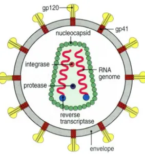
Figure I.1 – Simplified representation of the HIV-1. The reverse transcriptase, integrase, and
viral protease enzymes are packed in the viral capsid (in reality, many molecules of these enzymes are contained in each virion). The glycoprotein complex, at the surface of the viral envelope, is composed by the subunits gp120 and gp41. For simplicity, the other virus proteins were omitted (from Janeway et al., 2001).
An outline of the HIV-1 replication cycle is shown in Figure I.2. The gp120 on the surface of the virus binds to the CD4 receptor on the surface of the target cells activating the process that leads to membrane fusion and subsequent viral entry. Once inside the cell the viral RNA genome is transcribed into viral DNA by the action of the viral reverse transcriptase. The later is inserted into the host cell genome by the viral integrase. The RNA transcripts produced from the integrated viral DNA serve both as mRNA to the synthesis of the viral proteins (in the form of polyprotein precursors) and as the RNA genome of new viral particles. New viral particles escape from the cell after budding at the plasma membrane. Then, the viral protease acts cleaving the polyproteins to produce independent and functional viral proteins. The final maturation process leads to the formation of infectious virions (reviewed in Vaishnav and Wong-Staal, 1991; Turner and Summers, 1999).
Introduction
3 Figure I.2 – HIV replication cycle. The HIV life cycle presented shows the binding of the virus
to CD4 and coreceptor on the surface of the target cell, followed by membranes fusion and viral entry; reverse transcription, integration and protein synthesis processes are also shown; processing, virus assembly, budding and maturation of nascent virions are the last steps (adapted from Reeves and Piefer, 2005).
Currently, there are three U.S. Food and Drug Administration (FDA)-approved classes of drugs to the treatment of HIV-1 infection: 1) reverse transcriptase inhibitors (RTIs), including nucleoside or nucleotide and non-nucleoside molecules; 2) protease inhibitors (PIs) and 3) entry inhibitors (only one drug was approved). Both RTIs and PIs act on post-entry steps of the virus on the cells. Combination therapy with RTIs and PIs (called highly active antiretroviral therapy, HAART), involving at least three drugs, is the most common current treatment of HIV-1 infection (reviewed in Gulick, 2003; Stolk and Lüers, 2004; Yeni, 2006). Despite the success of this therapy in reducing morbidity and mortality of HIV-1 infected patients (Palella et al., 1998; Mocroft et al., 2000; Li et al., 2000; Louie and Markowitz, 2002), the adverse effects and the emergence of drug resistant HIV-1 strains have limited its application (Brinkman et al., 1998; Carr et al., 1999; Yerly et al., 1999; Hirsh et al., 1998; Carr, 2003). In an attempt to overcome these problems a third class of antiviral agents, the entry inhibitors, are receiving a lot of attention and large development. These inhibitors act extracellularly, targeting the first step in the HIV-1 replication cycle, i.e. preventing viral entry into target cells (reviewed
Introduction
4
in Eckert and Kim, 2001a; LaBranche et al., 2001; O’Hara and Olson, 2002; Moore and Doms, 2003).
1.2 HIV entry and its inhibition
The HIV-1 envelope glycoprotein, expressed on the surface of the viral membrane as a trimer (Center et al., 2002), is composed of two subunits noncovalently associated (gp120 and gp41). The gp120 subunit interacts with cellular receptors and the gp41 subunit is responsible for the fusion between the viral and cellular membranes (reviewed in Wyatt and Sodorski, 1998; Chan and Kim, 1998; Eckert and Kim, 2001a). The gp120/gp41 complex native conformation is not known in detail. Monomeric crystal structures of gp120 have been solved (Kwong et al., 1998; Wyatt et al., 1998) but no further information was achieved. Recent studies on the structure of the envelope glycoprotein complex showed contradictory results. For Zhu and colleagues a large globular domain comprises a gp120 trimer while the gp41 appears to form an open tripod (Zhu et al., 2006). The model derived by Zanetti et al. has the form of a mushroom with a more compact head domain and a gp41 stalk, rather than tripod legs (Zanetti et al., 2006). It was also proposed that the gp41 is folded in such way that brings the N- and C- terminal close to each other, stabilizing the native structure (Lorizate et al., 2006).
gp41 is composed of an ectodomain (extracellular domain), a transmembrane domain (TM) and an endodomain (intracellular domain or cytoplasm tail (CT)) (Figure I.3). Several functional domains have been identified in the ectodomain. The fusion peptide (FP) is located at the ectodomain N-terminal. This region, hydrophobic and rich in glycine residues, interacts with the target cell membrane and plays an important role in membrane fusion (reviewed in Tamm and Han, 2000; Epand, 2003). Two heptad repeat regions (HR) are also included: the first (NHR or HR1) near the N-terminal is adjacent to the FP; the second (CHR or HR2) is located at the ectodomain C-terminal. As observed for other viral fusion proteins, the regions that follow the FP, NHR and CHR, have the tendency to form α-helical coiled-coils (Chambers et al., 1990), denoted respectively, N- and C-helices. Peptides derived from these regions are referred to as N- and C-peptides, respectively. The two HR are separated by a loop region (LR) that contains an intramolecular cysteine bridge. At the C-terminal between the CHR and the
Introduction
5 TM is located a Trp-rich region, the membrane proximal region (MPR). Protein dissection combined with biophysical analysis demonstrated that the two HR regions within gp41 form a helical trimer of antiparallel dimers (Lu et al., 1995). The crystal structures of portions of the ectodomain (Chan et al., 1997; Tan et al., 1997; Weissenhorm et al., 1997) confirmed that the gp41 core tends to form a trimer of hairpins (or six-helix bundle (6HB)). Therefore, the N and C helices are arranged in a trimeric coiled coil. A central trimeric coiled coil formed by the N-peptide region is surrounded by three helical C-peptides that bind to conserved hydrophobic grooves on the coiled-coil surface in an antiparallel orientation. This structure represents the fusion-active conformation of gp41.
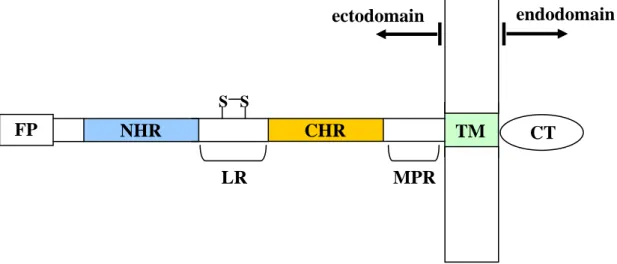
Figure I.3 – Schematic view of gp41 showing the location of the fusion peptide (FP), the N-
and C-terminal heptad repeat regions (NHR and CHR), the loop region (LR), the membrane proximal region (MPR), the transmembrane domain (TM) and the cytoplasm tail (CT).
HIV-1 entry into target cells (Figure I.4) is believed to be a multi-step and complex process (reviewed in Chan and Kim, 1998; Eckert and Kim, 2001a). The first step is the binding of gp120 to the target cell surface molecule CD4, which serves as the main receptor for HIV-1 (Maddon et al., 1986). However, CD4 alone is not sufficient for HIV-1 to fuse with the cells (Chesebro et al., 1990; Clapham et al., 1991). Two chemokine receptors, known as CCR5 and CXCR4, are the major HIV-1 coreceptors and all strains can use one (R5 and X4 viruses) or both (R5X4 viruses). CCR5 is particularly important as a coreceptor because R5 viruses are responsible for almost all cases of HIV-1 transmission and predominate during the early events of infection. X4 and R5X4 viruses emerge in many but not all infected individuals in the late stages of the infection (reviewed in Moore et al., 1997; Berger et al., 1999; Clapham and
FP NHR CHR S S LR TM MPR CT endodomain ectodomain
Introduction
6
McKnight, 2002; Douek et al., 2003). The gp120-CD4 binding induces conformational changes in gp120 leading to the exposure or the formation of the coreceptor binding site. gp120 binding to the CD4 and coreceptor results in further conformational changes that lead to gp41 activation into its fusion-active state. The gp41 conformational changes leads to the insertion of its FP into the target cell membrane and the formation of a prehairpin intermediate that bridges the viral and cellular membranes. Subsequent changes within the gp41 ectodomain involve the interaction of CHR and NHR, and a 6HB structure (also called hairpin structure) is formed. The hairpin formation brings the viral and cell membrane into closed proximity, allowing fusion of the membranes and then entry of the virus. It is likely that the free energy released with the 6HB formation must be sufficient to membranes fusion to occur. In fact it was demonstrated that fusion is caused by the movement of the protein into the 6HB rather than by the 6HB itself (Melikyan et al., 2000). Despite this widely accepted viral entry model, accumulation of new data indicates the model needs revision. Gallo et al. proposed that the gp120-CD4 interaction triggers the prehairpin intermediate formation and that coreceptor binding leads to rapid 6HB formation (Gallo et al., 2001; Gallo et al., 2004). Also, the formation of the 6HB might not be complete until the fusion pore complex is formed. Some data suggest that the formation of some 6HB occur after a fusion pore has formed and in this way the essential role of this structure is to stabilize the fusion pore against collapse and ensure its growth (Markosyan et al., 2003).
Introduction
7 Figure I.4 – HIV entry process. The fusion process for the trimeric envelope glycoprotein
complex is depicted in schematic form. The sequential binding of the gp120 to CD4 and a coreceptor on the cell membrane drives conformational changes in gp41. These changes cause the insertion of the FP into the cell membrane and the formation of the prehairpin intermediate. Subsequent gp41 conformational changes create the 6HB, and the viral and cellular membranes are brought into close proximity for fusion to occur (adapted from Liu et al., 2007b).
Each of the HIV-1 entry steps can be a target for entry inhibitors. The ones currently under development fall into three categories: gp120-CD4 binding inhibitors (or attachment inhibitors), gp120-coreceptors binding inhibitors (or chemokine coreceptors inhibitors) and fusion inhibitors (reviewed in De Clercq, 2004; Reeves and Piefer, 2005; Briz et al., 2006). Inhibitors that block the gp120-CD4 interaction include compounds that interact with the gp120 or CD4. PRO 542 (CD4-IgG2) is a recombinant tetrameric CD4-IgG2 fusion protein, which binds to gp120 (Allaway et al., 1995). BMS 806 (BMS 378806) is a small molecule inhibitor with potent antiviral activity. Firstly reported to binds directly to gp120 (Guo et al., 2003) it was proposed later that it should interfere with gp41 CD4-induced conformational changes (Si et al., 2004). TNX 355 (Hu5A8) is a CD4 specific MAb that binds to domain 2 of the CD4 receptor (Burkly et al., 1992). The antiviral agent cyclotriazadisulfonamide (CADA) exerts its effect by down-modulating the CD4 receptor (Vermeire et al., 2002). Several types of coreceptors inhibitors have been identified, namely low molecular weight CXCR4 and CCR5 antagonists. These include AMD 070 (Schols et al., 2003) and KRH-1636 (Ichiyama et al., 2003) for CXCR4 and Schering D (Tagat et al., 2004), TAK 220 (De Clercq, 2004) and AK602 (Maeda et al., 2004) for CCR5. Fusion inhibitors such as T20 (also known
Introduction
8
as DP-178, Enfuvirtide, or Fuzeon) and T-1249 (Tifuvirtide) are compounds whose mode of action involves binding to gp41 and interfere with the conformational changes that lead to the 6HB formation and membrane fusion. These kinds of inhibitors are the leading compounds, being T20 the most advanced and already approved by the FDA (Kilby and Eron, 2003; Robertson, 2003).
1.3 HIV fusion inhibitors
Some synthetic C-peptides, such as C34 and T20, are potent inhibitors of HIV-1 infection (Wild et al., 1994a; Kilby et al., 1998). Despite being more potent, C34 is not a good candidate to a drug due to its low solubility (Otaka et al., 2002). C34 has been used in studies of HIV entry blocking by peptides, while T20 was developed as a novel anti-HIV drug. C34 sequence includes residues at the N-terminal that precede the T20 sequence (Figure I.5). These residues include those that pack into the hydrophobic pocket on the surface of the N-peptide coiled coil in the 6HB conformation, and are critical for its antiviral activity (Chan et al., 1998). C34 lacks the T20 C-terminal residues that are crucial to T20 inhibitory activity. It has been proposed that these C-peptides act by interfering with the formation of the 6HB in a dominant negative fashion, by binding to the NHR region exposed in the prehairpin intermediate (Lu et al., 1995; Chen et al., 1995; Furuta et al., 1998; Chan and Kim, 1998; Kilgore et al., 2003). Despite synthetic N-peptides exhibit also inhibitory activity against HIV, they are less potent inhibitors than the C-peptides (Wild et al., 1992; Lu et al., 1995; Eckert and Kim, 2001b). It was proposed that one of the main reasons for this behaviour is their tendency to aggregate in solution (Lu et al., 1995). N36 and DP107 are examples of N-peptide inhibitors (Figure I.5). In their mode of action, N-peptides may either target the CHR region (Lu et al., 1995) or intercalate with the NHR region (Wild et al., 1992; Wild et al., 1994b; Weng and Weiss, 1998). Based on the knowledge gathered on the structure, activity and function of these inhibitors, several modified peptides have been created, in an attempt to achieve better fusion inhibitors (reviewed in Liu et al., 2007a). One of these is the rationally designed, second generation fusion inhibitor T-1249, more potent than T20 and active against T20 resistant HIV-1 isolates (Greenberg et al., 2002). Another line of research is the design of recombinant proteins containing sequences of the gp41 NHR and/or CHR regions, which may act in a similar way as the N- and peptides. 5-Helix inhibitor, for instance, consists of three N-peptides and two
Introduction
9 peptides connected by short linkers (Root et al., 2001). This inhibitor binds to the CHR region with high affinity and efficiently inhibits HIV-1 infection.
Figure I.5 – Peptide inhibitors from the NHR and CHR regions of gp41. The sequences of
N-peptides N36 (residues 546-581) and DP107 (residues 553-590) are derived from the NHR region. The sequence of C-peptide C34 (residues 628-661) is derived from the CHR region, while that of T20 (residues 638-673) is derived from the CHR region and the MPR.
The potency of the antiviral activity of these fusion inhibitors peptides targeting the gp41 stimulated the search for small molecules with the same function (reviewed in Liu et al., 2007a). Non-peptidic small molecules can be used as an alternative and have the advantage of being easier to mass-produce and purify, and may be orally bioavailable. Peptides lack oral bioavailability, and need to be injectable since they are degraded in the digestive track. Moreover, they can be subjected to metabolic degradation and have a high cost of production.
1.4 Anti-HIV antibodies
The antibody (Ab) response to HIV is directed to several viral proteins. In HIV-1 infected individuals Abs against different proteins, including gp120 and gp41, are detectable (Pellegrin et al., 1996; Pilgrim et al., 1997; Richman et al., 2003; Aasa-Chapman et al., 2004). However, only a fraction of the Abs are neutralizing (Wyatt and Sodroski, 1998), i.e. Abs that neutralize HIV-1 prior to its entry into target cells. On HIV-1 the envelope glycoprotein complex gp120/gp41 is the only viral target available for neutralizing Abs (Figure I.6). Neutralizing Abs target in the gp120, the CD4 binding
FP NHR CHR MPR TM CT
DP107 NNLLRAIEAQQHLLQLTVWGIKQLQARILAVERYLKDQ N36 SGIVQQQNNLLRAIEAQQHLLQLTVWGIKQLQARIL
C34 WMEWDREINNYTSLIHSLIEESQNQQEKNEQELL
Introduction
10
domain, carbohydrate structures, the CD4-induced coreceptor binding domains and in the gp41, structures that are exposed during membrane fusion (Wyatt and Sodroski, 1998). Nevertheless, due to the envelope glycoprotein complex features, such as the extensive glycosilation, the occlusion of conserved domains within the oligomeric structure, and the transiently exposure during the viral entry process, potent neutralizing monoclonal Abs (MAbs) targeting these proteins are rare (reviewed in Wyatt and Sodroski, 1998; Ferrantelli and Ruprecht, 2002). Neutralizing MAbs against gp120 are directed predominantly to the CD4 binding domain and both the second and third hypervariable loops (V2 loop and V3 loop, respectively). Those that have been found to bind the gp41 ectodomain, target the MPR (reviewed in Zolla-Pazner, 2004). However, only few human MAbs with potent and broadly neutralizing activity have been isolated (Figure I.6). Of these, 2G12 and b12 bind to gp120 while 2F5, 4E10 and Z13 are directed to the MPR within gp41 (reviewed in Zolla-Pazner, 2004; Phogat et al., 2007).
Figure I.6 – (A) The viral envelope glycoproteins gp120 and gp41 are targets for neutralizing
Abs. Neutralizing Abs may also bind to cellular receptors sites implicated in HIV-1 entry as CD4 and CCR5. (B) The two broadly neutralizing MAbs to gp41, 2F5 and 4E10, bind to epitopes in the MPR; whereas 2G12 and b12 are the two broadly neutralizing gp120 Abs (adapted from Phogat et al., 2007).
Introduction
11
2. BIOLOGICAL MEMBRANES
Biological membranes, consisting primarily of proteins and lipids, are essential for the integrity and functionality of cells, providing a barrier between the inside and outside environments. In eukaryotic cells internal membranes also form the boundaries of organelles. Despite the biomembranes complexity, a lipid bilayer is its primary structural element. Due to the amphipatic nature of the component lipids, the bilayer presents a hydrocarbon environment in the interior core with the polar groups oriented toward the water phase (Figure I.7). Biomembranes are not only physical barriers. They are highly selective permeability barriers that control the transfer of information and the transport of ions and molecules between the inside and the outside of the cell. Biomembranes serve as the matrix and support for several proteins, which are involved in important functions of the cell as energy and signal transduction, solute transport, protein targeting and trafficking (Edidin, 2003a).
Figure I.7 – Schematic depiction of a lipid bilayer. The lipid bilayer surrounds and protects the
cell from its environment. In a bilayer, the lipids hydrophilic head group of is oriented toward the water phase, while the hydrocarbon tails form the inner hydrophobic part (adapted from Menger et al., 2005).
2.1 Biological membranes lipid composition
Cell membranes are extremely complex having many different lipids. However, four main types of lipids occur in biological membranes: glycerophospholipids, phosphosphingolipids, sterols and glycolipids (reviewed in Gennis, 1989; Zubay, 1998; Loura and de Almeida, 2004).
Polar head groups
Introduction
12
Glycerophospholipids are the predominant class of lipids in biomembranes. Its hydrophobic section is composed of hydrocarbon chains esterified to sn-1 and sn-2 positions of the glycerol backbone. These hydrocarbon chains are diverse in length and unsaturation. The sn-3 position is esterified to phosphoric acid. If no more groups are linked to the molecule, the resulting compound is phospatidic acid (PA). However, the phosphate group is generally linked to other group as choline (Figure I.8 A), ethanolamine, serine, glycerol, and inositol. The corresponding phospholipids are phosphatidylserine (PS), phosphatidylethanolamine (PE), phosphatidylcholine (PC), phosphatidylglycerol (PG), and phosphatidylinositol (PI). As the phosphate group is always negatively charged, the net charge of the molecule depends on the charge of the polar group moiety. In this way PG, PS, PI are negatively charged, while PC and PE have a net neutral charge. PC is the most abundant phospholipid of animal cell membranes, whereas PE is the major component in bacterial membranes (Gennis, 1989; Loura and de Almeida, 2004).
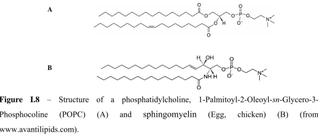
Figure I.8 – Structure of a phosphatidylcholine,
1-Palmitoyl-2-Oleoyl-sn-Glycero-3-Phosphocoline (POPC) (A) and sphingomyelin (Egg, chicken) (B) (from
www.avantilipids.com).
In phosphosphingolipids the hydrophobic group is a ceramide. Sphingolipids are rich in long and saturated hydrocarbon chains. Rarely found in plants and bacteria, sphingomyelin (SM) is the most abundant phosphosphingolipid in animal cell plasma membranes (Figure I.8 B). SM is structurally similar to PC. Both molecules have two long hydrocarbon side chains and a negatively charged phosphate group esterified to a choline.
The basic structure of sterols is a four-ring hydrocarbon. In cholesterol (Chol), the most important sterol in animal membranes, the molecular structure includes a tetracyclic
A
Introduction
13 fused ring skeleton, with a single hydroxyl group at carbon 3, a double bond between carbons 5 and 6, and a iso-octyl hydrocarbon side chain at carbon 17. The hydroxyl group gives Chol its slight amphipatic character and orients the molecule in membranes. Chol orients in a lipid bilayer with its polar hydroxyl group towards the aqueous phase and the hydrophobic ring system oriented parallel to, and buried in the hydrocarbon chains of the phospholipids. Other sterols are found in plant and yeast and other eukaryotic micro-organisms.
In glycoglycerolipids the sn-3 position of glycerol forms a glycosidic link to a carbohydrate. This kind of lipids is abundant in chloroplast membranes, blue algae and bacteria, but rarely found in animals. Glycosphingolipids have a glycosidic linkage to the terminal hydroxyl of ceramide. The simplest form of glycosphingolipid is a cerebroside that contains a single glycosidic residue. Glycosphingolipids are located in the outer surface of the plasma membrane and are particularly abundant in the nervous system.
2.2 Biological membranes asymmetry and heterogeneity
Lipids in the plasma membrane are asymmetrically distributed, i.e., the two membrane leaflets have a different lipid composition. The outer leaflet of an animal cell plasma membrane is mainly composed by SM and PC, while the inner leaflet has a mixture of predominantly PE and PS (Zachowski, 1993). Several functional roles for this asymmetric distribution have been suggested. These include several proteins that appear to localize in the cytoplasmic face of the membrane trough their interaction with PS (Palfrey and Waseem, 1985; Meers and Mealy, 1993; MacDonald, 1993; O’Toole et al., 1999). Disruption of lipid asymmetry leads to exposure of PS on the outer surface of the plasma membrane that serves as a trigger for macrophage recognition of apoptotic cells (Fadok et al., 1992; Bratton et al., 1997; Shiratsuchi et al., 1997).
In the Singer-Nicolson fluid mosaic model of biological membranes, bilayer lipids form a uniform and homogeneous fluid mixture, where lipid and proteins could move freely. Proteins are free to diffuse laterally or to rotate about an axis perpendicular to the plane of the membrane (Singer and Nicolson, 1972). However, the Singer-Nicolson model is now regarded as a simplistic description of the complex interactions and
Introduction
14
dynamics existent, namely by the presence of lateral heterogeneities like domains with distinct compositions. Examples of these are the apical and basolateral membranes of polarized epithelial cells that have distinct compositions, protein aggregation that result in domains that are enriched in a particular protein and lipid microdomains.
2.3 Lipid rafts
The fluidity of lipid bilayers is a function of temperature, pressure and lipid composition (length of the hydrocarbon chains and head group composition). The bulk of lipids in biomembranes are generally in the fluid liquid crystalline phase, and this state is essential for normal cell function (White et al., 2001). Pure phospholipid membranes show a temperature-dependent phase transition (Figure I.9). Phospholipid bilayers can exist in an ordered gel phase (So), where the lipid molecules are more
condensed and the hydrocarbon chains highly ordered. Dependent on the head group composition the gel phase is Lβ (for PE) or Lβ’ (for PC). In the Lβ and Lβ’ phases,
respectively, the chains are ordered parallel or show a tilt angle with the respect to the bilayer normal. Above a melting temperature (Tm), characteristic for each lipid, the
bilayer is present in a phase termed liquid-crystalline or fluid phase (Lα, Ld), in which
the phospholipid hydrocarbon chains are fluid and disordered (Gennis, 1989; Kranenburg and Smit, 2005). Chol has important effects on phase behaviour: decreases the order of the gel phase, increases the order of the fluid phase and stabilizes a new phase named liquid ordered (Lo). This Lo phase is characterized by high hydrocarbon
chain order in combination with high lateral mobility in the bilayer, that is, has characteristics from both gel- and fluid-phase (Ipsen et al., 1990; Spink et al., 1996). In mixtures that comprise appropriate amounts of sphingomyelin, unsaturated phospholipids and cholesterol, Lo and Ld phases can co-exist (de Almeida et al., 2003;
de Almeida et al., 2005).
Figure I.9 – Lipid bilayer gel Lβ (A) and Lβ’ (B) phases and the fluid phase (C) (adapted from Kranenburg and Smit, 2005).
Introduction
15 Lipid rafts are described as microdomains enriched in sphingolipids and Chol, that are present in the biological membranes organized in a way similar to a liquid ordered phase, more ordered that the surrounding fluid membrane (Simons and Ikonen, 1997; Edidin, 2003b; Simons and Vaz, 2004). It is believed that lipid rafts can facilitate selective protein-protein interactions, by excluding or including proteins. Examples of proteins that preferentially partition into raft domains are glycosylphosphatidylinositol (GPI)-anchored proteins and cholesterol-binding proteins such as caveolins (Rajendran and Simons, 2005). Raft domains are postulated to function as platforms involved in the lateral sorting of certain proteins during their trafficking within the cells and signal transduction events (Simons and Ikonen, 1997; Edidin, 2003b; Simons and Vaz, 2004). However, recent evidence points to an additional role of lipid and protein-protein interactions in lipid rafts formation and stabilization. These evidence suggests that rafts are initially small and unstable but can be captured and stabilized by proteins (Jacobson et al., 2007; Hancock, 2007). Several methods have been used to study lipid rafts in model and cell membranes (Pike, 2004). Isolation of detergent-resistant membranes (DRMs) and disruption of the lipid rafts structure based on Chol-depletion are two of the most widely used. However, these methods have limitations and undesired effects (Lichtenberg et al., 2005), and despite growing evidence showing the rafts existence and importance for cell functions, the presence of these domains in biological membranes still controversial (Munro, 2003). In spite of the possibility to visualize ordered domains in model membranes (Feigenson and Buboltz, 2001; Silvius, 2003; Veatch et al., 2004), visualize these domains in native cell membranes has revelled difficult. The small rafts size, its dynamic and unstable nature in cell membranes makes them difficult to be resolved by conventional microscopy techniques. Several techniques have been used to study the size of lipid rafts and their stability in vivo (Schütz et al., 2000; Pralle et al., 2000; Dietrich et al., 2002; Bacia et al., 2004) but no conclusive results were obtained so far.
2.4 Cholesterol and rafts role on HIV infection
Increasing amount of evidence now suggests that lipid rafts are involved in both early and late phases of the HIV-1 replication cycle (reviewed in Campbell et al., 2001), as well as in its infectivity. The HIV envelope is enriched in Chol and sphingolipids, which are characteristic of lipid rafts (Aloia et al., 1993; Brügger et al., 2006). It was
Introduction
16
shown that removal of Chol from viral particles almost completely eliminates HIV-1 infectivity (Campbell et al., 2002; Guyader et al., 2002; Graham et al., 2003; Campbell et al., 2004). The studies of the Chol and rafts role in viral entry have shown contradictory results. Some groups have shown that CD4 (Fragoso et al., 2003; Popik et al., 2002), CCR5 (Mañes et al., 1999; Popik et al., 2002) and, with reserve, CXCR4 (Popik et al., 2002) localize on lipid rafts. It was proposed that lipid rafts can serve as platforms for HIV entry, where the gp120 docking to receptors occurs (Mañes et al., 2000; Nguyen and Taub, 2002; Popik et al., 2002). However, coreceptors and CD4 may localize in different domains and it was suggested that initially HIV-1 binds to CD4 in rafts and subsequent interaction with CXCR4 requires the movement of the proteins complex out of the rafts (Kozak et al., 2002). A proposal based on Chol depletion of target cells indicates that although Chol is not required for viral entry, its depletion from cells with low coreceptor densities reduces the ability of gp120 to engage coreceptor clusters required to activate fusion (Viard et al., 2002). Additionally it was proposed that the presence of HIV-1 receptors in rafts is not required for viral infection and that Chol modulates the viral entry process independently of its ability to promote raft formation (Percherancier et al., 2003).
It has been suggested that viral budding occurs in cell membrane lipid raft domains (Nguyen and Hildreth, 2000, Ono and Freed, 2001; Pickl et al., 2001), what may explain the high level of Chol and sphingolipids in the viral membrane.
2.5 Membrane model systems
Given biomembranes complexity, the study of the lipid bilayer structure and properties is very important. The use of membrane model systems enables a simplified basis for these studies, while maintaining the main properties of the biomembranes. Models systems can be prepared with pure lipids or a mixture of lipids. There are different types of membrane model systems such as: lipid monolayers at an air-water interface; planar bilayers (also called black lipid membranes or BLMs); and lipid vesicles (or liposomes), which are probably the most popular models.
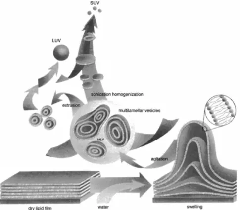
Liposomes preparation (Figure I.10) requires solubilization of lipids in organic solvents, followed by drying into a film and ressuspension in an aqueous environment. Suspensions of liposomes prepared in this manner form multilamellar vesicles (MLVs). MLVs are constituted by a heterogeneous mixture of vesicular structures which contain
Introduction
17 multiple bilayers forming groups of concentric shells. Due to the size of the MLVs particles light scattering is very significant, which could difficult its application in photophysical studies. In this way, different strategies were created in order to obtain unilamellar liposomes (Menger et al., 2005). The so called small unilamellar vesicles (SUVs), unilamellar vesicles of small diameter (30-50nm), are mainly obtained by sonication of MLVs suspensions. Regarding their small diameter, their light scattering activity is minimal. However, the small radius of curvature of SUVs results in packing difficulties of lipids. This curvature is much higher than the curvature of cell membranes, resulting in poor mimics of the properties of biomembranes. Additionally, SUVs are not stable and fusion occurs easily. Larger unilamellar vesicles (LUVs) can be produced by extrusion of MLVs through polycarbonate filters (with a defined pore size) that allow to obtain vesicles with a diameter near the pore size of the filter used. These vesicles closely resemble the lipid packing density and curvature of biomembranes. Giant unilamellar vesicles or GUVs (5-200μm) can be prepared by gentle hydration and electroformation (Rodriguez et al., 2005). These vesicles are particularly suitable for microscopy applications as their size enables visualization.
Figure I.10 – Schematic illustration of liposomes preparation that leads to MLVs, SUVs and
Introduction
18
3. PEPTIDES
3.1 Peptides as models
Proteins complexity may difficult their functional and structural characterization as a whole. The study of protein-protein and protein-lipid interactions for instance is frequently important to understand the function of proteins and their role in supra-molecular systems. The use of a peptide, whose sequence derives from a specific protein region can be a valuable strategy to study that specific protein sequence. Synthetic peptides are easier to manipulate, to obtain in large quantities, and can be mutated in relation to the primary sequence with extensive mutation possibilities. These peptides can be used to the study of a particular protein characteristic and understand the role of a particular amino acids sequence.
In HIV-1 entry studies the envelope glycoprotein complex role is of crucial importance. Recently acquired knowledge of the gp41 and gp120 (Di Bello et al., 2004) function and behaviour was achieved by the study of the whole protein but also by the use of synthetic peptides derived from the proteins sequence. The study of gp41 synthetic derived peptides allows to identify and to study different functional domains. Namely, taking into account the importance of the gp41 in the membranes fusion process, several studies use different peptides to unravel the membrane-active regions (Mobley et al., 2001; Moreno et al., 2004; Moreno et al., 2006). As the gp41 FP is responsible for the initial insertion of the gp41 on target cells membrane, the use of synthetic peptides to study the membrane binding properties of this region is very common (reviewed in Nieva and Agirre, 2003). The identification of key residues was possible due to the use of synthetic FP with different lengths and amino acids mutations (Pécheur et al., 1999). The use of peptides derived from the gp41 NHR and CHR regions is widely used to study the interactions of both regions involved in the formation of the 6HB structure (Chan et al., 1997; Tan et al., 1997; Lu et al., 1999). Additionally these peptides are also frequently studied to assess the involvement of the correspondent regions in membrane binding and destabilization, and consequently in the fusion process (Rabenstein and Shin, 1995; Peisajovich et al., 2000; Kliger et al., 2000; Sackett and Shai, 2002; Sackett and Shai, 2003; Shnaper et al., 2004; Pascual et al., 2005a; Korazim et al., 2006). A direct role of the gp41 LR in the fusion process was also proposed based on studies with synthetic peptides (Santos et al., 1998; Contreras et
Introduction
19 al., 2001; Pascual et al., 2005b). It has been proposed that the gp41 MPR is essential for the fusion process and the study with peptides has provided additional evidence for this function (Suárez et al., 2000a; Suárez et al., 2000b; Cirion et al., 2002; Sáez-Cirion et al., 2003). Even the gp41 CT was shown to interact with membranes by the use of peptides derived from this region (Kliger and Shai, 1997).
3.2 Peptide-membrane interactions
Understanding the interactions between peptides and membranes is important since such interactions play a key role in many biological processes. Antimicrobial peptides interact with microbial membranes as part of their mechanism of action and selectivity (Epand and Vogel, 1999; Hancock and Diamond, 2000; Zasloff, 2000), cell-penetrating peptides (CPPs) are able to translocate cell membranes non-endocytically (Derossi et al., 1998; Lindgren et al., 2000), and viral protein fragments are responsible for membranes fusion (Peisajovich and Shai, 2002; Tamm et al., 2002).
Both entropic, so called “hydrophobic” (Wimley and White, 1993) and electrostatic effects play a central role in peptide partition from water into membranes. Hydrophobic interactions are related with the energetic requirements for insertion of nonpolar residues in the aqueous environment. Electrostatic interactions occur because basic amino acid residues will be attracted electrostatically to the membrane surface containing negatively charged lipids. This effect is of particular importance for positively charged amphipatic peptides, such as antibacterial peptides, which interact and disrupt the negatively charged bacterial membrane (Matsuzaki, 1999; Epand and Vogel, 1999). However, if the lipids are neutral the electrostatic influence is insignificant and if both lipids and peptides present negative charges, the electrostatic contribution becomes repulsive. The partition of peptides into membranes can promote changes on their conformation. It is frequent that peptides that in the aqueous environment have a random coil structure adopt an α-helical conformation when associated with the lipid bilayer (Ladokhin and White, 1999; Wieprecht et al., 1999). The change to an amphipatic structure at the membrane surface reduces the free energy and is in this way an important contribution for membrane binding. The aromatic amino acids Trp, Tyr and Phe are hydrophobic and important in defining the presence of the peptides in the bilayer. Trp amino acid residues have preference to being inserted into lipid bilayers at the membrane-water interface (Yau et al., 1998; Persson et al., 1998;
Introduction 20 W W S, L L S, p /V n /V n K =
Killian and von Heijne, 2000; de Planque et al., 2003). Hydrophobicity scales (Wimley and White, 1996; White and Wimley, 1998) are composed of experimentally determined free energies of transfer between phases for each amino acid. These scales are a good tool in hydropathy analyses and in the prediction of peptide interactions with the bilayer. Two scales are used: one for partitioning from water to the bilayer interface and one for partitioning into n-octanol. The octanol scale appears to be a good measure of the partitioning to the bilayer interior (Wimley and White, 1996; White and Wimley, 1998). The parameter that describes the partition of a peptide between the lipid and water phases is the partition coefficient (Kp):
where nS,i are the moles of solute present in each phase (i = W, aqueous phase; i = L,
Membranes role on the mode of action of the HIV-1 fusion inhibitors T20 and T-1249
21
Chapter II
MEMBRANES ROLE
ON THE MODE OF
ACTION OF THE HIV-1
FUSION INHIBITORS
T20 and T-1249
1. INTRODUCTION
HIV infection most common treatment comprises RTIs and PIs. However, limitations such as treatment failure, toxicity, intolerance and HIV drug resistance, demand for new classes of antiviral agents. Entry inhibitors are a new class of drugs; the first antiviral agents that act extracellularly. Fusion inhibitors are the leading compounds of this class and T20 is the only approved so far by the FDA for clinical use (Robertson, 2003).
T20 is a synthetic 36 amino acids peptide derived from the C-terminal region of HIV-1 gp41 (Wild et al., 1994a; Wild et al., 1995). Its sequence contains residues from the CHR C-terminal and MPR N-terminal parts (Figure II.1). This fusion inhibitor is indicated for use as part of a combination therapy with other antiretroviral agents. Candidates for this therapy are treatment-experienced individuals with evidence of HIV-1 replication despite ongoing antiretroviral therapy, and is not generally applied in treatment-naïve patients. The recommended dosage of T20 in adults is 90mg twice daily, administered by subcutaneous injection (Dando and Perry, 2003). The addition of T20 to an optimized antiretroviral background regimen improved the virological
Membranes role on the mode of action of the HIV-1 fusion inhibitors T20 and T-1249
22
suppression and immunological benefit in treatment-experienced HIV-infected patients (Lalezari et al., 2003; Lazzarin et al., 2003). The most common adverse effects associated with its use are local injection sites reactions, but this does not seem to be a limitation to the treatment (Dando and Perry, 2003). Potent synergy was observed for T20 in combination with other entry inhibitors in vitro (Nagashima et al., 2001; Tremblay et al., 2002).
Figure II.1 – T20 sequence (residues 638-673) is derived from gp41 CHR and MPR.
The mode of action by which T20 inhibits viral fusion is still unclear, but several proposals have been presented involving different target sites in gp41 and gp120. The most currently accepted mechanism (Figure II.2) is the one proposed for C-peptides in general, involving interaction with the gp41 NHR region in an early intermediate of fusion, thus preventing the conformational changes that lead to the fusion-active arrangement (Wild et al., 1994a; Wild et al., 1995; Chen et al., 1995; Lawless et al., 1996; Rimsky et al., 1998; Kliger and Shai, 2000). In agreement, viral resistance to T20 is associated with mutations in the GIV sequence of the NHR region (Rimsky et al., 1998; Wei et al., 2002), and the LLSGIV sequence is important for the binding of T20 to gp41 (Trivedi et al., 2003). Nevertheless, T20 lacks some N-terminal residues present in other gp41 inhibitory C-peptides (e.g. C34) postulated as essential for the binding to the NHR region and inhibiting HIV-1 entry (Wild et al., 1994a, Chan et al., 1998). Blumenthal and co-workers (Muñoz-Barroso et al., 1998) proposed the existence of a second binding site for T20 on gp41, involving the contact site of gp41 oligomers cluster to form a fusion pore, which is required for the occurrence of the complete fusion process (Blumenthal et al., 1996; Chan and Kim, 1998). In fact, it was suggested that T20 binding affinity to the NHR region cannot justify its strong inhibitory activity and should have at least two different interactions modes with gp41 (Ryu et al., 1999). In addition Shai and colleagues (Kliger et al., 2001) showed that T20 can bind to membranes and oligomerize on its surface, at variance with aqueous solution, it cannot
FP NHR CHR MPR TM CT
YTSLIHSLIEESQNQQEKNEQELLELDKWASLWNWF T20
Membranes role on the mode of action of the HIV-1 fusion inhibitors T20 and T-1249
23 interact with the NHR region in the membrane environment. Therefore there are two possible T20 target sites proposed in gp41, both contributing to fusion inhibition: interaction with the NHR region in aqueous solution prevents the formation of the 6HB structure while interaction with the gp41 C-terminal region in the membrane environment inhibits fusion pore formation (Ryu et al., 1999; Muñoz-Barroso et al., 1998; Kliger et al., 2001). When T20 is bound to membrane-anchored proteins expressed in genetically modified target cell of HIV-1 infection, replication of the virus was inhibited more than 100-fold (Hildinger et al., 2001). A less accepted, but also proposed T20 mode of action is its binding to the gp41 FP, preventing the insertion of the last into the target cell membrane and thus membrane fusion (Mobley et al., 2001; Jiang et al., 2002).
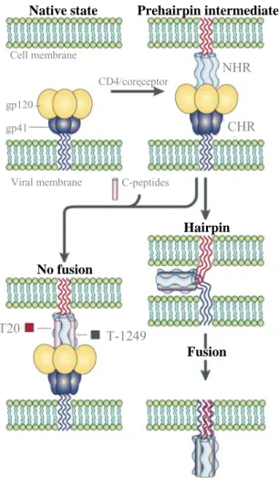
Figure II.2 – Mode of action of T20 and T-1249. Schematic of the most accepted mechanism
by which T20 and T-1249 inhibit viral entry into target cells. Both inhibitor peptides bind to the NHR region in a prehairpin intermediate, preventing the formation of a fusion active structure and thereby fusion and viral entry (adapted from Moore and Stevenson, 2000).
CD4/coreceptor
Native state Prehairpin intermediate
Cell membrane gp120 gp41 Viral membrane NHR CHR C-peptides Hairpin Fusion No fusion T20 T-1249 CD4/coreceptor