Journal of Applied Microbiology1998, 84, 523–530
The microstructure and distribution of micro-organisms
within mature Serra cheese
M.L. Parker, P.A. Gunning, A.C. Macedo1
, F.X. Malcata2
and T.F. Brocklehurst
Institute of Food Research, Norwich Research Park, Colney, Norwich, UK,1Instituto Superior da Maia, Maia and 2
Escola Superior de Biotecnologia, Universidade Cato´lica Portuguesa, Porto, Portugal 6240/05/97: received 21 May 1997, revised 10 July 1997 and accepted 14 July 1997
M . L . P A R K E R , P . A . G U N N I N G , A . C . M A C E D O , F . X . M A L C A T A A N D T . F . B R O C K L E H U R S T . 1998.
The distribution of micro-organisms in mature Serra, a traditional Portuguese
cheese made from unpasteurised ewes’ milk without added starter culture, was examined
by light microscopy and electron microscopy. Four populations of micro-organisms were
recognized according to their position within the cheese: (i) those present as apparently
axenic colonies within the curd matrix; (ii) bacteria growing along curd junctions; (iii) yeasts
and bacteria present in the smear on the surface of the cheese and (iv) bacteria found
in cracks which penetrated the outer part of the cheese from the rind. Two types
of crystals were observed, together with contaminants of vegetable origin and somatic
cells originating from the milk.
I N T R O D U C T I O N
Serra cheese is probably the most popular traditional Por-tuguese cheese. This semi-soft cheese has been produced for centuries on farms in the interior mountainous regions of Serra da Estrela, and is much in demand due to its unique organoleptic properties. It is manufactured from raw ewes’ milk using dried flowers of the thistle (Cynara cardunculus L.) as coagulant, without addition of any starter or secondary microflora (Macedo et al. 1993).
Detailed descriptive work on the microbiology of Serra cheese has been undertaken recently. This includes deter-mination of the viable numbers of major groups of micro-organisms (e.g. mesophilic lactic acid bacteria, coliforms, sta-phylococci, pseudomonads and yeasts) as a function of the length of the ripening period of the cheese, the time of manufacture within the lactation season and the axial location in the cheese (Macedo et al. 1996). The major species in these groups, as a function of the length of the ripening period and the time of manufacture within the lactation period, were also identified (Macedo et al. 1995). This type of information is essential if, in the future, legislation demands tighter control of the manufacturing process and conditions, and perhaps the use of well-defined starter/non-starter strains.
Growth of bacteria in food has recently been shown to be influenced by the physical structure of the food matrix,
Correspondence to: Dr F. Xavier Malcata, Director, Escola Superior de Biotecnologia, Rua Dr. Anto´nio Bernardino de Almeida, P-4200 Porto, Portugal.
particularly in close-packed emulsion systems (Brocklehurst
et al. 1995; Parker et al. 1995) such as full-fat cheese. As there
have been no previous studies on the microstructure of Serra cheese, it is not known whether the diverse wild microflora is distributed evenly throughout the cheese matrix, or con-fined to specific regions as is known to occur in other cheeses, such as Cheddar (Dean et al. 1959; Rammell 1960) and St Nectaire (Marcellino and Benson 1992). Nor is it clear why the final flavour characteristics of Serra are influenced by the degree of manual working of the curd (Macedo et al. 1993), so that cheeses of differing flavours can be produced by a cheese-maker using the same batch of milk and the same ripening conditions.
The main objective of this investigation was to establish the distribution of micro-organisms and other inclusions in mature examples of the two types of Serra, short-ripened (or soft, buttery) cheese with a ripening period of approxi-mately 40 d, and long-ripened (or hard, piquant) cheese rip-ened for at least 6 months. Tentative relationships between the cheese-making protocol and the cheese microstructure, distribution of micro-organisms, bacterial growth and even-tual development of flavour are also discussed.
M A T E R I A L S A N D M E T H O D S Cheese manufacture
Ewes are milked by hand twice a day into a small open vessel. The milk is filtered into a coagulation vat through a fine,
clean cloth to remove large impurities such as straw and wool, then placed by the fireplace to attain and maintain a coagulation temperature of around 28 °C, the temperature being assessed by the immersion of the cheese-maker’s finger tips into the milk. A plant coagulant extract is prepared by macerating dried thistle flowers into a paste with a little tap water and crude kitchen salt, then adding more water to extract the enzymes before filtering through a cloth. When the milk is at the correct temperature, generally after 30– 60 min, the rennet-like extract is added to the milk and mixed in with a long wooden spoon. Coagulation takes about 1 h, and the consistency of the curd is assessed visually by a slight agitation of the coagulation vat. The curds are cut into irregular shapes and sizes by manually stirring the coagulum with the bare hand for about 1 min, then they are poured into perforated plastic cheese moulds. The curds are then thoroughly smashed and pressed by hand to express the whey, and this may be followed by a 3–24-h period of pressing using stones placed on top of the curd in the mould. The most common processes of salting are either to add crude kitchen salt to milk before adding coagulant, or to rub the top and bottom surfaces of the pressed cheese with salt.
The cheeses are then taken to the maturation room and placed on wooden shelves which are covered with cotton cloths that are regularly washed but seldom sterilized. The humidity and temperature of the maturation room are not artificially controlled, so the ripening conditions are chiefly determined by the weather prevailing in the Serra da Estrela mountains (ca. 2000 m high); such weather, usually snowy and windy, is characterized by temperatures typically around 12 °C and relative humidities typically around 90%, and frequent opening of the ripening chambers allows equal-ization to some extent of inner and outer environmental conditions. Each cheese is turned over daily and, after 8–15 d, a reddish ‘smear’ appears on the outside of the cheese. The cheese is then washed with warm water for the first time, then a band of clean cotton cloth is wound around the cheese and tied with a small knot; this band keeps the cheese in shape as proteolysis proceeds. The cheese is then moved to a second, cooler (approximately 8 °C) maturation room and turned over daily, the cloth band being adjusted as necessary. The cheeses are washed weekly. The short-ripened, soft but-tery type cheeses are ripened for 30–45 d and the long-ripened, hard piquant types have a ripening period of at least 6 months. In order to reduce stickiness of the outside of long-ripened cheeses, particularly when the cheese is wrapped in paper, flour is sometimes rubbed onto the surface.
Cheese sampling
Short-ripened and long-ripened Serra cheeses were pur-chased from selected farm cheese-makers in Portugal, and immediately sent by express delivery, within appropriate
packaging, to the Institute of Food Research (Norwich, UK). On receipt, samples were cut from the outer part of each cheese including the rind (outer), from the centre of the cheese (inner) and from an intermediate region (mid). All samples were stored in formal saline (10 ml of 40% (w/v) formalin with 90 ml of 0·94% (w/v) sodium chloride) as described by Dean et al. (1959). The remaining cheese was stored in a refrigerator.
Cryostat sections
Sections, nominally 10 mm thick, were cut from formal saline-fixed cheese using a Cryocut E freezing microtome (Reichert-Jung, Vienna, Austria) and some of the sections were then defatted by immersing them in 100% acetone. Sections were stained either in very dilute toluidine blue (C.I. 52040; BDH, UK) or in equal volumes of 0·1% (w/v) acridine orange (C.I. 46005; BDH) and 1% (v/v) acetic acid. Cheese sections were examined by bright-field microscopy (LM) and fluorescence microscopy (FM) using a Leitz Ortholux II microscope with an HBO 50 W mercury arc lamp and an exciter and barrier filter combination with transmission of 450–490 nm and ×515 nm, respectively.
Resin-embedding for light microscopy
Blocks of the formalin-fixed cheese (in fixative for 2 months) were cut into smaller pieces, dehydrated in an ethanol series and infiltrated and embedded in the acrylic resin LR White (London Resin Co. Ltd, Reading, UK). The resin was poly-merized at 60 °C for 24 h. Sections, nominally 0·4 mm thick, were cut using glass knives and stained with 1% (w/v) tol-uidine blue in 1% (w/v) borax at pH 11, or acridine orange/ acetic acid for FM as above.
Resin-embedding for transmission electron microscopy
Pieces of cheese from the original supply, kept refrigerated since receipt, were fixed in 3% (v/v) glutaraldehyde (Agar Scientific Ltd, Stansted, UK) in 0·05 mol l−1 cacodylate
buffer (pH 7·4) overnight. After three washes in buffer, the cheese was post-fixed in 1% (w/v) osmium tetroxide (Agar Scientific Ltd), dehydrated in an ethanol series, transferred to 100% acetone, and then infiltrated and embedded in Spurr resin (Agar Scientific Ltd). Sections, approximately 80 nm thick, were cut using a diamond knife and collected onto copper grids. After sequential staining with uranyl acetate (saturated solution in 50% (v/v) ethanol) and lead citrate (Reynolds 1963), the sections were examined and photo-graphed in a JEOL 1200EX transmission electron microscope (JEOL, Tokyo, Japan).
M I C R O - O R G A N I S M S I N S E R R A C H E E S E 525
Scanning electron microscopy
Pieces of the formalin-fixed cheese were fractured, dehy-drated in an ethanol series and then transferred to 100% acetone and critical point dried (CPD) (Polaron, Watford, UK). Some of this dried cheese was also fractured. The pieces of cheese, fracture surfaces or rind uppermost, were mounted on aluminium stubs with silver-conducting paint (Agar Scientific Ltd), sputter-coated with a layer of gold approxi-mately 25 nm thick (Emitech Ltd, Ashford, UK) and exam-ined and photographed under a Leica Stereoscan 360 scanning electron microscope (LEO, Cambridge, UK).
R E S U L T S
Cryostat sections cover an area of about 1 cm2and are ideal
for investigating the large-scale morphology of the cheeses and the overall distribution of micro-organisms within them. However, because the sections are relatively thick, and there is a large difference in scale between the gross structure and the micro-organisms, it is difficult to illustrate all the features together. Staining cryostat sections with very dilute toluidine blue delineated the protein-rich curd junctions and revealed substantial areas of dark-staining micro-organisms on the surface rind of both soft (short-ripened) and hard (long-ripened) cheeses. Similarly, staining with acridine orange revealed the heavy microbial load in Serra with both yeasts and bacteria fluorescing orange-red within a green back-ground of casein, although the curd junctions were less well defined.
In hard cheese, cracks extended from the crust into the outer layer of the cheese, and these too were heavily con-taminated. Within the bulk of the cheese, micro-organisms appeared as numerous dark-staining streaks lining the curd junctions, and also as more compact masses within the curd matrix. Also visible, particularly in the outer region of the cheese, were numerous pink-tinged crystalline spheres approximately 50 mm in diameter. Fragments of plant cells, pollen grains and rust teleutospores were distributed throughout the cheese, but no fungal hyphae were seen in any part of the cheese.
Details of the micro-organisms were clearly seen in cheese embedded in LR White resin. The fat component of the cheese was not retained by this method, but its location is assumed to be the non-staining voids within the casein matrix. Part of the ‘smear’ covering the outer surface of the cheese is shown in section in Fig. 1a; the most conspicuous organisms within the smear were yeasts. In the outer layer of both hard and soft (Fig. 1a) cheeses, the fat-containing voids in the casein matrix were flattened, the protein appeared as strands and free fat was present in the smear, possibly as a result of proteolytic and lipolytic activity by microbes. Pockets of yeast cells were also found just below the rind within the casein
matrix (Fig. 1a). Examination of the smear by scanning elec-tron microscopy (SEM) revealed that populations of elongate yeasts (Fig. 1b), spherical yeasts (Fig. 1c), bacilli and cocci (Fig. 1b) coexist. Plate-like fat crystals were also present (Fig. 1c).
The cracks running from the rind into the outer region of the cheese usually followed the line of curd junctions and were colonized by bacteria (Fig. 1d). Deeper in the cheese, in places where curd junctions were not pressed tightly together, there were extensive colonies of bacteria (Fig. 1e) and some free fat. Fractures through CPD cheese revealed both face-views and cross-sections of the curd junctions. In face-view (Fig. 1f), some bacteria were seen embedded in the protein matrix. In Fig. 1g, a cross-section of a curd junction, bacilli and cocci can be seen to coexist.
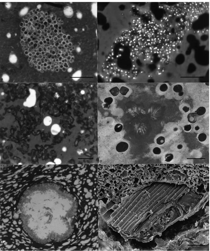
In both soft and hard Serra cheeses, considerable numbers of bacteria were present within colonies embedded through-out the casein matrix (Fig. 2a–f). These colonies were roughly spherical and, as far as can be judged from the morphology of the bacteria within them, each was occupied by a single species. These ranged from closely packed small cocci (Fig. 2a,b), to bacilli in both longitudinal and cross-section (Fig. 2c,d) and larger diplococci (Fig. 2e,f). Some colonies encompassed spherical fat globules (Fig. 2b,f). With experi-ence, colonies were easily distinguished from the surrounding protein matrix in fractured CPD material by the slight dif-ference in granularity between matrix and colony (Fig. 2b). This may reflect the differences in the amount of pro-teinaceous material present between the organisms in colon-ies, some apparently having very little (Fig. 2a,d,e) and others considerably more (Fig. 2c,f).
Colonies of yeast cells were also present in the protein matrix of the outer and mid-regions of both hard and soft (Fig. 3a) cheese. In the inner part of the cheese, groups of only a few yeast cells were seen. The most effective preparative procedure for visualizing small groups or single cells of both bacteria and yeasts was acridine orange staining of acrylic sections approximately 0·4 mm thick, visualized by FM (Fig. 3b).
Two types of crystalline inclusions were seen in Serra cheese. The mid- and inner regions of the cheeses, par-ticularly the protein-rich areas of the soft cheese, were rich in aggregates of kidney-shaped masses (Fig. 3c). At higher magnification, these were seen to consist of groups of radially arranged needles (Fig. 3d), each group approximately 2 mm in diameter. Numerous larger spherical crystalline inclusions (Fig. 3e) were visible to the naked eye as white specks in the outer regions of both types of cheese.
Fragments of vegetable material (Fig. 3f), including pollen grains, were present within the cheese and probably come from the thistle-flower extract added as coagulant, or from the straw from the milking areas. Other inclusions seen were signet cells and somatic cells with processes (not illustrated)
(d) Crack, running from surface of hard cheese, containing bacteria; LM, scale bar 50 mm. (e) Curd junctions in the outer region of hard cheese showing proliferation of bacteria; LM, scale bar 10 mm. (f) Curd junction, fractured in face-view, showing bacilli impressed onto the casein matrix in outer part of hard cheese; SEM, critical point dried (CPD), scale bar 5 mm. (g) Fracture across curd junction in inner region of hard cheese showing mixed population of bacilli and cocci; SEM, CPD, scale bar 5 mm
M I C R O - O R G A N I S M S I N S E R R A C H E E S E 527
Journal of Applied Microbiology
Fig. 2 (a) Part of a colony of small cocci in the outer region of soft cheese; light microscopy (LM), scale bar 10 mm. (b) Fracture through inner region of soft cheese showing outline of colony within the casein curd; scanning electron microscopy (SEM), critical point dried (CPD), scale bar 50 mm. (c) Part of a colony of bacilli in the mid-region of soft cheese; LM, scale bar 10 mm. (d) Fracture through colony of bacilli in inner region of hard cheese; SEM, CPD, scale bar 5 mm. (e) Part of a colony of large diplococci in the outer region of soft cheese; LM, scale bar 10 mm. (f) Fracture through colony of large diplococci in inner region of soft cheese; SEM, CPD, scale bar 5 mm
Fig. 3 (a) Colony of yeast cells in mid-region of soft cheese; light microscopy (LM), scale bar 10 mm. (b) Individual bacteria in the mid-region of soft cheese fluoresce after acridine orange staining; LM, scale bar 10 mm. (c) Crystalline deposits in protein-rich matrix of the mid-region of soft cheese; LM, scale bar 10 mm. (d) Needle-like structure of crystalline deposits in inner region of soft cheese; transmission electron microscopy, scale bar 2 mm. (e) Spherical crystal from outer region of soft cheese; LM, scale bar 25 mm. (f) Fragment of vegetable material in fractured inner region of hard cheese; scanning electron microscopy, critical point dried, scale bar 25 mm
M I C R O - O R G A N I S M S I N S E R R A C H E E S E 529
which probably originate from the mammary tissue of the ewes.
D I S C U S S I O N
Serra cheese is traditionally manufactured in farmhouses under poor conditions of hygiene, and is therefore subject to wide variations in quality (Macedo et al. 1993). As no added starter is employed, the naturally occurring micro-organisms in the unpasteurised milk represent both starter and reference flora. The present investigation is the first to demonstrate that the large and varied population of micro-organisms con-tained in Serra cheese is not evenly distributed throughout the curd, but occurs in well-defined areas or colonies related to the morphology of the cheese. Bacterial colonies within Serra cheese are more numerous and extensive than those previously described for Gouda (Kalab 1977), Cheddar (Dean
et al. 1959; Rammell 1960; Brooker 1979; Yiu 1985) and St
Nectaire (Marcellino and Benson 1992) cheeses. The micro-organisms in Serra cheese can be defined in terms of four putative populations (A, B, C and D).
Population A consists of those micro-organisms, pre-dominantly bacteria, that are found in compact, apparently axenic colonies within the curd matrix. The organisms are trapped within the curd as it coagulates. The sources of Population A organisms include the natural flora of the milk, the ewes’ mammary glands and wool, the stable and milking bowl, the filter cloth, coagulation vessel and wooden spoon, the thistle flowers and tap water used as a coagulant source, the shepherd’s hands during milking and temperature-testing and the fireside environment. These micro-organisms are able to multiply freely within the raw milk, particularly if milk is bulked until sufficient is available for cheese-making (Macedo et al. 1993), and also during the coagulation process and the early stages of cheese ripening if growth conditions and physical constraints allow. These single-species colonies are similar to those that develop within the first few days of maturation in St Nectaire, a soft cheese also made from unpasteurised milk without starter organisms (Marcellino and Benson 1992).
Population B consists of the micro-organisms, mostly bac-teria, found at the curd junctions often as mixed-species colonies. The contamination of these surfaces can take place during the breaking up of the curds and the subsequent working of the curds for periods of 10–45 min to expel the whey (Macedo et al. 1993). Possible sources of these con-taminants are the environment, the cheese-maker’s hands, the cloth bag in which the curd is sometimes worked and also any Population A bacteria which are exposed when the curds are broken up. The smaller the fragments of curd, the greater the exposed curd surface available for bacterial growth, and the more frequently Population A bacteria will contaminate these surfaces. Small curds also facilitate drainage of the whey
from the casein matrix and allow the curds to be pressed tightly together.
The identity of the Population A and B micro-organisms found in 1-d-old curds of cheeses made in the Autumn, Winter and Spring has been established (Macedo et al. 1995) and includes the lactic acid bacteria Enterococcus faecium,
Lactococcus lactis ssp. lactis, Lactobacillus paracasei ssp. para-casei, Lact. plantarum, Leuconostoc lactis and Leuc. mesen-teroides ssp. mesenmesen-teroides/dextranicum, the coliforms Hafnia alvei, Klebsiella oxytoca, Escherichia coli and Citrobacter freun-dii, and the staphylococci Staphylococcus aureus, Staph. xylosus, Staph. epidermidis, Staph. simulans and Staph. hominis.
Yeasts found include Sporobolomyces roseus, Kluyveromyces
marxianus, Rhodotorula aurantiaca, Yarrowia lipolytica, Pichia membranaefaciens, Trichosporum beigelii, Leucosporidium scottii
and Debaryomyces hansenii. The lactic acid bacteria ferment lactose to lactic acid and reduce the pH in the curd, and so they are able to outgrow other bacteria because lactose is more rapidly metabolized than protein or fat.
Population C is primarily a secondary flora composed of the numerous yeasts and bacteria in the reddish ‘smear’ which develops on the surface of the cheese during ripening. It originates from the curds, the environment of the maturation rooms, the cheese-maker’s hands, the cloths on which the cheese is stood and the cotton bands which keep the cheese in shape and the water used for the weekly washing procedure. This population is subject to most change because of the washing procedure, which constantly removes micro-organisms and introduces others, and the surface salting process, which exerts a preferential pressure towards the more halophilic strains. Filamentous fungi were not observed in either short-ripened or long-ripened Serra in this study, although filamentous fungi overgrow the yeasts and bacteria on the rind and penetrate into the outer part of St Nectaire cheese (Marcellino and Benson 1992).
Population D consists of those micro-organisms found in the short cracks running into the outer part of the cheese from the rind, particularly in hard cheese. They often follow the curd junctions, and contain micro-organisms from Popu-lation B and C. Considering that extensive pressing has been found to prevent excessive development of cracks and to improve cheese quality (Lopes et al. 1963), the existence and characteristics of Population D can be seen as directly related to the pressing process.
Cheese ripening involves the molecular breakdown of pro-teins, fat and lactose by bacterial enzymes and naturally occurring enzymes in the unpasteurised milk, as well as in the coagulant aqueous extract; this leads slowly to changes in the chemical composition and consequently changes in the texture and flavour of the cheese. Although the changing microbial profile of the outer, inner and mid-region of rip-ening Serra cheese has been identified (Macedo et al. 1996), the range of species within each of the populations identified
in the present work is unknown, but could be assessed, albeit laboriously, by in situ immunolabelling using antibodies raised to each species. Clearly, the level of nutrients, salt, oxygen, acidity, toxins and interspecies competition experienced by each population during ripening will differ. Different species may flourish better as a component of one population than of another; for instance, starter bacteria added to milk as Population A may have a different growth potential to starter added to the curds as Population B (Rammell 1960). The contribution of each species to the flavour and texture of the cheese may also differ between populations. Relating data on the microbial profile of ripening cheese with morphological observations of bacteria in colonies is not straightforward as the former measures viable colony-forming units and the latter reveals the presence of bacteria, but not their viability. For example, most pathogenic micro-organisms cannot sur-vive conditions within cheese ripened for 45 d at temperatures around 10 °C (Macedo et al. 1993) but their colonies may still be visible in mature cheese.
The presence of crystals of calcium phosphate and calcium lactate in cheese is well documented (Brooker et al. 1975; Kalab 1977; Brooker 1979; Bottazzi et al. 1982; Yiu 1985). By comparison, the large crystals visible to the naked eye in the outer regions of Serra are probably calcium lactate, whilst the smaller crystalline aggregates which are particularly abundant in the centre of the cheese are probably calcium phosphate.
A C K N O W L E D G E M E N T S
Financial support was provided by the Ministry of Agri-culture, Fisheries and Food (UK) for M.L.P, P.A.G. and T.F.B. Financial support for the research work was partly obtained from PAMAF (PROTOLACTIS: PROduc¸a˜o, por Tecnologias Optimizadas, de LACticı´nios TradicionaIS cer-tificados, Portugal) and PRAXIS XXI (IMPACTO: Invest-igac¸a˜o dirigida ao Melhoramento do Processo de produc¸a˜o de queijo serrA por integrac¸a˜o de abordagens Cientı´ficas e TecnOlo´gicas, Portugal).
R E F E R E N C E S
Bottazzi, V., Battistotti, B. and Bianchi, F. (1982) The microscopic
crystalline inclusions in Grana cheese and their X-ray micro-analysis. Milchwissenschaft 37, 577–580.
Brocklehurst, T.F., Parker, M.L., Gunning, P.A., Coleman. H.P. and Robins, M.M. (1995) Growth of food-borne pathogenic bac-teria in oil-in-water emulsions: II Effect of emulsion structure on growth parameters and form of growth. Journal of Applied Bacteriology 78, 609–615.
Brooker, B.E. (1979) Milk and its products. In Food Microscopy, ed. Vaughan, J.G, pp. 273–311. London: Academic Press.
Brooker, B.E., Hobbs, D.G. and Turvey, A. (1975) Observations on the microscopic crystalline inclusions in Cheddar cheese. Jour-nal of Dairy Research 42, 341–348.
Dean, M.R., Berridge, N.J. and Mabbitt, L.A. (1959) Microscopical observations on Cheddar cheese and curd. Journal of Dairy Research 26, 77–82.
Kalab, M. (1977) Milk gel structure. VI Cheese texture and micro-structure. Milchwissenschaft 32, 449–458.
Lopes, G., Pires, A.M., Saramago, J. and Rebelo, A. (1963) Uma nova te´cnica de fabrico de queijo de ovelha. Agricultura 20, 14– 27.
Macedo, A.C., Costa, M.L. and Malcata, F.X. (1996) Changes in the microflora of Serra cheese: evolution throughout ripening time, lactation period, and axial location. International Dairy Jour-nal 6, 79–94.
Macedo, A.C., Malcata, F.X. and Hogg, T.A. (1995) Micro-biological profile in Serra ewes’ cheese during ripening. Journal of Applied Bacteriology 79, 1–11.
Macedo, A.C., Malcata, F.X. and Oliveira, J.C. (1993) The tech-nology, chemistry, and microbiology of Serra cheese: a review. Journal of Dairy Science 76, 1725–1739.
Marcellino, N. and Benson, D.R. (1992) Scanning electron and light microscopic study of microbial succession on Bethlehem St. Nectaire cheese. Applied and Environmental Microbiology 58, 3448–3454.
Parker, M.L., Brocklehurst, T.F., Gunning, P.A., Coleman, H.P. and Robins, M.M. (1995) Growth of food-borne pathogenic bac-teria in oil-in-water emulsions: I Methods for investigating the form of growth. Journal of Applied Bacteriology 78, 601–608. Rammell, C.G. (1960) The distribution of bacteria in New Zealand
Cheddar cheese. Journal of Dairy Research 27, 341–351. Reynolds, E.S. (1963) The use of lead citrate at high pH as an
electron-opaque stain in electron microscopy. Journal of Cell Biology 17, 208–212.
Yiu, S.H. (1985) A fluorescence microscopy study of cheese. Food Microstructure 4, 99–104.