SYSTEMATICS, MORPHOLOGY AND PHYSIOLOGY
Morphology and Distribution of Antennal Sensilla of Gryon gallardoi
(Brèthes) (Hymenoptera: Scelionidae) Females
L
UCIANE DAR
OCHA¹, G
ILSONR.P. M
OREIRA²
ANDL
UIZAR. R
EDAELLI¹
¹Depto. Fitossanidade, Faculdade de Agronomia – UFRGS, Av. Bento Gonçalves 7712, 91540-000, Porto Alegre, RS ²Depto. Zoologia, Instituto de Biociências – UFRGS, Av. Bento Gonçalves 9500
91501-970, Porto Alegre, RS Neotropical Entomology 36(5):721-728 (2007)
Morfologia e Distribuição das Sensilas Antenais das Fêmeas de Gryon gallardoi (Brèthes) (Hymenoptera: Scelionidae)
RESUMO - Usando a microscopia eletrônica de varredura, foram descritas e ilustradas a morfologia externa e a distribuição das sensilas antenais da fêmea de Gryon gallardoi (Bréthes), um importante parasitóide de ovos de coreídeos. As antenas são genículo-clavadas, dotadas de 12 antenômeros. Ao ORQJRGRÀDJHORIRUDPUHJLVWUDGDVJOkQGXODVGRUVDLVHVHWHWLSRVGHVHQVLODVSDSLOiULDVFDpWLFDVGH dois tamanhos - uma mais alongada e outra mais curta, estilocônicas, cavilhas, falciformes e tricóides, FRPYDULDomRQXPpULFDHHVSDFLDOHQWUHRVÀDJHO{PHURV$VSHFWRVUHIHUHQWHVjIXQomRGDVVHQVLODV em associação ao comportamento de tamborilamento antenal de G. gallardoi são discutidos Assim, a descrição e a determinação do padrão de distribuição das sensilas antenais de G. gallardoi, no presente estudo, é um avanço no sentido de elucidar os mecanismos envolvidos nos comportamento de seleção de hospedeiros dessa espécie.
PALAVRAS-CHAVE: Ultraestrutura, seleção de hospedeiros, parasitóide de ovos
ABSTRACT - With the aid of scanning electron microscopy, the external morphology and distribution of the antennal sensilla were described for females of Gryon gallardoi (Bréthes), an egg-parasitoid of coreids. TheJHQLFXODWHGDQGFODYDWHGDQWHQQDHDUHFRPSRVHGRIDQWHQQRPHUHV$ORQJWKHÀDJHOXP we registered dorsal glands and seven types of sensilla: papillary, chaetica of two sizes – long and short types, styloconic grooved-peg, sickle-shaped and trichodea with numerical and spatial variation DPRQJWKHÀDJHOORPHUHV)XQFWLRQDODVSHFWVRIWKHVHQVLOODDUHGLVFXVVHGLQWKHFRQWH[WRIG. gallardoi antennal drumming behavior. Thus, the determination and the description pattern of antennal sensilla in G. gallardoi, in the present study, is an advance regarding the elucidation of the mechanisms involved in the host selection behavior of this species.
KEY WORDS: Ultrastructure, host selection, egg parasitoid
Gryon gallardoi (Brèthes) is solitary egg parasitoid, associated to the tobacco-grayish-bug, Spartocera dentiventris (Berg) (Hemiptera: Coreidae) in Rio Grande do Sul State (Santoset al. 2001) and to Leptoglossus zotanus (Dallas) (Hemiptera: Coreidae) in São Paulo State, Brazil (Souza & Amaral-Filho 1999, Solis et al. 2001). During oviposition, G. gallardoi females continuously touch the surface of the host egg with their antenna, a behavior known as drumming. Such behavior seems to be determinant in the choice processes for this parasitoid, being also registered for other species of Scelionidae (Weber et al. 1996, Field & Keller 1999, Dasilao & Arakawa 2004).
In many species of egg parasitoids, the mechanisms involved in the drumming behavior are not yet clear. Nevertheless, some important features can be inferred by means of descriptive studies of the antennal sensilla.
These structures can exhibit different sensorial roles as touch (mechanosensilla), smell and taste (chemosensilla) (Schneider 1964, Zacharuck 1985), either isolated or grouped. Because the antennal sensilla bear specialized receptors, they perceive stimuli and can determine female behavior, mainly in the processes involving host selection such as habitat location, recognition, discrimination, and acceptance (Isidoro et al. 1996, Vinson 1998, Isidoro et al. 2001).
Especially in Hymenoptera, the antennal sensilla present conspicuous morphological differences, besides showing diverse patterns of distribution. Thus, they might be utilized in taxonomic studies at the species level (Norton & Vinson 1974, Villa & Mineo 1990a), and even to establish phylogenetic relationships among species (Basibuyuk & Quicke 1998).
aporous (Zacharuck 1985). Those bearing pores are usually associated to chemoreception (smell and taste). In this case, the pores could be numerous and distributed all over their wall (multiporous sensilla) or restricted to their tip (uniporous sensilla) (Zacharuck 1985). In some cases, especially in Scelionidae, the porous sensilla can be associated to exocrine glands, known as accessory glands (Isidoro et al. 1996, Isidoroet al. 2001). The aporous sensilla are associated to mechanoreception roles as touch, thermo, and hygroreception (McIver 1985).
Considering the paucity of information about the antennal sensilla of grionineous as a whole, the present study aimed to describe the external morphology and the distribution of those structures in G. gallardoi females, with the aid of scanning electron microscopy. This knowledge provides the morphological basis for the understanding of the process of recognition, discrimination and host acceptance involved in the drumming behavior, already investigated for this species in laboratory conditions (Rocha 2005).
Material and Methods
Gryon gallardoi females were obtained from a mass rearing on S. dentiventris eggs kept under controlled abiotic conditions (25 ± 1°C, and 12h photophase) at the Laboratório de Biologia, Ecologia e Controle Biológico de Insetos of the Faculdade de Agronomia, Universidade Federal do Rio Grande do Sul (UFRGS), Porto Alegre, RS, Brazil. Only
QHZO\HPHUJHGLQGLYLGXDOVZHUHXVHGWKH\ZHUH¿[HGDQG preserved in Dietrich solution (600 ml of alcohol 96º, 300 ml of distilled water, 100 ml of phormaldehyde 40% and 20 ml of acetic acid). For scanning electron microscopy (SEM), whole specimens were critical point dried (BALZERS®, model
CPD030), mounted with double-side tape on aluminum stubs, sputter-coated with gold particles (BALZERS®,
model SCD050), observed and photographed in a scanning electron microscope (JEOL®, model JSM 58000), at the
Centro de Microscopia Eletrônica of UFRGS. The employed terminology was adapted from Villa & Mineo (1990a, b).
Results
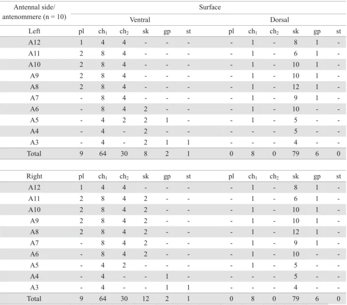
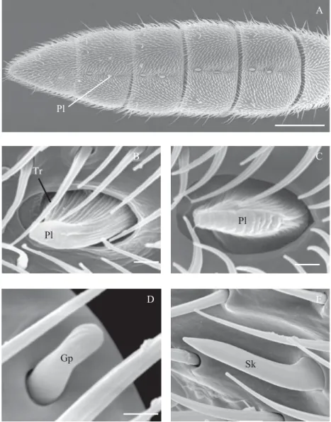
The antenna of G. gallardoi females is geniculated, bearing 12 antennomeres (A): scape (A1), pedicel (A2) and WHQÀDJHOODU\DQWHQQRPHUHV ÀDJHOORPHUHV$±$ )LJ7KH¿UVWDQWHQQRPHUH$LVORQJHUDQGQDUURZHU relative to the others, which gradually broaden towards the antennal tip. The last (A12) has a triangular shape. Along the ÀDJHOOXPZHREVHUYHGGRUVDOJODQGVGJDQGVHYHQW\SHVRI sensilla: papillary (pl), chaetica of two sizes-long (ch1) and short (ch2), styloconic (st), grooved pegs (gp), sickle-shapped (sk) and trichoids (tr). They showed numerical and spatial variation among the antennomeres (Fig. 1A-D; Table 1).
The papillary sensilla were found only on the distal antennomeres or club (A8-A12), along the median region of the ventral surface, being one in A12 and two from A11 to
A8 (Table 1; Figs. 1A, B and 2A). They have a protruding DQGÀDWWHQHGVXUIDFHEHDULQJWUDQVYHUVDOJURRYHV7KHODWHUDO margins are folded in, with an expanded base, forming a nearly ellipsoid pit (Figs. 2B and C). The exposed region of the sensillum is 5.4ȝPLQOHQJWKE\ȝPLQZLGWKEHLQJ shorter in A12 (approximately 2.1ȝPLQOHQJWK)LJ%
Two types of chaetica sensilla (ch1 and ch2) are equally distributed on the left and right antennae (Figs. 1A and B). Both are setiform and bear longitudinal grooves and a pore at the apex as well (Figs. 2D and E). The ch1 sensilla, longer and VWLIIPHDVXUHDERXWȝPLQOHQJWK)LJ'7KHFKW\SH shorter and lightly bent towards the antennal apex, measure DSSUR[LPDWHO\ȝPLQOHQJWK)LJ(7KHORFDOL]DWLRQRI the chaetica sensilla is the same on the left and right antennae (Table 1). The ca1 type ones are present laterally between the dorsal and the ventral antennal surfaces, from A3 to A12, and
near the middle surface of the dorsal surface, from A5 to A12 (Figs. 1A and D). The ch2 sensilla were found only on the ventral portion, from A5 to A12, two at each side of the middle portion, near to the antennomere apex (Fig. 1B).
Each antenna has only one styloconic sensillum, located on the anterior ventral surface, near the A3 base (Table 1; Figs. 1A and B). Its surface is apparently smooth and there is no evidence of the existence of pores neither grooves. They KDYHDURXQGHGDSH[EURDGHUWKDQWKHEDVHDUH¿WWHGWRWKH FDYLW\DQGPHDVXUHDERXWȝPLQOHQJWK)LJ$
The grooved-peg sensilla were found either on the ventral and dorsal surface. At the ventral surface, they show a different distribution pattern between the left and right antennae (Table 1; Figs. 1A and B). At the right antenna, they are positioned in A3 and A4 and at the left in A3 and A5. In both, they are positioned near the apex of each antennomere,
Surface Antennal side/
antenommere (n = 10) Ventral Dorsal
Left pl ch1 ch2 sk gp st pl ch1 ch2 sk gp st
A12 1 4 4 - - - - 1 - 8 1
-A11 2 8 4 - - - - 1 - 6 1
-A10 2 8 4 - - - - 1 - 10 1
-A9 2 8 4 - - - - 1 - 10 1
-A8 2 8 4 - - - - 1 - 12 1
-A7 - 8 4 - - - - 1 - 9 1
-A6 - 8 4 2 - - - 1 - 10 -
-A5 - 4 2 2 1 - - 1 - 5 -
-A4 - 4 - 2 - - - 5 -
-A3 - 4 - 2 1 1 - - - 4 -
-Total 9 64 30 8 2 1 0 8 0 79 6 0
Right pl ch1 ch2 sk gp st pl ch1 ch2 sk gp st
A12 1 4 4 - - - - 1 - 8 1
-A11 2 8 4 2 - - - 1 - 6 1
-A10 2 8 4 2 - - - 1 - 10 1
-A9 2 8 4 2 - - - 1 - 10 1
-A8 2 8 4 2 - - - 1 - 12 1
-A7 - 8 4 2 - - - 1 - 9 1
-A6 - 8 4 2 - - - 1 - 10 -
-A5 - 4 2 - - - - 1 - 5 -
-A4 - 4 - - 1 - - - - 5 -
-A3 - 4 - - 1 1 - - - 4 -
-Total 9 64 30 12 2 1 0 8 0 79 6 0
at the middle portion (Figs. 1A and B). At the dorsal surface, their distribution is the same in both antennae (Table 1), locating parallel to the apex, at the lateral posterior portion, from A7 to A12 (Fig. 1D). These sensilla are short (around 2.8 ȝPLQOHQJWKZLWKDURXQGHGDSH[DQGVKRZLQJORQJLWXGLQDO grooves. They are inserted in a rounded cavity, slightly enlarged compared to the sensillum base (Fig. 3B).
The sickle-shaped sensilla were found on both surfaces of the antennomeres (Table 1). They are curved towards the antennal apex and lightly compressed laterally (Fig. 3C). Tiny pores cover its surface. These sensilla are more numerous at the dorsal portion (Table 1), being distributed near the apical and basal margins of A3 to A12 (Fig. 1C). Those located near the apical margin extend distally, surpassing the membranous area existing between the antennomeres. At the ventral portion, they are less numerous and distributed in a different
manner at the left and right antenna (Table 1). At the right DQWHQQDWKH\RFFXULQSDLUVRQÀDJHOORPHUHV$WR$RQH at each lateral side, near to the apical margin (Fig.1B).
7KHWULFKRGHDVHQVLOODZHUHUHFRUGHGRQDOOÀDJHOORPHUHV showing considerable diversity in size and curvature. The great majority show a longitudinally grooved surface and are insert at an enlarged cavity. In the proximal antennomeres, these sensilla are more scattered and less abundant (Fig.3D). From the antennomere A7 they become gradually more abundant towards the distal region. At the ventral portion, they are shorter and decumbent, curved to the antennomere middle portion (Fig. 3E). At the club, they are found facing the papillary sensilla.
The dorsal glands (dg) are represented by a shallow and striated groove, located along the median-dorsal region of WKHÀDJHOORPHUHV$WR$)LJ)
Pl
C
D
Gp
E
Sk Pl
A
Pl
B Tr
Fig. 2A-D. Scanning electron micrographs of the antennal of G. gallardoi female. A – broad view of the ventral surface of the club of the right antenna showing the antennomeres A8 to A12. B and C – papillary (pl) and trichoid (tr) sensilla on the antennomeres A12 and A9, respectively. D – chaetica 1 sensillum (ch1) on the dorsal portion of the antennomere A10. E – chaetica 2 sensillum
Discussion
Concerning the number of antennomeres, the antennae of G. gallardoi females agree with the descriptions for other grionines (Masner 1979, Villa & Mineo 1990a,b). In Telenominae, including the genera Trissolcus Ashmed and Telenomus Haliday, the antennae have from 10 to 11 antennomeres (Bin 1981).
By showing various pores, the papillary sensilla belong to the multiporous category, as proposed by Zacharuck (1985). But different terminologies are employed to them. They were ¿UVWGHVFULEHGIRU6FHOLRQLGDHE\%LQDVSODWHV7KH terms multiporous plate (Barlin & Vinson 1981, Basibuyuk & Quicke 1998), multiporous grooved plate (Zacharuck 1985) and basiconic sensillum (Cave & Gaylor 1987) also were used to designate these structures.
According to Bin (1981), the distal antennomeres, being broader and endowed with papillary sensilla, delimitate a region in the Scelionidade female antenna named clava. The distribution of these sensilla in the antennomeres varies among species. For this reason, this author suggests the expression ‘claval sensilla formula’ to designate the ordinate array of papillary sensilla in each antennomere of a given club and the utilization of this criterium as a WRROIRUWD[RQRPLFLGHQWL¿FDWLRQRIJHQHUDJURXSVSHFLHV or species. In this way, the claval sensilla formula of the papillary sensilla found in G. gallardoi female antennae was 1-2-2-2-2 (respectively, from A12 to A8), corroborating what has been recorded for others scelionids as Trissolcus spp. (Bin 1981), and the grionines G. boseli (Villa & Mineo 1990a) and G. pennsylvanicum (Villa & Mineo 1990b). In
Fig. 3A-F. Scanning electron micrographs of antennal sensilla of G. gallardoi females: A – styloconic sensillum (st). B – grooved-peg sensilla (gp) on the dorsal surface of the antennomere A10 (left antenna). C – sickle-shapped sensilla (sk) on the dorsal surface of the antennomere A10 (right antenna). D and E – broad view of the trichoid sensilla (tr) on the ventral portion of DQWHQQRPHUHV$DQG$UHVSHFWLYHO\) FORVHYLHZRIWKHGRUVDOJODQGVGJRQWKHDQWHQQRPHUH$6FDOHEDUV$%) PP& PP'( Pm.
dg
F tr
D
E tr
B
gp
C
sk
A
most species of the genus Telenomus, Bin (1981) registered a claval formula 1-2-2-1.
The shape described for the G. gallardoi papillary sensilla agrees with those found by Bin (1981) for a great number of Telonominae, and by Cave & Gaylor (1987) for Telenomus reynoldsi Gordh & Coker. Nonetheless, in a small group ofTrissolcus VSHFLHV%LQYHUL¿HGWKDWWKHSDSLOODU\ sensilla had a more elongated surface, with the apex little HOHYDWHGEHLQJPRUHÀDWWHQHGWUDQVYHUVHO\DQGLQVHUWHGLQ a shallow pit.
Regarding their role, the papillary sensilla seem to be associated to tasting, being also named multiporous gustatory sensilla in Platygastridae and Scelionidae (Isidoro et al. 1996). +RZHYHUWKHLUJXVWDWRU\UROHGH¿QLWLRQKDVVROHO\EHHQEDVHG upon morphological and behavioral studies, since there is a lack of electrophysiological work concerning such sensilla.
The morphological evidences for the gustatory role rely on the location of their pores inside grooves, lobes or spherical protuberances (Isidoro et al. 2001), and on the presence of a great number of sensorial neurons associated to the pores (Zacharuck 1985, Isidoro et al. 1996). Considering this, Isidoro et al. (2001) suggest that during the drumming behavior, chemical compounds of the egg might be moved through the grooves or lobes towards the pores, allowing the recognition and host discrimination by tasting. In G. gallardoi, the employed SEM technique did not allow to verify the presence of pores. According to Isidoro et al. (1996), pore visualization is only possible when the specimens are previously treated with protease to remove secretions that cover the sensilla surface. These secretions, produced by exocrine glands associated to the papillary sensilla, seem to be related to the dissolution and/ or degradation of substances left on the host eggs, mediating their recognition by parasitoids (Isidoro et al. 2001).
The behavioral evidences that suggest the gustatory role of the papillary sensilla are related to the way females examine their hosts (Bin 1981, Isidoro et al. 1996). Behavioral studies carried out with G. gallardoi demonstrated that females evaluate and choose their hosts by drumming (Solis et al. 2001, Wiedenmann et al. 2003), partially rejecting eggs either already parasitized or old (Rocha 2005). These studies demonstrated that during drumming, the antennal portion that stays in touch with the host is the club, strongly suggesting that it is related to the perception of chemical stimuli by contacting the egg, as recorded for other Scelionidae species (Bin 1981, Isidoro et al. 1996, Isidoro et al. 2001). In studies in which the antennal club of parasitoid females was removed, they become unable to discriminate their egg hosts (Vinson 1998).
The grooved-peg sensilla are also named multiporous grooved sensilla (Zacharruck 1985, Cave & Gaylor 1987). Due to their insertion in an enlarged pit, they are also referred as coeloconica (Scheneider 1964, Ochieng et al. 2000). According to Slifer (1970), these sensilla are associated to an olfactory role, perceiving long distance stimuli. Their IXQFWLRQDO VSHFL¿FLW\ VHHPV WR GHSHQG RQ WKH WKLFNQHVV of their wall (Zacharuck 1985). They might also play a mechanoreception role, considering their association to a basal dendrite (Zacharuck 1985), involving both thermal and hygroreception (Altner & Loftus 1985).
The grooved-peg sensilla found in G. gallardoi females occur in the same antennomeres registered for G. boseli
(Villa & Mineo 1990a) and G. pennsylvanicum (Villa & Mineo 1990b). However, these authors registered a different distribution for them on the dorsal surface of the antennomeres. In such cases, they were distributed in pairs from A8 to A12 in the left antenna, in a number of two in A12, and of one from A11 to A7 in the right antenna. In T. reynoldsi, Cave & Gaylor (1987) registered the peg type only in the dorsal surface, near the apical external margin of the antennomers A6, A4, and A11, and referred them as multiporous grooved sensilla.
Cave & Gaylor (1987) suggest that the peg type sensilla do not engage in host recognition and discrimination in the egg parasitoid Scelionidae, because they are not present in the antennal club of these insects.
The sickle-shaped sensilla are also referred as multiporous punctured sensilla (Zacharuck 1985). Cave & Gaylor (1987) described them as horn-like sensilla, ascribing also the term “trichoid curvata” to them in T. reynoldsy. In other egg parasitoid families, as for example Trichogrammatidae, they correspond to the multiporous trichoidea sensilla (Olson & Andow 1993).
In Scelionidae, the sickle-shaped sensilla are encountered LQ DOO ÀDJHOODU\ DQWHQQRPHUHV HVSHFLDOO\ RQ WKH GRUVDO surface; nevertheless, its distribution may show variations among species (Cave & Gaylor 1987, Basibuyuk & Quicke 1998). In G. gallardoi, the distribution pattern of these sensilla is different between the ventral regions of the right and left antennae. In the same way, but in both surfaces, Villa & Mineo (1990b) pointed out to the existence of differences in the distribution of sensilla between the left and right antennae of G. pennsylvanicum. In most works, the authors did not compare the sensilla distribution between the right and left antennae. Thus, erroneous conclusions could be attained in such cases, especially when the number and distribution of sensilla are used with taxonomical purpose.
Zacharuck (1985) describes the chaetica sensilla as hairs or spines, but with a ticker wall when compared to the trichodea sensilla. In general, the sensilla described as chaetica bear a terminal pore connected to sensorial dendrites, thus belonging to the uniporous category (Zacharuck 1985). They are also named grooved basiconic (Norton & Vinson 1974, Ochieng et al. 2000) or uniporous gustatory sensilla (Isidoroet al. 1996). In G. boseli, Villa & Mineo (1990a) registered them only on the ventro-lateral surface, where they occur in a number of two per antennomere, at the apical portion, from A7 to A11, and of eight on A12. Based upon the description of these authors, such sensilla correspond to the ch1, described in the present work for G. gallardoi. In the same way, sensilla corresponding to this type were found in G. pennsylvanicum on the dorsal surface from A8 to A12 in the left antenna, and from A9 to A12, in the right antenna, being two in A12 and one in the other antennomeres. The ch1 sensilla were also similar to those encountered in T. reynoldsi por Cave & Gaylor (1987). The ch2 sensilla of G. gallardoi seem to match those registered for G. pennsylvanicum in the ventral surface, from A5 to A12. In this case, they show the same distribution pattern, except for A12, where six of them were registered.
The chaetica sensilla function as both mechano and chemoreceptors (Zacharuck 1985). Such a double function is ascribed to the existence of a dendrite not attached to the pore (McIver 1985, Isidoro et al. 1996). The chemoreceptive function of the chaetica sensilla seems to be associated to the perception of contacted liquid stimuli, being thus gustative (Zacharuck 1985). Their gustative function has been evidenced through behavioral studies. Bin (1981) inferred that these sensilla, by being stiffer than the others, tend to touch the VXEVWUDWH¿UVWDQGVRPLJKWGHWHFWLQIRUPDWLRQGLUHFWO\IURP the host. Ultrastructural studies associated to behavioral ones, carried out by Isidoro et al. (1996) in several parasitoid families, including scelionids such as T. basalis, corroborated that. Due to the occurrence in great abundance on antennal areas that touch either the substrate, the host, or the opposite sex during mating, they were called gustative sensilla by these authors. In T. nubilale, the chaetica sensilla are localized in the DQWHQQDODSH[DORFDOWKDWWRXFKWKHVXEVWUDWH¿UVW7KH\DUH also associated to host recognition (Olson & Andow 1993).
The styloconic sensilla, classified as aporous, are associated to thermal and hygroreceptive function in many insects (Zacharuck 1985). Altner & Loftus (1985) ascribed such functions to the absence of pores on them, to their narrow cavity, and to the presence of dendritic segments externally at their base. Studies regarding their sensitivity to changes in temperature and humidity should be carried out WRFRQ¿UPVXFKIXQFWLRQVIRUWKHPLQG. gallardoi.
Location of the styloconic sensilla is variable among species of Scelionidae. In T. reynoldsi, Cave & Gaylor (1987) registered them on the dorsal surface of the antennomeres A4 and A10. Villa & Mineo (1990b) mentioned a stlyloconic sensillum in G. pennsylvanicum on the ventral surface of the antennae, but in a different number and distribution per antennomere. In these species, the authors found one in A5 of the right antenna, two and one respectively for the A5 and A6 of the left antenna.
The trichoid sensilla, which were considered aporous by Zacharuck (1985), are also referred as setiform (Zacharuck 1985, Cave & Gaylor 1987). According to Villa & Mineo (1990a,b), these sensilla correspond to short structures, spine shaped, which occur on the pedicel base and on the ventral surface of the scape. According McIver (1985), these sensilla have mechanoreceptive function, considering the presence of a single sensorial neuron. Therefore, these sensilla would be associated to a tactile function, being able to perceive differences in substrate texture, movement and wind direction (Isidoroet al. 1996).
According to Isidoro et al. (1996), the thrichodea sensilla that occur in the antennal club and are associated to the papillary and to the chaetica type, in conjunction IRUPDIXQFWLRQDOSRUWLRQRIWKHDQWHQQDGH¿QHGDV³WRXFK and taste area”. This area would be responsible for host recognition and acceptance, detecting chemical substances and diverse textures present either on the host surface or substrate during the drumming behavior. In behavioral studies carried out with G. gallardoi females, the drumming behavior lasted longer when either eight or twelve day-old eggs were utilized (Rocha 2005). This fact could be related to a supposed chemical or physical change at the egg surface, which would hinder its recognition by the club sensilla. Nevertheless, eletrophysiological studies as well as chemical and morphological analysis of the S. dentiventris egg surface are required to validate this hypothesis.
The dorsal glands are distributed in the same manner as in Gryon pennsylvanicum (Ashmead) (Villa & Mineo 1990b), but different from G. boseli Mineo et Szabo, where they were registered from A12 to A7 (Villa & Mineo 1990a). In Trissolcus basalis (Wollaston), Isidoro et al. (1996) registered these structures from A7 to A12. According to these authors, the internal wall of this dorsal depression bears numerous pores, which correspond to the opening of the glands. The corresponding function is not clear, and in most studies, it is not even mentioned. Nevertheless, Isidoro et al. (1996), while observing the mating behavior of T. basalis, related them to sexual recognition.
The description and determination of the distribution pattern of antennal sensilla in G. gallardoi females accomplished on the present work is an advance regarding the elucidation of the mechanisms involved in the host selection behavior of this species. In view of the kind of sensilla found in the antennal club, one can infer that chemical compounds present in the host, besides its texture, have an important role in the host recognition and acceptance. Therefore, additional studies, at the ultrastructural level, using transmission electron microscopy should be carried out, in association with electrophysiological ones, in order to elucidate the real function of these structures in relation to the egg parasitoids.
Acknowledgments
References
Altner, H. & R. Loftus. 1985. Ultraestructure and function of insect thermo and hygroreceptors. Annu. Rev. Entomol. 30: 273-295.
Barlin, M.R. & S.B. Vinson. 1981. Multiporous plate sensilla in the antennae of the Chalcidoidea (Hymenoptera). Int. J. Insect Morphol. Embryol. 168: 97-108.
Basibuyuk, H.H. & D.L.J. Quicke. 1998. Gross morphology of multiporous plate sensilla in the Hymenoptera (Insecta). Zool. Scr. 28: 51-67.
%LQ)'H¿QLWLRQRIIHPDOHDQWHQQDOFODYDEDVHGRQLWVWKH plate sensilla in Hymenoptera Scelionidae Telenominae. Redia 64: 245-261.
Cave, R.D. & M.J. Gaylor. 1987. Antennal sensilla of male and femaleTelenomus reynoldsi Gordh and Coker (Hymenoptera: Scelionidae). Int. J. Insect Morphol. Embryol. 16: 27-39.
Dasilao, A.O. & R. Arakawa. 2004. Reproductive capacity and host handling behavior of Gryon philippinense (Asmead) (Hymenoptera: Scelionidae), a solitary egg parasitoid of the winter cherry bug, Acanthocoris sordius Thunberg (Hemiptera: Coreidae). Appl. Entomol. Zool. 39: 263-269.
Field, S.A & M.A. Keller. 1999. Short-term host discrimination in the parasitoid wasp Trissolcus basalis Wollaston (Hymenoptera: Scelionidae). Aust. J. Zool. 47: 19-28.
Higuchi, H. & Y. Suzuki. 1996. Host handling behaviour of the egg parasitoidTelenomus triptus to the egg mass of the stink bug
Piezorus hybneri. Entomol. Exp. Appl. 80: 475-479.
Isidoro N., F. Bin, S. Colazza & S.B. Vinson. 1996. Morphology of antennal gustatory sensilla and glands in some parasitoids Hymenoptera with hypothesis on their role in sex and host recognition. J. Hym. Res. 5: 206-239.
Isidoro, N., R. Romani & F. Bin. 2001. Antennal multiporous sensilla; their gustatory features for host recognition in female wasps (Insecta, Hymenoptera: Platygastroidea). Microsc. Res. Tech. 350-358.
Masner, L. 1979. The variicornis-group of GryonHaliday (Hymenoptera: Scelionidae). Can. Entomol. 111: 791-805.
McIver, S.B. 1985. Mechanorreception, p. 71-132. In G.A. Kerkut & L.I. Gilbert. (eds.), Comprehensive insect physiology biochemistry and pharmacology, v. 6, Oxford, Pergamon Press, 8336p.
Navasero, R & E. Oatman. 1989. Life history immature morphology and adult behaviour of Telenomus solitus (Hymenoptera: Scelionidae). Entomophaga 34: 165-177.
Norton, W.N. & S.B. Vinson. 1974. Antennal sensilla of three parasitic Hymenoptera. Int. J. Insect Morphol. Embryol. 3: 305-316.
Ochieng, S.A., K.C. Park, J.W. Zhu & T.C Baker. 2000. Functional morphology of antennal chemoreceptors of the parasitoid
Microplitis croceipes (Hymenoptera: Braconidae). Arthrop. Struct. Dev. 29: 231-240.
Olson, D.M. & D.A. Andow. 1993. Antennal sensilla of female
Trichogramma nubilale (Erthe and Davis) (Hymenoptera: Trichogrammatidae) and comparison with other parasitic Hymenoptera. Int. J. Insect Morphol. Embryol. 22: 507-520.
Rocha, L. 2005. Resposta de Gryon gallardoi (Brèthes) (Hymenoptera: 6FHOLRQLGDHjTXDOLGDGHGRVRYRVGHSpartocera dentiventris
(Berg) (Hemiptera: Coreidae) e evidências dos mecanismos de seleção envolvidos. Tese de doutorado, Universidade Federal do Rio Grande do Sul, Porto Alegre, 145p.
Santos, S.S.R., L.R. Redaelli & L.M.G. Diefenbach. 2001. Ocorrência de parasitismo em ovos de Spartocera dentiventris
(Berg) (Hemiptera: Coreidae) em cultura de fumo. Neotrop. Entomol. 30: 731-733.
Schneider, D. 1964. Insect antennae. Annu. Rev. Entomol. 9: 103-122.
Slifer, E.H. 1970. The structure of arthropod chemorreceptors. Annu. Rev. Entomol. 15: 121-142.
Solis, S.R., G.G. Fagundes & B.F. Amaral Filho. 2001. Comportamento de oviposição de Gryon gallardoi em ovos deLeptoglossus zonatus. Rev. Agric. 76: 451-462.
Souza, C.E.P. & B.F. Amaral Filho. 1999. Ocorrência natural de parasitóides de Leptoglossus zonatus (Dallas) (Heteroptera: Coreidae). An. Soc. Entomol. Brasil 28: 757-759.
Strand, M.R. & S.B. Vinson. 1983. Host acceptance behaviour ofTelenomus heliothids (Hymenoptera: Scelionidae) toward
Heliothis virescens (Lepdoptera: Noctuidae). Ann. Entomol. Soc. Am. 76: 781-785.
Villa, L. & G. Mineo. 1990a. Morphology of antennal structures of
Gryon boseli Mineo and Szabo (Hymenoptera: Scelionidae): A scanning electron microscope study. Frustula Entomol. n.s. 13: 9-17.
Villa, L. & G. Mineo. 1990b. Mapping of the antennal structures ofGryon pennsylvanicum (Ashmead): A SEM study (Hym.: Scelionidae). Frustula Entomol. n.s. 13: 225-235.
Vinson, S.B. 1998. The general host selection behaviour of parasitoid hymenoptera and a comparasion of initial strategies utilized by larvaphagous and oophagous species. Biol. Control 11: 9-96.
Weber, C.A., J.M. Smilanick, L.E. Ehler & F.G Zalom. 1996. Ovipositional behaviour and host discrimination in three scelionid egg parasitoid of stink bug. Biol. Control 6: 245-252.
Zacharuck, R.Y. 1985. Antennae and sensilla, p.1-69. In G.A. Kerkut & L.I. Gilbert (eds.), Comprehensive insect physiology biochemistry and pharmacology, v. 6, Oxford, Pergamon Press, 8336p.