Universidade de Aveiro Ano 2020
Departamento de Biologia
ANA RITA PEREIRA
GOUVEIA
TÉCNICAS AVANÇADAS DE BIOLOGIA
MOLECULAR E TRANSCRITÓMICA APLICADAS A
ACTINOMYCETES
ADVANCED TECHNIQUES IN MOLECULAR
BIOLOGY AND TRANSCRIPTOMICS APPLIED TO
ACTINOMYCETES
Universidade de Aveiro Ano 2020
Departamento de Biologia
ANA RITA PEREIRA
GOUVEIA
TÉCNICAS AVANÇADAS DE BIOLOGIA
MOLECULAR E TRANSCRITÓMICA APLICADAS A
ACTINOMYCETES
ADVANCED TECHNIQUES IN MOLECULAR
BIOLOGY AND TRANSCRIPTOMICS APPLIED TO
ACTINOMYCETES
Dissertação apresentada à Universidade de Aveiro para cumprimento dos requisitos necessários à obtenção do grau de Mestre em Biologia Molecular e Celular, realizada sob a orientação do Doutor Antonio Rodríguez García, Investigador do Instituto de Biotecnologia de León e sob a coorientação científica da Doutora Sónia Mendo, Professora Auxiliar com Agregação do Departamento de Biologia da Universidade de Aveiro
o júri
presidente Prof. Doutor António José de Brito Fonseca Mendes Calado professor auxiliar da Universidade de Aveiro
Prof. Doutora Ana Sofia Direito dos Santos Duarte professora auxiliar da Universidade Católica Portuguesa
Prof. Doutora Sónia Alexandra Leite Velho Mendo Barroso professora auxiliar com agregação da Universidade de Aveiro
agradecimentos A toda a comunidade de León, que me aceitou de braços abertos e me fez sentir em casa, ainda que, pacientemente, me tivessem que incutir que não se almoça antes das 14 horas nem se janta antes das 21.
A todas as pessoas da Inbiotec, que nunca disseram não a conversar comigo, que nunca disseram não a fazerem me sentir um pouco menos sozinha, mesmo com o meu “portunhol” a atrapalhar.
Ao Antonio, à Rosma e à Maite que sempre me ajudaram quando não sabia o que fazer, que sempre me tiraram as dúvidas, mesmo as mais parvas, e sempre me perdoaram, ainda que tivesse estragado alguns dias de trabalho. Agradeço-vos toda a paciência dos últimos meses que, sem dúvida, tornaram-me um pouco mais apta a seguir esta carreira profissional.
À senhora professora Sónia Mendo, que nunca me abandonou e sempre se mostrou pronta a fazer as oito horas de viagem para ir ter comigo. Agradeço, também, por me ter falado da Inbiotec e, por isso, ser a responsável por esta minha aventura.
Por fim, mas não menos importantes, agradeço aos meus pais por todo o apoio e paciência, já lá vão há 23 anos. Obrigada pelos abraços, pelos castigos, pelas conversas e pelas palmadas que me ajudaram a crescer e a ser quem sou. Este trabalho não teria sido feito sem vocês, não teria sido feito se vocês não tivessem concordado em deixar-me sair de casa, e até do país, por meio ano. Só por causa de vocês é que consegui chegar até aqui. E independente do número de palavras que escreva aqui, elas nunca serão suficientes para agradecer por tudo o que fizeram.
palavras-chave sRNAs, Streptomyces, metabolitos secundários, PhoR-PhoP.
resumo Hoje em dia, a resistência a antibióticos está a tornar-se numa das maiores preocupações do século. Esta problemática está a arriscar a nossa saúde pública e pode vir a tornar-se, no futuro, num dos problemas de saúde mais letais. Portanto, uma solução é necessária.
Streptomyces é conhecido como o género que é responsável por imensos antibióticos. Porém, nestes últimos anos, o número de antibióticos que chegaram a uso clínico diminuiu, devido a várias razões.
Metabolismo secundário é composto de vários processos que, ainda que não sejam fundamentais para a sobrevivência da célula, dão-lhe imensas
vantagens. A maior parte destas vantagens originam-se a partir dos metabolitos secundários, alguns deles conhecidos como antibióticos. Por outro lado, fósforo é um dos elementos mais importantes para qualquer organismo e, por isso, necessita de um mecanismo que se assegure que ele está sempre regulado. Um destes sistemas depende de PhoR-PhoP. Com a descoberta de um novo tipo de RNAs, os sRNAs (que têm entre 50 a 400 nucleótidos), a investigação de novos compostos com importância farmacêutica e industrial pode continuar a avançar, dado que algumas destas moléculas podem funcionar como reguladoras do metabolismo secundário e, por isso, estar relacionadas com antibióticos.
O objetivo deste trabalho é identificar sRNAs que estão implicados na regulação de fosfato, a principal forma de assimilação de fósforo. A minha função era experienciar o que era trabalhar num laboratório, ao aprender algumas técnicas de biologia molecular e transcritómica, como introdução de DNA em células e extração de ácidos nucleicos, necessárias para este projeto.
keywords sRNAs, Streptomyces, secondary metabolites, PhoR-PhoP.
abstract Nowadays, the resistance to antibiotics is turning to be one of the biggest concerns of this century. It is risking our public health and might be, in the future, one of the deadliest health issues. Therefore, a solution is needed. Streptomyces is known as a genus that is responsible for many antibiotics. However, in the latest years, the number of antibiotics that reached the clinical use diminished, due to various reasons.
The secondary metabolism is composed by a number of processes that, even though aren’t of extreme importance to the cell survival, can give it several advantages. Most of those advantages come as secondary metabolites, and some are known as antibiotics.
On the other hand, phosphorus is one of the most important elements to any organism, and, therefore, a mechanism is needed to make sure that this element is regulated. One of the systems that does that depends on PhoR-PhoP.
With the discovery of a new type of RNAs, the sRNAs (which have between 50 and 400 nucleotides) the investigation of new compounds with pharmaceutical and industrial importance may continue to go forward, since some of those molecules may function as regulators of the secondary metabolism and, therefore, be related to antibiotics.
The aim of this work is to identify sRNAs that are implicated in the phosphate regulation, which is the main assimilation form of phosphorus. My objective in this study was to experience what was like to work in a laboratory, by learning some molecular biology and transcriptomic techniques, such as DNA
palabras clave sRNAs, Streptomyces, metabolitos secundarios, PhoR-PhoP.
resumen La resistencia a los antibióticos es uno de los grandes problemas de este siglo. Pone en riesgo la salud pública y se prevé que en el futuro sea una de las mayores causas de mortalidad, por lo que se hace necesaria una solución. El género Streptomyces es conocido por ser capaz de producir muchos tipos de antibióticos diferentes. Sin embargo, en los últimos años, el número de antibióticos que llegaran a la práctica clínica ha disminuido, debido a varias razones.
El metabolismo secundario está formado por muchos procesos que, aunque no sean totalmente necesarios a la supervivencia de la célula, le confiere
ventajas. La mayoría de esas ventajas son los metabolitos secundarios, algunos conocidos como antibióticos.
Por otro lado, el fósforo es uno de los elementos más importantes para cualquier organismo y por eso necesita tener uno mecanismo que regule su metabolismo. Uno de esos mecanismos depende de PhoR-PhoP.
Con el descubrimiento de un nuevo tipo de ARN, los ARNp (que tienen entre 50 y 400 nucleótidos), la investigación de nuevos compuestos con importancia farmacéutica y industrial puede seguir avanzando, ya que algunas de estas moléculas pueden funcionar como reguladoras de metabolismo secundario y, por eso, estar relacionadas con los antibióticos.
El objetivo de este trabajo es identificar ARNp que están implicados en la regulación por fosfato, la principal forma de asimilación de fósforo. Mis objetivos en este trabajo han sido experimentar lo que es trabajar en
laboratorio y aprender algunas de las técnicas transcriptómicas y de biología molecular, como la introducción de ADN en células y extracción de
nucleótidos, necesarias para este estudio. .
Index
INTRODUCTION ... 16 1.1. Streptomyces ... 17 1.1.1. General characteristics ... 17 1.1.2. Life cycle ... 17 1.1.3. Genetic aspects ... 18 1.1.4. Streptomyces coelicolor ... 191.1.5. Importance of the Streptomyces genus ... 20
1.1.6. Difficulties when dealing with the Streptomyces genus ... 20
1.2. Secondary metabolism ... 21
1.3. Two-component system PhoR-PhoP ... 23
1.4. Small RNAs ... 26
1.5. Aim ... 27
MATERIALS AND METHODS... 28
Methods in Microbiology ... 29
1. Melting of solid media ... 29
2. Inoculation and plating of microorganisms ... 30
3. Identification of microorganism* ... 35
4. Ligation reaction ... 37
5. Transformation (chemical procedure) ... 40
6. Transformation (electrical procedure)* ... 43
7. Plasmid Extraction ... 45
7.1. Minipreparations... 46
7.2. E.Z.N.A. kit ... 49
7.3. Alkaline lysis* ... 52
8. Conjugation ... 56
9. Cup plate method* ... 60
Methods in Molecular and Cellular Biology ... 61
1. Plasmid DNA Digestion ... 61
2. Electrophoresis... 63
3. Recovery of DNA from agarose gel ... 68
4. Spectrophotometric measurements ... 69
5. Electrophoresis of RNA* ... 73
6. Polymerase chain reaction (PCR)* ... 77
Methods in Bioinformatics ... 82
1. Annotation ... 83
a) Profile of sRNA ... 84
b) Length of sRNA ... 85
c) Types of sRNA ... 86
d) Naming the sRNAs ... 88
e) If not a sRNA, then what is it? ... 88
2. Prioritization ... 88
3. Searching for targets ... 89
4. Prioritization of targets ... 91
RESULTS AND DISCUSSION ... 94
Results obtained for methods in Microbiology ... 95
7. Plasmid Extraction ... 95
7.1. Minipreps ... 95
7.2. E.Z.N.A. kit ... 98
9. Cup plate method* ... 99
Results obtained for methods in Molecular and Cellular Biology ... 100
5. Electrophoresis of RNA* ... 100
1. Annotation ... 105
2. Prioritization ... 108
3. Searching for targets ... 109
4. Prioritization of targets ... 110
CONCLUSION ... 115
List of figures
IntroductionFig. 1. The life cycle of Streptomyces. ... 18
Fig. 2. A model, in Streptomyces, for the evolution of linear chromosome. ... 19
Fig. 3. S. coelicolor secondary metabolites in relation to their chromosomal location ... 20
Fig. 4. The various phases of the bacterial growth curve ... 22
Fig. 5. Cluster situated regulators (CSRs) and global regulatory systems ... 23
Fig. 6. A model for controlling, in E. coli, the balance between PhoR phosphatase and autokinase activities ... 25
Fig. 7. The DRus in the Pho boxes of S. coelicolor and S. avermitilis ... 26
Fig. 8. Two general biogenesis pathways of sRNAs from the 3’ region of mRNA loci. ... 27
Materials and Methods Fig. 9.Petri dishes with E. coli (A and B) and Streptomyces (C and D) ... 32
Fig. 10. Difference between sticky (A) and blunt ends (B)... 37
Fig. 11. Schematic representation of the plasmid constructed. ... 38
Fig. 12. Schematic representation of transformation and conjugation. ... 40
Fig. 13. Flowchart summing up the studies done with the two strains of E. coli. ... 41
Fig. 14. Main steps of electroporation ... 43
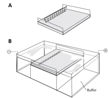
Fig. 15. Schematic representation of a chamber used for agarose gel electrophoresis. ... 64
Fig. 16. The equipment needed for “NanoDrop” ... 70
Fig. 17. Graph given by “NanoDrop” for a DNA sample ... 71
Fig. 18. A Bioanalyzer chip for RNA electrophoresis ... 73
Fig. 19. Exponential amplification in the PCR technique ... 78
Fig. 20. Representation of the three-step principle followed by PCR ... 79
Fig. 21. Representation of the two studies done in PCR ... 79
Fig. 22. An example of a profile for a sRNA ... 84
Fig. 23. The most usual seen profiles. ... 85
Fig. 24. Template of IGV ... 86
Fig. 25. The most usual seen types of sRNAs. ... 87
Fig. 26. Example of three sequences in the FASTA format... 90
Fig. 27. Example of a profile of a gene (A) and its differential profile (B). ... 92
Fig. 28. Example of part of the results given by “CopraRNA” (A) and by “IntaRNA” (B)... 93
Results Fig. 29. Electrophoresis of the first digestion done to the minipreps (A) ... 96
Fig. 30. Electrophoresis of the second digestion done to some of the minipreps (A) ... 97
Fig. 31. Result from the electrophoresis done for two plasmids extracted. ... 98
Fig. 32. Result from the cup plate method ... 99
Fig. 33. Electropherogram of a perfect sample (RIN of 10) for eukaryotes ... 101
Fig. 34. Electropherogram of the ladder used. ... 102
Fig. 35. Some of the electropherograms of the samples obtained in this study. ... 102
Fig. 36. Electrophoresis of the samples after PCR ... 104
Fig. 37. Information for the analysis of Candidate “A”. ... 105
Fig. 38. Information for the analysis of Candidate “B” ... 106
Fig. 39. Information for the analysis of Candidate “C” ... 107
Fig. 40. Information for the analysis of Candidate “D”. ... 108
Fig. 42. Profile that might not be as interesting as it seems for cases of candidates with profile DA. ... 111 Fig. 43. Targets that didn’t have an IT where the software said it should be ... 111 Fig. 44. Profiles of gene A, B and C and their annotation. ... 112
List of tables
Table 1. Example of the cutsites for three enzymes ... 62
Table 2. Purity values in DNA samples given by “NanoDrop” and its significance ... 71
Table 3. Value given to the candidates depending on their characteristics ... 83
Table 4. Results on “NanoDrop” of the extraction of plasmid by E.Z.N.A. kit ... 98
Table 5. Representation of the results gotten for the prioritization of sRNAs ... 109
Table 6. Example of homologues given by “GLASSgo” and their analysis. ... 109
Table 7. Summary of the results gotten for those sRNAs that had different homologues/lengths searched ... 113
List of abbreviations
Abbreviation Meaning
5’PPP 5’ triphosphate
ADB Agarose Dissolving Buffer
BGC Biosynthetic Gene Cluster
bp base pair
CCC Covalently Closed Circular
CIA Chloroform-Isoamyl Alcohol
CSR Cluster Situated Regulator
DNA Deoxyribonucleic Acid
DNApol DNA polymerase
DNAse Deoxyribonuclease
dNTP Deoxyribonucleotide Triphosphate
E-test Epsilometer test
EDTA Ethylenediaminetetraacetic Acid
GTE Glucose-Tris-EDTA
ID Identification
IT Transcription Start Site
kb kilobase
LA Luria Agar medium
LB Luria Broth medium
Mb Megabase
min minute
ms millisecond
Miniprep Minipreparation
mRNA messenger RNA
OC Open Circular
OD600 Optical Density at a wavelength of 600 nm
PCR Polymerase Chain Reaction
pH potential for Hydrogen
RBS Ribosome-Binding site
RCF Relative Centrifugal Force
RIN RNA Integrity Number
RNA Ribonucleic Acid
RNA-seq RNA sequencing
RNase Ribonuclease
rpm revolutions per minute
s second
SARP Streptomyces Antibiotic Regulatory Protein
SDS Sodium Dodecyl Sulfate
SOB Super Optimal Broth medium
sRNA small RNA
TAE Tris-Acetate-EDTA medium
TB Terrific Broth medium
Ter Terminator
TE Tris-EDTA medium
TPM Transcripts Per Million
tra transfer
TSA Tryptic Soy Agar medium
TSB Tryptic Soy Broth medium
Tsr Thiostrepton
U Unit
UTR Untranslated Region
Introduction
1.1. Streptomyces
1.1.1. General characteristics
Actinomycetes are a heterogeneous group of bacteria that have high guanine and cytosine content (more than 50 %) in their deoxyribonucleic acid (DNA) and are Gram-positive [1], [2]. The high content of guanine and cytosine might come from an adaptative response, since it enables them to fight certain bacteriophages using rare codons [3]. They have at least 350 genera, which makes them one of the biggest bacterial phyla [1]. From this group, Streptomyces is the most prevailing genus [4].
These bacteria are classified as both mesophilic and neutrophilic, since their optimal temperature is around 25-35 ºC, and their optimal potential for hydrogen (pH) is around 6.5-8 (but they can live in pHs higher than 9) [5], [6]. They are also considered strict aerobic, but Streptomyces coelicolor (S. coelicolor) has enzymes that allow it to breathe nitrate (this might also be an adaptative response to living in a place as variable as the soil) [7].
They live normally in the soil (since they need nutrients that come from plants’ degradation), but they can also be found in water, air, and other places (including extreme environments as glaciers and deserts) [8], [9]. This happens because they are able to grow in different sources of carbon and nitrogen [10]. In order to be able to adapt to the many changes that the soil suffers, Streptomyces has a lot of sigma factors [11].
Terrestrial Streptomyces has been over-exploited by various companies and, therefore, its investigation has now turned to its relatives that can be found in those other environments, to those that invade plant tissues and even to non-Streptomyces actinomycetes [4].
1.1.2. Life cycle
In this section, the complex life cycle of Streptomyces cultivated in solid medium will be explained (since, in liquid, it is not completed, since Streptomyces is not able to sporulate in this type of medium) [12]. The first stage, spores’ sporulation, is characterized by presenting hydrophobic pigmented cells. When the optimal conditions are achieved, germ tubes are originated, that create branched hyphae (which are polynuclear and capable of getting nutrients by penetrating the substrate) [13], [14]. When the hyphae turn into a cell aggregation, a first mycelium is created, which has the proteins responsible for primary metabolism or vegetative mycelium (Fig. 1) [15].
The second or reproductive mycelium, or the precursor of aerial mycelium in solid media, is originated after some central cells of the primary mycelium go through apoptosis (Fig. 1). The second mycelium might be characterized as early or late, depending on the hydrophobic covers typical of aerial hyphae; if it has the covers, it is a late secondary mycelium. The secondary mycelium is crucial since it has the proteins needed for secondary metabolism [15].
After this stage, another apoptosis happens in order to create spores, that will be able to reinitiate the cycle [12].
Even though this explanation only refers secondary metabolism in the secondary mycelium, it is also possible for it to happen, and secondary metabolites being originated, in the stage of primary mycelium [12].
Fig. 1. The life cycle of Streptomyces. (1) represents a spore, which germinates (2, 3) and produces the primary mycelium (4). (5, 6) have hyphae, that in (7) starts producing initial cells (8). Then, these cells germinate into the secondary mycelium (9, 10). In that mycelium, there are pairs of chromosomes (11, 12) able to develop into spores (13, 14). Taken from [16].
1.1.3. Genetic aspects
Streptomyces has its genome organized in a large linear chromosome [with more than 7 megabase (Mb)], which doesn’t happen in the majority of the actinobacteria [17]. This linearity has been explained by a recombination between a linear plasmid and an ancestor with a circular chromosome, since, when this happens, the result is always a linear molecule (Fig. 2) [18]. This chromosome would have its core originated by the ancestor chromosome and its arms by the linear plasmid [19].
In the central coreof this chromosome, genes related to primary and central metabolism and, therefore, essential and that are conserved in a lot of species, are present [20]. On the other hand, the genes considered not essential, such as the ones related with secondary metabolism, are located in the arms of the chromosome [21].
There are also elements outside of the chromosome, such as linear and circular plasmids, that are able to replicate in other hosts with variable size and number of copies [22]. They also have a high rate of mutations, most of them negative or neutral, that occur in the terminal regions. These mutations can be explained by the need that these bacteria have to adapt to the different environments [18].
Fig. 2. A model, in Streptomyces, for the evolution of the linear chromosome. Here, it can be seen that, during the evolution, circular chromosomes (in green) opened up through recombination with the linear plasmid. Taken from [19].
1.1.4. Streptomyces coelicolor
Streptomyces coelicolor is the model organism of the actinomycetes, because it is easy to manipulate, and it also was the first species of Streptomyces to have its genome sequenced. Therefore, a lot of known genetic tools that can be used in this species have been already studied [16], [23]. Moreover, it has more codifying sequences than most eukaryotes, which only shows how much it has to adapt. It also has three plasmids: SCP1 (linear and produces methylenomycin); SCP2 (circular and smaller); SLP1 (circular and able to integrate in the chromosome, which enables it to replicate in other species of the genus) [24].
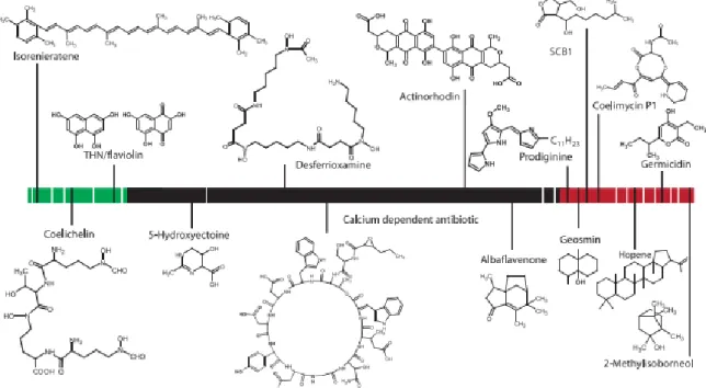
One reason why this species facilitates its studies, compared to other species of Streptomyces, is due to the production of pigmented antibiotics, which are easy to see and, therefore, easy to confirm if the bacteria are producing metabolites [25]. If not for this, it would be harder to conclude the same thing. S. coelicolor is able to produce various antibiotics, for instance, actinorhodin, produced by genic cluster act, which only acts on Gram-positive bacteria and is an acid-base indicator, since in acidic/neutral conditions it has the colour red and in alkaline conditions it is blue [26]. On the other hand, undecylprodigiosin, another antibiotic produced by this species, is red and is produced by the genic cluster red [27]. Both of these are detected when there is no phosphate in the medium [28].
There are also antibiotics that depend on calcium levels present in the medium, such as the so-called calcium dependent antibiotic produced by the genic cluster cda [29]. There are
specially in genus Proteus in acid conditions), and coelimycin P1, originated from cpk, able to affect Bacillus subtilis, Escherichia coli (E. coli) and Kocuria rhizhophila, and has the colour yellow (Fig. 3) [30], [31].
Fig. 3. S. coelicolor secondary metabolites in relation to their chromosomal location. Most of the biosynthetic genes are located outside the highly conserved core region (in black). The green and red represent, respectively, the left and right arm. Taken from [25].
1.1.5. Importance of the Streptomyces genus
Actinobacteria are one of the most notable group of microorganisms, since they alone, and specially the Streptomyces genus, represent a tremendous source of valuable chemicals. This genus is responsible for around two-thirds of all naturally derived antibiotics used in various areas, from medicine to agriculture [1], [32]. Until 2005, rare actinomycetes were responsible for the discovery of 2250 new bioactive secondary metabolites, approximately [4].
Although antibiotics are of extreme importance, they are not the only bioactive molecules that the genus Streptomyces has provided, but also antiprotozoals, antifungals and antivirals [4].
All of this is only a small percentage of the value of this genus. Until now, only the medical importance has been described. But these bacteria also have a huge industrial value, because their genome is big (which might indicate that more biosynthetic gene clusters, BGCs, can be present), and an ecological value as well, since they are important heterotrophics, which are able to degrade plant biomass (an important carbon source in the terrestrial environment) [33]. None of these, even though the medical importance will be the one discussed the most in this work, should be underestimated.
1.1.6. Difficulties when dealing with the Streptomyces genus
Natural product research, including the one that aims to discover new drugs that come from Streptomyces’ secondary metabolites, has been decreasing for various reasons, causing less products to reach the marketplace and have a clinical use. Low production concentration and a challenging isolation of the bacteria, which makes a large-scale cultivation very difficult to achieve, are some of the causes [1], [4].
Everyone thought that this genus had nothing more to offer than these bioactive secondary metabolites that are already known. However, it was discovered that Streptomyces might have a plethora of secondary metabolite encoded in the genome that weren’t found until now, probably because their products might not be able to be detected by widely used analytical methods or because these genes were not expressed under conventional laboratory conditions (in a laboratory, it is difficult to mimic some aspects of the environment). Therefore, BGCs, contiguous genes that assemble the secondary metabolites, that have remained inhibited under standard cultivation conditions, might be a potential source of new scaffolds to create new antimicrobials [34].
But the difficulties of dealing with Streptomyces don’t stop there. The mandatory phases to get an antibiotic to clinical use, from preclinical testing to approval for human use, don’t facilitate, at all, the process, since it takes around 10-15 years for it to reach the market. This doesn’t happen only in the investigation that uses this genus, but with all. There are also economic and scientific factors that keep delaying the appearance of new antibiotics. In general, 1 in each 1000 potential drugs proceed to clinical trials and, of those, around 90 % will fail in the human testing phase [1].
1.2. Secondary metabolism
Metabolism is defined as the set of reactions or processes needed by an organism to maintain life.
The primary metabolism consists of the reactions that aim to produce energy. These normally happen during the exponential phase of growth of the cell (Fig. 4) [35]. Without the metabolites that come from this process, there would be no life.
On the other hand, the secondary metabolism is responsible for the production of substances, known as secondary metabolites, that are not crucial for the organism survival, but give it several advantages, and it normally happens during the stationary growth phase (Fig. 4) [12], [36]. One of the most important secondary metabolites, and the most crucial one for this work, are antibiotics, which is originated from a microorganism and acts in low doses.
The synthesis of secondary metabolites is related with the morphological differentiation, since, for instance, the activation of secondary metabolism happens when the secondary mycelium is developed [15]. Both processes depend on extracellular signals and environmental changes [37].
Fig. 4. The various phases of the bacterial growth curve. Taken from [38].
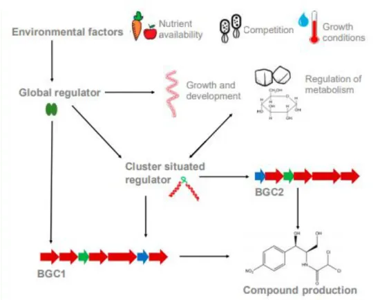
In order to survive, these bacteria, as all living organisms, need to supress their pathogens. In the case of Streptomyces, this can be done by producing the antibiotics, antiprotozoals, antifungals and antivirals referred earlier [4]. These natural inhibitors come from secondary metabolites, which are assembled in adjacent chromosomal genes, located in terminal zones of the chromosome (Fig. 5) [32], [39]. These contiguous genes are organized in clusters called BGCs, which are able to, individually, codify 35 secondary metabolites [32]. Normally, these clusters are constituted by resistance, transport, structural and regulator genes [40]. These last normally act by enhancing transcription of the structural genes that are in the same cluster as them, which explains why they are called cluster-situated regulators (CSRs) [41].
Most of the BGCs are responsible for the origin of everything (enzymes, regulatory proteins and transporters) needed to generate, process and transport a metabolite, which includes genes with regulatory functions, which are called CSRs (Fig. 5) [32], [39]. One BGC has normally one or more CSRs, and the most common one, among Streptomyces, is Streptomyces antibiotic regulatory protein (SARP) [39].
Outside of these BGCs, there are pleiotropic regulators responsible for controlling, for example, the morphological development [39].
The secondary metabolism needs, as all processes, to be regulated. That regulation is, mainly, done by transcription control. How this control works has already been theorized in two different ways. The first one, the hierarchical theory, says that there are different layers of regulation: an inferior layer, where CSRs control their own metabolites in their own biosynthetic cluster, and a superior one where the CSRs and one or more secondary metabolites are regulated by pleiotropic or global regulators (localized outside of the cluster) [42]. These pleiotropic regulators are capable of also affecting the morphological differentiation, the capture of nutrients, the secondary metabolism, and other [43]. CSRs, pleiotropic and central metabolic genes are regulated by global regulators spread throughout the chromosome. Every single one of them might regulate the biosynthesis of antibiotic [39].
The second theory says that, more than a hierarchy, the regulation of the secondary metabolism is a web and that the different regulators are all connected (Fig. 5) [44].
Fig. 5. Cluster situated regulators (CSRs) and global regulatory systems. These last are influenced by external environmental factors and both control the regulation of the expression of the genes related to secondary metabolism. Taken from [45].
1.3. Two-component system PhoR-PhoP
Phosphorus is important and it’s present in DNA, ribonucleic acid (RNA), in compounds needed for central metabolism (like ATP), signalling and in various other molecules. Moreover, it is involved in post translational regulation and phosphorylation/dephosphorylation of proteins [46], [47].
Therefore, an efficient supply of phosphorus is needed for the cell to survive. However, there are not, for Streptomyces, any systems known to deliver organic phosphate in the interior of the cell. So, when there is not enough phosphate, phosphate transporters and enzymes are activated to obtain it. This element is maintained by two strategies: intracellular storing and, under phosphor limiting conditions, scavenging it with the help of enzymes [46].
Phosphorus can be assimilated by bacteria in its inorganic form, phosphate, by, normally, two systems. An exception is, for instance, S. coelicolor that has three systems: pitH1 (expressed when there is a lot of phosphate), pitH2 and pstSCAB, which stands for phosphate specific transport and is the main phosphate transport system in conjugation with pitH2, and both are expressed when there is not much phosphate. These last two optimize the transport of phosphate and are regulated by the system PhoR-PhoP [48].
The system PhoR-PhoP (known as PhoR-PhoB in E. coli) is a two-component signal transduction system and the main regulator in phosphate metabolism control (mainly in cases
of limitation of phosphate). It is also responsible for the signal transduction from the outside to the inside of the cell, which enables adaptation of bacteria to environmental changes [49].
In this system, PhoR acts as a sensor kinase that receives sensory input, since it has a large extramembrane domain, and has the characteristics of a transmembrane sensor protein needed to adapt the cell to the phosphate limited state [50].
The other member of this system, PhoP, is the response regulator, and as so, has a regulator domain and an effector domain (this last one is responsible for the bound with DNA, making PhoP also a transcription factor) [51]. It is also responsible for the primary and secondary metabolism and morphological development, since it activates/inactivates genes implicated in phosphate metabolism and nitrogen assimilation (and therefore, in the balance between phosphate/nitrogen) [52].
This member, when phosphorylated, binds to DNA sequences known as Pho boxes, which are located in the promotor region of PhoBR regulon genes and is the consensus sequence that is shared by the regulatory areas of the genes in the phosphate (Pho) regulon, which is controlled by this two component system [46], [53]. The Pho regulon is important since it is involved in the response to conditions of limitation of phosphate [54]. These boxes, in these conditions, are activated by either phosphate transporter pstSCAB or phoU deletion and answer by increasing expression of phosphate transporters (such as pstSCAB), of enzymes needed to scavenge phosphate or by amplifying responses through positive autoregulation of the phoPR operons [46].
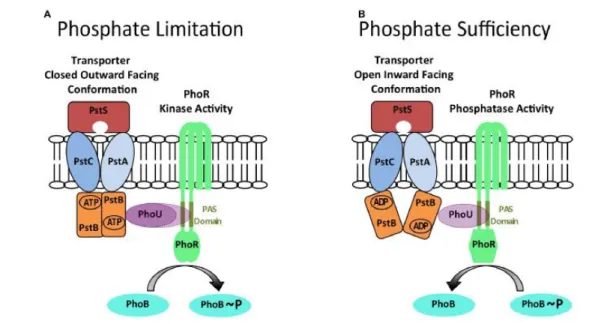
The transporter pstSCAB is also important since its structure determines the autokinase and phosphatase modes of PhoR activity: when it is outward facing closed structure, it promotes autokinase activity (needed in conditions of limited phosphate); and when pstSCAB is in an inward facing open structure, the phosphatase activity is promoted (essential for condition of sufficient phosphate) (Fig. 6) [46].
In E. coli, this system was already studied in extreme conditions, which created the Wanner’s Model [55]. In this model, it is explained that, in conditions of excess of phosphate, PstS (protein that unites with phosphate and its promotor is regulated by phosphate dependent of PhoB, PhoP alike) creates an inhibitor complex with transport system pstSCAB, protein PhoU and PhoR. Here, PhoR is inhibited and shows phosphatase activity towards PhoB, making sure that the last one is not activated. When phosphate is limited, pstSCAB changes its conformation and the inhibition complex is freed [46]. Then, PhoR self-phosphorylates, by transferring its phosphate to PhoB. This response regulator binds to Pho boxes, activating about 30 phosphate-regulated genes (Fig. 6) [28].
Fig. 6. A model for controlling, in E. coli, the balance between PhoR phosphatase and autokinase activities. The autokinase mode exists during conditions of phosphate limitation (A), while the phosphatase mode happens during phosphate sufficiency (B). Those alternate states are proposed to be determined by the different conformations of the pstSCAB transporter, which are relayed by PhoU to PhoR, in order to determine its activity. For instance, when pstSCAB is in a closed outward facing conformation (A), PhoU adaptor protein is unable to interact with PhoR and the autokinase activity is promoted. On the other hand, in (B) the pstSCAB is in an open inward facing conformation, which enables the contact between the PhoU adaptor and the PAS domain of PhoR, promoting the phosphatase activity. This way, the PhoB response regulator is dephosphorylated. Taken from [46].
In Streptomyces, the proteins from Wanner’s model have been described and it is known that, in these bacteria, for the PhoR-PhoP system to work, PhoP needs to be recognized and bounded to DNA. To do that, as explained before, there are DNA sequences, called the Pho boxes, that are described, for Streptomyces, as two direct repeat units (DRus) of 11 nucleotides (in which the first seven, “GTTCACC” for S. coelicolor, are the most conserved ones and have consensus sequence) (Fig. 7). Moreover, the strength of this bond depends on the conservation of the sequence (the more conserved it is, the stronger the bond is) [50].
When the genus Streptomyces doesn’t have enough phosphate, it can suffer effects on a number of processes, for instance, its growth, its morphological differentiation and its production of secondary metabolites [28], [50]. These metabolites are regulated negatively by the phosphate concentration in the medium, which explains why they are, normally, only produced in limiting concentrations of this substance [28]. This might happen due to the fact that, if there is less phosphate, the organism is probably in conditions where it is difficult to grow (it is stressed), and these secondary metabolisms might be an advantage to, for instance, inhibit the competition, or act as biochemical cross talk signals. Therefore, it is a strategy to survive [28]. All of these are reasons why the PhoR-PhoP system is crucial and why it needs to be investigated.
Fig. 7. The DRus in the Pho boxes of S. coelicolor and S. avermitilis. The height of the letter represents the frequency of that nucleotide in each position. Taken from [50].
1.4. Small RNAs
Small RNAs (sRNAs) are small-sized RNA molecules, of length between 50 nucleotides (nt) and 400 nt in prokaryotes (in eukaryotes they are even smaller), that, normally, don’t encode proteins and have regulator properties capable of modulating genetic expression by means of interaction with messenger RNA (mRNA) or, less frequently, with proteins [56], [57]. Most of sRNAs are transcriptionally induced under specific conditions, such as cold and heat shock, iron homeostasis, membrane remodelling and sugar and nitrogen metabolism, between others [58].
To the cell, creating regulatory sRNAs instead of proteins is beneficial because sRNAs are synthetized the fastest, since they are smaller and they don’t need to be translated [57].
Every single sRNA can have various action mechanisms, which influence every step of a gene expression: since the structure of DNA until the translation [57]. They might inhibit transcription, if they bind with mRNA that’s being synthesized [59]. They also act in the translation, by inhibiting it, if: they unite between positions -20 and -15 of the mRNA; or if they target 5’-untranslated regions (UTR) close or in the ribosome-binding site (RBS), thus competing with the 30S ribosomes [59], [60], [61]. On the other hand, they might enhance it, if they bind in the untranslated extremes of the mRNA and leave the mRNA bond site freer for the ribosome. They can also do it by stabilizing the mRNA or even by binding with translational enhancers [62]. Finally, sRNA are also responsible for the posttranscriptional regulation of physiology, stress responses, metabolism and virulence [63].
Even though transcription factors might have similarities with sRNAs, there are crucial differences: transcription factors cannot keep a noise-free silent state and have less recognition sites [63].
sRNAs can be characterized depending on their position in relation to their targets. The antisense sRNAs, or cis-acting sRNAs, are responsible for the regulation of the gene in the opposite chain and are completely complementary to the target gene of the mRNA in one of the extremes, in the middle, or with the complete gene [64].
There are different types of cis-acting sRNAs. One of them is called intragenic sRNAs, which are codified in the reading frame of a coding gene [65]. There are different types of intragenic sRNAs. They can be, for instance, 3’ sRNAs, which are codified, as the name itself suggests, in the region non translated 3’ of a mRNA. They can be classified as type I, if their promotor is inside the 3’ UTR or the mRNA coding sequence, and type II, if they come from
the processing of their parental mRNA. Both types share the 3’ end with their corresponding mRNA. On the other hand, their 5’ end is different, since there is a 5’ triphosphate (5´PPP) in type I and a 5’ P in the type II (Fig. 8) [66].
Fig. 8. Two general biogenesis pathways of sRNAs from the 3’ region of mRNA loci. Type I, in which the sRNA is transcribed from an mRNA-internal promotor and type II, in which the sRNA is processed from its parental mRNA. Adapted from [66].
On the other hand, riboswitches, another type of cis-acting sRNAs, are located in the non-translated 5’ region of the mRNA [63].
Finally, intergenic sRNAs, or trans sRNAs, are codified in a different chromosome than its target. Its complementarity, as opposed to the antisense sRNAs, is not perfect, which might explain why one intergenic sRNA has more than one target. In model Gram-negative bacteria, most of intergenic sRNAs need the chaperone Hfq to have a good regulatory activity, since it can unfold and stabilize RNAs and recruit ribonuclease (RNase) E [64]. Since they have multiple targets, their target gene regulation is determined by the ratio between the number of target RNA and sRNA transcription sites [63].
In Gram-positive bacteria, the sRNAs regulation has some differences, when compared with the regulation in Gram-negative bacteria. For instance, in the Gram-negative, the sRNAs cannot regulate translation and the Hfq chaperone is more important than it is in the Gram-positive bacteria [67].
1.5. Aim
The research group I was integrated in was working in a project that aimed to identify, by using transcriptomic techniques and manual annotation, the sRNAs from S. coelicolor that were implicated in the regulation by phosphate, dependently or independently of PhoP. These might also be characterized as regulators of the secondary metabolism and, therefore, might be related to the biosynthesis of antibiotics and other secondary metabolites, that show pharmacological activities and industrial interest.
The main objective of my internship with them was learning the routine of a laboratory. In order to do that, a wide variety of techniques, such as techniques of DNA introduction into cells, nucleic acid extraction and gel-analysis were learned. Tasks involving bioinformatics, crucial here for the annotation and evaluation of candidate genes for experimental confirmation, were also done.
Materials and Methods
In this section, there are protocols, and their detailed steps, that were done and others that were only seen. Those last will be differentiated from the others with a “*” in front of the name of the technique.
Methods in Microbiology
1. Melting of solid media
Theoretical FoundationMedium can either be liquid and be used for inoculating microorganisms, or solid, and be used to plate in petri dishes. Both have to be sterilized in an autoclave before being used to prevent unwanted contaminations.
Material
The material here presented is the one needed to make sure that the solid medium is melted and split into petri dishes in a sterilized manner, in order not to contaminate the media.
For situations when little medium is needed ➢ Bottles of medium;
➢ Microwave.
For situations when much medium is needed ➢ Bottles of medium;
➢ Pressure cooker; ➢ Water;
➢ Metal tube rack;
Used when plastic material is placed inside the pressure cooker. ➢ Electric heater.
Procedure
Normally, two methods can be used to melt media:
a) If there is not the need for much medium (less than 6 bottles with 100 mL of it), a microwave can be utilized. While using it, to prevent burns, gloves should be used.
1. Before the bottles were placed in the microwave, the lid should be opened slightly, in order to ensure that some of the pressure was taken from it.
2. The moment the medium started to boil, it should be taken from the microwave.
So that none of it leaves the bottle.
3. The lid should be opened a bit more and the bottle agitated.
This is done to liberate a bit more of pressure, that was accumulated thanks to the boiling medium.
4. This must be repeated until the medium was completely melted and there was nothing floating in it.
b) The other method is used to melt more than 6 bottles of medium and it involves a pressure cooker.
1. In this one, the bottles and water, enough to be as tall as the thickness of a finger, should be placed in the cooker.
If what is put in the pressure cooker is made of plastic, something, like a metal tube rack, should be placed under the plastic material.
2. After that, the pressure cooker was put on top of an electric heater, the maximum temperature is chosen, and the pressure cooker was turned on.
3. The pressure regulator at the top of the lid of the pressure cooker would rise with the pressure. The moment the pressure regulator was as high as it could be, the heat in the electric heater was decreased a bit for 5 minutes (min).
4. After that period of time, the cooker was taken from the heater, already turned off, and when the pressure regulator decreased completely, the lid could be open, and the bottles could be taken from the pressure cooker.
This pressure cooker is also used to sterilize medium.
The same as the medium, all material needed for the procedures should be sterile, in order to not contaminate the samples. From here on out, all the material mentioned is sterile.
2. Inoculation and plating of microorganisms
Theoretical FoundationWhen studying microorganisms, their living conditions, such as nutrients and oxygen, need to be assured, in order to be able to learn more about them. Therefore, they are grown in media, which, theoretically, have all nutrients needed for them to survive and multiply.
Two microorganisms were studied a great deal in this work: Streptomyces and E. coli. Even though only those two were worked with, many differences were seen, particularly while plating and inoculating them.
Each microorganism has different needs. For instance, there are some that need oxygen and others to which that element is lethal. One difference between the necessities of the two mentioned microorganisms has to do with the fact that Streptomyces, since it is filamentous, needs baffled flasks to provide more agitation, while E. coli doesn’t. That is why a brief study on the microorganism should be done before starting working with it.
Media can be differentiated between solid (which is split between different petri dishes) or liquid (that is maintained in either tubes or flasks). Even though the first one is called solid, in order to use it, it needs to be firstly in a liquid state, or else it can’t be split into petri dishes. Normally, the way to differentiate them is in the name: if the name ends with an “A”, it might
mean that it has agar and, therefore, it’s solid; on the other hand, if it ends with an “B” it can be liquid (example, tryptic soy agar, TSA, and tryptic soy broth, TSB).
The methods of placing the microorganism in the medium have different names depending on the type of medium used. When liquid medium is used, it’s called inoculation, while with solid it’s called plating the microorganism.
There are also differences in the methods when different microorganisms are used, because they can have different growth rates. While plating Streptomyces, compared to E. coli, more cautions are needed, since it grows slower, which means that is easier to contaminate. Since E. coli grows fast, not many other microorganisms that might be in the petri dish or in the flask can keep up with it.
Since Streptomyces grows slower, the thickness of the medium in the petri dishes should be higher than the ones used for microorganisms with faster growth rates, because, in the dry oven, the media tend to dry when inside it for some days.
The texture of the colonies is also different. E. coli, for instance, has a so-called creamy texture, which means that the colonies easily stick to the toothpick, and touching them is enough to have sufficient amount of cells. On the other hand, Streptomyces needs a lot of pressure to be done in the colony, which easily breaks into pieces, in order to have some sample in the toothpick (Fig. 9).
Plating for both of them is also different. In E. coli, a single line done in the medium with the toothpick with bacteria is enough to, in a few hours, have fully grown colonies. While, in Streptomyces, after making sure that the toothpick or the wire loop has some cells, a square or a circle should be done in the medium, and it should be done over and over again, to ensure that some cells are transferred to the new medium (Fig. 9).
While inoculating, some differences are also seen when working with both bacteria. If the inoculation is also done with a toothpick, in E. coli the toothpick with the sample staying a few seconds in the medium is enough, while with Streptomyces it needs to stay for more time and the toothpick should also be moved in order to help the colony separate itself from the toothpick.
Since Streptomyces has many morphologies, there are also differences when some of those are used. For instance, mycelium is harder to take a representative volume with the pipette and its absorbance value may not represent the reality. On the other hand, using spores also present some issues, since they develop much slower.
Fig. 9. Petri dishes with E. coli (A and B) and Streptomyces (C and D). Material
For inoculation ➢ Flow hood;
➢ Samples of the microorganism; ➢ Flask or tube;
➢ Incubator shaker; ➢ Micropipette and tips;
➢ Medium for the microorganism; ➢ Wire loop or toothpicks.
For plating
➢ Samples of the microorganism; ➢ Petri dishes;
➢ Dry oven;
➢ Micropipette and tips; ➢ Flow hood;
➢ Medium for the microorganism;
➢ Wire loop, Digralsky spreader or toothpicks. Procedure
These procedures might be done in a flow hood, depending on the microorganism used and its growth rate (for instance, when dealing with Streptomyces this precaution should be considered).
For inoculation
1. Enough medium was placed in a flask or in a tube.
Normally, TSB is used for Streptomyces and terrific broth (TB) for E. coli.
When inoculating microorganisms, some space should be left without medium, for there to be oxygen for them to breath. For instance, in Falcon tubes (with the capacity of 50 mL) only 5 to 10 mL of their volume should be used.
2. The flask/tube was tilted and the antibiotic, if needed, was added.
When small volumes are added, the plunger button of the micropipette should be pressed up and down several times.
3. A colony was taken with a toothpick from one petri dish where it grown.
Only individual colonies that are grown enough should be picked. This should be done, because if colonies that are joined together are used, there is a chance that there will be different genomes. This shouldn’t happen, because all colonies are clones, have the same genome, but this fact is not always true.
4. The flask/tube was tilted and the tip of the toothpick with the colony was placed in the medium.
Tilting is done to ensure that only the tip of the toothpick touches the medium to discard the microorganism.
5. The flask/tube was placed in the incubator shaker at the right conditions.
The optimal temperature for Streptomyces is around 30 ºC, while for E. coli is at 37 ºC at 250 revolutions per minute (rpm).
To stop the cells from growing more, they should be taken out from the shaker and placed on ice.
The minute the incubations leave the shaker, their cells start to die. Therefore, they should only be taken out when they are needed and when everything else is prepared.
For plating
1. The medium needed was melted and split into petri dishes.
Slipping medium into petri dishes should be done quickly, to avoid its solidification.
If antibiotic is needed, it needs to be added to the medium before it is split into petri dishes and when it’s not too hot, or else the antibiotics can be degraded.
2. The medium was let alone to solidify in the petri dishes.
3. This step could be done in various ways, depending on where the culture of bacteria was and what was needed:
a. When the culture was in a liquid medium:
a.1. Some volume of it was added with a micropipette to the petri dish.
If the culture comes from a flask, only what is in the liquid should be used, not what is in the walls.
a.2. Then a spreader was used immediately to spread the bacteria around all the petri dish, in order to have equal conditions.
a.3. Then, they needed to dry.
If more than one volume of culture is plated in different petri dishes, the micropipette should start with the one with less volume. The same thing needs to be done with the spreader. If the volume of sample added is too little, pure water should also be added to avoid it drying before it is spread.
b. When the culture was in another petri dish:
b.1. An individualized colony was picked either by a toothpick or a wire loop. The first is normally used when the colony is plated in a small place inside the petri dish, while the second is used to spread the colony around all of it.
b.2. The toothpick or the wire loop spread the colony in the petri dish.
In this method, no bigger pieces of colonies should stay behind, in order for it not interfering in the results.
4. The petri dishes were placed in the dry oven at the optimized temperature for each microorganism.
For E. coli is 37 ºC while for Streptomyces is from 28 ºC to 30 ºC. Normally, E. coli is also able to grow in less than 16 h, while Streptomyces might take days, depending on the medium used.
After the microorganism is grown, the medium with it can only be saved one or two weeks in the fridge.
3. Identification of microorganism*
Theoretical FoundationSometimes other microorganisms can be seen along with our samples in petri dishes. If that happens, identifying which microorganism is contaminating the sample might be important.
In this study, samples of microorganisms that were found in wine barrels of a company that produces wine were sent to analyse. This procedure was important in this case because the owner of the company needed to know if the wine that was in those contaminated barrels was drinkable, and, therefore, could be sold, or if the microorganism was nefarious and the wine needed to be discarded in order not to case any health issue.
Material
➢ Samples; ➢ Petri dishes; ➢ Flow hood;
➢ Bottles with sterile TSA medium;
Used since it is a general medium that can be utilized for most bacteria. ➢ Specific medium for lactic acid bacteria;
Only used because it is known that these are lactic acid bacteria.
Two media were used because in TSA different bacteria and contaminants can grow. ➢ 10 mL tubes;
➢ Test tube rack;
➢ Platform to balance the petri dishes; ➢ Micropipettes and tips;
➢ Digralsky spreader; ➢ Water bath;
➢ Computer; ➢ Dry oven. Procedure
This procedure must be done in a flow hood. 1. TSA medium was placed in petri dishes.
2. From the samples gotten, two dilutions (10-1 and 10-2) were done for each sample and
This step was done in order to ensure that individual colonies would be obtained in some of the petri dishes.
3. The same amount of sample not diluted was plated in the same conditions. 4. These petri dishes were placed in the dry oven at 28 ºC.
This temperature was chosen because most bacteria can grow in these conditions.
5. The bottles of the medium specific for lactic acid bacteria were placed in a water bath at 52 ºC.
This temperature was chosen because it ensured that the medium continued in a liquid state and that the bacteria, when placed in it, survived.
6. From the dilutions and the original tubes of samples, another 100 µL were taken and added to 10 mL of the specific medium. This mix was then split into petri dishes.
This step needs to be done rapidly, in order for the medium not to solidify. Therefore, only one bottle of medium at a time can be taken from the water bath and only when everything else is already ready.
7. After doing the last step to every dilution and every sample, the petri dishes were let alone to dry.
8. While the last petri dishes filled with sample and medium were still wet, the first ones, already dried, were filled with more 10 mL of medium without bacteria.
Again, the necessary quantity of medium is only taken from the water bath when everything else is prepared, so it doesn’t solidify.
This step is necessary to ensure that much oxygen doesn’t exist, which is preferred by this kind of bacteria. If these were anaerobic, the process would be different*.
9. The petri dishes were placed in a dry oven at 28 ºC.
*If these bacteria were anaerobic, which they are not, more cautions would be needed in order to ensure that much oxygen doesn’t enter in contact with them. Working with bacteria that are intolerant to oxygen is difficult and needs a specific incubation chamber for anaerobiosis. On the other hand, a special compound that degrades oxygen is also placed in the petri dish.
The procedure of identifying the microorganism doesn’t end here. After the last step, and after colonies have growth in both media, the morphology of each of them is analysed. This is done since a different morphology might mean that different microorganisms are present.
Then, every different morphology is isolated, and its DNA is extracted. After having the DNA, it is sequenced and the obtained sequence is then compared to the other from the different morphologies, to conclude if they indeed come from the same microorganism or not (here the software “ContigExpress” might be a reliable option).
The sequencing procedure produces a file with “electropherogram”, or “electrophoregram”, which is a record or chart produced by electrophoresis, and it has different peaks and different colours for each nucleotide. The file also computes a quality value, which represents the reliability that the program has that that peak is indeed that nucleotide. When the value is “0”, caution is needed. There is also the possibility of having two peaks in the same position, which might be caused by having two templates during polymerization. Smaller peaks at the end can also occur, which might be explained by the loss of intensity that the fluorescent chemical has in that moment. Normally, beginning and endings of the sequence are of low quality. The
vector NTI (Invitrogen) tool recognized by “ContigExpress” can be used to visualize the sequencing files.
Finally, the different sequences, that correspond to different microorganisms, are then run by “Nucleotide BLAST” (“Blastn”), which will finally be able to identify, by comparison with a database, with a certain percentage of identity, the microorganism. Here, the “closest” organism is decided by the “Query Cover”, which is the alignment percentage between the two sequences, and the “E value”, that is the number of alignments with similar quality gotten randomly.
Some more information on the organism can be retrieved through the program “Mycobank”.
4. Ligation reaction
Theoretical FoundationA ligation reaction is the process used to join two specific DNA molecules (insert/fragment and vector), to generally form a plasmid. Only some ligations, those that will originate a functional plasmid, will enter in the cell and multiply [68].
To bind insert (fragment of interest) to vector, there is the need to cut them. In order to do that, restriction enzymes are used. These are proteins that can recognize specific small sequences and can cut them or near them. While cutting them, two different ends can be originated by the enzymes (Fig. 10):
● Blunt ends: Straight cuts that don’t produce salient ends.
● Sticky ends: Cuts that produce salient ends that are complementary. These provide better results in a ligation experiment.
Fig. 10. Difference between sticky (A) and blunt ends (B). Taken from [69].
The plasmid originated, if the ligation and transformation are successful, will be similar to the one represented below (Fig. 11).
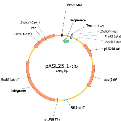
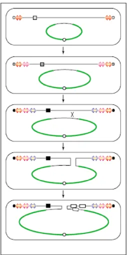
Fig. 11. Schematic representation of the plasmid constructed.
As seen in the image, the vector has resistance genes (in this case to thiostrepton, identified as “tsr” in the image, for Streptomyces, and to apramycin for E. coli, identified as “aac(3)IV”), that function as selecting markers. It also has one origin of replication for E. coli (“pUC18 ori”), a transfer sequence (“RK2 oriT”) which enables the mobilization of the plasmid from a donor strain (E. coli) to the host strain (Streptomyces). There is also a site where the plasmid will bind with the genomic DNA (“attP”) of Streptomyces and an enzyme to do that bonding, an integrase.
Finally, the plasmid contains an insert (“Promoter”-“Sequence”-“Terminator”) that will originate an antisense of the sRNA of interest.
This scheme is also useful to know the size of the fragments gotten in an electrophoresis after digestion with restriction enzymes, since they have the coordinates where the enzyme cuts [for instance, here EcoRI cuts in the coordinate 4835 and in 381 base pairs (bp)]. This way, and by knowing that the whole plasmid has a length of 6883 bp (as seen in the figure), the sizes expected in the electrophoresis can be calculated.
The plasmid was obtained after ligation of a BclI-XbaI vector and a BamHI-SpeI insert with sticky ends, but these restriction enzymes are unable to recognize those sites again. This happens because when the vector and insert ends are united by a ligase, the sequence obtained is not palindromic like the original one was.
After transforming, to confirm if the ligation was successful, extracting the plasmid and digesting it needed. The ultimate verification is done afterwards, through an electrophoresis. Material
➢ Vector; ➢ Insert DNA;
➢ T4 DNA Ligase; ➢ Buffer for the ligase; ➢ Restriction enzymes; ➢ Pure water;
➢ Eppendorf tubes; ➢ Centrifuge tube rack; ➢ Heat block;
➢ Incubator shaker; ➢ Micropipettes and tips. Procedure
The procedure can have some small changes depending on the insert and the vector used. To increase the probability of joining the insert to the vector, ligations are normally done with small volumes.
1. Vector and insert were mixed together.
It is important to know the proportion between the quantities of insert and vector used and that is often of 3:1 or 5:1.
2. Ligase buffer and ligase were also added.
3. Two enzymes with different regions of cut (one for each of the vector targets) were used to originate distinguished ends where the insert would bind.
In this type of ligation, the insert can only bind with the vector from one direction and only one ligase is used because it is not specific and doesn’t care if the cut regions are the same or not.
4. The samples were incubated.
The temperature and the duration of the incubation depends on which type of cut will happen: • Since blunt ends are more difficult to bind, the temperature normally used is between 4 and 6 ºC and the ligation incubates overnight.
• On the other hand, sticky ends are normally bound at room temperature (about 22 ºC) for 2 or 3 hours.
5. After this, the ligation was completed and the enzyme was inactivated by placing the samples for 10 min at 65 ºC.
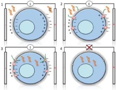
5. Transformation (chemical procedure)
Theoretical FoundationTransformation is a method in which a foreign DNA present in the medium is introduced into a cell (Fig. 12) [70]. Even though this occurs naturally in some bacteria, its efficiency changes depending on the species.
Fig. 12. Schematic representation of transformation and conjugation. Adapted from [70].
In the laboratory, this technique is very helpful, since it allows almost every plasmid, no matter if they are in their circular or in their supercoiled form, to be introduced in nearly all bacteria.
In order for the transformation to happen, the bacteria must be in a so-called competent state, which is only achieved in certain physiological conditions. This state is crucial for the success of the technique because it ensures that both the wall and cell membrane present changes that allow the entrance of nucleic acids in the cell.
Even though there are many species that are not able to present this state, some methods, based on physical and chemical treatments, were created in the laboratory to induce it. These treatments produce micropores in the cell, which allow the introduction of the foreign DNA in an efficient way.
To detect if the transformation was successful or not, the plasmid has a selective marker which will make the bacteria present characteristics, such as resistance to an antibiotic, that will facilitate the differentiation between the cells that were transformed and the ones that weren’t. Here, E. coli must present resistance to apramycin, as seen before, in order for the transformation to be concluded as successful. However, having the resistance gene as a selective marker has some problems because it implicates adding antibiotic, which can lead to secondary effects.
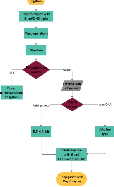
This protocol was done to two different strains of E. coli, since every strain has a specific function. One of them, E. coli DH5-alpha, always used first, methylase positive, is used to
confirm if the ligation was indeed well done, if the plasmid is built the way wanted. This step is needed because the second strain, E. coli ET12567 pUZ8002, the one used to conjugate with Streptomyces, already has a plasmid (pUZ8002), which makes it harder to analyse the result of a ligation. This plasmid that this strain has is also important for the conjugation, since it has transfer (tra) genes responsible for the transference of vectors that have “RK2 oriT” transference origin (Fig. 11). Another characteristic that makes this strain apt for conjugation is the fact that it is methylase negative, and, if it wasn’t, Streptomyces would recognize the plasmid as foreign and would reject it. Therefore, when it is planned to introduce a construction in Streptomyces, first the result of the ligation is transformed in E. coli DH5-alpha, then the transformants are analysed by minipreparations (minipreps). After that, the desired plasmid is isolated and transformed in E. coli ET12567 pUZ8002 and the derived strain is used to conjugate with Streptomyces (Fig. 13).
![Fig. 4. The various phases of the bacterial growth curve. Taken from [38].](https://thumb-eu.123doks.com/thumbv2/123dok_br/15740520.1072500/22.893.225.716.128.511/fig-various-phases-bacterial-growth-curve-taken.webp)