Clinical Case Study
GE Port J Gastroenterol 2017;24:89–94 DOI: 10.1159/000450872
Paraduodenal Pancreatitis: Three Cases
with Different Therapeutic Approaches
Diana Carvalho Rafaela Loureiro Verónica Pavão Borges Pedro Russo
Carlos Bernardes Gonçalo Ramos
Gastroenterology and Hepatology Department, Hospital Santo António dos Capuchos, Centro Hospitalar de Lisboa Central, Lisbon , Portugal
Pancreatite Paraduodenal: Três Casos Com Diferentes Abordagens Terapêuticas
Palavras Chave
Pancreatite crónica · Dor abdominal crónica ·
Pancreaticoduodenectomia · Somatostatina · Obstrução duodenal
Resumo
Introdução: A pancreatite paraduodenal constitui uma causa rara de dor abdominal crónica, caracterizada por processo inflamatório com consequente cicatrização no sulco entre a cabeça do pâncreas e a parede duodenal. Para além de dor abdominal, sintomas como vómitos e perda de peso são frequentes. Actualmente, com o desen-volvimento dos métodos diagnósticos imagiológicos e endoscópicos, pode ser identificada sem necessidade de confirmação histológica, embora a diferenciação com o adenocarcinoma pancreático possa ser desafiante. Diver-sas opções terapêuticas encontram-se disponíveis incluin-do tratamento farmacológico, enincluin-doscópico ou cirúrgico. Métodos: Os autores descrevem três casos de pancreatite paraduodenal com diferentes atitudes terapêuticas. Re-sultados e Conclusão: Estes demonstram que esta patolo-gia deve ser considerada no diagnóstico diferencial de massas pancreáticas com infiltração duodenal e que a sua abordagem deve ser individualizada e judiciosa.
© 2016 Sociedade Portuguesa de Gastrenterologia Publicado por S. Karger AG, Basel
Key Words
Pancreatitis · Chronic abdominal pain ·
Pancreaticoduodenectomy · Somatostatin · Duodenal obstruction
Abstract
Background: Paraduodenal pancreatitis is a rare cause of chronic abdominal pain characterized by an inflammatory process and scarring in the groove area between the pancre-atic head and the duodenal wall. Besides abdominal pain, symptoms such as vomiting and weight loss are common. Currently, advances in radiological and endoscopic diagnos-tic methods allow it to be identified without histological confirmation, although the differentiation from pancreatic adenocarcinoma could be challenging in some cases. Many therapeutic options are available nowadays including phar-macological, endoscopic, or surgical treatment. Methods: We report 3 cases of paraduodenal pancreatitis that had dif-ferent therapeutic approaches. Results and Conclusion: They show that this pathology should be taken into account in the differential diagnosis of pancreatic masses with duo-denal infiltration, and that its management should be indi-vidualized and judicious.
© 2016 Sociedade Portuguesa de Gastrenterologia Published by S. Karger AG, Basel
Received: March 31, 2016 Accepted after revision: July 3, 2016 Published online: November 22, 2016
Dr. Diana Carvalho
Gastroenterology and Hepatology Department, Hospital Santo António dos Capuchos © 2016 Sociedade Portuguesa de Gastrenterologia
Introduction
Paraduodenal pancreatitis (PP) is a pathological
con-dition resulting from fibrotic inflammation that involves
the duodenal wall closely to the minor papilla, the
adja-cent pancreatic parenchyma, and the area between them
[1] . Its pathophysiology remains unclear, although it is
believed that alcohol consumption and the presence of
ectopic pancreatic tissue in the duodenal wall may play a
major role in its development [2] . Through the last years,
its treatment has been a topic of discussion in the
litera-ture, with the proposal of a wide range of conservative
and surgical approaches [3] . Nowadays, the consensus is
to initially prefer medical or endoscopic treatment,
sur-gery being reserved to patients with uncertain diagnosis
or symptoms that do not improve with other measures
[4] .
We present 3 cases of PP that were managed with 3
different therapeutics procedures: supportive treatment,
use of octreotide, and surgery with
pancreaticoduodenec-tomy.
Case 1
A 55-year-old male patient, with daily consumption of 130 g of alcohol and tobacco, was admitted to the Gastroenterology De-partment for severe epigastric abdominal pain accompanied by anorexia, nausea, vomiting, and weight loss (3 kg). An upper gas-trointestinal endoscopy was performed, revealing a bulb deformed by a vegetating lesion, not allowing visualization of the second du-odenal portion (D2); histology was negative for malignancy.
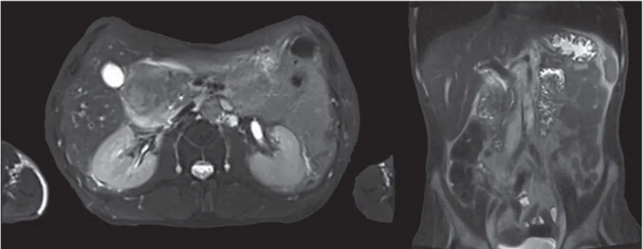
During hospitalization, he improved with analgesia, intrave-nous fluids, and bowel rest. Laboratory evaluation showed carbo-hydrate antigen (CA) 19.9 of 61 ng/mL, with no other changes. Magnetic resonance imaging (MRI) showed a large abdominal le-sion involving the bulb and D2, which was predominantly hyper-intense on T2 and hypohyper-intense on T1, without significant contrast enhancement. In the pancreatic head, a cystic lesion was observed ( Fig. 1 ).
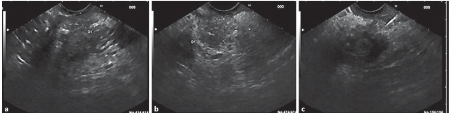
Endoscopic ultrasound (EUS) revealed thickening of D2 (9 mm) with microcysts in the wall, chronic pancreatitis and a cyst (21 mm) in the pancreatic head. Endoscopically, no irregularities of the duodenal mucosa were observed and passage to D2 was pos-sible ( Fig. 2 ). Fine-needle aspiration (FNA) of the pancreatic cyst revealed an amylase of 108,643 U/L and carcinoembryonic antigen (CEA) 44.5 ng/mL on cytochemical examination.
Reassessment imaging by computed tomography exhibited a significant regression of previous duodenal and pancreatic lesions. A presumptive diagnosis of PP was made. After multidisciplinary
a b
a b c
Fig. 1. MRI revealing an extensive oval lesion involving the duodenum bulb as well as D2 ( a ) and a pancreatic cyst ( b ).
Fig. 2. EUS showing thickening of the D2 wall ( a ) and chronic pancreatitis ( b ). FNA of the pancreatic cyst ( c ).
team discussion, close monitoring was proposed with radiological examination, abstinence of alcohol, and smoking cessation. Dur-ing the 13 months of follow-up, the patient remains asymptom-atic, with no laboratory or radiological abnormalities.
A close follow-up was decided in this case because of the favor-able clinical response and the patient’s adherence to alcohol and tobacco abstinence. It is important to state that all current thera-peutic alternatives (medical, endoscopic, or surgical) carry some degree of risk, which was deemed unwarranted in this case.
Case 2
A 58-year-old female patient was referred to the Gastroenterol-ogy Department for recurrent epigastric pain not related to food ingestion, with irradiation to the right abdominal flank, followed by nauseas. The patient had no history of addictive behavior. Blood tests revealed cholestatic changes (alkaline phosphatase 222 U/L and gamma-glutamyltransferase 82 U/L) and normal IgG4 levels. She was treated with ursodeoxycholic acid (1,000 mg/day) and pancreatin without clinical improvement.
A MRI was performed, showing parietal concentric thickening of D2 with apparent origin in the submucosal layer. Endoscopi-cally, the duodenal mucosa was edematous, with decreased lumi-nal caliber at this level; biopsies did not reveal any pathological changes. An EUS confirmed a slight hypoechoic thickening of the D2 wall (5 mm), especially for the submucosa, without cystic areas or other abnormalities in the echo structure. The pancreatic paren-chyma was regular, but with marked atrophy of the body/tail seg-ments ( Fig. 3 ). PP was diagnosed, and she started therapy with octreotide (20 mg with interval of 4 weeks). She had marked pain reduction, with a sustained clinical response after 1 year of fol-low-up.
The treatment’s choice was due to the persistence of pain de-spite conservative measures (i.e. alcohol abstinence) and the ab-sence of complications that could justify an endoscopic approach. In order to avoid a more invasive approach, the somatostatin ana-log octreotide was considered the best option for symptomatic re-lief.
Case 3
A 56-year-old male patient with chronic consumption of alco-hol (50 g/day) and tobacco (36 pack-year) was referred to the Gas-troenterology Department for intense epigastric postprandial pain with 4 months of evolution. He also mentioned nauseas, vomiting, and weight loss (15 kg). Laboratory tests only showed hyperamy-lasemia (312 U/L). He was treated with omeprazole 40 mg twice daily, domperidone 10 mg/day, and pregabalin 150 mg/day, with-out symptomatic relief.
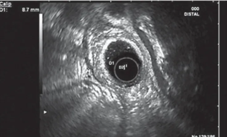
For clarification, an abdominal computed tomography was done, revealing D2 thickening (12 mm) with wall heterogeneity. EUS evidenced a heterogeneous circumferentially thickened duo-denal wall (8.7 mm), mainly the third layer (submucosa), without focal lesions and a hypoechoic but homogeneous parenchyma in the pancreatic head ( Fig. 4 ).
Over the next 2 months, the symptoms worsened, with con-tinuous epigastric pain, uncontrollable vomiting, and weight loss (>20 kg). For this reason, the patient was admitted to the Gastro-enterology Department and treated with fluid therapy and analge-sia. He showed tolerance to mild oral diet but suffered moderate abdominal pain. Magnetic resonance cholangiopancreatography showed D2 thickening with heterogeneous contrast enhancement, areas of hyperintense signal, remaining cyst or necrosis, and mild heterogeneity of the pancreatic head contour ( Fig. 5 ).
Our diagnostic hypotheses were PP versus duodenal malignan-cy and, in a multidisciplinary meeting, it was decided to perform a pancreaticoduodenectomy (Transverse-Longmire). The surgery was performed without complications, and histological examina-tion of the surgical specimen revealed cysts in the muscular layer coated with mucinous epithelium in the region of the minor pa-pilla. Ectopic pancreatic tissue, Brunner’s gland hyperplasia, ab-scesses, proliferation of smooth muscle cells, and fibrosis were also identified. These aspects confirmed the diagnosis of PP ( Fig. 6 ).
Fifteen months after surgery, the patient remains asymptom-atic with progressive increase of weight. In this patient, the defini-tive diagnosis remained uncertain after the diagnostic work-up, making the surgical approach the best option for both treatment and etiologic confirmation.
Discussion
PP is a rare cause of chronic abdominal pain that has
been described for the first time by Becker in 1973 as a
segmental pancreatitis [5] . Later, in the early 1990s, it was
classified into the following 2 forms: pure, which involves
only the groove area (space between the duodenal wall
and pancreatic head), and segmental, with inflammation
extending to the adjacent pancreatic head. In practice,
this differentiation is not always possible to be clearly
de-fined [6] .
Its prevalence varies largely from 2.7 to 24.5% in
surgi-cal specimens of patients with chronic pancreatitis [5]
and is most frequently found in men during the fifth
de-cade [7] . Risk factors include severe alcohol consumption
and heavy smoking [8] and in two-thirds, it is associated
with chronic pancreatitis [9] .
Various factors are implicated in PP’s etiology:
pan-creatic ectopic tissue in the duodenal wall with localized
inflammation and scarring; absence or functional
ob-struction of the minor papilla; increased fluid viscosity,
Brunner gland hyperplasia and intraductal pressure
caused by chronic consumption of alcohol, as well as
modifications of local anatomy secondary to
gastroduo-denal ulcers, gastrectomy, or biliary diseases [8] .
Usually patients present with upper abdominal pain
associated with nausea and recurrent postprandial
vomit-ing [10] . With chronicity, significant weight loss appears
and eventually jaundice, diarrhea, and diabetes mellitus
[4, 11] .
In laboratory evaluation, a slight elevation of
pancre-atic and heppancre-atic enzymes is frequently found [5] . The
tu-mor markers CEA and CA 19.9 are normal, with few
cas-es dcas-escribing a small raise.
c b
a
Fig. 5. D2 thickening with heterogeneous areas of hyperintense signal on magnetic resonance cholangiopancre-atography.
Fig. 6. Histology showing Brunner’s gland hyperplasia ( a ), cysts in the muscular layer, ectopic pancreatic tissue ( b ), proliferation of smooth muscle cells, and fibrosis ( c ).
Diverse pathologies are included in the differential
di-agnosis: pancreatic adenocarcinoma, cystic tumors,
auto-immune pancreatitis [8] , duodenal carcinoma,
cholan-giocarcinoma, and acute relapsing pancreatitis. The first
one is particularly challenging even with resource to
ima-giology and endoscopic modalities [1] .
Imaging exams, especially MRI, are very important to
confirm the diagnosis of PP. The spectrum of findings is
large but the typical presentation involves focal thickening
of the D2, cystic degeneration, and a “sheet-like” mass of
fibrotic tissue within the groove [3] . Other changes that
can be found include duct abnormalities (dilation,
atro-phy, and irregularity) and signs of chronic pancreatitis [3] .
Nowadays, besides MRI, EUS is also considered a gold
standard for PP diagnosis [2] . It reveals a hypoechoic area
between the duodenal wall and the head of the pancreas,
thickening and stenosis of the duodenal wall, intramural
cysts and a slightly narrowing of the main biliary duct.
Eventually, in cases of segmental PP, it is possible to see
infiltration and calcification of pancreatic parenchyma,
pseudocysts, and dilation of pancreatic duct [1, 4] .
EUS-FNA has limited value in this setting and depends on the
biopsy site and its interpretation, being difficult to
ex-clude a subjacent tumor [6] . Upper gastrointestinal
en-doscopy usually shows a swollen and polypoid duodenal
mucosa and stenosis; however, biopsies are frequently
in-conclusive [8] . Endoscopic retrograde
cholangiopancrea-tography can be difficult or even impossible due to
duo-denal stenosis. When successful, it may reveal distal
ste-nosis of the main biliary duct or irregularities of Wirsung
and Santorini ducts [4] .
Histologically, the most characteristic findings consist
of myofibroblastic and neural expansion into the groove
area, thickening of the duodenal wall with fibrosis and
Brunner’s gland and smooth muscle hyperplasia,
occur-rence of dilated ducts as well as pseudocysts within the
duodenal wall, and the presence of hypercellullar
granu-lation tissue and foreign body type giant cells [1, 12] .
Cy-tology from fragments obtained by EUS can show a wide
range of results, frequently with observation of spindled
stromal cells. Although the majority is interpreted as
neg-ative for malignancy, the results can be misinterpreted as
a tumor, especially if there is an abundance of Brunner
gland hyperplasia and spindle cells [6, 12] .
Currently, its treatment is based on a stepwise
ap-proach, which can be effective with an acceptable
compli-cation rate [9] . Conservative measures involve the
absti-nence of alcohol and tobacco, pancreatic rest, and
anal-gesics, the first one being crucial for long-term results [8] .
Recently, somatostatin analogs were considered an
alter-native medical option. However, few reports are
de-scribed in literature and symptomatology recurrence
seems to be frequent after treatment cessation [4] .
Endoscopic treatment, such as stricture dilation and
cysts or pancreatic ductal drainage, is an important
non-surgical approach with good results. In a study conducted
by Arvanitakis et al. [9] involving 51 patients, this
ap-proach, together with medical treatment, had a high rate
of clinical success (nearly 80%) and showed low adverse
effects.
If symptoms do not improve with the previous
strate-gies, complications appear, or when a suspicion of a
neo-plasm is present, surgery must be the treatment of choice.
The technique usually performed is the Whipple
proce-dure [4] , but some authors sustain that more conservative
interventions, such as suprapapillary segmental duodenal
resection, should be considered in cases of isolated
duo-denal dystrophy [13] . Apart of the procedure used, it is
proved that surgery highly improves quality of life and
contributes to pain cessation [2] .
In conclusion, PP should be considered in the
differ-ential diagnosis of pancreatic head masses with duodenal
infiltration. Its treatment should be individualized to
each case, choosing a conservative management
when-ever possible.
Statement of Ethics
This study did not require informed consent or review/approv-al by the appropriate ethics committee.
Disclosure Statement
The authors declare that they have no competing interests.
1 Arora A, Dev A, Mukund A, Patidar Y, Bhatia V, Sarin SK: Paraduodenal pancreatitis. Clin Radiol 2014; 69: 299–306.
2 Casetti L, Bassi C, Salvia R, Butturini G, Gra-ziani R, Falconi M, et al: ‘‘Paraduodenal’’ pan-creatitis: results of surgery on 58 consecutives patients from a single institution. World J Surg 2009; 33: 2664–2669.
3 Hungerford JP, Magarik MAN, Hardie AD: The breadth of imaging findings of groove pancreatitis. Clin Imag 2015; 39: 363–366. 4 Pallisera-Lloveras A, Ramia-Ángel JM,
Vi-cens-Arbona C, Cifuentes-Rodenas A: Groove pancreatitis. Rev Esp Enferm Dig 2015; 107: 280–288.
5 Black TP, Guy CD, White RR, Obando J, Bur-bridge RA: Groove pancreatitis: four cases from a single center and brief review of the literature. ACG Case Rep J 2014; 1: 154–157. 6 Raman SP, Salaria SN, Hruban RH, Fishman
EK: Groove pancreatitis: spectrum of imaging findings and radiology-pathology correla-tion. AJR Am J Roentgenol 2013; 201:W29– W39.
7 Rebours V, Lévy P, Vullierme MP, Couvelard A, O’Toole D, Aubert A, et al: Clinical and morphological features of duodenal cystic dystrophy in heterotopic pancreas. Am J Gas-tro 2007; 102: 871–879.
8 DeSouza K, Nodit L: Groove pancreatitis: a brief review of a diagnostic challenge. Arch Pathol Lab Med 2015; 139: 417–421.
9 Arvanitakis M, Rigaux J, Toussai E, Eisen-drath P, Bali MA, Matos C, et al: Endotherapy for paraduodenal pancreatitis: a large retro-spective case series. Endoscopy 2014; 46: 580– 587.
10 Brosens LAA, Leguit RJ, Vleggaar FP, Veld-huis WB, Van Leeuwen MS, Offerhaus GJA: EUS-guided FNA cytology diagnosis of para-duodenal pancreatitis (groove pancreatitis) with numerous giant cells: conservative man-agement allowed by cytological and radiolog-ical correlation. Cytopathology 2015; 26: 122– 125.
11 Gravito-Soares M, Gravito-Soares E, Alves A, Gomes D, Almeida N, Tralhão G, et al: Groove pancreatitis with biliary and duodenal stric-ture: an unusual cause of obstructive jaun-dice. GE Port J Gastroenterol 2016; 23: 170– 174.
12 Chute DJ, Stelow EB: Fine-needle aspiration features of paraduodenal pancreatitis (groove pancreatitis): a report of three cases. Diagn Cytopathol 2012; 40: 1116–1121.
13 Egorov VI, Vankovich AN, Petrov RV, Sta-rostina NS, Butkevich AT, Sazhin AV, et al: Pancreas-preserving approach to ‘‘paraduo-denal pancreatitis’’ treatment: why, when, and how? Experience of treatment of 62 pa-tients with duodenal dystrophy. Biomed Res Int 2014; 2014: 185265.
Erratum
In the article by Carvalho et al. entitled “Paraduodenal Pancreatitis: Three Cases with Dif-ferent Therapeutic Approaches” [GE Port J Gastroenterol 2017;24:89–94, DOI: 10.1159/ 000450872], figures 1 and 2 have erroneously been exchanged. Figure legend 1 refers to figure 2 (MRI), and figure legend 2 refers to figure 1 (EUS).