UNIVERSIDADE DE LISBOA FACULDADE DE MEDICINA DENTÁRIA
“MICRO-TENSILE BOND STRENGTH TO DENTINE OF A
SELF-ETCH AND A UNIVERSAL ADHESIVE SYSTEM IN SELF-SELF-ETCH
MODE”
Ana Catarina Palmeirinha Pinto
Dissertação
Mestrado Integrado em Medicina Dentária
UNIVERSIDADE DE LISBOA FACULDADE DE MEDICINA DENTÁRIA
“MICROTENSILE BOND STRENGTH TO DENTINE OF A
SELF-ETCH AND A UNIVERSAL ADHESIVE SYSTEM IN SELF-SELF-ETCH
MODE”
Ana Catarina Palmeirinha Pinto
Orientação por:
Mestre Ana Catarina Coito
Co - orientação por:
Mestre Ana Luísa Silva
Dissertação
Mestrado Integrado em Medicina Dentária
“In a time of turbulence and change, it is more true than ever that
knowledge is power”
AGRADECIMENTOS
Em primeiro lugar, à Dr.ª Ana Catarina Coito por me ter dado o privilégio de ser minha orientadora nesta caminhada, pela disponibilidade, pelo apoio, estímulo e incentivo para fazer sempre mais e melhor em todas as etapas percorridas. Todo o conhecimento por si partilhado será, por certo, uma mais valia no futuro.
Ao Professor Doutor Alexandre Cavalheiro pela imprescindível ajuda na análise estatística e por toda a disponibilidade e ensinamentos durante a realização deste trabalho.
À Professora Doutora Manuela Lopes e à Dr.ª Ana Luísa Silva por toda a disponibilidade e ajuda no laboratório.
A todos os elementos do Departamento de Dentisteria Operatória da Faculdade de Medicina Dentária de Lisboa, pelo conhecimento transmitido ao longo dos últimos anos, pela forma calorosa com que me receberam e pela ajuda na realização deste trabalho.
À Joana, Tatiana e Mafalda pela indispensável ajuda durante a realização do procedimento experimental.
À Madalena, pela amizade, carinho e apoio nos bons e maus momentos dentro e fora da faculdade.
À Soraia, pela amizade e por ter encurtado a distância entre a Suécia e o laboratório, contribuindo para a realização deste trabalho.
À Patrícia, tia de coração, e à Filipa, amiga de sempre, pela disponibilidade e ajuda na reta final deste trabalho.
À Inês, Catarina e Marialice pela amizade, carinho, inspiração e pelos bons momentos que passámos nos últimos cinco anos. Sendo especial o agradecimento à Catarina, minha dupla de todas as horas, com quem aprendi a ser mais calma e ponderada.
Aos meus pais, simplesmente por existirem. Sou grata por tudo o que me ensinam, pelas oportunidades únicas que me proporcionam, por me incentivarem e inspirarem a querer sempre mais e melhor e por acreditarem que sou capaz. Espero estar sempre a altura dos valores que me transmitiram.
Aos meus avós, por cuidarem de mim. Ao João, por caminhar sempre ao meu lado.
Micro-Tensile Bond Strength To Dentin Of A Self-Etch And A Universal Adhesive System In Self-Etch Mode vii GENERAL INDEX
GENERAL INDEX ...vii
TABLES INDEX ... viii
GRAPHICS INDEX ...ix
FIGURES INDEX ... x ABREVIATIONS ...xi ABSTRACT ... xiii RESUMO ... xv I – LITERATURE REVIEW ... 1 1. ADHESION PRINCIPLES ... 1 1.1 Adhesion to Dentine ... 2
2. ADHESIVE S YSTEMS CLASSIFICATION ... 2
2.1 Etch-and-Rinse Strategy... 3
2.2 Etch-and-Dry Strategy ... 4
3. UNIVERS AL ADHESIVES ... 7
II - PURPOSE... 8
III - MATERIALS AND METHODS... 9
Type of study ... 9
Design of the study ... 9
Teeth selection and preparation ... 9
Distribution and treatment of the crown segments ... 12
Bonding Procedures ... 12
Restorative Procedures ... 14
Specimens preparation for micro-tensile tests ... 15
Micro-tensile bond strength tests (μTBS) ... 16
Statistical Analysis ... 17
IV - RESULTS ... 18
V - DISCUSSION ... 22
VI - CONCLUSION ... 30
Limitations of the study ... 30
VII – REFERENCES ... 31 APPENDIX ... I APPENDIX 1 ... II a. Materials And Components... III b. Manufacturer‟s Instructions ...IV
Micro-Tensile Bond Strength To Dentin Of A Self-Etch And A Universal Adhesive System In Self-Etch Mode viii TABLES INDEX
Table 1 - Number of sticks (N); micro-tensile bond strengths (μTBS) mean values; standard deviation (Std. Deviation) and standard error mean (Std. Error Mean)….…...18 Table 2 – Test of Normality: Kolmogorov-Smirnov and Shapiro-Wilk tests…….…...18 Table 3 – Results of Levene‟s Test and t-test………..…...19 Table 4 – T-test for equality of means………..…..20 Table 5 – Number of sticks in each failure mode: A – adhesive failure; CC– composite cohesive failure; CD – dentine cohesive failure; M – mixed failure…...21 Table 6 - Materials used, components, manufacturers and lot numbers ………...…….III
Micro-Tensile Bond Strength To Dentin Of A Self-Etch And A Universal Adhesive System In Self-Etch Mode ix GRAPHICS INDEX
Graphics 1 and 2 – Tests of Normality for the SBU SE D and CL SE D………….…19 Graphic 3 – Box-whisker plot of the μTBS for SBU SE D and CL SE D: x axis represents the group and y axis represents the MPa………20 Graphic 4 – Failure mode distribution: A – adhesive failure; CC– composite cohesive failure; CD – dentine cohesive failure; M – mixed failure………..21
Micro-Tensile Bond Strength To Dentin Of A Self-Etch And A Universal Adhesive System In Self-Etch Mode x FIGURES INDEX
Figure 1 - Diamond Wafering Blade - 10,2cmx0,3mm - Series 15HC, Buehler Ltd.,
Lake Bluff, IL, USA………..………9
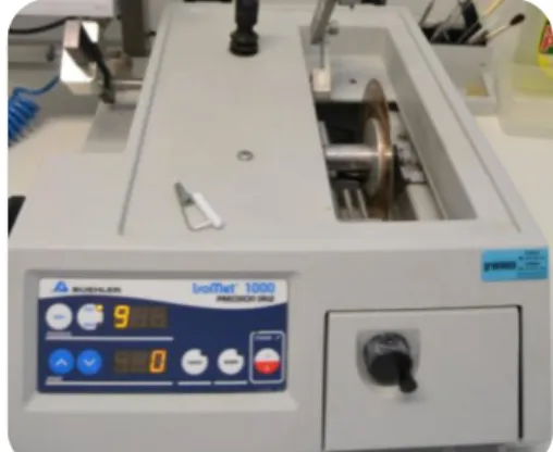
Figure 2 - IsometTM 1000 Precision Saw, Buehler Ltd. Ltd., Lake Buff, IL, USA…..…9
Figure 3 - Tooth fixed to an acrylic holder with sticky wax………..…10
Figure 4 - First cut 1-2 mm below the CEJ………..…..10
Figure 5 - Pulp chamber‟s exposure………...…10
Figure 6 - Removal of pulp tissues……….………..…..11
Figure 7 - Filling the pulp chamber with cyanoacrylate glue (737 Black Magic Toughened adhesive, Permabond, Hampshire, UK)………...…11
Figure 8 - Crowns fixed with cyanoacrylate glue to the acrylic holder……….11
Figure 9 - Removing the occlusal enamel and superficial dentine……….………11
Figure 10 - Mid-coronal dentine surface………....…11
Figure 11 - Mechanical grinder (Lunn Major, Struers, Denmark)………...12
Figure 12 - Scotchbond Universal Adhesive.………...13
Figure 13 - Clearfil SE Bond (Kuraray Co, Osaka, Japan)……….…...14
Figure 14 - Resin composite ENAMEL plus HRi………..14
Figure 15 - Resin composite buil-up with 6 mm………14
Figures 16 and 17 - Teeth after being sectioned in both „x‟ and „y‟ directions……...15
Figure 18 - Sticks………18
Figure 19 - Sticks attached to Geraldeli‟s jig with cyanoacrylate glue………..16
Micro-Tensile Bond Strength To Dentin Of A Self-Etch And A Universal Adhesive System In Self-Etch Mode xi ABREVIATIONS
% - per cent
10-MDP - 10-Methacryloyloxydecyl dihydrogen phosphate 4-MET – 4-methacryloyloxyethyl trimellitic acid
AD-concept – Adhesion-Decalcification Concept Bis-GMA - bisphenol A diglycidyl methacrylate CEJ – Cementoenamel junction
CL SE D – Clearfil SE Bond (Kuraray, Okayama, Japan) applied to dentine in self-etch mode
cm – Centimeters Et al. – Et alli
HEMA - 2-hydroxyethyl methacrylate
ISO/TR - International Organization for Standardization/ Technical Report mm – Millimeters
mm/min – Millimeter per minute mm2- Square millimeter
MMP – matrix metalloproteinase MPa - MegaPascal
mW/cm2 - milliWatt per square centimeter N – Newton
nm - Nanometer p – Significance value pH – Power of hydrogen
Phenyl-P – 2-(methacryloyloxyethyl)phenyl hydrogenphosphate
SBU SE D – Scotchbond Universal Adhesive (3M ESPE, St. Paul, MN, USA) applied to dentine in self-etch mode
SEM – Scanning Electron Microscopy
SPSS - Statistical Package for the Social Sciences TEM – Transmission Electron Microscopy μm – Micrometer
Micro-Tensile Bond Strength To Dentin Of A Self-Etch And A Universal Adhesive System In Self-Etch Mode xii
Micro-Tensile Bond Strength To Dentin Of A Self-Etch And A Universal Adhesive System In Self-Etch Mode xiii ABSTRACT
The evolution of adhesive systems over the last years has had a strong influence in the actual restorative concepts. The growing need for less time consumer procedures and less sensitive techniques, led manufacturers to release a new type of dental adhesives known as „universal‟, „multi-mode‟ or „multi-purpose‟. This concept of adhesives gives dentists the possibility to choose which approach they prefer to use: etch-and-rinse or self-etch strategy. So far, only few studies have been done with the purpose to know the performance of these adhesives.
Purpose: Evaluating micro-tensile bond strength to dentine of a universal adhesive system (Scotchbond Universal Adhesive, 3M ESPE, St Paul, MN – SBU SE D) in self-etch mode, with a control group (Clearfil SE Bond, Kuraray, Okayama, Japan – CL SE D),
Materials and Methods: Six human teeth (n=6) were used to obtain crown segments by exposing middle dentine and then randomly distributed into two groups according to the different adhesive systems used: Scotchbond Universal applied as a one-step self-etch adhesive and Clearfil SE applied as a two-step self-etch adhesive, both per manufacturer‟s instructions. After all teeth have received a composite restoration, sticks with 1mm2 of cross sectional area were obtained, by sectioning longitudinally in both „x‟ and „y‟ directions with a low speed diamond disk, and stored in distilled water (37º/24h). Subsequently, the specimens were tested using micro-tensile tests (μTBS) to assess dentine bond strength. Data were analyzed with a parametric paired-sample t test when the assumption of normality was valid.
Results: SBU SE D showed higher μTBS mean (41.03±19.31MPa) than CL SE D (36.70±17.77MPa), nevertheless the comparison between these two adhesive systems revealed no significant statistical differences (p > 0,05).
Conclusions: Despite the limitations of this study, it can be concluded that SBU SE D seems to have a similar performance to the control group, regarding to μTBS to dentine.
Keywords: universal adhesives; self-etch mode; dentine; micro-tensile bond strength.
Micro-Tensile Bond Strength To Dentin Of A Self-Etch And A Universal Adhesive System In Self-Etch Mode xiv
Micro-Tensile Bond Strength To Dentin Of A Self-Etch And A Universal Adhesive System In Self-Etch Mode xv RESUMO
A evolução dos sistemas adesivos ao longo dos últimos anos tem demonstrado uma forte influência nos conceitos da Dentisteria Restauradora atual. Como alternativa aos métodos mecânicos e mais invasivos de reter as restaurações, os sistemas adesivos permitem a preparação de cavidades mais conservadoras.
De um modo geral, a adesão entre a superfície dentária e o adesivo ocorre através de um mecanismo que envolve a substituição da porção mineralizada dos tecidos por monómeros da resina adesiva. A desmineralização da superfície dentária pelo condicionamento ácido cria microporosidades que, posteriormente, são preenchidas pela resina adesiva, existindo então uma retenção micromecânica. Recentemente, foi estudada a existência de uma interação química entre a superfície dentária e o adesivo que pode estar relacionada com o aumento da durabilidade do mesmo.
Enquanto que a adesão ao esmalte é fiável e previsível, a adesão à dentina permanece um desafio devido à sua complexidade e heterogeneidade.
Atualmente, os sistemas adesivos são classificados em etch-and-rinse ou self-etch de acordo com a sua interação com a estrutura dentária, sendo cada um subdividido consoante o número de passos executados. Com os adesivos etch-and-rinse é executado um primeiro passo de condicionamento ácido seguido de aplicação do primer e do adesivo separadamente and-rinse de três passos) ou combinados num frasco (etch-and-rinse de dois passos). Por outro lado, com os adesivos self-etch não é feito um condicionamento prévio da estrutura dentária uma vez que estes contêm monómeros acídicos que permitem condicionamento simultâneo à aplicação do primer. Assim, é feito um primeiro passo de aplicação do ácido e primer que estão juntos num frasco seguidos da aplicação do adesivo (self-etch de dois passos) ou os componentes (ácido, primer e adesivo) podem estar todos juntos num único frasco sendo realizada apenas uma aplicação (self-etch de um passo).
A necessidade crescente de procedimentos simplificados, com menor consumo de tempo e técnicas menos sensíveis levou os fabricantes a desenvolverem uma nova família de adesivos dentários conhecidos como adesivos universais, „multi-mode’ ou „multi-purpose‟. Este conceito versátil de adesivos possibilita a escolha da estratégia de adesão que mais se adequa em cada situação clínica, nomeadamente: etch-and-rinse ou self-etch.
Micro-Tensile Bond Strength To Dentin Of A Self-Etch And A Universal Adhesive System In Self-Etch Mode xvi O Scotchbond Universal Adhesive é um destes adesivos universais. Este contém na sua composição as moléculas de 10-MDP e um copolímero de ácido polialquenóico que desempenham um papel fundamental no processo de adesão. O 10-MDP é um monómero funcional específico que contém grupos carboxilo e fosfato com capacidade de formar ligações iónicas com o cálcio da hidroxiapatite. O ácido polialquenóico também tem a capacidade de estabelecer ligações químicas com o cálcio da hidroxiapatite podendo mesmo competir com o 10-MDP para o estabelecimento destas mesmas ligações.
Até agora poucos estudos foram realizados no sentido de conhecer o desempenho deste novo tipo de adesivos.
Objetivo: Avaliar as forças de adesão à dentina, através de testes de microtração, do adesivo universal Scotchbond Universal Adhesive (3M ESPE, St. Paul, MN, USA – SBU SE D), aplicado segundo as instruções do fabricante em modo self-etch e utilizando como grupo controlo o Clearfil SE Bond (Kuraray, Okayama, Japan – CL SE D). A hipótese nula testada neste estudo foi de que não existem diferenças nas forças de adesão à dentina entre o adesivo universal Scotchbond Universal Adhesive em modo self-etch e o adesivo self-etch de dois passos Clearfil SE Bond, ambos utilizados segundo as instruções do fabricante.
Materiais e Métodos: Um total de seis terceiros molares (n=6) recentemente extraídos, intactos e livres de cárie ou restaurações foram armazenados em Cloramina T 0,5% (Sigma Chemical Co., St Louis, MO, USA) a 4ºC durante uma semana e depois deixados em água destilada a 4ºC não mais do que três meses. A partir de cada dente foram obtidos segmentos de coroas através de dois cortes paralelos à face oclusal e com alguns milímetros de distância, utilizando para isso um disco diamantado a baixa velocidade (Diamond Wafering Blade - 10,2cmx0,3mm - Series 15HC, Buehler Ltd., Lake Bluff, IL, USA) sob irrigação constante com água destilada, num micrómetro de tecidos duros (IsometTM 1000 Precision Saw, Buehler Ltd. Ltd., Lake Buff, IL, USA): 1) 1-2 mm abaixo da junção amelocementária para remover as raízes; 2) remoção do esmalte oclusal e exposição da dentina média. Foi realizado polimento da superfície dentinária com tira de lixa de sílica-carboneto grão 600 (Ultra-Prep, Buehler Ltd., Lake Bluff, IL, USA), durante 60 segundos, para criação de smear-layer padronizada
Micro-Tensile Bond Strength To Dentin Of A Self-Etch And A Universal Adhesive System In Self-Etch Mode xvii semelhante à obtida em condições clínicas. Posteriormente, os segmentos de coroa foram aleatoriamente distribuídos em dois grupos de acordo com os diferentes sistemas adesivos utilizados: o Scotchbond Universal aplicado em modo self-etch de um passo e o Clearfil SE aplicado como adesivo self-etch de dois passos, ambos segundo com as instruções do fabricante. Após a aplicação do sistema adesivo, todos os segmentos de coroa receberam uma restauração em resina composta com ENAMEL plus HRi (Micerium S.p.A. Avegno (GE), Italy) cor UD4 (6 mm polimerizados em incrementos de 2 mm e com polimerização adicional de 10 segundos em cada uma das faces mesial, distal, vestibular e lingual). A superfície externa de todos os dentes foi pintada, com cores diferentes, com tinta à prova de água por forma a excluir todos os palitos em que a adesão era feita ao esmalte. Obtiveram-se palitos com área aproximada de 1 mm2 através de secções longitudinais segundo o eixo „x‟ e „y‟ com um disco diamantado a baixa velocidade e irrigação constante com água destilada, seguidamente armazenados em água destilada (37º/24h). Os espécimes foram testados um a um para avaliar as forças de adesão (MPa) à dentina utilizando testes de microtração (μTBS), numa máquina de teste universal (Instron® 4502 Series, Serial no. H3307, Instron Corporation, Canton, MA, USA) a uma velocidade de 1mm/min até ocorrer fratura. Com uma craveira digital foram medidas as arestas dos palitos para calcular a área de adesão (mm2). As forças de adesão (μTBS) foram calculadas a partir da divisão entre a força (N) no momento da fratura e a área de cada palito. O tipo de fratura foi analisado, pelo mesmo observador, utilizando um estereomicroscópio com ampliação de 10x e classificadas em: 1) adesivas (fratura ocorre na interface adesivo/compósito); 2) coesiva de compósito ou de dentina (fratura ocorre exclusivamente no compósito ou na dentina, respetivamente) ou 3) mista (fratura envolve a dentina e o compósito). Os dados foram analisados recorrendo ao teste paramétrico de amostras emparelhadas Teste t, após ser verificada a existência de uma distribuição normal.
Resultados: Um total de 101 (cento e um) palitos foram testados: 54 (cinquenta e quatro) pertencentes ao grupo do SBU SE D e 47 (quarenta e sete) do grupo do CL SE D, ambos segundo instruções do fabricante. Após verificação da existência de uma distribuição normal em cada grupo através dos testes de Kolmogorov-Smirnov e Shapiro-Wilk, foi realizado um teste paramétrico de amostras emparelhadas, o Teste t. Para avaliar a homogeneidade das variâncias foi executado um teste de Levene e, uma
Micro-Tensile Bond Strength To Dentin Of A Self-Etch And A Universal Adhesive
System In Self-Etch Mode xviii vez que o valor de p foi superior a 0,05, as variâncias foram assumidas como iguais. O SBU SE D (41.03±19.31 MPa) apresenta um valor médio de forças de adesão à dentina superior ao CL SE D (36.70±17.77 MPa). No entanto, a análise estatística através do Teste t não revela diferenças estatisticamente significativas entre os grupos, visto que o valor de p é superior a 0,05 (p = 0,247). Assim, por outras palavras, pode afirmar-se que não existem diferenças significativas entre os grupos, com um intervalo de confiança de 95%.
Conclusões: Os resultados obtidos levaram a que a hipótese nula fosse aceite. Apesar das limitações deste estudo, pode concluir-se que o adesivo universal Scotchbond Universal Adhesive quando aplicado em modo self-etch na dentina segundo as instruções do fabricante, parece exibir uma performance favorável e similar à do Clearfil SE, no que respeita às forças de adesão à dentina utilizando testes de microtração. Em estudos futuros, recomenda-se a avaliação não só das forças de adesão imediatas mas também após um período de envelhecimento de forma a que possa ser reportada a performance deste adesivo a longo prazo. Recomenda-se também a utilização de uma amostra maior por forma a que os resultados obtidos neste estudo sejam confirmados e, assim, estes possam ser extrapolados para a prática clínica.
Palavras-chave: adesivos universais; modo self-etch; dentina; testes de microtração.
Micro-Tensile Bond Strength To Dentin Of A Self-Etch And A Universal Adhesive System In Self-Etch Mode 1
2014
I – LITERATURE REVIEW
Since the first experimental study about adhesion was carried out, in 1952 (Kramer IRH & McLean JW, 1952), and followed by the introduction of enamel chemical etching, in 1955 (Buonocore MG, 1955), the mechanical methods of retaining restorations were progressively abandoned and replaced by conservative adhesive methods.
Nowadays, we are in a new era of restorative concepts, the era of „adhesive dentistry‟, in which the manufacturers are constantly challenged to create simpler, user-friendly and less technique-sensitive adhesive systems (Van Meerbeek B et al., 1998; Peumans M et al., 2005).
1. ADHESION PRINCIPLES
The adhesion between the adhesive agent to enamel or dentine is achieved by an exchange process in which the inorganic material from the hard dental tissue is replaced by resin monomers that, after polymerization, become micro-mechanically interlocked in the retentions previously created (Nakabayashi N et al., 1982; Van Meerbeek B et al., 2003). Nakabayashi et al. (1982) first described this process that is called „hybridization‟ or the formation of the „hybrid layer‟.
Recently, the potential benefit of a supplementary chemical interaction between the tooth structure and the functional monomers of the adhesives has attracted attention because it could improve the bond stability through time (Van Meerbeek B et al., 2003; Yoshida Y et al., 2004).
Adhesion-Decalcification Concept (AD-concept)
The „AD-concept‟ explains the way that molecules interact with hydroxyapatite (Yoshida Y et al., 2001). Specifically in the adhesives systems, molecules like 10-MDP (a functional monomer included in self-etch adhesives) can chemically bond to calcium of hydroxyapatite: it‟s an ionic bond with concomitant release of phosphate and hydroxide ions (Van Meerbeek B et al., 2011). Due to the stability of formed calcium salt, the molecule will remain bond occurring only slight decalcification of the surface (Van Meerbeek B et al., 2011).
Micro-Tensile Bond Strength To Dentin Of A Self-Etch And A Universal Adhesive System In Self-Etch Mode 2
2014
1.1 Adhesion to Dentine
Whereas the adhesion to enamel is reliable and predictable when etched with phosphoric acid, the adhesion to dentine is still considered a challenge and less predictable because it is a heterogeneous substrate (Swift EJ et al., 1995). This can be explained by the intrinsic dentine wetness (Pashley DH & Pashley EL, 1991), the organic material content (Swift EJ et al., 1995) and variabilities regarding to dentine depth and permeability (Tagami J et al., 1990).
Dentine hydrophilicity is straightly related with the closeness to the pulp tissue across numerous tubules, which results in a positive pulpal fluid pressure (Swift EJ et al., 1995; Van Meerbeek B et al., 1998). This characteristic remains one of the most important challenges of the adhesion to dentine, which induced manufacturers to create dentine adhesives compatible with humid environments.
On the other hand, etching dentine is aggressive as it dissolves and removes hydroxyapatite exposing the collagen matrix (Van Meerbeek B et al., 2003; Pashley DH et al., 2011). This exposed collagen is susceptible to hydrolytic and enzymatic degradation processes due to water sorption (De Munck J et al., 2009; Van Meerbeek B et al., 2010; Van Meerbeek B et al., 2011). Thus, water sorption seems to be the main accountable reason for degradation of the adhesive-tooth interface (De Munck J et al., 2009).
2. ADHESIVE SYSTEMS CLASSIFICATION
Currently, the dental adhesives can be classified, according to the adhesion strategy, in two main groups: Rinse adhesives (or Total-Etch) and Etch-and-Dry adhesives (also called Self-Etch) (Van Landuyt KL et al., 2007; Breshi L et al., 2008; Cardoso MV et al., 2011; Pashley DH et al., 2011; Van Meerbeek B et al., 2011). The term total-etch is now considered less proper because self-etch adhesives can also etch and demineralize tooth surface (Van Meerbeek B et al., 2005).
Although the different number of bottles of which adhesive can consist of, they all contain similar ingredients but in different proportions (Van Landuyt KL et al., 2007), namely: acrylic resin monomers to guarantee a covalent bond between the adhesive and the composite; organic solvents (water, acetone or ethanol); initiators and inhibitors; and sometimes filler particles (Van Landuyt KL et al., 2007).
Micro-Tensile Bond Strength To Dentin Of A Self-Etch And A Universal Adhesive System In Self-Etch Mode 3
2014
2.1 Etch-and-Rinse Strategy
Depending on the number of steps, etch-and-rinse adhesives can be classified in three-steps etch-and-rinse adhesives or two-steps etch-and-rinse adhesives (Van Meerbeek B et al., 2003; De Munck J et al., 2005; Pashley DH et al., 2011). While the first maintain the etching, priming and bonding separated, the second combine the primer and the bond into one application.
Etch-and-Rinse adhesives require an initial etching step with phosphoric acid (35-37%), etching enamel and dentin at the same time (Pashley DH et al., 2011). The aim of this step is to remove the smear layer, clean the tubules and create a micro porous surface (Van Meerbeek B et al., 2005). This acid-etching step promotes dentine demineralization over a depth of 5-8μm, exposing the collagen fibrillar matrix almost without hydroxyapatite (Van Meerbeek B et al., 1992; Pashley DH et al., 2011). After that, the surface should be rinsed off removing all the reaction products and gently dried just to remove the excess of water (Peumans M et al., 2005; Pashley DH et al., 2011). Towards increasing the strength of the resin-dentine bonds, the demineralized dentine surface has to be wet so the collapse of unsupported collagen is prevented (Kanca J, 1992).
The next step consists of applying a primer, which contains specific resin monomers, such as 2-Hydroxy ethyl methacrylate (HEMA) dissolved in a solvent (water, acetone or ethanol). HEMA is a monomer with simultaneously hydrophobic and hydrophilic properties that is responsible for transforming the hydrophilic dentine surface into a hydrophobic surface (Van Meerbeek B et al., 1998; Van Landuyt KL et al., 2007). On the other hand, the solvent allows the penetration of monomers in the collagen matrix and removes the remaining water from the dentine surface ensuring a good wetting (Van Landuyt KL et al., 2007).
Finally, the adhesive resin is applied on the prepared surface and penetrates into the exposed collagen matrix and the dentine tubules (Cardoso MV et al., 2011). This results in the formation of the hybrid layer and resin tags into the dentinal tubules providing micromechanical retention (Van Meerbeek B et al., 1993; Van Meerbeek B et al., 1998).
Micro-Tensile Bond Strength To Dentin Of A Self-Etch And A Universal Adhesive System In Self-Etch Mode 4
2014
2.2 Etch-and-Dry Strategy
In a different way from the etch-and-rinse approach, the self-etch adhesives do not require a separate etching step since they contain acidic monomers that can etch and prime the dental surface at the same time (Van Meerbeek B et al., 2005; Cardoso MV et al., 2011; Van Meerbeek B et al., 2011). These monomers are less acidic than the phosphoric acid used in etch-and-rinse adhesives (Van Meerbeek B et al., 2003). The surface is not rinsed away after this first step, which means that the dissolved smear-layer and demineralization products are incorporated in the adhesion process (Van Meerbeek B et al., 2005; Cardoso MV et al., 2011).
Self-etch adhesives can be classified according to: 1) their application procedures as „two-step‟ and „one-step‟ (known as „all-in-one‟) adhesives, or 2) their acidity as strong (pH ≤ 1), intermediate (pH ≈ 1.5) and mild (pH ≥ 2) (Van Meerbeek B et al., 2003; Van Meerbeek B et al., 2005; Cardoso MV et al., 2011; Van Meerbeek B et al., 2011).
While a two-step self-etch adhesive consists in the application of an acidic primer (hydrophilic) followed by the adhesive resin (hydrophobic), the one-step self-etch adhesive combines self-etching, priming and bonding into one single solution (Van Meerbeek B et al., 2005; Van Meerbeek B et al., 2011). The main concern about the one-step self-etch adhesives is the considerably lower bond strength due to their high hydrophilicity which make them capable to attract water from the intrinsically wet dentine and so they are regarded as semi-permeable membranes (Tay F et al., 2002; De Munck J et al., 2005; Van Meerbeek B et al., 2005). The water will be retained in the hydrophobic composite layer leading to the formation of water blisters which result in the loss of adhesion between the adhesive and composite (Tay F et al., 2002; Perdigão J et al., 2013).
Strong self-etch (pH ≤ 1) adhesives show morphological similarities with the etch-and-rinse adhesives producing a deep demineralization in both enamel and dentine (Cardoso MV et al., 2011; Van Meerbeek B et al., 2011). A thick hybrid layer devoid of hydroxyapatite and with resin tags at dentine are seen with TEM images (Van Meerbeek B et al., 2005). It differs from the etch-and-rinse adhesives because the dissolved calcium phosphates are not rinsed and they are probably unstable in an aqueous environment (Van Meerbeek B et al., 2011).
Micro-Tensile Bond Strength To Dentin Of A Self-Etch And A Universal Adhesive System In Self-Etch Mode 5
2014
Mild-self etch adhesives (pH ≥ 2) only allow a shallow demineralization (≈ 1μm) in the dentin surface, leaving hydroxyapatite crystals around the collagen fibrils and do not remove completely the smear plugs (Van Meerbeek B et al., 2005; Van Meerbeek B et al., 2011). Besides the collagen being protected by hydroxyapatite, this also allows an additional chemical interaction which leads to a two-fold micro-mechanical and chemical bonding mechanism (Van Meerbeek B et al., 2005; Cardoso MV et al., 2011; Van Meerbeek B et al., 2011). This chemical bonding is achieved by the presence of specific functional monomers, such as 10-MDP, 4-MET and phenyl-P, which contain carboxyl and phosphate groups that are able to form ionic bonds with the calcium of the hydroxyapatite and it should be stable in an aqueous environment (Van Meerbeek B et al., 2003; Yoshida Y et al., 2004). Therefore, water is an indispensable ingredient of self-etch adhesives that allows adequate ionization of functional monomers (Van Landuyt KL et al., 2005; Van Landuyt KL et al., 2007; Perdigão J et al., 2014). However, the excess of water can lead to phase separation between adhesive ingredients, by inhibiting the optimal copolymerization of the adhesive monomers (Jacobsen T & Söderholm KJ, 1995; Van Landuyt KL et al., 2005).
Despite the thin hybrid layer and the near absence of resin tags, the mild-self etch adhesives can achieve satisfactory results with respect to bond strength, since the finding that the thickness of the hybrid layer and the length of resin tags do not interfere in bonding effectiveness and stability (Van Meerbeek B et al., 2003; Van Meerbeek B et al., 2005).
Many authors (Perdigão J et al., 2006; Toledano M et al., 2006; Brackett WW et al., 2008; Muñoz MA et al., 2013) have been doing in vitro studies with Clearfil SE Bond (Kuraray, Okayama, Japan), which is a mild two-step self-etch adhesive. They reported reliable both in vitro and clinical results with regard to bonding effectiveness. This may be in part attributed to the presence of 10-MDP (Van Landuyt KL et al., 2007). Actually, CL SE is nowadays considered as the „gold standard‟ for self-etch adhesives in both laboratory and clinical situations (Perdigão J et al., 2012).
The self-etch approach, when compared with etch-and-rinse, has been considered more user-friendly, because it has less steps and reduces the chairside time (Van Meerbeek B et al., 2005; Van Meerbeek B et al., 2011); less technique-sensitive since it does not use the „wet-bonding‟ and critical steps as rinsing and drying are eliminated (Van Meerbeek B et al., 2005; Van Meerbeek B et al., 2011); and less
post-Micro-Tensile Bond Strength To Dentin Of A Self-Etch And A Universal Adhesive System In Self-Etch Mode 6
2014
operative sensitivity (Van Meerbeek B et al., 2011). Commonly, self-etch adhesives can concurrent demineralize and infiltrate dentine surface to the same depth, which theoretically ensures the complete penetration of the adhesive into the exposed collagen matrix (Van Meerbeek B et al., 2005; Van Meerbeek B et al., 2011).
A disadvantage of self-etch adhesives, particularly of one-step adhesives, is the reduction in enamel bonding effectiveness once the increase in surface area in intact and ground enamel obtained with these adhesives is lower than that achieved with etch-and-rinse adhesives (Pashley DH & Tay F, 2001).
To improve the performance of these adhesives to enamel, a prior etching step can be performed (Van Landuyt KL et al., 2006). On the other hand, intentionally etching dentine with phosphoric acid prior to the application of a self-etch adhesive can result in decreased bond strength due to the formation of an unsatisfactory hybrid layer prone to nanoleakage (Van Landuyt KL et al., 2006). This becomes a challenge because clinically it is difficult to selectively etch the enamel with phosphoric acid without it flowing back to dentine (Van Landuyt KL et al., 2006; Perdigão J et al., 2012; Muñoz MA et al., 2013).
Peumans et al. (2005) did a systematic review including 85 published clinical trials in which adhesive systems were tested in selected class-V cavities. They concluded that three-step etch-and-rinse adhesives and two-step self-etch adhesives are clinically reliable and have a predictably good clinical performance .
After a comparison of contemporary adhesives, De Munck et al. (2005) concluded that the three-step etch-and-rinse adhesives remain the “gold standard” in terms of adhesion durability and only two-step self-etch adhesives can closely approach those.
Micro-Tensile Bond Strength To Dentin Of A Self-Etch And A Universal Adhesive System In Self-Etch Mode 7
2014
3. UNIVERSAL ADHESIVES
Recently, manufacturers have launched a new type of adhesives called „multi-mode‟, „multi-purpose‟ or „universal‟ adhesives (Muñoz MA et al., 2013; Perdigão J et al., 2014). These systems contain one bottle and give the dentist the opportunity to choose which adhesive strategy seems to be the most appropriate to the clinical situation: etch-and-rinse or etch-and-dry (Hanabusa M et al., 2012; Mena-Serrano A et al., 2013; Perdigão J et al., 2014). This versatile new concept advocates the use of the simplest strategy in each situation.
The capability of using these adhesive systems in different application modes makes possible a prior selective enamel etching, which allows to combine the benefit of the etch-and-rinse technique on enamel and the easier etch-and-dry approach on dentine (Perdigão J et al., 2012; Marchesi G et al., 2014).
As previously mentioned, it was reported that one-step self-etch adhesives behave as permeable membranes which allow water to pass through the bond interface. This can happen with universal adhesives too, once both of them have equivalent water content (Perdigão J et al., 2014). Because of that, degradation of bonding interface can also occur affecting their clinical durability.
Scotchbond Universal Adhesive (3M ESPE, St. Paul, MN, USA) is one of these new universal adhesives. It contains 10-MDP and a polyalkenoic acid copolymer in its composition (Perdigão J et al., 2012; Muñoz MA et al., 2013; Muñoz MA et al., 2014). Polyalkenoic acid copolymer may compete with 10-MDP as both of them bond chemically to hydroxyapatite‟s calcium (Mena-Serrano A et al., 2013; Muñoz MA et al., 2013; Muñoz MA et al., 2014). Marchesi et al. (2014) and Muñoz et al. (2014) found similar values regarding to immediate micro-tensile bond strength of Scotchbond Universal Adhesive used as etch-and-rinse or etch-and-dry adhesive.
Muñoz et al. (2013) studied the micro-tensile bond strength of three universal adhesives applied to dentine using the etch-and-rinse and the self-etch strategies. They concluded that the bond strengths using those universal adhesives were higher using etch-and-rinse than self-etch approach.
So far, only few studies have been carried out with the purpose of knowing the performance of this new type of adhesives.
Micro-Tensile Bond Strength To Dentin Of A Self-Etch And A Universal Adhesive System In Self-Etch Mode 8
2014
II - PURPOSE
This is an in vitro experimental study with the aim of:
Evaluating dentine bond strength of a universal adhesive (Scotchbond Universal Adhesive, 3M ESPE, St. Paul, MN, USA) in self-etch mode, using micro-tensile tests;
Comparing the micro-tensile bond strength values with a two-step self-etch adhesive system (Clearfil SE Bond, Kuraray, Okayama, Japan).
The following null hypothesis was tested in this study:
1. There is no difference in bond strength to dentine between the universal adhesive Scotchbond Universal in self-etch mode (per manufacturer‟s instructions) and the two-step self-etch adhesive Cleafil SE Bond (per manufacturer‟s instructions), using micro-tensile tests.
Micro-Tensile Bond Strength To Dentin Of A Self-Etch And A Universal Adhesive System In Self-Etch Mode 9
2014
III - MATERIALS AND METHODS
Type of study
This is an in vitro experimental study with the purpose of evaluating and comparing dentine micro-tensile bond strength between a universal adhesive system in self-etch mode and a self-etch adhesive system, both as per manufacturer‟s instructions.
Design of the study
A total of six recently extracted human third molars, intact and without macroscopic evidence of caries or restorations, were used in this study. Before preparation, the teeth were randomly selected from a group of teeth, firstly stored in 0,5% Chloramine T (Sigma Chemical Co., St Louis, MO, USA) at 4ºC for one week and after that, left in distilled water at 4ºC, as required from the ISO TR 11405 standard, no more than three months. All teeth were cleaned under running water using a periodontal scaler before preparation.
Teeth selection and preparation
From each tooth, a crown segment was obtained exposing middle dentin by sectioning the crowns with two cuts, a few millimeters apart, parallel to the occlusal surface, with a low-speed precision diamond disk (Diamond Wafering Blade - 10,2cmx0,3mm - Series 15HC, Buehler Ltd., Lake Bluff, IL, USA – Figure 1), on a hard tissue microtome (IsometTM 1000 Precision Saw, Buehler Ltd. Ltd., Lake Buff, IL, USA – Figure 2) under constant distilled water irrigation, in the following way:
Micro-Tensile Bond Strength To Dentin Of A Self-Etch And A Universal Adhesive System In Self-Etch Mode 10
2014
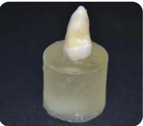
1. The teeth crowns were attached to an acrylic holder with sticky wax, perpendicular to the long axis of the tooth (Figure 3);
Figure 3: Tooth fixed to an acrylic holder with sticky wax.
2. The first cut was made parallel to the occlusal surface 1-2 mm below the cementoenamel junction to remove the roots (Figure 4) and expose the pulp chamber (Figure 5);
Figure 4: First cut 1-2 mm below the CEJ. Figure 5: Pulp chamber‟s exposure.
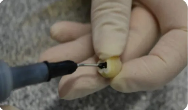
3. The crowns were detached from the acrylic holders and the pulp tissues were removed from the pulp chamber with a dentin curette (Figure 6). The pulp chamber was then filled with cyanoacrylate glue (737 Black Magic Toughened adhesive, Permabond, Hampshire, UK – Figure 7) and the crowns were fixed with the same glue to the acrylic holders, by the sectioning surface (Figure 8).
Micro-Tensile Bond Strength To Dentin Of A Self-Etch And A Universal Adhesive System In Self-Etch Mode 11
2014
Figure 6: Removal of pulp tissues.
Figure 8: Crowns fixed with cyanoacrylate glue to the acrylic holder.
4. Mid-coronal dentin surfaces were obtained by removing the occlusal enamel and superficial dentine of the molar crowns (Figure 9 and 10), perpendicular to the long axis of tooth, using a diamond disk at low speed, under constant water irrigation.
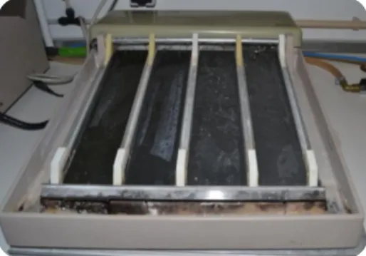
With the purpose of creating a uniform smear layer obtained in similar conditions to those occurring in clinic situations, the dentin surface was polished with 600-grit silica-carbide (SiC) sandpaper (Ultra-Prep, Buehler Ltd., Lake Bluff, IL, USA) on a mechanical grinder (Lunn-Major, Struers, Denmark) during 60 seconds under
Figure 7: Filling the pulp chamber with cyanoacrylate glue.
Figure 9: Removing the occlusal enamel and superficial dentine.
Figure 10: Mid-coronal dentine surface.
Micro-Tensile Bond Strength To Dentin Of A Self-Etch And A Universal Adhesive System In Self-Etch Mode 12
2014
water irrigation (Figure 11). The crown segments were kept in distilled water during this procedure.
Figure 11: Mechanical grinder (Lunn Major, Struers, Denmark).
Distribution and treatment of the crown segments
The six crown segments were randomly distributed into two groups (n=6) according to the different adhesive systems used. The order in which the crown segments were treated was random, to avoid a possible bias due to any particular sequence of treatment.
Bonding Procedures
All the treatment procedures were performed by the same operator in the following way as described:
Group 1 – Scotchbond Universal (3M ESPE, St. Paul, MN, USA) (Figure 12) as per manufacturer‟s instructions - Self-etch strategy on dentine (SBU SE D):
1. The occlusal surface was rinsed with water. The excess of water was removed from the dentin surface using a moist cotton pellet, so that the surface remained shiny and visibly moist.
2. The adhesive was applied at the tooth surface by using a disposable microbrush, scrubbing lightly for 20 seconds.
3. The surface was then gently air-dried until it ceases to show any movement and the solvent was evaporated completely, forming a homogenous and slightly shiny film. Beginning with a soft blow of air from a distance of approximately 10 cm, the air pressure was increased while decreasing distance,
Micro-Tensile Bond Strength To Dentin Of A Self-Etch And A Universal Adhesive System In Self-Etch Mode 13
2014
finishing at a distance of approximately 1-2 mm from the surface at maximum air pressure.
4. Finally, the surface was polymerized for 10 seconds.
Figure 12: Scotchbond Universal Adhesive.
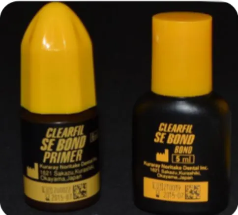
Group 2 - Clearfil SE Bond (Kuraray, Okayama, Japan) (Figure 13) as per manufacturer‟s instructions – Self-etch strategy on dentin (CL SE D):
1. The occlusal surface was rinsed with water. The excess of water was removed from the dentin surface using a moist cotton pellet, so that the surface remained shiny and visibly moist.
2. The primer was applied to tooth surface with a disposable microbrush. Waited for 20 seconds.
3. To evaporate the volatile ingredients, the surface was gently air-dried. Then the adhesive was applied to the entire surface with a disposable microbrush. A thin and uniform adhesive layer was left, by removing the excess with the same microbrush and using a gentle air stream. Beginning with a soft blow of air from a distance of approximately 10 cm, the air pressure was increased while decreasing distance, finishing at a distance of approximately 1-2 mm from the surface at maximum air pressure.
Micro-Tensile Bond Strength To Dentin Of A Self-Etch And A Universal Adhesive System In Self-Etch Mode 14
2014
Figure 13: Clearfil SE Bond.
Restorative Procedures
After the bonding procedures, all crown segments received a composite restoration with ENAMEL plus HRi (Micerium S.p.A. Avegno (GE), Italy), color UD4 (Figure 14), applied in increments of 2mm each, until a height of 6mm (Figure 15). Each layer was light cured for 20 seconds, according to manufacturer‟s instructions. Additional light polymerization was performed on mesial, distal, facial and lingual surfaces for 10 seconds each.
Figure 14: Resin composite ENAMEL plus HRi.
All light curing was performed with a light intensity of 600 mW/cm2 using a halogen light-activation unit (ELIPAR S10, 3M ESPE, St. Paul, MN, USA), with the 13 mm light guide held 1-2 mm from the treatment surface. The output of the curing light was periodically verified at > 600 mW/cm2 with a radiometer (Curing Radiometer 100, Serial No. 1279, Demetron Research Corporation, Danbury, USA).
Figure 15: Resin composite build-up with 6 mm.
Micro-Tensile Bond Strength To Dentin Of A Self-Etch And A Universal Adhesive System In Self-Etch Mode 15
2014
Specimens preparation for micro-tensile tests
All teeth were painted with waterproof ink in different colors in order to exclude all the sticks in which the bonding procedure was made to enamel. The restored teeth were stored in distilled water at 37ºC during 24h, in an incubator. The date and time of restoration were registered.
Then, the teeth were longitudinally sectioned in both „x‟ and „y‟ directions (Figure 16 and 17), with a low-speed diamond disk (Diamond Wafering Blade – 10,2cm*0,3mm, Series - 15HC, Buehler Ltd, Lake Bluff, IL, USA) under water irrigation, and using a hard tissue microtome (Isomet® 1000 Precision Saw, Buehler Ltd, Lake Buff, IL, USA), to obtain sticks with approximately 1mm2 of a cross sectional area.
A final cut was made at the base of the root, perpendicular to the long axis of the tooth, to separate the sticks from the acrylic holders (Figure 18).
Figure 18: Sticks.
Micro-Tensile Bond Strength To Dentin Of A Self-Etch And A Universal Adhesive System In Self-Etch Mode 16
2014
Debonded or lost sticks were registered: debonded sticks were those separated in the adhesive interface during the cutting procedure; lost sticks were those, which were lost or fractured during test preparation.
The sticks were stored in distilled water for a maximum of 24h until the micro-tensile tests were performed.
Micro-tensile bond strength tests (μTBS)
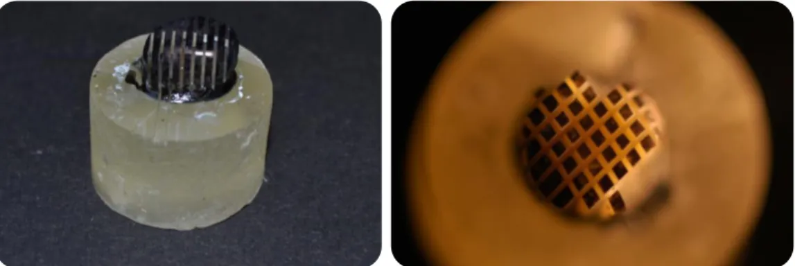
The sticks were individually attached to a stainless-steel grooved Geraldeli‟s jig with cyanoacrylate glue (737 Black Magic Toughened adhesive, Permabond, Hampshire, UK) (Figure 19) and tested one by one under a tension load using a universal testing machine (Instron® 4502 Series, Serial no. H3307, Instron Corporation, Canton, MA, USA) (Figure 20), at a crosshead speed of 1mm/min until fracture occurred, with the stress to failure expressed in MPa.
The cross section of each fractured stick was measured with a digital caliper (Ficher Darex®, 0-150mm, France) to calculate the bonding area (mm2). The μTBS (MPa) values were calculated by dividing the load (N) at failure by the area (mm2) of each stick.
Failures were analyzed by the same observer, under a stereomicroscope (Nikon, Japan) at 10x magnification to determine the mode of failure. The failure modes were classified as: 1) cohesive when the failure occurred exclusively in dentin (CD) or in composite (CC); 2) adhesive (A) when failure occurred in the dentin-resin interface; and 3) mixed (M) when it involved both dentin and resin.
Figure 19: Sticks attached to Geraldeli‟s jig with cyanoacrylate glue.
Figure 20: Instron® 4502, universal testing machine.
Micro-Tensile Bond Strength To Dentin Of A Self-Etch And A Universal Adhesive System In Self-Etch Mode 17
2014
Statistical Analysis
The statistical analysis of the results was performed through descriptive and inference methods using the software program SPSS Statistics for MAC Version 20 (SPSS Inc., Chicago, IL, USA). A Levene‟s Test was done to verify the homogeneity of the variances. A parametric paired-sample t test was performed when the assumption of normality was valid.
Pre-testing failures that occurred during specimen preparation were previously excluded and not taken into account for the statistic analysis.
Micro-Tensile Bond Strength To Dentin Of A Self-Etch And A Universal Adhesive System In Self-Etch Mode 18
2014
IV - RESULTS
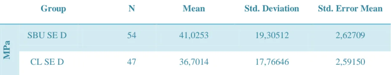
The number of sticks per group, micro-tensile bond strength (μTBS) mean values in MPa and the respective standard deviations among the adhesives are listed in Table 1.
A total of 101 (one hundred and one) sticks were analyzed: 54 (fifty four) using the Scotchbond Universal Adhesive in self-etch mode (SBU SE D, N=54) and 47 (forty seven) using the Clearfil SE Bond adhesive (CL SE D, N=47), both as per manufacturer‟s instructions.
Group N Mean Std. Deviation Std. Error Mean
MPa
SBU SE D 54 41,0253 19,30512 2,62709 CL SE D 47 36,7014 17,76646 2,59150 Table 1: Number of sticks (N); Micro-tensile bond strength (μTBS) mean values; Standard
deviation (Std. Deviation) and Standard Error Mean (Std. Error Mean).
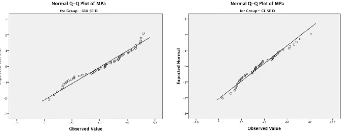
The Kolmogorov-Smirnov test and Shapiro-Wilk test (Table 2) were used to assess if the data followed a normal distribution. A paired-sample t-test was performed, as the assumption of normality in each group was valid.
Group Kolmogorov-Smirnov Shapiro-Wilk
Statistic df Sig. Statistic Df Sig.
MPa SBU SE D CL SE D ,102 ,097 54 47 ,200 ,200 ,952 ,974 4 7 ,032 ,359 Table 2: Test of Normality.
Micro-Tensile Bond Strength To Dentin Of A Self-Etch And A Universal Adhesive System In Self-Etch Mode 19
2014
Graphics 1 and 2: Tests of Normality for the SBU SE D and CL SE D.
To verify the homogeneity of the variances a Levene‟s Test was performed (table 3). Since the significance value (p) is superior to 0,05, the variances were assumed as equal.
Levene’s Test for
Equality of Variances t-test for Equality of Means
F Sig. t df Sig. (2-tailed) Mean Difference M Pa Equal variances assumed ,895 ,346 1,165 99 ,247 4,32389 Equal variances not assumed 1,172 98,677 ,244 4,32389
Table 3: Results of Levene‟s Test and t-test.
The distribution of μTBS is shown in Graphic 3, where the central line of the box represents the median μTBS.
Micro-Tensile Bond Strength To Dentin Of A Self-Etch And A Universal Adhesive System In Self-Etch Mode 20
2014
Graphic 3: Box-whisker plot of the μTBS for SBU SE D and CL SE D: x axis represents the
group and y axis the MPa.
Although the statistical analysis revealed no significant differences between immediate bond strengths of SBU SE D (group 1) and CL SE D (group 2) (p > 0,05), SBU SE D resulted in higher μTBS mean (41.03±19.31MPa) than CL SE D (36.70±17.77MPa), with a 95% confidence interval (Table 4).
t-test for Equality of Means
Std. Error Difference 95% Confidence Interval of the
Difference Lower Upper M P a Equal variances assumed 3,71166 -3,04085 11,68863 Equal variances not assumed 3,69019 -2,99855 11,64633
Table 4: T-test for equality of means.
Pre-testing failures were excluded from further statistical analysis. Failure mode distribution is shown in table 3 and graphically represented in graphic 4. The majority
Micro-Tensile Bond Strength To Dentin Of A Self-Etch And A Universal Adhesive System In Self-Etch Mode 21
2014
of SBU SE D specimens showed composite cohesive failures while CL SE D had more mixed failures.
Failure Mode A CC CD M
SBU SE D 7 21 9 17
CL SE D 16 8 5 18
Table 5: Number of sticks in each failure mode: A- adhesive failure; CC- Composite cohesive
failure; CD- dentine cohesive failure; M – mixed failure
Graphic 4: Failure mode distribution: A- adhesive failure; CC- Composite cohesive failure; CD-
dentine cohesive failure; M – mixed failure. 0 5 10 15 20 25 A CC CD M 7 21 9 17 16 8 5 18 N u m b e r o f st ic ks Failure Mode SBU SE D CL SE D
Micro-Tensile Bond Strength To Dentin Of A Self-Etch And A Universal Adhesive System In Self-Etch Mode 22
2014
V - DISCUSSION
Recently, manufacturers released a new family of adhesives: the universal adhesive systems. These adhesives have a new concept as they can be used with etch-and-rinse or self-etch strategy. Scotchbond Universal Adhesive is one of these universal adhesives.
Only few studies about these adhesives have been carried out so far and both laboratory and clinical studies are needed to evaluate the performance of them, comparing with those that are accepted as “gold standards”.
This experimental study evaluated the bond strength to dentine of a new universal adhesive system (SBU SE D – group 1) used in self-etch mode as per manufacturer‟s instructions, with a control group (CL SE D - group 2).
Clearfil SE is a two-step mild self-etch adhesive system and was used in this study as a control group since a great number of studies have already evaluated its clinical and laboratory effectiveness, regarding to dentine bond strengths, with excellent results in both performances (Perdigão J et al., 2006; Mine A et al., 2009; Van Landuyt KL et al., 2009; Peumans M et al., 2010; Sarr M et al., 2010; Mena-Serrano A et al., 2013).
In a previous clinical trial the authors concluded that the success obtained, at eight years, with Clearfil SE was not only due to the presence of 10-MDP molecule in the primer, but also due to the high-quality mechanical properties and high converse rate of the separate particle-filled hydrophobic resin (Peumans M et al., 2010). In fact, De Munck et al. (2012) reported that the second best performing adhesive was Clearfil SE only after the three-step etch-and-rinse adhesives, which are considered the “gold standard”.
The teeth selected were stored in 0,5% chloramine T at 4ºC for one week and after that, left in distilled water at 4ºC no more than three months, as is required from the ISO TR 11405 standard and as was done in some other studies (Perdigão J et al., 2012; De Munck J et al., 2013; Marchesi G et al., 2014; Muñoz MA et al., 2014; Taschner M et al., 2014).
An important parameter to consider is how dentine is prepared before bonding procedures. In the clinical practice, when rotary instruments are used to perform cavity preparations, the surface becomes covered by smear-layer which plays an important role
Micro-Tensile Bond Strength To Dentin Of A Self-Etch And A Universal Adhesive System In Self-Etch Mode 23
2014
in adhesion, particularly when the self-etch approach is used (Van Meerbeek B et al., 2011). On the other hand, in experimental studies, different grinding patterns can result in size and structures‟ variations of the smear-layer (Van Meerbeek B et al., 2011). De Munck et al. (2012) reported in their meta-analytical review that, among the analyzed studies, the most used preparations methods were: 1) carbide or diamond dental bur; 2) silicon-carbide paper. It was reported that using rotary instruments or abrasive paper may produce different bond strengths of resins to dentine and that would be advantageous to prepare dentine with dental burs in laboratory (Tagami J et al., 1991). This differs from Tao & Pashley (1988) results, who found only a small difference in bond strength when the smear layer was created with dental burs or sandpaper, thus validating the use of sandpaper . In the current study, a standardized and uniform smear-layer was created by polishing the exposed dentine with 600-grit silica-carbide abrasive paper (Buehler, Lunn Major, Struers Denmark) under running water during 60 seconds, on a mechanical grinder (Lunn Major, Struers, Denmark). This had the purpose of creating a smear-layer similar to that obtained in clinical situations. The same procedure to create a standardized smear-layer was realized in other studies (Pashley DH et al., 1988; Sano H et al., 1994; Perdigão J et al., 2006; Perdigão J et al., 2012; Muñoz MA et al., 2013; Muñoz MA et al., 2014; Perdigão J et al., 2014).
Both of the adhesive systems were applied as per manufacturer‟s instructions by the same operator. However, it was necessary to detail some of the steps in order to standardize the bonding procedures. The manufacturer‟s instructions are displayed in appendix 1 and the bonding procedures used in this study were specified in the Materials and Methods chapter.
The restorative procedures were performed using the ENAMEL plus HRi composite that according to the manufacturer‟s instructions should be polymerized for 20 seconds. Nevertheless, an additional light polymerization was performed on mesial, distal, facial and lingual surfaces, for 10 seconds each, in order to avoid composite cohesive failures. In a previous study carried out by Perdigão et al. (2006), they polymerized the resin composite (Filtek Z250, shade A2, 3M ESPE, St. Paul, MN, USA) for 40 seconds instead of the 20 seconds as is recommended by the manufacturer, thus obtaining less composite cohesive failures.
Micro-tensile test was used to assess the dentine bond strength of the resin-dentine interface. It should be considered as an “immediate” bond strength test because
Micro-Tensile Bond Strength To Dentin Of A Self-Etch And A Universal Adhesive System In Self-Etch Mode 24
2014
it was carried out after a maximum of 24 hour storage in distilled water (Hanabusa M et al., 2012).
Although clinical trials remain the ultimate tests to collect scientific evidence of the bonding effectiveness (Van Meerbeek B et al., 2003; Peumans M et al., 2005; Van Meerbeek B et al., 2010), in vitro studies are quite popular mainly due to the rapid evolution of adhesive systems that often leads manufacturers to launch new products without even clinically testing their antecessors (Van Meerbeek B et al., 2010). Nevertheless, laboratory tests as bond strength tests can gather valuable results to predict clinical effectiveness (Van Meerbeek B et al., 2003; Van Meerbeek B et al., 2010).
Bond strength tests are the most used method to evaluate the bonding effectiveness to enamel and dentine, among which stand out the shear and micro-tensile bond strength tests (De Munck J et al., 2005; De Munck J et al., 2013). Even so, it is important to refer that the bond strength values are not a specific material property (De Munck J et al., 2005; De Munck J et al., 2013).
Nowadays, approximately 60% of the scientific papers use the micro-tensile bond strength approach (Van Meerbeek B et al., 2010). Actually, according to the meta-analytical review developed by De Munck et al. (2012), among the two major tests present in literature, micro-tensile test had higher discriminative power than the macro-shear test.
Sano et al. (1994) created the micro-tensile bond strength test with the purpose of measuring the bond strengths of samples with small bonded surface areas (≤ 1mm2). These authors found that smaller surfaces are associated with higher tensile bond strengths while larger surfaces are associated with lower tensile bond strengths. Once the cross-sectional area influences strongly the bond strength, it is important that the sticks in different groups have similar cross-sectional areas (Sano H et al., 1994; Pashley DH et al., 1999). In the present study, teeth were longitudinally sectioned to obtain sticks with approximately 1mm2 of cross sectional area because it was previously studied that specimens with this cross sectional area are easier to manipulate, standardize and preserve a uniform stress distribution (Poitevin A et al., 2010).
Some advantages of the micro-tensile tests are described when compared with the macro-shear tests, namely: the possibility of obtaining multiple specimens from one tooth; better control of regional differences (peripheral versus central dentine); better
Micro-Tensile Bond Strength To Dentin Of A Self-Etch And A Universal Adhesive System In Self-Etch Mode 25
2014
stress distribution (avoiding cohesive failures in dentine or composite) (Van Meerbeek B et al., 2010). This makes the micro-tensile bond strength test more versatile, reliable and discriminative (Van Meerbeek B et al., 2010; De Munck J et al., 2013).
The longitudinal sections done in order to obtain the sticks were performed based on existing literature, including other in vitro studies where the teeth were prepared for microtensile tests (Pashley DH et al., 1999; Perdigão J et al., 2006; Sarr M et al., 2010; Scholtanus JD et al., 2010; Perdigão J et al., 2012; Perdigão J et al., 2014). These prepared specimens were „non-trimmed‟ which means that the sticks were cut out from the restored tooth and directly used in the universal testing machine (Perdigão J et al., 2012; Muñoz MA et al., 2013; Marchesi G et al., 2014; Muñoz MA et al., 2014)
When specimens are prepared for micro-tensile tests they can be „trimmed‟ or „non-trimmed‟. In the trimmed specimens, a constriction at the interface is shaped by using a dental hand piece or, more recently, using a semi-automatic device as MicroSpecimen Former (University of Iowa, Iowa City, IA, USA) (Sarr M et al., 2010; Van Meerbeek B et al., 2010). This preparation creates an hourglass-shaped specimen (Van Meerbeek B et al., 2010). Although trimmed micro-specimens may concentrate the stress better, this process may induce interfacial defects which may lead to premature failures during the micro-tensile tests (Sarr M et al., 2010; Van Meerbeek B et al., 2010). Besides that, „non-trimmed‟ specimens are easier to prepare and less dependent on the operator‟s experience (Sarr M et al., 2010).
In this study was used a crosshead speed of 1mm/min in the universal testing machine as was suggested by Poitevin et al. (2010). In that study, the authors reported no statistical differences when a crosshead speed of 0,01mm/min, 0,1mm/min and 1mm/min was used. They also found that using a crosshead speed of 1mm/min allows a more uniform stress-time pattern.
The debonded and lost sticks during the preparation for the micro-tensile tests were registered and considered as pre-testing failures.
Pre-testing failures are frequently recorded when micro-tensile bond strength tests are used (Van Meerbeek B et al., 2010). The correct approach for pre-testing failures is controversial and some options are described in literature, namely: a) consider the μTBS as 0 MPa to each pre-testing failure; b) exclude all the pre-testing failures from the μTBS mean calculation; c) assuming a pre-determined value to each
Micro-Tensile Bond Strength To Dentin Of A Self-Etch And A Universal Adhesive System In Self-Etch Mode 26
2014
pre-testing failure, for example, the lowest μTBS measured within the respective group (Mine A et al., 2009; Van Meerbeek B et al., 2010).
In this study, the pre-testing failures were excluded from further statistical analysis as was done in other studies (Marchesi G et al., 2014; Perdigão J et al., 2014; Taschner M et al., 2014). This approach may overestimate the bond strength values but, on the other hand, considering the bond strength value has 0 MPa severely penalizes the adhesive performance because the product as a certain bond strength (Van Meerbeek B et al., 2010). Nowadays, special procedures are described in order to avoid pre-testing failures as using alginate or gypsum to fill the space between the sticks, after the first longitudinal cut (Mine A et al., 2009; Scholtanus JD et al., 2010; Van Meerbeek B et al., 2010; Walter R et al., 2012).
In this in vitro study SBU SE D showed higher μTBS mean values (41,03±19,31MPa) than CL SE D (36,70±17,77MPa). However, there are no statistical differences in dentine μTBS between the two adhesive systems tested, since p > 0,05. Thus, the null hypothesis was accepted in this study. The obtained results may suggest that SBU SE D has similar performance when compared to CL SE D regarding to μTBS.
Both of tested adhesives have the capacity of partially demineralizing dentine surface, leaving hydroxyapatite crystals around the collagen fibrils (Van Meerbeek B et al., 2003; Van Meerbeek B et al., 2005; Cardoso MV et al., 2011; Mena-Serrano A et al., 2013). This allows not only a micro-mechanical interlocking but also a chemical bonding mechanism, which plays an important role in bonding stability and longevity (Van Meerbeek B et al., 2003; Van Meerbeek B et al., 2011).
In the same line that Clearfil SE adhesive, Scotchbond Universal is a 10-MDP containing adhesive (Perdigão J et al., 2012; Mena-Serrano A et al., 2013; Muñoz MA et al., 2013; Muñoz MA et al., 2014), although in proportionally less amount than the first (Perdigão J et al., 2012; Mena-Serrano A et al., 2013). 10-MDP, as well as 4-MET and phenyl-P, is a specific functional monomer present in self-etch adhesives composition, which contain carboxylic/phosphate groups that are able to ionically bond to the hydroxyapatite‟s calcium (Yoshida Y et al., 2004; Van Meerbeek B et al., 2011). It is known that Ca-10-MDP salt provides a more efficient and stable chemical bonding than 4-MET and phenyl-P (Yoshida Y et al., 2004).