I
Inês Alexandra Marreiros Silva
Degree in Biochemistry
Evaluation of Chemotherapeutic Potential
of Natural Extracts Using 3D Models of
Colon Cancer
Dissertation to obtain master degree in Biotechnology
Supervisor: Ana Teresa Serra, Ph.D, IBET/ITQB-UNL
Co-supervisor: Catarina Brito, Ph.D, IBET/ITQB-UNL
Jury:
President: Prof. Doutora Ana Cecília Afonso Roque
Arguer: Prof. Doutora Maria Paula Amaro de Castilho Duarte Supervisor: Doutora Ana Teresa de Carvalho Negrão Serra
III
Inês Alexandra Marreiros Silva
Degree in Biochemistry
Evaluation of Chemotherapeutic Potential of Natural
Extracts Using 3D Models of Colon Cancer
Dissertation to obtain master degree in Biotechnology
Supervisor: Ana Teresa Serra, Ph.D, IBET/ITQB-UNL
Co-supervisor: Catarina Brito, Ph.D, IBET/ITQB-UNL
Jury:
President: Prof. Doutora Ana Cecília Afonso Roque
Arguer: Prof. Doutora Maria Paula Amaro de Castilho Duarte Supervisor: Doutora Ana Teresa de Carvalho Negrão Serra
I Copyright
Evaluation of Chemotherapeutic Potential of Natural Extracts Using 3D Models of
Colon Cancer
Inês Alexandra Marreiros Silva FCT/ UNL
UNL
III
Acknowledgments
Gostaria de expressar os meus sinceros agradecimentos a todos os que me apoiaram e contribuíram direta e indiretamente para o desenvolvimento deste trabalho.
Em primeiro lugar, à Doutora Ana Teresa Serra por todo o apoio e orientação prestada ao longo deste último ano. Agradeço por todo o acompanhamento, motivação e pelo apoio incondicional demonstrado mesmo nos momentos de maior adversidade. Obrigada por toda a contribuição para o meu desenvolvimento científico e pessoal.
Agradeço à Doutora Catarina Brito por me ter recebido e ter acreditado neste trabalho, pela disponibilidade e apoio, por tudo o que ensinou e por toda a ajuda prestada na procura de soluções no decorrer do trabalho.
À Doutora Catarina Duarte e Doutora Paula Alves por me terem dado a oportunidade de desenvolver o meu trabalho no laboratório de Nutracêuticos e Libertação Controlada e no laboratório de Tecnologia de Células Animais do IBET.
A todos os membros do grupo de Nutracêuticos e Libertação Controlada, especialmente ao Agostinho Alexandre, Mário Bordalo, à Ana Nunes, Joana Poejo, Sara Nunes e Janine Diogo agradeço por todo o apoio, comentários, sugestões e companhia. Agradeço à Doutora Maria do Rosário Bronze e à Elsa Mecha pela ajuda prestada na parte analítica.
Agradeço aos membros do laboratório de Tecnologia de Células Animais, Marta Silva e Ana Paula Terrasso por a ajuda e tempo despendido. Um especial agradecimento à Marta Estrada por todo o conhecimento transmitido e pela grande contribuição para o desenvolvimento de uma importante fase deste trabalho, agradeço muito todo o auxílio, paciência e todo o tempo despendido comigo.
Agradeço a todos os meus amigos próximos e aos meus colegas de tese ITQB, foram mesmo um grande apoio, como vocês por perto tudo se tornou mais fácil.
V
Abstract
Recently there is a growing interest in cancer treatment through the use of natural compounds. In particular, phenolic compounds and monoterpenes found in fruits and vegetables are very attractive in the prevention and chemotherapy of various types of cancer. However, many promising compounds previously tested in in vitro cell models fail to demonstrate activity when evaluated in vivo. Therefore, there is an emerging need to
develop more robust and reliable cellular models for pre-clinical evaluation of new chemotherapeutic agents.
The main goal of this thesis was the evaluation of the chemotherapeutic potential of natural extracts, rich in bioactive compounds, using 3D models of colon cancer. For this purpose, a 3D model of human colorectal cancer cell line HT29 was developed.
By culturing HT29 cells in a stirred culture system, it was possible to obtain 3D cellular spheroids with different size diameter during culture time. It was verified phenotypic changes within the spheroid along culture, such as formation of apoptotic core and altered expression of stem and epithelial markers in different spheroid areas, which are typical features of tumor progression.
After an initial screening of the antiproliferative potential of 14 natural extracts performed in a 2D model of HT29 cells, the most promising samples were selected for further analysis in the 3D model. Cherry and orange extracts showed potential anticancer effect in HT29 aggregates through the inhibition of cell proliferation, induction of apoptosis and cell cycle arrest. A decrease on the bioactive effect was verified with the increase of aggregate diameter, probably due to limited diffusion. The anticancer activity was correlated with the phytochemical composition of natural extracts. For cherry extract, perillyl alcohol was the main bioactive compound identified whereas for orange extract, compounds like nobiletin, tangeretin and sinensetin were highlighted.
Results of this thesis demonstrated that natural extracts of cherry and orange contain bioactive molecules with promising application on the development of new therapies for colon cancer treatment. The use of 3D cell models is a valuable tool for the study and evaluation of the effect of new chemotherapeutic compounds.
VII
Resumo
Recentemente tem surgido um interesse crescente no tratamento de cancro através do uso de compostos naturais. Nomeadamente, os compostos fenólicos e os monoterpenos, presentes em frutas e vegetais, são muito atrativos na prevenção e quimioterapia de diversos tipos de cancro. Muitos dos compostos promissores, previamente testados em modelos celulares in vitro, apresentam falhas quando avaliados in vivo. Como tal, existe
uma necessidade emergente de desenvolvimento de novos modelos celulares, mais robustos e mais representativos do tecido in vivo, para avaliação pré-clínica de novos
agentes quimioterapêuticos.
Neste trabalho pretendeu-se avaliar o potencial quimioterapêutico de extratos naturais, ricos em compostos bioativos, usando modelos 3D de cancro do colon. Para tal, foi necessário proceder ao desenvolvimento do modelo usando uma linha celular humana de cancro do colon (HT29).
Foi possível obter, com reprodutibilidade, um modelo celular 3D (agregados) de cancro do colon usando um sistema de cultura agitado, tendo estes apresentado um fenótipo variável ao logo do tempo de cultura. Estas alterações, tais como, a formação de um centro apoptótico e alterações na expressão de marcadores estaminais e epiteliais, são também características da progressão tumoral.
Após um estudo inicial onde foi avaliada a capacidade antiproliferativa de 14 extratos naturais num modelo 2D de células HT29, foram selecionadas as amostras mais promissoras para posterior análise no modelo 3D. Os extratos de cereja e laranja apresentaram potencial efeito anticancerígeno no novo modelo desenvolvido, através da inibição da proliferação celular, da indução da apoptose e paragem do ciclo celular, tendo-se registado uma diminuição do efeito bioativo com o aumento do agregado. A atividade anticancerígena foi correlacionada com a composição fitoquímica dos extratos. Para o extrato de cereja foi enfatizada a relevância do álcool perílico e, no caso, do extrato de laranja, compostos como a nobiletina, tangeretina e sinensetina foram destacados.
Os resultados obtidos demonstraram o potencial promissor dos compostos naturais no desenvolvimento de novas terapias aplicadas ao tratamento do cancro do colon. O uso de modelos celulares em 3D constitui uma boa ferramenta para o estudo e avaliação do efeito quimioterapêutico de novos compostos.
IX
Contents
1 Introduction ... 1
1.1 Colon Cancer ... 1
1.1.1 Risk Factors ... 3
1.1.2 Metastization ... 4
1.1.3 Chemotherapy ... 6
1.2 Natural compounds and colon cancer ... 8
1.2.1 Olive ... 12
1.2.2 Orange ... 13
1.2.3 Fruits of Prunus genus ... 14
1.3 Cancer Cellular Models ... 16
1.3.1 Three Dimensional Models ... 17
1.4 Thesis Goal ... 21
2 Experimental Procedure ... 23
2.1 Natural extracts ... 23
2.2 Phytochemical characterization ... 24
2.2.1 Terpenes analysis by thin layer chromatography (TLC) ... 24
2.2.2 Phenolic analysis by high performance liquid chromatography (HPLC) ... 24
2.3 Cell-based assays ... 25
2.3.1 Cell lines and culture ... 25
2.3.2 Antiproliferative assay using 2D cell culture system ... 25
2.3.3 Cytotoxicity assay ... 25
2.3.4 Cell culture and spheroids formation ... 26
2.3.5 Cell viability and total cell count ... 26
2.3.6 Analysis of spheroids size and shape ... 27
2.3.7 Flow cytometry immunophenotyping ... 27
2.3.8 Immunofluorescence microscopy ... 28
2.3.9 Antiproliferative assay using 3D culture systems ... 29
2.3.10 Apoptotic activity ... 30
2.3.11 Cell cycle assessment ... 31
3 Results and Discussion ... 33
3.1 Screening of the antiproliferative effect of natural extracts ... 33
3.1.1 Olive seed extracts ... 33
3.1.2 Fruit residues extracts ... 35
X
3.2.1 Generation of 3D model of colon cancer ... 40
3.2.2 Phenotypic characterization ... 44
3.3 Evaluation of anticancer potential of natural extracts using 3D cell models ... 51
3.3.1 Antiproliferative activity ... 51
3.3.2 Cell cycle analysis ... 54
3.3.3 Apoptosis induction ... 55
4 Conclusion ... 59
5 References ... 61
6 Appendix ... 71
XI
Figure Index
XII
Figure 3.3: Phenolic profiles of all orange peel fractions. The results were obtained by HPLC-DAD-UV recorded at 280 nm. Legend: 1- ferulic acid; 2- sinensetin; 3- nobiletin; 4- tangeretin ... 35 Figure 3.4: Antiproliferative effect after 24 hours of incubation with orange peel fractions collected at different extraction times on HT29 cell line. Results were mean ± SD (n=3). .. 36 Figure 3.5: Phenolic profiles of plum and peach (A) and both cherry extracts (B). The results were obtained by HPLC-DAD-UV recorded at 280 nm. Legend: 1- vanillin; 2- naringenin; 3- sakuranin. ... 37 Figure 3.6 : Antiproliferative activity of Brooks and Sweet Heart cherry extracts in HT29 colon cancer cell line (incubation time=24 h; results are mean ± SD (n=3))... 38 Figure 3.7: Antiproliferative effect of plum and peach extracts on HT29 cells (incubation time=24 h; results are mean ± SD (n=3)) ... 38 Figure 3.8: HT29 spheroid culture characterization in stirred culture systems. Monitoring of spheroids diameter, concentration and total cell concentration in culture. Data are mean ± SD of four independent experiments. ... 40 Figure 3.9 : HT29 spheroid size distribution along culture time in stirred culture system. Spheroid feret diameter was determined as described in section 3.4.3. Results were mean ± SD of four independent experiments. All the means present significant difference with P<0.0001 by one-way ANOVA analysis. ... 41 Figure 3.10 : Monitoring of 3D HT29 cultures, along culture time. Phase contrast and fluoresce microscopy images. Viable cells were stained with FDA (green) and non-viable cells were stained with PI (red). Data is from one representative 3D culture of four experiments (scale bar=300 µm) ... 42 Figure 3.11: Flow cytometry analysis of CD44 expression in HT29 cells cultured in 2D (A) and 3D conditions at day 3 of culture (B). Results from one representative experiment of 3 independent assays. ... 44 Figure 3.12 : Characterization of HT29 3D cultures by immunofluorescence microscopy.
Detection of β-catenin, E-cadherin, F-actin, cytokeratin 18 and CD44 along spheroids
growth process (day 3, 7 and 12). DAPI was used to stain nuclei. ... 45 Figure 3.13: Characterization of HT29 spheroids by immunofluorescence microscopy. High
magnification images of β-catenin and F-actin detection. Nuclei were labeled with DAPI.
XIII
Figure 3.15 : Characterization of 3D cultures by immunofluorescence microscopy. Detection of early apoptotic cells using M30cytodeath marker, along culture time (day 3, 7 and 12). DAPI was used to stain nuclei ... 48 Figure 3.16: Dose-response curves of orange peel (A) and Brooks cherry (B) on HT29 cell spheroids with different sizes (incubation time=24; results are mean ± SD (n=6)). ... 51 Figure 3.17: Dose-response curves of perillyl alcohol on HT29 cell spheroids with different sizes (incubation time=24; results are mean ± SD (n=6)). ... 52 Figure 3.18: EC50 values of antiproliferative effect on HT29 tumor spheroids, after 24 and 72 hours of incubation with orange peel (A) and cherry Brooks (B) extract. Data are mean ± SD of nonlinear curve fitting (n=6). ... 53 Figure 3.19 : Cell cycle distribution on HT29 cells in the tumor spheroid (diameter=500 µm) after incubation with natural extracts (A) and perillyl alcohol (B) (incubation time=24 h). Results are mean ± SD of four independent experiments. The significant differences are expressed in asterisks (* P<0.5 and**** P<0.0001) by two-way ANOVA analysis. ... 54 Figure 3.20: Evaluation of apoptotic activity on HT29 cells in tumor spheroids incubated with natural extracts by fluorescence microscopy (incubation time=24 h). Detection of capase-3 (green) and mitochondrial activity (red) for spheroids with 300, 400 and 500 µm of diameter. ... 55 Figure 3.21: Evaluation of apoptotic activity on HT29 cells in tumor spheroids incubated with orange peel extract by fluorescence confocal microscopy (incubation time=24 h). Detection of capase-3 (green) and mitochondrial activity (red) for spheroids with 300, 400 and 500 µm of diameter. ... 56 Figure 6.1: (A) Standard curves for PrestoBlue® regent (300 µm – r2=0.9315; 400 µm –
r2=0.9767; 500 µm – r2=0.9845) and for (B) CellTiter 96 Aqueous One Cell Proliferation
Assay (300 µm – r2=0.9930; 400 µm – r2=0.9426; 500 µm – r2=0.9754) using different
XV
Table Index
Table 1.1: Phenotypic alterations associated with EMT in colorectal carcinoma [25]. ... 5
Table 1.2: Reported anticancer activity of phenolic compounds from olive. ... 12
Table 1.3: Reported anticancer activity of orange peel flavonoids. ... 13
Table 1.4: Reported anticancer activity for cherry, plum and peach phytochemical compounds. ... 15
Table 1.5: Genetic alterations and mutated genes in several colon cancer cell lines [120]. 16 Table 1.6: Major advantages and disadvantages of common 3D culture methods. ... 20
Table 2.1 : Extraction conditions used to obtain fruit residues extracts with high pressure technology... 23
Table 2.2 : List of antibodies and respective dilutions used for immunofluorescence microscopy assays ... 29
Table 3.1 : Phytochemical characterization of olive extracts. ... 34
Table 3.2: Total phenolic content of orange peel extracts ... 36
Table 3.3: Phenolic characterization of Prunus extracts ... 37
Table 3.4: EC50 values of natural extracts (incubation time=24 h). ... 39
Table 3.5: Cell markers localization along the culture time. ... 49
Table 3.6: EC50 values of natural extracts and perillyl alcohol determined using 2D and 3D cell models (incubation time= 24 h). ... 52
XVII
Abbreviations
ABC ATP binding cassette
Ajs Adherens junctions
APC Adenomatous polyposis coli
ATP Adenosine triphosphate
CK Cytokeratin
CDK Cyclin-dependent kinase
CSC Cancer stem cell
CSE Conventional solvent extraction
DAD Diode array detector
DNA Deoxyribonucleic acid
DAPI 4’,6-diamidino-2-phenylindole
ECM Extracellular matrix
EC50 Half maximal effective concentration EDTA Ethylenediamine tetraacetic acid EMT Epithelial-mesenchymal transition
EtOH Ethanol
FACS Fluorescence activated cell sorting
FAP Familial adenomatous polyposis
FBS Fetal bovine serum
FDA Fluorescein diacetate
FITC Fluorescein isothiocyanate
FSG Fish skin gelatin
GAE Gallic acid equivalent
HNPCC Hereditary nonpolyposis colon cancer HPLC High performance liquid chromatography
K Kelvin degree
XVIII mg/ml Milligrams per mililiter
mM Milimolar
MMP Metalloproteinase
MMR Mismatch repair
MPa Mega Pascal
OE Oleuropein equivalent
PBS Phosphate saline buffer
PE R-Phycoerythrin
PFA Paraformaldehyde
PI Propidium iodide
PMFs Polymethoxylated flavones
PLL Poly-L-lysine
POH Perillyl alcohol
ROS Reactive oxygen species
RPMI Roswell Park Memorial Institute medium
RWV Rotating-wall vessel
SD Standard deviation
TLC Thin layer chromatography
TPC Total phenolic content
VEGF Vascular endothelial growth factor
v/v Volume per volume
w/v Weight per volume
µl Microliter
µm Micrometer
1
1
Introduction
1.1
Colon Cancer
Cancer is the primary cause of death in economically developed countries and the second in developing countries, representing around 13% (7.6 million) of total deaths. Most of these deaths are due to lung, stomach, liver, colon and breast cancer [1].
Particularly, colon cancer is one of the most common and lethal diseases in developed countries (Figure 1.1), being the third most common cause of cancer in men and the second cause in women worldwide. In Europe, this malignancy kills 230,000 citizens every year and it is the second most common malignant tumor. Colon cancer continuous to be one of the most incident cancers, however, over the past 10 years, death rates have declined 3% in US [2].
2
their longevity and self-renewing capacity [6]. Apoptosis is a crucial mechanism to control and promote normal crypt homeostasis [7].
Colon cancer arises from colon cells and can be designated colorectal cancer, if neoplasms are found from caecum to rectum. This abnormal growth of cells caused by several gene mutations, result in a dysregulated balance between cell proliferation and death. Ultimately, these cells can invade tissues and metastasize to distant sites which is typical from malignant cancer cells [8].
The development of colon cancer can occur with different proliferation rates, but, in general, it is a slow process that evolves several years. Most cases begin as polyps, which are benign tumors made from the excessive proliferation of the inner lining cells. Some polyps remain as non-cancerous tumors (hiperplastic and inflammatory polyps) and others can evolve into a pre-malignant type, designated as adenoma or adenomatous polyps. This type of polyps originates adenocarcinomas (malignant type), which represents more than 98% of total colorectal cancers (Figure 1.2). These tumors arise from glandular epithelial cells of mucosa, which produces mucus that lubricates the lumen of the large bowel. Adenocarcinoma formation process can take 5-10 years to occur and only around 10% of adenomas become cancer [9]. There are other types of colon tumors which are less prevalent, such as carcinoid and gastrointestinal stromal tumors, lymphomas and sarcomas [10]
Figure 1.2:Colorectal adenocarcinoma progression stages. The lower stage indicates a smaller tumor with less proliferation and the higher stage represents a bigger tumor with invasion (adapted from National Cancer Institute) [11].
3
suppressor gene, becomes inactivated by mutation. Then, it progresses with more genetic mutations in other tumor-suppressing genes such as K-ras (Figure 1.3). This events,
combined with DNA modulation by methylation, leads to the inactivation of repair genes or the amplification of oncogenes activation. All these genetic damages are morphologically related with the developing process of polpys and adenomas [13].
Figure 1.3: Multiple step model of sporadic colorectal carcinogenesis and progression (adapted from [8]).
One prominent genetic change occurs in p53, a tumor suppressor gene. The p53 has been
implicated in 80% of sporadic colon cancer cases. It is the most frequently mutated gene found in solid tumors [14]. Other tumor suppressor genes have been reported, such as MCC,
DCC and DPC-4 [15].
1.1.1
Risk FactorsAs mention above, developed countries appears to have a higher incidence of colon cancer and it can be linked with modern diet (high dietary intake of fat, refined carbohydrates, animal protein and low intake of fiber) and lifestyle, combined with lack of physical activity [16]. The majority of colon cancer cases are sporadic, which mean that they are not related to genetic inheritance or family history. The major risk factor is age and, in fact, 90% of these tumors are diagnosed after the age of 50 years. This profile may be due to a lifetime of biochemical injuries resulting from the risk factors previously described. Other risk factors involved in sporadic type includes: prior personal colon cancer history, inflammatory bowel disease, radiation exposition and acromegaly [17].
Another strong factor for the development of colorectal cancer is the genetic predisposition. There are two known genetically inherited syndromes: familial adenomatous polyposis (FAP) and hereditary non-polyposis colon cancer (HNPCC). These are the most common familial colon cancer syndromes, but together they account for only about 5% of colorectal cancer cases [18]. HNPCC can be caused by a mutation in one of several DNA mismatch repair (MMR) genes. MMR system prevents incorrect base pairing and when they are inactivated there is an accumulation of spontaneous mutations in DNA microsatellites [17]. FAP is the most characterized familial syndrome and it is caused by the mutation of the APC
gene [17].
APC K-ras DCC/DPC4 p53
Normal
Epithelium AdenomaEarly
Intermediate Adenoma
Late Adenoma
4
1.1.2
MetastizationMetastasis is the dissemination of malignant cells from the primary tumor to a distant organ and it constitutes the leading cause of death for cancer patients. It is a complex molecular mechanism that includes cancer cell migration and invasion into adjacent tissues and intravasation (cells enter in blood space to travel to distant organs) into blood/ lymphatic vessels [19]. Despite the clinical importance, the biological process of metastasis remains poorly understood. However, several studies have shown that tumor cells penetrate adjacent tissues by different ways. They can disseminate as individual cells, such as leukemia, lymphomas and most solid stromal tumors, or they can expand in solid cell strands, sheets files or clusters, commonly observed in epithelial tumors. Usually, the lower differentiation stages are more likely to spread by individual cells [20].
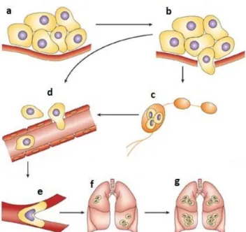
Metastasis formation involves a multiple step process. First, there is the development of a blood supply that can support the metabolic demands, a process called angiogenesis. These new blood vessels provide a route by which the malignant cells can escape from the primary tumor and enter into the blood stream. Cancer cells can also get access into circulatory system indirectly, through the lymphatic system. Once in the circulation, they extravasate to the surrounding tissue, starting a new proliferation process (Figure 1.4) [21]. During the invasion process, cancer cells penetrate the basement membrane and interstitial stroma through proteolysis of extracellular matrix (ECM). Metalloproteinases (MMPs) play a crucial role for this EMC proteolysis [22].
Figure 1.4: Metastatic process schematization. (a) in situ cancer. (b) invasion begins with changes in
cell-cell and cell-EMC adherence, destruction of stroma and motility. The cells can migrate (c) via
lymphatic or (d) directly in the blood circulation. (e) survival, arrest of metastatic cancer cells and
5
Epithelial-mesenchymal transition (EMT) plays an important role in the invasive cascade because it marks the transition from a collective to a single-cell migration mechanism. The development of metastasis requires the movement of cancer cells, driving the progression of carcinomas to an invasive and metastatic phonotype. In general, EMT process occurs during embryonic development in order to escape from structural constraints imposed by tissue architecture, adopting a more flexible cell movement capacity. Briefly, during EMT, epithelial cancer cells lose their polarity and some cell-cell and cell-ECM adhesion systems, resulting in reduced intercellular interactions and increased migratory character as mesenchymal cells. In the metastatic process, EMT enables cancer cells to disseminate from a primary tumor and promotes their self-renewal, required for metastases progression. The loss of expression of structural epithelial proteins (E-cadherin, plakoglobin and cytokeratins) was verified, when epithelial cells become invasive. E-cadherin is an adhesion molecule essential for the maintenance of adherents junctions, thus, the loss of their expression is a hallmark characteristic of EMT [24]. These phenotypic alterations, in general, are accompanied by the expression of vimentin and other mesenchymal characteristic proteins [25]. In Table 1.1 are summarize the main phenotypic alterations associated with EMT in colon carcinoma.
Table 1.1: Phenotypic alterations associated with EMT in colorectal carcinoma [25].
The role of β-catenin in E-cadherin-mediated cell-cell adhesion is essential for the study of
the molecular basis of EMT and malignant cancer development. In colon epithelia, β-catenin
links cadherin to the actin in cytoskeleton. The dismantling of cell-cell adhesion (loss of
E-cadherin) during EMT releases β-catenin which displays an important role in the regulation
of EMT. Indeed, it is usual to find β-catenin accumulation in the cell nucleus of the invasive carcinoma cells, instead of being located in the plasmatic membrane [25].
Once cancer cells have been established in the tissue, the new stroma environment lacks signaling to undergo an EMT. This allows cancer cells to revert the prior process and occurs
Epithelial Mesenchymal
Polarization
Polarized with differences between apical and basal membrane
constituents
No apical-basal membrane polarity, elongated morphology Cell-cell
contacts
Strong cell-cell junctions as tight and adherent and desmosomes
Weak cell-cell contacts discernible Structural
proteins E-cadherin, cytokeratins Vimentin
Extracellular environment
Basement membrane containing type IV collagen and laminin
Expression of type I collagen, fibronectin and metalloproteinases
Motility Non-motile cells Highly motile cells often
6
a mesenchymal-epithelial transition [26]. Thus, EMT process makes possible cancer cell dissemination and enables a self-renewal capacity to metastatic cancer cells [27].
1.1.3
ChemotherapyThe success of chemotherapy in the treatment of malignant diseases has been dramatic. In the most common carcinomas (breast, colon and lung) the effectiveness of chemotherapeutic agents has been disappointing [28].
Usually, cancer therapies, including surgery, chemotherapy and radiotherapy, have a limited but important role in the overall treatment of most solid tumors. In early-stage disease, low risk patients are often cured with surgery alone, but in many other cases a combination of treatments is required. The use of combined therapies or agents, which act through different molecular mechanisms, has been considered as a promising strategy [29]. These mechanisms are summarized in Figure 1.5.
Figure 1.5: Therapeutic targeting of the Hallmarks of Cancer. Drugs that interfere with each of the acquired abilities necessary for tumor growth and progression have been developed and are in clinical trials [30].
7
Chemotherapy drugs cause apoptosis by directly interfering with DNA or by targeting the key proteins involved in cell division [31].
Cell cycle is controlled by several checkpoints. These points are regulated by the transient association of cyclin and cyclin-depend kinase in order to determine the progression into next stage of division. Cyclins appear to act as oncogenes in some type of cancers. Hence, there are multiples aberrant pathways which can be exploited by drug design. In addition, different phases of the cell cycle or different signaling pathways are targeted to achieve maximal cell kill and the less likelihood of resistance [31, 32].
The most successful drug used in chemotherapy of colon cancer is 5-fluorouracil (5-FU). Particularly, this type of cancer is usually treated with a combinatory therapy with other drugs. It has been reported various mechanisms of action, including inhibition of thymidylate synthase and incorporation into RNA and DNA. Pyrimidine de novo synthesis is
impossible without thymidylate synthase activity and their inhibition results in a decrease of DNA synthesis and repair [33].
In general, standard anticancer drugs show low efficacy in providing long term resolutions in epithelial tumors. Recently, this limited effectiveness of anticancer therapies has been attributed to the existence of cancer stem cells (CSCs), which confers a highly drug resistant phenotype to the tumor. CSCs are characterized by their unlimited ability to self-renew, to be undifferentiated and seed new tumors with the same cellular heterogeneity. In the metastasis process, disseminated cancer cells require self-renewal capability, very similar to that exhibited by stem cells, in order to generate metastases in distant parts of the body [27]. Further, it is believed that common chemotherapy kills most cells in a tumor but leaves CSCs behind. It has already been shown that drug transporters protect CSCs from
chemotherapeutic agents. This drug’s resistance mechanism is due to the expression of high
8
1.2
Natural compounds and colon cancer
Over 200 years ago, Friedrich Sertäurner, a pharmacist´s apprentice, isolated the first pharmacologically active pure compound from a plant. This compound was morphine from opium, produced by cutting the seed pods of the poppy, Papaver somniferum [35]. After this
event, it was started a new era where drugs from plants could be purified, studied and administered. In 1990 about 80% of drugs were natural products or analogs inspired by them, causing a revolution in medicine. However, during the 90’s, it was observed an expansion in the synthetic medicinal chemistry, driving to a decrease of proportion of new drugs based on natural products to 50% [36, 37]. Nevertheless, natural products continue to provide unique structural diversity in analogy to standard chemistry. Since less than 10%
of world’s biodiversity has been evaluated for potential biological activity, many useful
natural compounds await discovery [38].
The World Health Organization estimates that 80% of worldwide population chooses traditional medicine for primary health care [39]. Essential oils from plants, nutraceutical products, dietary supplements, herbal remedies, teas and infusions are the main selected products [36].
As a source of new pharmaceuticals, natural products and derived molecules play an important role due to the great variety of functionally relevant metabolites of microbial and plant species. The development of improved products and new drugs can provide to drug development the scientific tools needed to perform a robust study of natural compounds and traditional medicines [40]. To be eligible as a medicinal plant or compound, it is necessary to take considerations about phytochemical analysis and pharmacological screening, which includes studies in vitro and in animal models. It needs further verification
in order to identify the most bioactive components and their mechanisms of action, activity and cytotoxicity profiles. In 2008, over 100 natural products-derived compounds were undergoing clinical trials and, at least, 100 products in preclinical development. Most of them were derived from plants and microbial sources and these products were predominantly being study for the use in cancer or as anti-infectious agents[36, 41].
In the context of colon cancer, multiple natural products and their bioactive phytochemicals have been found to exert anticancer effects by inducing cell-cycle arrest and apoptosis, decreasing cell proliferation and angiogenesis while inhibiting cancer cell invasion and migration. Besides, phytochemical compounds can also modulate several signals transduction and pathways involved in colon cancer development, including COX-2/PGE2
and Wnt/β-catenin [42]. Various in vitro studies in a wide range of colon cancer cell lines
9
Figure 1.6: Different mechanisms of action of natural products in colon cancer prevention and therapy (adapted from [43]).
Several developments in organic chemistry have been inspired by natural products, including the advances in methodologies and the possibility for synthesizing analogues of the original compound with enhanced pharmacological proprieties [46]. Natural products
can provide ‘privileged’ structures in terms of their ability to be the basis of new drugs. Natural products libraries are basically based on 3 major groups of phytochemical compounds, namely alkaloids, phenolics and terpenoids. The application of several techniques allows the creation of analogues and derivatives of these phytochemical compounds, with the possibility of being patented. Actually, with the development of analytical techniques, it has become easy to isolate, purify and determine structures of relevant compounds present in natural products [36].
Phenolic compounds, found in a large variety of plants, have shown to inhibit or attenuate the initiation, progression and spread of cancer cells, both in vitro and in animal models in vivo. This phytochemicals are secondary metabolites and can be monophenolic (one phenolic aromatic ring) or polyphenolic (more than one aromatic ring). Up to now, over 8000 different phenolics in plants were identified and classified in multiple sub groups of compounds (Figure 1.7). Flavonoids are the most abundant and numerous plant phenolics and can be found in high concentrations in the leaf epidermis and fruit skins due to their important role in pigmentation, UV protection and disease [47].
Natural Products
Cell cycle arrest Down-regulation of
Wnt/β-catenin pathway
Apoptosis indution Modulation of
signal trasduction
Down-regulation of COX-2/PGE2
pathway
Inhibition of angiogenesis
↓Bcl-2
↑Bax
Caspases activation ↓↓VEGFMMPs ↓HIF1-α
↑CDK Inhibitor ↓Cyclins ↓β-catenin
↓Cyclin D1 ↓C-Myc
↓ERK
↓MAPK
↓JNK
10
Figure 1.7: Schematic classification of phenolic compounds reported to have health benefits (adapted from [47]).
Phenolic compounds play a significant role in the suppression of tumor initiation, as well as in the promotion and progression of cancer. They can act in the regulation of growth factors-receptor interactions and in cell signaling cascades, such as kinases and transcription factors, which determines cell cycle arrest, cell survival and apoptosis. Aberrant growth factor expression results in an uncontrolled cell proliferation. Furthermore, phenolic compounds can enhance immune system to recognize and destroy cancer cells, inhibit angiogenesis and reduce the metastatic potential. The antiangiogenic effect appears to be linked with attenuation of MMPs and inhibition of vascular endothelial growth factor (VEGF) [47].
Terpenoids or terpenes are the largest group of natural compounds and many of them present bioactivity in the treatment of human diseases. Among the terpene-based pharmaceuticals already commercialized, it can be found a successful anticancer drug, the Taxol®. Besides anticancer properties, terpenes display a wide range of other biological
activities against inflammation and infectious diseases. They can be structurally classified as monoterpenes (C10), sesquiterpenes (C15), diterpenes (C20) and sesterterpenes (C25) [48].
11
Figure 1.8: Actions of monoterpenes in cancer prevention and therapy [49].
Alkaloids are a diverse group of compounds that contain one or more nitrogen atoms, usually, in a heterocyclic ring. They are widely distributed in the plant kingdom, mainly in higher plants. Alkaloids are among the most important bioactive components and some of them have already been successful in cancer chemotherapy [50]. Vinca alkaloids
(vinablastine, vincristine and vinerolbine) isolated from Madagascar periwinkle are anticancer agents in clinical use [51]. Recently, other anticancer relevant alkaloids have been studied, such as berberine, matrine and evodiamine. They act in several cancer mechanisms, including apoptosis, cell cycle arrest, angiogenesis, metastasis, and others [50]. Despite a period of decrease in their use, there are multiple natural compounds candidates for new drugs in current development. Phytochemical compounds provide the core scaffolds for future drugs due to an increasing acceptance of public opinion. In the future, the continuing development of natural products and chemical libraries based on natural products will continue to grow in drug research [36].
Several fruits have been reported as strong sources of phytochemical compounds, as previously described. The high content in these potential anticancer compounds makes this natural extracts as having therapeutic interest. The use of the fruit wastes as a source of bioactive compounds might be an approach to recover phenolic compounds and terpenoids. Fruits as olives, oranges, plums, peaches and cherries present several anticancer molecules that can be selectively extracted in order to obtain an extract with anticancer activity [52-55].
Monoterpenes
Carcinogen detoxification
Inhibition of tumor initiation/promotion
Induction of phase I and II carcinogen-metabolizing enzymes
↓tumor proliferation
↑apoptosis
12
1.2.1
OliveOlive tree (Oleaceae) has a great economic and social role in Mediterranean countries and it
is one of the most important agricultural activities in this area. Oil and olives are the main products of olive-related industry, which generates large volumes of by-products such as crude olive cake, vegetation water, twigs and leaves [56, 57]. Olive products have shown beneficial effects on human health mostly due to the presence of antioxidants such as phenolic compounds, carotenoids and tocopherols that assume an important function in disease prevention [58, 59]. Studies demonstrate the presence of anticancer active phenolics in olive fruit, including, luteolin, apigenin, oleuropein, rutin and hydroxytyrosol (Table 1.2) [60]. The anti-colon cancer activity of olive fruit skin terpenoids-rich extracts have been also reported [61].
The stone represents 18-22% of total olive weight. It is mainly formed by lignocellulosic material with commercial interest for production of solid, liquid or gas biofuel. Despite the relevance for the biofuel industry, olive stone is a rich source of valuable components due to their chemical and physical properties. Like the olive fruits, the presence of phenolic compounds in olive seed is also important. Several anticancer compounds have been already identified, including, tyrosol, oleuropeina and their derivatives (Table 1.2) [62].
Table 1.2:Reported anticancer activity of phenolic compounds from olive.
Phenolics Mechanism of anticancer activity Cancer model Ref
Tyrosol ↑ apoptosis in vitro
(cell-based assays) [63]
Hydroxytyrosol G1 arrest, ↑ apoptosis, ↓ ROS (cell-based assays) in vitro [63]
Oleuropein
S arrest, ↓cell proliferation, ↓ invasion, ↓ migration, angiogenesis
inhibition in vitro (cell-based assays) in vivo (mouse model) [64, 65]
Verbascoside ↓ ROS, ↑ tumor differentiation (cell-based assays)in vitro [66, 67]
Apigenin G2/M arrest, ↓cell proliferation, ↑ apoptosis, carcinogens detoxification
in vitro (cell-based assays) in vivo (mouse model) [68-70] Luteolin
↓ ROS, G2/M arrest,↓cell proliferation, ↑ apoptosis, angiogenesis inhibition, carcinogens
13
1.2.2
OrangeCitrus fruits have commercial relevance due to their nutritional value and special flavor. About 34% of orange production was used for juice production, where a large amount of residues is produced, constituted mainly by orange peels [52]. Thus, large amounts of peels are produced every year; this residue is the primary waste fraction.
Sweet orange (Citrus sinensis) peel is an interesting source of phenolic compounds such as
polymethoxylated flavonoids and terpenoids, mainly limonene and linalool [52]. Polymethoxyflavones (PMFs) have been of particular interest because these flavonoids display a large spectrum of biological activities, including, anti-inflammatory, anticancer (Table 1.3) and antioxidant. These compounds are particularly abundant in sweet orange [73-76]. PMFs are lipophilic molecules due to the hydrophobic nature of methoxy groups, in comparison to hydroxyl groups, as observed in quercetin, luteolin and narigenin. Consequently, PMFs have shown a higher absorbance through the small intestine and are easily absorbed into the blood circulation system [52]. All of these beneficial actions suggest new value-added uses for these compounds as nutraceuticals, generating considerable interest to the citrus industry.
Tangeretin and nobiletin are among the most effective citrus flavonoids at inhibition of human cancer cell proliferation [70]. In addition, in vivo antitumor studies of PMFs reveal
their safe pharmacological use [73].
Table 1.3: Reported anticancer activity of orange peel flavonoids.
Flavonoids Mechanism of anticancer activity Cancer model Ref
Tangeretin G1 arrest, ↓tumor invasion, ↓↓cell proliferation, cytostatic, ↑ tumor differentiation
in vitro
(cell-based assays)
in vivo
(rat model)
[73, 77]
Nobiletin G1 arrest, ↓↓cell proliferation, ↑ apoptosis
in vitro
(cell-based assays)
in vivo
(rat model)
[70, 74, 77]
Hesperidin carcinogens detoxification, ↓ ↑ apoptosis, tumor invasion
in vitro
(cell-based assays) [70, 78, 79]
Hesperetin G1 arrest, ↓cell proliferation, ↓ metastasis
in vitro
(cell-based assays)
in vivo
(mouse model)
[70, 80-82]
Sinensetin G2/M arrest ,↓cell proliferation, ↑ apoptosis, angiogenesis inhibition
in vitro
(cell-based assays)
in vivo
(zebrafish model)
[70, 83, 84]
Naringenin ↓cell proliferation, ↑ apoptosis, ↓ metastasis
in vitro
(cell-based assays)
in vivo
(mouse model)
14
1.2.3
Fruits of Prunus genusPrunus genus comprises more than 400 species of flowering shrubs and trees. Almond,
peach, plum, cherry and apricot are the most economically relevant cultivars.
Plums (Prunus domestica), peaches (Prunus persica) and sweet cherries (Prunus avium) are botanically and compositionally similar fruits. Previous works have reported their phenolic compounds, such as flavonols, anthocyanins, catechins and hydroxycinnamates in both peel and flesh [86, 87]. Besides, bioactive terpenoides have been also detected in several varieties of these fruits including perillyl alcohol, lutein and zeaxanthin [53, 88-90].
In recent years, cherry products have been in a continue demand in the food market because of their potential positive effects on health [91]. Sweet cherries contain significant amounts of phenolic compounds such as anthocyanins, which display a wide range of health promoting benefits [92, 93]. These nutraceutical compounds are not uniformly distributed in fruit tissue. High amounts of polyphenolics and anthocyanins are observed in the fruit skin due to their functions as photoprotective agents and attractants for seed dispersal [94]. Prior researches have shown that anthocyanin members have anticancer activity [95, 96]. Antiproliferative activity in colorectal cancer cell lines was recently reported for several varieties of tart cherries [91]. Moreover, the value of cherries as fresh fruit is too high to be used for anthocyanins extraction [97]. The use of cherry wastes such as skins and pits from juice-processing industry represents a feasible alternative for these purposes.
Besides, cherries exhibit a high content of perillyl alcohol, a monoterpene molecule which has chemotherapeutic proprieties against colorectal cancer [53, 98]. Perillyl alcohol has been shown to inhibit cancer cells migration and angiogenesis, two important processes in cancer dissemination [99, 100](described in section 1.3).
Plums and peaches appear to have a wide range of health benefits, mostly due to their high content in antioxidant molecules such as neochlorogenic acid, protocatechuic acid and rutin [101-104]. Peach and plum extracts that have a high anthocyanin content can inhibit cancer proliferation and exert a differentiating effect [54]. This anthocyanins have demonstrated anti-tumorigenic effects by several mechanisms, including apoptosis induction and cell cycle arrest at G1 phase [105].
15
Table 1.4: Reported anticancer activity for cherry, plum and peach phytochemical compounds.
Phytochemicals Mechanism of anticancer activity Cancer model Ref
Te rpe n o ids Lutein and Zeaxanthin
Carcinogens detoxification, ↑ apoptosis, angiogenesis inhibition, ↑
differentiation, ↓ ROS, modulation of immune response
in vitro
(cell-based assays)
in vivo
(mouse and rat models)
[106]
Perillyl alcohol
Carcinogens detoxification, ↑ differentiation, ↑ apoptosis, ↓proliferation, G1 arrest, angiogenesis
and migration inhibition
in vitro
(cell-based assays)
in vivo
(mouse and rat models) [49, 99, 100, 107] P hen o lics Catechin
↓Proliferation, ↑ apoptosis, ↓ ROS, angiogenesis and invasiveness
inhibition
in vitro
(cell-based assays)
in vivo
(rat model and humans)
[108, 109]
Rutin ↓Proliferation, ↓ DNA damage, ↑ apoptosis
in vitro
(cell-based assays)
in vivo
(mouse and rat models) [110, 111] Chlorogenic and Neochlorogenic acid
↓Proliferation, invasion and migration inhibition in vitro (cell-based assays) [112, 113] Protocatechuic acid
Carcinogens detoxification, prevent DNA adduct formation, ↑ apoptosis ,
metastasis inhibition in vitro (cell-based assays) in vivo (mouse model) [101, 114, 115] Anthocyanins
G1 arrest, Carcinogens detoxification,↑ apoptosis, ↓Proliferation, ↑ differentiation, invasion and
angiogenesis inhibition
in vitro
(cell-based assays)
in vivo
(mouse and rat models)
16
1.3
Cancer Cellular Models
Drug development process typically comprises three major steps: i) drug discovery process; ii) pre-clinical studies and iii) clinical trials (phase I, II and III). Pre-clinical studies are the stage that precedes clinical trials with is testing the drug in humans. Discovery and pre-clinical development is a continuous process resulting in preliminary pharmacology and toxicology studies [118]. In this phase the main goal is to evaluate product bioactivity, effectiveness, cytotoxicity, pharmacodynamics and pharmacokinetics. In the last two decades, several attempts have been made to develop cellular models to minimize the gap between cell-based assays and animal studies frequently used in pre-clinical phase. This allows a reducing of experimental uncertainties arising from traditional culture systems and, consequently, the cost of drug screening process [119].
Cellular models for colorectal cancer can display a unique tool to study tumors and their metastatic capacity. There are several adenocarcinoma cell lines derived from colon tumors at diverse points of differentiation and development stages. Tumor cell lines can be an important resource for understanding cancer initiation and progression. They present an identical spectrum of genetic alterations as primary tumors (described in section 1.1) [120]. In Table 1.5 is present the most common genetic alteration found in eight colon cancer cell lines.
Table 1.5: Genetic alterations and mutated genes in several colon cancer cell lines [120].
Cell line Sex/age APC K-ras p53 β-catenin MMR
Lovo M56 + + - - +
HCT116 M? - + - + +
SW48 F82 - - - + +
HCT15 M? + + + - +
LS1034 M54 + - + - -
HT29 F44 + - + - -
SW480 M51 + + + - -
Colo320 F55 + + + - -
During discovery phase and preclinical development of anticancer drugs, these are tested for their efficacy and potential, usually using cells grown in vitro on culture plates as
monolayers (2D). Traditionally, 2D cell culture involves growing cells on solid, impermeable adherent surfaces. These monolayer cultures are easy and convenient to set up with high cell viability and have contributed greatly to our understanding of several diseases processes [121, 122]. However, these cellular models have many limitations and usually drugs do not work as effectively in vivo as in 2D model [121, 123]. Cell culture in 2D systems
is a poor physiological model in comparison with in vivo systems, mainly due to the
geometric differences between 2D culture and tissues, which are three-dimensional (3D) [124, 125]. Thus, the cells lose the capacity to respond to the molecular gradients in 2D [126]. It is, therefore, necessary to improve in vitro cell-based testing methods for a better
17
The lack of stroma and structural architecture are the main limitations of 2D monolayer models. Organ cultures could provide a better model, however, it is difficult to obtain specimens and the poor viability of the tissues cultured are major obstacles [128].
1.3.1
Three Dimensional Models3D cultures or multicellular tumor spheroids (MCTS) were first described, at the beginning of last century [129]. They were considered an improved in vitro model to simulate biological characteristics of tumor and consisted in 3D structures with a certain complexity [130]. The use of 3D in vitro systems has been recognized as a potential link to minimize the
gap between monolayer cultures and animal model studies [127].
Tumor cells in spheroids show a higher degree of morphological and functional differentiation than in 2D culture [131]. One major advantage of 3D cancer models is their reproducible concentric arrangement of different cell populations which is an inherent property of solid tumors [132]. This gives a stratified composition, with proliferating cells in the outer rim, followed by a quiescent layer of cells and, eventually, necrotic cells in the spheroid center (Figure 1.9) [133]. Beyond altered gene expression and resistance to cancer treatment, the cell-cell and cell-matrix contacts in compact spheroid can also limit the diffusion of nutrients, oxygen and other compounds [125, 134]. In addition, poor efficiency of mass transport also leads to a metabolic waste accumulation inside the spheroids, like in vivo tumors [135].
Figure 1.9: Multicellular tumor spheroid scheme. Cells are organized forming a sphere spheroid with different zones, a proliferative, quiescent and central necrotic area. Oxygen, nutrients and proliferation gradients are indicated [136].
Necrotic core
Quiescent zone
Proliferative zone
P
ro
lif
e
ra
tio
18
Co-cultivation of multiple cell types is possible in 3D cultures, allowing the study of the interactions between epithelial and stromal cells, such as fibroblasts, which regulate normal and neoplastic development. The interactions and support from stromal elements and ECM plays a crucial role in tumor biology. These parameters can determine the behavior and gene expression of the cells [137]. Cell shape and tissue architecture depends on various adhesion molecules, consequently, every alteration in these molecules has relevance to cancer environment [138]. Therefore, recapitulating these in vitro can potentially minimize
the relevance of in vitro models and better mimic of the in vivo like response to cancer
therapies. The culture of colon cancer spheroids has been described since 1986 and has already been used for multiple in vitro studies [139-141]. However, co-cultures of colon
cancer cells with cancer associated fibroblasts have been described only recently [142, 143]. The cell-cell and cell-matrix interactions in the complex 3D model not only affects drug action and penetration but it is also essential in the distribution and function of physiologically occurring factors. These biochemical effectors that play an important role in the regulatory mechanisms of cell growth, differentiation and death include hormones and growth factors. This microenvironment is important for the regulation of normal cell function and it may be useful to understand how it regulates tumorigenic phenotypes, structural and biochemically. Despite the complexities of 3D spheroids co-cultures, these models provide a strong complementary use to the animal models [137].
There are several methodologies to achieved 3D culture from cell lines, primary cells and organs, including spontaneous aggregation, liquid overlay cultures, and stirred culture systems [137]. The advantages and disadvantages for each culture systems, depending on the type of experiment and the final model. It is important to take in consideration the increased time and cost of experimental set-up and the model optimization and validation (Table 1.6) [122].
Traditionally, spheroids were obtained by spontaneous cell aggregation, generating spherical cellular conglomerates. Spheroids formed by this technique are analogous to avascular tumor nodules in terms of growth kinetic, with populations of proliferating, quiescent and necrotic cells. These different cell populations appear mostly due to mass transport limitations and also recreate the situation observed in intravascular microregions of large tumors or micrometastases prior to vascularization [144]. Other similar way to get 3D spheroids is ensuring conditions in which the adhesive forces between the cells are greater than the substrate plated on. Liquid overlay techniques (forced-floating and hanging drop methods) promotes a spontaneous aggregation, preventing cell matrix deposition. Thus, spheroids formations can even be induced in cell lines which are difficult to aggregate (Figure 1.10 (a) and (b)) [145].
Stirred culture systems present great advantages to grow 3D spheroids. These methods include gyratory rotation, rotary cultures, rotating-wall vessel bioreactor (RWV) and stirred-tank systems such as spinner flasks. In contrast to the static environment of liquid overlay cultures, which are useful to study the individual spheroid, the dynamic suspension
of spinner flasks allows the production of larger numbers of spheroids. The spinner’s
19
multicellular tumor spheroids. However, other successful methods, including gyratory shakers have also been used, as well as, RWV bioreactor which maintains cells in a dynamic fluid suspension mixed by minimal hydrodynamic forces. The culture flask rotates whole on its horizontal axis, providing the mixing of the cells. The vessel is completely filled with medium minimizing fluid turbulence and shear forces with a semi-permeable membrane providing aeration and the elimination of bubbles (Figure 1.10 (c)) [146, 147].
Figure 1.10: Common methods for multicellular spheroid formation: (a) forced floating, (b) hanging drop, (c) rotating-wall vessel bioreactor and using (d) spinner vessel bioreactor. (adapted from [148])
Finally, 3D cultures in environmental controlled stirred-tank bioreactors working in perfusion mode have been developed for culture and analysis of 3D tissues. These systems allow an automatic replenishment of exhausted media and control the addition of fresh medium. Further, it enables a tight regulation of extracellular environment including fluid shear and the concentration gradients of biochemical molecules. Besides the control of temperature, pH, oxygen, nutrients and metabolites, bioreactors allow the automatic feeding and effective mass transfer [149, 150].
In addition to the spheroid culture, various alternative cell culture strategies are available as tools for engineering 3D models, including the use of microcarrier beads as scaffolds. Microcarriers have provided many advantages to cell culture such as supporting the aggregation of attachment dependent cells and cell lines which do not spontaneously spheroid. Furthermore, microcarriers can be used as a platform to co-culture different cell types [137, 151]. Scaffolds are often used with the possibility of adding growth and regulatory factors. In the past decade, enhanced scaffolds have been introduced as improved support in natural molecules (collagen composite), synthetic/semi-synthetic polymers (polyethylene glycol or hydrogels) or a combination of these materials. The major advantage of these scaffolds is the great potential to recreate the natural physical and structural environment of living tissue. Has been demonstrated to promote signalling pathways that regulate migration, proliferation and differentiation [152-154] .
Liquid overlayer techniques
Stirred systems Non adhesive coating
(a) (b)
Centrifugation
Spheroids in hanging drop
20
In Table 1.6 the main advantages and disadvantages of common 3D cell culture systems are summarized.
Table 1.6: Major advantages and disadvantages of common 3D culture methods.
Culture system Advantages Disadvantages
Static systems
Relatively simple Inexpensive Suitable for high-throughout
Spheroids production are easily accessible
Labor intensive
Small culture volumes difficult medium exchange without disturbing cells
No control over size of spheroid
Stirred systems
Simple to culture Large-scale production easily achievable
Better nutrient transport Spheroids production are easily accessible
21
1.4
Thesis Goal
The major goal of this thesis is the evaluation of the anticancer effect of phytochemical extracts, using 3D cellular model of colon cancer. For this propose a 3D model using HT29 cells growing as spheroids was developed and characterized.
The work was divided in three major parts as schematically presented in Figure 1.11.
Figure 1.11: Structure of the thesis.
In the first part, the antiproliferative effect of natural extracts obtained from cherries, peaches, plums, oranges and olive seeds were tested in 2D monolayer cultures. 2D dose-response curves were performed to determine the effective dose value (EC50) of each extract. In this part, phytochemical characterization of the extracts was also carried out. The second part was focused in the development of 3D cell model of colon cancer using dynamic stirred culture system. Tumor spheroids were grown in spinner flasks and several culture parameters were optimized. Phenotypic assessment was also performed in order to better characterize the 3D cell model.
In the third part of the work, chemotherapeutic potential of the most promising phytochemical-rich extracts were evaluated using 3D model of colon cancer and results were compared with 2D monolayer results.
Part 1
Part 2
Screening of natural extracts
3D Model development
Evaluation of anticancer effect of natural extracts
using 3D cell models
•Antiproliferative effect using 2D cell cultures
•Phytochemical characterization
•HT29 spheroids generation
•Phenotypic characterization
•Antiproliferative effect
•Cell cycle arrest
23
2
Experimental Procedure
2.1
Natural extracts
Olive seeds extracts from six olive trees varieties (Cobrançosa, Zambujeiro, Picual, Arbosana, Koroneiki and Arbequina) were performed by conventional solvent extraction (CSE) in the
context of an ongoing project [155].
Initially, the seeds were obtained by breaking the olive stones. The solid-liquid extraction was carried out using 2 g of each seed sample and 10 ml of solvent mixture containing EtOH:H2O (80:20,v/v). Samples were mixed by vortex during 5 minutes and then
centrifuged for 15 min at 5000 rpm. The extraction procedure was repeated once again and finally supernatant was filtered and concentrated by rotary evaporator. Samples were dissolved in EtOH:H2O (80:20,v/v) and stored at -20 0C after being filtered through a 0.45
µm filter Acrodisc® (Pall, USA).
Extracts of cherry, plum, peach and orange residues were performed by Nutraceuticals and Delivery group of IBET (Oeiras, Portugal) using high pressure technology. All of the raw-materials used for extraction are residues of fruit juice production (peels, seeds and core), or crop residue (cherry culls). The extracts were obtained under variable extractions conditions and using diverse CO2/EtOH ratios.
In Table 2.1 are summarized the extraction conditions used to obtain high pressure natural extracts.
Table 2.1 : Extraction conditions used to obtain fruit residues extracts with high pressure technology.
Extract CO2 pre treatment
Pressure (MPa)
Temperature (K)
Time (min)
% CO2/EtOH
Ref
Orange peel - 25 323 30; 60;
90;120 80/20 [156]
Cherries 60 min 25 323 30 90/10 [90]
Peach 60 min 25 323 30 90/10 [90]
24
2.2
Phytochemical characterization
2.2.1
Terpenes analysis by thin layer chromatography (TLC)The detection of terpenes, such as perillyl alcohol and linalool, was performed by thin layer chromatography (TLC).
TLC analysis was carried using silica gel plates with a 254 nm fluorescent indicator with aluminum base (20 cm x 20 cm) (Macherey-Nagel, Duren, Germany). Dichoromethane was used as mobil phase and for detection a mixture of 1% ethanolic vanillin + 10% ethanolic sulphuric acid + Burchard reagent (1% acetic anhydride and 1% suphuric acid ethanolic) was applied. At the end of elution, the plate was sprayed with prepared solutions and heated at 110 0C for 10 minutes. Then, the plate was revealed with Burchard reagent and
the visualization was performed under 254 nm UV light.
2.2.2
Phenolic analysis by high performance liquid chromatography (HPLC)The phenolic HPLC analysis of natural extracts was performed by Analytical Group of IBET (Oeiras, Portugal) coordinated by Dr. Rosário Bronze. Briefly the analysis was carried out using a Surveyor apparatus form Thermo Finnigan with a diode array detector (Thermo Finnigan-Surveyor, San Jose, CA, USA) and an electrochemical detector (Dionex, ED40) [157]. Separations were performed at 35 0C in a LiChrospher C18 (5 µm, 250 mm x 4 mm
25
2.3
Cell-based assays
2.3.1
Cell lines and cultureHuman colon cancer cell lines, HT29 and Caco2 were provided by American type Culture Collection (ATCC, USA) and Deutsche Sammlung von Microorganismen und Zellkulturen (DSMZ, Germany), respectively. Both cell lines were cultured in RPMI 1640 medium supplemented with 10% heat-inactivated fetal bovine serum (FBS) and 2 mM of glutamine. Cells were kept at 37 0C in a humidified incubator with 5% CO2 and routinely grown in 75 or
175 cm2 culture flasks. Cell culture medium and supplements were obtained from
Invitrogene (Gibco, Invitrogene Corporation, Paisley, UK). The cell lines were split once or twice a week, the morphology and growth of cells were monitored daily.
2.3.2
Antiproliferative assay using 2D cell culture systemAntiproliferative assays were performed on human colon adenocarcionoma cell line (HT29) growth as monolayer.
The assay was carried out in 96-well microplates as described by Serra et al. [53]. Briefly cells were seeded at a cellular density of 1x10⁴ cell/well. After 24 hours of incubation in 5%
CO2 humidified atmosphere at 37 OC the medium was removed and cells were incubated
with natural extracts diluted in RPMI medium supplemented with 0.5% FBS. After 24 hours, the medium was removed and cell viability was assessed using commercial PrestoBlue®
Viability Reagent (Molecular Probes, Invitrogen, US). Briefly, PrestoBlue® regent (diluted
1:10) was added to each well and the microplate was incubated for 2 hours. Finally the fluoresce was quantified on a microplate fluorescent reader (FL800, Bioteck Instruments, USA) with filter appropriate for 580 nm excitation and 595 nm emission maxima. The results were expressed as a percentage of cell viability relative to the control (cells incubated with culture medium only, without natural extracts). The experiment was performed in triplicate. EC50 values (the concentration of sample necessary to decrease 50% of cell viability) were obtained from dose-response curves using software GraphPad Prism (GraphPad Software, Inc., La Jolla, CA) fit.
![Figure 1.1: World age-standardized mortality rates of colorectal cancer, 2008 data [3]](https://thumb-eu.123doks.com/thumbv2/123dok_br/16627897.740555/23.892.171.647.521.858/figure-world-standardized-mortality-rates-colorectal-cancer-data.webp)
![Figure 1.2: Colorectal adenocarcinoma progression stages. The lower stage indicates a smaller tumor with less proliferation and the higher stage represents a bigger tumor with invasion (adapted from National Cancer Institute) [11]](https://thumb-eu.123doks.com/thumbv2/123dok_br/16627897.740555/24.892.191.683.585.896/colorectal-adenocarcinoma-progression-indicates-proliferation-represents-national-institute.webp)
![Figure 1.5: Therapeutic targeting of the Hallmarks of Cancer. Drugs that interfere with each of the acquired abilities necessary for tumor growth and progression have been developed and are in clinical trials [30]](https://thumb-eu.123doks.com/thumbv2/123dok_br/16627897.740555/28.892.164.697.519.917/therapeutic-targeting-hallmarks-interfere-abilities-necessary-progression-developed.webp)
![Figure 1.6: Different mechanisms of action of natural products in colon cancer prevention and therapy (adapted from [43])](https://thumb-eu.123doks.com/thumbv2/123dok_br/16627897.740555/31.892.145.719.99.464/figure-different-mechanisms-natural-products-prevention-therapy-adapted.webp)
![Figure 1.7: Schematic classification of phenolic compounds reported to have health benefits (adapted from [47])](https://thumb-eu.123doks.com/thumbv2/123dok_br/16627897.740555/32.892.111.751.103.339/figure-schematic-classification-phenolic-compounds-reported-benefits-adapted.webp)

![Table 1.5: Genetic alterations and mutated genes in several colon cancer cell lines [120]](https://thumb-eu.123doks.com/thumbv2/123dok_br/16627897.740555/38.892.141.723.592.828/table-genetic-alterations-mutated-genes-colon-cancer-lines.webp)