Raquel Patrícia Gomes Silvestre Vinhas
Mestre em Biologia CelularGold nanoparticles for nanotheranostics in
leukemia
–
Addressing Chronic Myeloid Leukemia
Dissertação para obtenção do Grau de Doutor em Biociências Moleculares
Orientador: Prof. Doutor Pedro Miguel Ribeiro Viana Baptista, Professor
Catedrático, Faculdade de Ciências e Tecnologia da Universidade Nova
de Lisboa
Co-orientadora: Prof. Doutora Maria Alexandra Núncio de Carvalho
Ramos Fernandes, Professora Auxiliar, Faculdade de Ciências e
Tecnologia da Universidade Nova de Lisboa
Júri:
Presidente: Prof. Doutor Paulo Manuel Assis Loureiro Limão-Vieira Vogais: Prof. Doutora Cecília Maria Pereira Rodrigues
Prof. Doutor José Alexandre de Gusmão Rueff Tavares Prof. Doutor Pedro Miguel Ribeiro Viana Baptista Prof. Doutor João Nuno Sereno de Almeida Moreira
iii COPYRIGHT
Autorizo os direitos de copyright da minha tese de doutoramento, com o título: “Gold nanoparticles for nanotheranostics in leukemia –
Addressing Chronic Myeloid Leukemia”
v
ACKNOWLEDGEMENTS
This thesis was most definitely a joint effort. Each person that crossed my path during this journey had a part in it and the following words cannot express the amount of gratitude I share with you.
First, I acknowledge my supervisors, Prof. Pedro Baptista and Prof. Alexandra Fernandes, for having accepted me for a lab rotation, four years ago, without knowing the first thing about me. That week turned into four fruitful years, which could not have happened if it was not for your trust and belief in my abilities. I still feel very lucky to be in the intersection of your work and for all the autonomy and opportunities you gave me. Though our discussions on scientific issues were enlightening, our best conversations far surpassed the limits of science and occasionally involved a glass of wine, which, as we all know, I never refuse!
Prof. Pedro Baptista, thank you for taking a chance on me, pushing me further and, most of all, for the ambitious directions you always envisioned for this project. Your positivity was paramount and throughout the years that same positivity and scientific spirit passed on to me.
Prof. Alexandra Fernandes, thank you for looking at this project with such joy. Most of all, thank you for the many conversations that made me persevere. You always had the right words at the right time. More than a supervisor, you were a guiding friend.
I express my gratitude to the staff at Hospital dos Capuchos: Dr. Aida Botelho de Sousa, Dr. Patrícia Ribeiro, Alexandra Lourenço and Susana Santos. The clinical perspective you provided during this thesis made me look at results in a different way. It is all about the patients! The most rewarding feeling I could have found through these years is that our collaboration might have helped a few people.
I acknowledge all collaborators at CEMOP and CENIMAT, specially Prof. Hugo Águas and Pedro Alves, for providing all the required resources and knowledge for the microfluidics experiments. Pedro Alves, thank you as well for your kindness, advice and, above all, I praise your patience.
vi
Andreia, the time that we have known each other has taught me many things. One of them is that comfort can come from the most unusual places. Never had I thought that we would be flatmates for this long and how much fun we were going to have together. I am sure many adventures are still yet to come.
I thank my family and closest friends for always expressing how proud they are of me. This achievement is mostly yours. It is almost impossible to fail when one is surrounded by such caring, strong and resilient people. I thank my father for the enormous support along the way. I emphasize, as well, the major part my sister Cláudia had in this period, particularly when I doubted everything. You know me better than anyone. After all, you have 34 years of experience on the job. Thank you so much!
Throughout the course of these PhD thesis, I have learnt a lot on cancer genetics and nanotechnology, however I have to confess that my most important knowledge has been given to me by my mother since the very beginning. You showed me how to be kind and independent. You are my best role model.
I acknowledge the Department of Life Sciences of Faculdade de Ciências e Tecnologia, UCIBIO and ITQB, from Universidade Nova de Lisboa, for hosting the Molecular Biosciences PhD program and providing all the necessary means for this academic effort.
vii
RESUMO
A leucemia é um tipo de cancro que se inicia na medula óssea e resulta na produção desregulada de glóbulos brancos imaturos (células leucémicas). A leucemia mieloide crónica (LMC) é o seu subgrupo mais homogéneo, afetando aproximadamente 1.5 milhões de pacientes no mundo. Praticamente todos os casos apresentam a translocação t(9;22)(q34.1;q11.2) e a consequente fusão genética BCR-ABL1,
que codifica a tirosina cinase BCR-ABL1. O sucesso do tratamento da LMC advém do seu diagnóstico precoce e do desenvolvimento de inibidores de tirosina cinase eficazes.
A nanotecnologia apresenta um enorme potencial para suprimir as lacunas dos procedimentos convencionais usados no combate à LMC. As nanopartículas de ouro (AuNPs), em particular, possuem propriedades óticas relevantes para deteção ex vivo de biomarcadores, mas podem funcionar também in vivo numa estratégia de teranóstico, combinando tratamento e diagnóstico de acordo com o perfil
molecular do paciente.
Neste estudo, foi otimizado um ensaio colorimétrico, usando nanossondas de ouro, para a deteção da isoforma mais frequente de BCR-ABL1 (e14a2), tendo sido posteriormente validado em amostras
clínicas. Este método simples e direto permitiu a deteção de amostras de RNA que expressam e14a2, com precisão e especificidade. O ensaio foi ainda incorporado num chip de microfluídica, traduzindo-se em resultados mais rápidos recorrendo a volumes mais baixos de amostra, devido à escala e estrutura do dispositivo.
Adicionalmente, foi desenhada uma nova estratégia terapêutica para combater os mecanismos de resistência ao tratamento de primeira linha para LMC: imatinib (IM). O silenciamento in vitro de BCR-ABL1 foi eficaz, usando AuNPs funcionalizadas com polietilenoglicol e oligonucleotídos de DNA em
cadeia simples, antisense e em formato hairpin. A nanoformulação permitiu redução da dose de IM,
numa estratégia combinada, e potenciou um decréscimo da viabilidade de células K562 resistentes a IM.
Os resultados desta tese demonstram a adequação e flexibilidade das AuNPs como ferramentas para nanoteranóstico, diagnóstico e tratamento personalizado de LMC.
Termos chave:
ix
ABSTRACT
Leukemia is a type of cancer that initiates in the bone marrow and results in the unregulated production of immature white blood cells (leukemic cells). The most homogenous subgroup of the disease is chronic myeloid leukemia (CML) accounting for nearly 1.5 million patients worldwide. Virtually all cases harbor the genetic translocation t(9;22)(q34.1;q11.2) resulting in the BCR-ABL1 gene
fusion, that encodes for BCR-ABL1 tyrosine kinase. CML treatment success relies on an early diagnosis and the intense research towards developing effective tyrosine kinase inhibitors (TKI).
Nanotechnology offers unprecedent advantages to tackle the shortcomings of conventional procedures for the management of CML. Gold nanoparticles (AuNPs) have unique optical properties suitable for ex vivo biosensing applications, but can also function in vivo as nanocarriers in a theranostic
approach that links treatment with diagnosis according to patient’s molecular profile.
A gold nanoprobe (Au-nanoprobe) colorimetric assay was optimized for the detection of the most frequent BCR-ABL1 isoform (e14a2) and was validated on fully characterized clinical samples. This
simple and cheap method enabled the direct detection of e14a2-expressing RNA samples, with accuracy and high specificity. The Au-nanoprobe assay was translated onto a microfluidics chip, resulting in a faster outcome with smaller sample volumes, due to the scale and design of the device.
Additionally, a new therapeutic strategy was designed to overcome CML resistance to first line therapy, such as imatinib (IM). BCR-ABL1 gene silencing was effectively achieved in vitro, using
AuNPs functionalized with polyethylene glycol and a hairpin-shaped antisense single stranded DNA (ssDNA) oligonucleotide. Furthermore, the nanoconstruct allowed to reduce the dose of IM, when tested in a combined approach, and potentiated a viability decrease of K562 cells resistant to IM.
The results of this thesis strongly suggest that AuNPs are a suitable and flexible tool for CML nanotheranostics, improving detection and a personalized treatment strategy.
Keywords:
xi
LIST OF PUBLICATIONS
Included in this thesis:
• Vinhas R, Cordeiro M, Carlos FF, Mendo S, Fernandes AR, Figueiredo S, Baptista PV. 2015. Gold nanoparticle-based theranostics: disease diagnostics and treatment using a single nanomaterial. Nanobiosensors in Disease Diagnosis. 4:11-23.
• Larguinho M, Canto R, Cordeiro M, Pedrosa P, Fortuna A, Vinhas R, Baptista PV. 2015. Gold nanoprobe-based non-crosslinking hybridization for molecular diagnostics. Expert Review of Molecular Diagnostics. 15(10):1355-1368.
• Pedrosa P, Vinhas R, Fernandes AR, Baptista PV. 2015. Gold nanotheranostics: proof-of-concept or clinical tool? Nanomaterials. 5(4):1853-1879.
• Vinhas R, Tolmatcheva A, Canto R, Ribeiro P, Lourenço A, Sousa AB, Baptista PV, Fernandes AR. 2016. A novel mutation in CEBPA gene in a patient with acute myeloid leukemia. Leukemia
& Lymphoma. 57(3):711-713.
• Vinhas R, Correia C, Ribeiro P, Lourenço A, Sousa AB, Fernandes AR, Baptista PV. 2016. Colorimetric assessment of BCR-ABL1 transcripts in clinical samples via gold nanoprobes.
Analytical and Bioanalytical Chemistry. 408(19):5277-5284.
• Veigas B, Pinto J, Vinhas R, Calmeiro T, Martins R, Fortunato E, Baptista PV. 2017. Quantitative real-time monitoring of RCA amplification of cancer biomarkers mediated by a flexible ion sensitive platform. Biosensors and Bioelectronics. 91:788-795.
• Vinhas R, Cordeiro M, Pedrosa P, Fernandes AR, Baptista PV. 2017. Current trends in molecular diagnostics of chronic myeloid leukemia. Leukemia & Lymphoma. 58(8):1791-1804.
• Vinhas R, Fernandes AR, Baptista PV. 2017. Gold nanoparticles for BCR-ABL1 gene silencing:
Improving tyrosine kinase inhibitor efficacy in chronic myeloid leukemia. Molecular Therapy - Nucleic Acids. 7:408-416.
• Vinhas R, Mendes R, Fernandes AR, Baptista PV. 2017. Nanoparticles – Emerging potential for managing leukemia and lymphoma. Frontiers in Bioengineering and Biotechnology. 5(79):1-10.
xii
nanoprobe based RNA detection – application to Chronic Myeloid Leukemia. Scientific reports. 8(1):381.
• Matias AS, Vinhas R, Mendes R, Fernandes AR, Baptista PV. 2018. Nanoparticles as emerging diagnostic tools in liquid tumours. European Medical Journal. 2(1):80-87.
• Vinhas R, Lourenço A, Santos S, Ribeiro P, Silva M, Sousa AB, Baptista PV, Fernandes AR. A double Philadelphia chromosome-positive chronic myeloid leukemia patient, co-expressing P210BCR-ABL1 and P195BCR-ABL1 isoforms. [Submitted to Haematologica]
• Vinhas R, Lourenço A, Santos S, Lemos M, Ribeiro P, Sousa AB, Baptista PV, Fernandes AR. A novel BCR-ABL1 mutation in a patient with Philadelphia chromosome-positive B-cell acute
xiii
LIST OF ABBREVIATIONS AND SYMBOLS
ABC –ATP-binding-cassette
ABL1 Abelson murine leukemia 1 gene
Abs – absorbance
AFC 7-amino-4-trifluoromethyl coumarin ALL – acute lymphocytic leukemia
AlloSCT – allogeneic stem cell transplantation AML – acute myeloid leukemia
AP – accelerated phase AUC – area under the curve Au-nanobeacon – gold nanobeacon Au-nanoconjugate – gold nanoconjugate Au-nanoprobe – gold nanoprobe
AuNP – gold nanoparticle
AuNP@PEG – gold nanoparticles functionalized with polyethylene glycol
AuNP@PEG@e14a2 – gold nanoparticles functionalized with polyethylene glycol and e14a2 antisense ssDNA oligonucleotide
AuNP@PEG@MYC – gold nanoparticles functionalized with polyethylene glycol and c-MYC
antisense ssDNA oligonucleotide B-ALL – B-cell acute lymphocytic leukemia
BAX – Bcl-2-associated X gene
BC – blast crisis
BCL-2 – B-cell lymphoma 2 gene BCR breakpoint cluster region gene
bZIP basic leucine zipper domain
C/EBP CCAAT/enhancer binding protein CCyR – complete cytogenetic response
CEBPA CCAAT/enhancer binding protein gene
CEMOP Center of Excellence in Microelectronics Optoelectronics and Processes CENIMAT – Materials Research Center
CHLC – Centro Hospitalar de Lisboa Central CHR – complete hematologic response CLL – chronic lymphocytic leukemia CML – chronic myeloid leukemia
xiv
CP – chronic phase
CT – computed tomography Ct – threshold cycle
CTC – circulating tumor cell dB decibel
DEPC diethyl pyrocarbonate DLS dynamic light scattering
DMEM Dulbecco's modified Eagle medium DMR – deep molecular response
DMSO dimethyl sulfoxide DTT dithiothreitol
EDTA – ethylenediaminetetraacetic acid EGF – epithelial growth factor
ELN – European LeukemiaNet EMA European Medicines Agency EPR – enhanced permeability and retention FBS – fetal bovine serum
FDA – US food and drug administration FISH – fluorescence in situ hybridization
FRET - Förster resonance energy transfer GRP – gastrin-releasing peptide
IM –imatinib IR – infrared
IRIS – Study of Interferon and STI571 IS – international scale
K562-IM – K562 cells resistant to imatinib LED light-emitting diode
LOD – limit of detection MB – methylene blue
MCyR – major cytogenetic response MDR – multidrug resistance
MMR – major molecular response MRD – minimal residual disease MRI – magnetic resonance imaging
xv NCL – non-crosslinking
NGS – next generation sequencing NIR – near infrared
NNI – National Nanotechnology Initiative NP – nanoparticle
NPV negative predictive value
OCT1– organic cation transporter 1 gene
PAT – photoacoustic tomography PBS – phosphate buffer saline PDMS – polydimethylsiloxane PDT – photodynamic therapy PEG – polyethylene glycol
PET – positron emission tomography Ph – Philadelphia chromosome pHLIP –pH low insertion peptides PI – polydispersity index
PMSF phenylmethylsulfonyl fluoride PNB – plasmonic nanobubbles
POC – point-of-care
PPV positive predictive value PS – photosensitizer
PTT – photothermal therapy PTD – photodynamic therapy QD – quantum dot
qPCR – quantitative polymerase chain reaction rAbs absorbance ratio
RCA – rolling circle amplification RGD – arginylglycylaspartic acid
rhTNF – recombinant human tumor necrosis factor RPMI Roswell Park Memorial Institute medium Rs detection response
RT-PCR – reverse transcription polymerase chain reaction
SDS-PAGE sodium dodecyl sulfate polyacrylamide gel electrophoresis SERS – surface-enhanced Raman scattering
xvi
SNR – signal-to-noise ratio
SPECT – single-photon positron emission tomography SPIO – super-paramagnetic iron oxide
SPR – surface plasmon resonance SSC – side scatter intensity SSC-A – side scatter peak area TAD – transactivation domain
TBST tris-buffered saline with tween 20 TEM transmission electron microscopy Tf – transferrin
TKI – tyrosine kinase inhibitors
UCIBIO – Research Unit on Applied Molecular Biosciences US – ultrasound
UV ultraviolet
xvii
TABLE OF CONTENTS
ACKNOWLEDGEMENTS ... v
RESUMO... vii
ABSTRACT ... ix
LIST OF PUBLICATIONS ... xi
LIST OF ABBREVIATIONS AND SYMBOLS ... xiii
TABLE OF CONTENTS ... xvii
LIST OF FIGURES ... xix
LIST OF TABLES ... xxi
CHAPTER1–INTRODUCTION ... 1
1.1 LEUKEMIA ... 3
1.1.1 Classification ... 3
1.1.2 Chronic myeloid leukemia ... 6
1.1.2.1 Molecular biology and disease progression ... 6
1.1.2.2 Diagnostics and disease follow-up ... 8
1.1.2.3 Therapeutics ... 11
1.1.2.4 Current trends ... 13
1.2 NANOMEDICINE ... 21
1.2.1 Focus on gold ... 21
1.2.2 Gold nanoparticle-based diagnostics ... 22
1.2.2.1 Non-cosslinking assay optimization ... 24
1.2.2.2 Non-crosslinking assay application to biomolecular detection... 26
1.2.2.3 Point-of-need devices ... 29
1.2.3 Gold nanoparticle-based theranostics ... 30
1.2.3.1 Targeting and delivery ... 31
1.2.3.2 Therapeutic agents ... 33
1.2.3.3 Phototherapy ... 34
1.2.3.4 Multimodal Imaging ... 35
1.2.3.5 From research lab to the clinic ... 36
1.2.4 Nanotechnology commercial impact ... 37
1.3 NANOTECHNOLOGY TOWARDS LEUKEMIA MANAGEMENT ... 39
1.3.1 Clinical trials ... 42
1.4 SCOPE OF THE THESIS ... 43
CHAPTER 2 – MOLECULAR CHARACTERIZATION OF LEUKEMIA CLINICAL SAMPLES ... 47
xviii
2.2 INTRODUCTION ... 49
2.3 PATIENTS AND METHODS ... 52
2.4 RESULTS AND DISCUSSION ... 60
2.5 CONCLUDING REMARKS ... 73
CHAPTER3–DETECTIONOFBCR-ABL1TRANSCRIPTSIN CLINICALSAMPLESVIA GOLDNANOPROBES ... 75
3.1 ABSTRACT ... 77
3.2 INTRODUCTION ... 77
3.3 MATERIAL AND METHODS ... 79
3.4 RESULTS AND DISCUSSION ... 81
3.5 CONCLUDING REMARKS ... 88
CHAPTER4–MICROFLUIDIC CHIPFORGOLD NANOPROBE-BASEDBCR-ABL1RNA DETECTION... 91
4.1 ABSTRACT ... 93
4.2 INTRODUCTION ... 93
4.3 MATERIAL AND METHODS ... 96
4.4 RESULTS AND DISCUSSION ... 98
4.5 CONCLUDING REMARKS ... 109
CHAPTER 5 – GENE SILENCING USING GOLD-NANOPROBES: TOWARDS A NANOTHERANOSTICSAPPROACHFORCML ... 111
5.1 ABSTRACT ... 113
5.2 INTRODUCTION ... 113
5.3 MATERIAL AND METHODS ... 115
5.4 RESULTS AND DISCUSSION ... 120
5.5 CONCLUDING REMARKS ... 131
CHAPTER6–CONCLUSIONSANDFUTUREPERSPECTIVES ... 137
6.1 FINAL CONSIDERATIONS ... 139
6.2 FUTURE PERSPECTIVES ... 143
REFERENCES ... 149
xix
LIST
OF
FIGURES
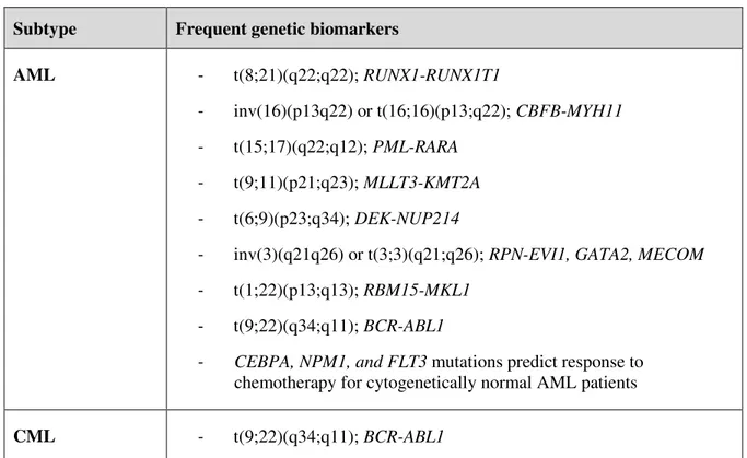
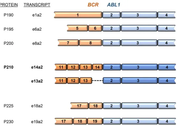
Figure 1.1 Structure of BCR-ABL1 isoforms ... 7
Figure 1.2 Methodologies for CML cytogenetic and molecular screening ... 16
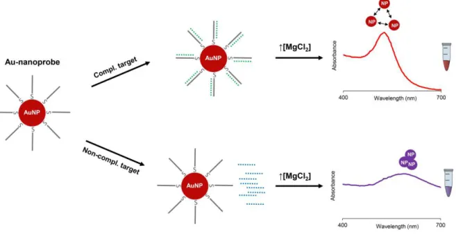
Figure 1.3 Au-nanoprobe non-crosslinking assay ... 24
Figure 1.4 Precision nanomedicine for the management of hematological disorders ... 41
Figure 2.1 Most recurrent amino acid substitutions in BCR-ABL1... 50
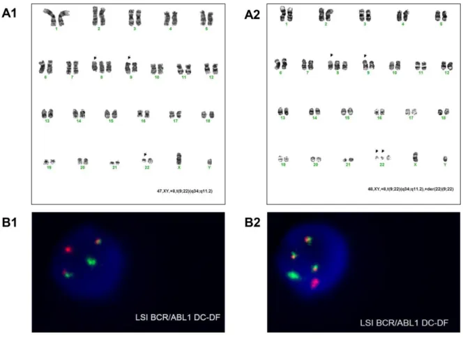
Figure 2.2 Cytogenetic analysis for clinical case 44 ... 66
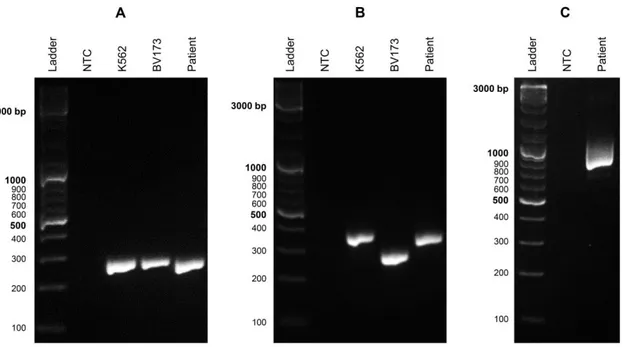
Figure 2.3ABL1 and BCR-ABL1 molecular analysis for clinical case 44 ... 67
Figure 2.4 Sequence analysis of BCR-ABL1 from clinical case 44... 67
Figure 2.5 Cytogenetic analysis for clinical case 54 ... 69
Figure 2.6ABL1 and BCR-ABL1 molecular analysis for clinical case 54 ... 69
Figure 2.7 P190BCR-ABL1 protein structure and identified mutation on clinical case 54. ... 70
Figure 2.8 C/EBP protein structure and identified mutation on clinical case 7 ... 72
Figure 3.1 Genetics of CML ... 79
Figure 3.2 Au-nanoprobe assay using synthetic oligonucleotides ... 83
Figure 3.3 Au-nanoprobe assay using total RNA from leukemia cell lines ... 84
Figure 3.4 Au-nanoprobe assay using total RNA from clinical samples ... 86
Figure 3.5 Schematic for CML colorimetric detection via Au-nanoprobe assay ... 90
Figure 4.1 Concept of the lab-on-chip as a point-of-care application ... 95
Figure 4.2 Microfluidic chip design.. ... 100
Figure 4.3 Microfluidic setup circuits. ... 101
Figure 4.4 Salt saturation study for Au-nanoprobes ... 103
Figure 4.5 Signal-to-noise ratio of the microfluidic device. ... 104
Figure 4.6 Colorimetric results via microfluidic chip ... 107
Figure 4.7 Sensitivity between positive and negative results via microfluidic chip ... 107
Figure 4.8 Limit of detection of BCR-ABL1 using Au-nanoprobes (on-chip vs off-chip) ... 108
xx
Figure 5.2 Au-nanoconjugate AuNP@PEG@e14a2 effectively silences BCR-ABL1 ... 122
Figure 5.3 Side scattering analysis of K562 cells after exposure to AuNP@PEG@e14a2 ... 123
Figure 5.4 Cell proliferation assays upon exposure to AuNP@PEG@e14a2 ... 125
Figure 5.5 Apoptosis induced by silencing nanoconjugate. ... 127
Figure 5.6 Triggering of apoptotic cascade via AuNP@PEG@e14a2 ... 129
Figure 5.7 Viability of K562 cells resistant to imatinib after exposure to AuNP@PEG@e14a2 ... 131
Figure 5.8 Schematic for AuNP-based BCR-ABL1 gene silencing. ... 132
Figure 5.9In vitroc-MYC gene silencing on K562 cells using AuNP@PEG@MYC ... 133
xxi
LIST OF TABLES
Table 1.1 Common genetic abnormalities associated to leukemia ... 4
Table 1.2 Expected levels of CML patients response after TKI therapy initiation ... 9
Table 1.3 TKIs in CML management ... 12
Table 1.4 Advantages and disadvantages of new approaches in CML molecular characterization. .... 17
Table 1.5 The different applications of the non-crosslinking nanoprobe assay ... 27
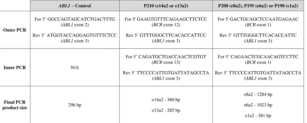
Table 2.1 Nested-PCR primers for ABL1 and BCR-ABL1 molecular analysis ... 58
Table 2.2 Nested-PCR primers for BCR-ABL1 mutational analysis ... 59
Table 2.3 Clinical samples cytogenetic and molecular characterization. ... 61
Table 2.4 Disease progression for clinical case 44... 65
Table 3.1 Oligonucleotide probe and synthetic oligonucleotide targets ... 80
Table 3.2 AuNPs and Au-nanoprobe characterization ... 82
Table 3.3 Performance of Au-nanoprobe diagnostic test. ... 87
Table 4.1 AuNPs and Au-nanoprobe characterization ... 102
Table 4.2 Experimental conditions used the microfluidic chip assay ... 106
Table 5.1 AuNPs, AuNP@PEG and AuNP@PEG@e14a2 characterization ... 121
xxiii Para aquela que me ensinou a rir, Mãe
"Well I can ease your pain Get you on your feet again Relax
I'll need some information first Just the basic facts."
1
CHAPTER 1
–
INTRODUCTION
Part of the literature review presented in this chapter has been published, whole or in part, elsewhere. The author of this thesis reviewed and critically discussed the references cited in the text.
• Vinhas R, Cordeiro M, Carlos FF, Mendo S, Fernandes AR, Figueiredo S, Baptista PV. 2015. Gold nanoparticle-based theranostics: disease diagnostics and treatment using a single nanomaterial. Nanobiosensors in Disease Diagnosis. 4:11-23.
• Larguinho M, Canto R, Cordeiro M, Pedrosa P, Fortuna A, Vinhas R, Baptista PV. 2015. Gold nanoprobe-based non-crosslinking hybridization for molecular diagnostics. Expert Review of Molecular Diagnostics. 15(10):1355-1368.
• Pedrosa P, Vinhas R, Fernandes AR, Baptista PV. 2015. Gold nanotheranostics: proof-of-concept or clinical tool? Nanomaterials. 5(4):1853-1879.
• Vinhas R, Cordeiro M, Pedrosa P, Fernandes AR, Baptista PV. 2017. Current trends in molecular diagnostics of chronic myeloid leukemia. Leukemia & Lymphoma. 58(8):1791-1804.
• Vinhas R, Mendes R, Fernandes AR, Baptista PV. 2017. Nanoparticles – Emerging potential for managing leukemia and lymphoma. Frontiers in Bioengineering and Biotechnology. 5(79):1-10.
3
1.1
Leukemia
Hematologic malignancies are the most common type of cancer among children and young adults, and comprise diseases such as leukemia, lymphoma and myeloma, which affect the bone marrow, lymphatic system and blood cells.1 The sequential stages of hematopoietic differentiation provide multiple occasions for the occurrence of mutations and other disruptive events that lead to distinct tumor subtypes and clinical presentations.2 In fact, the heterogeneity of tumors of the hematopoietic system presents unique challenges for current diagnosis and treatment.3–6
Leukemia is a clonal disorder originated in the bone marrow during hematopoiesis and is characterized by the unregulated proliferation of poorly differentiated white blood cells. Prognosis and treatment of the disease depend highly on the type of leukemia, the extent of the disease, age and patient history. Most patients are treated with chemotherapy/targeted therapy, although a small portion ultimately undergoes radiation therapy and/or bone marrow transplantation.7
Though hematological malignancies constitute no more than 10 % of all cancers worldwide, leukemia research has attracted considerable attention. This is believed to result from the combination of several factors: 1) the blood-borne nature of the disease and accessibility of relevant tissues; 2) the fact that hematopoiesis has been thoroughly dissected, thus prompting modelling of leukemogenesis; 3) the emotional impact of a lethal cancer of childhood, since acute leukemia is the most common pediatric cancer in developed societies, which engrossed massive funding opportunities; and 4) the early identification of chromosomal translocations and fusion genes in leukemia karyotypes.7
1.1.1
Classification
Classification of leukemia is based on the type of cell affected (myeloid or lymphoid) and the degree of cell proliferation (acute or chronic), resulting in four main categories.2 Acute myeloid leukemia (AML) is the most common type in adults and acute lymphocytic leukemia (ALL) is more prevalent among pediatric patients.1 Chronic myeloid leukemia (CML) is a myeloproliferative disorder with an annual incidence of 1-2 cases per 100 000 adults, accounting for 15-20 % of newly diagnosed cases of leukemia in adults.1,8
4
Following clinical, morphological and immunophenotypic diagnosis,10 genetics plays an important role in the classification of these diseases and commonly assist risk stratification, from poor to favorable prognosis (Table 1.1).11,12
AML is the most clinically and biologically heterogeneous group of leukemias. Many of the variations that underlie the pathogenesis of the disease include chromosomal additions, deletions, and translocations, as well as small-scale mutations in oncogenes (Table 1.1). Since disease progression and treatment response are largely dependent on these genetic events, proper cytogenetic and molecular testing is imperative for patient evaluation and provide insights into critical pathways involved in AML pathogenesis. Small-scale mutations on genes encoding for nucleophosmin 1 (NMP1), fms-related
tyrosine kinase 3 (FLT3) and CCAAT/enhancer-binding protein (CEBPA) are in fact used to define
AML sub-classes according to World Health Organization (WHO) guidelines. CEBPA encodes for a
transcription factor that is critical for myeloid differentiation. Point mutations on this gene confer a relatively good prognosis for AML patients, but this seems only true for cases with biallelic variations.13–16
CML is a good example of how hematological diseases have greatly benefited from the advance of cytogenetic and molecular methodologies. It was the first cancer in which a unique chromosomal abnormality was identified, t(9;22)(q34.1;q11.2) – Philadelphia chromosome (Ph) - and the associated
BCR-ABL1 gene, providing a specific target for disease treatment (Table 1.1).17 Tyrosine kinase
inhibitors (TKIs) target the dysregulated kinase activity of the fusion protein encoded by BCR-ABL1.18,19
Table 1.1 Common genetic abnormalities associated to leukemia.11,20 Subtype Frequent genetic biomarkers
AML - t(8;21)(q22;q22); RUNX1-RUNX1T1
- inv(16)(p13q22) or t(16;16)(p13;q22); CBFB-MYH11
- t(15;17)(q22;q12); PML-RARA
- t(9;11)(p21;q23); MLLT3-KMT2A
- t(6;9)(p23;q34); DEK-NUP214
- inv(3)(q21q26) or t(3;3)(q21;q26); RPN-EVI1, GATA2, MECOM
- t(1;22)(p13;q13); RBM15-MKL1
- t(9;22)(q34;q11); BCR-ABL1
- CEBPA, NPM1, and FLT3 mutations predict response to
chemotherapy for cytogenetically normal AML patients
5 B-cell ALL - t(9;22)(q34;q11); BCR-ABL1
- t(v;11q23); KMT2A
- t(12;21)(p13;q22); ETV6-RUNX1
- t(5;14)(q31;q32); IL3-IGH
- t(1;19)(q23;p13); TCF3-PBX1
- Hyperdiploidy - Hypodiploidy
T-cell ALL - TAL1 gene rearrangements (1p32)
- TLX1 gene rearrangements (10q24)
- LMO2 gene rearrangements (11p13)
- t(5;14)(q35;q32); TLX3- BCL11B
- t(10;11)(p12;q14); PICALM- MLLT10
- NUP214- ABL1 fusion and amplification
- KMT2A gene rearrangements (11q23)
CLL (or SLL) - del(13q) - trisomy 12 - del(11q); ATM
- del(17p); TP53
ALL, acute lymphocytic leukemia; AML, acute myeloid leukemia; CLL, chronic lymphocytic leukemia; CML, chronic myeloid leukemia; SLL, small lymphocytic leukemia.
Genes: ABL1, Abelson murine leukemia 1; ATM, ataxia-telangiectasia mutated; BCL11B, B-cell
lymphoma/leukemia 11B; BCR, breakpoint cluster region; CBFB, core-binding factor ß; CEBPA,
CCAAT/enhancer-binding protein ; DEK, DEK proto-oncogene; ETV6, erythroblast transformation-specific
translocation variant 6; EVI1, ecotropic virus integration site 1; FLT3, fms-related tyrosine kinase 3; GATA2,
GATA-binding factor 2; IGH, Immunoglobulin heavy-chain; IL3, interleukin-3; KMT2A, lysine methyltransferase
2A; LMO2, LIM-domain only 2; MECOM, MDS1 and EVI1 complex locus; MKL1, megakaryoblastic leukemia
1; MLLT10, histone lysine methyltransferase DOT1L cofactor; MLLT3, super elongation complex subunit;
MYH11, myosin heavy chain 11; NPM1, nucleophosmin 1; NUP214,nucleoporin 214; PBX1, pre B cell leukemia
homeobox 1; PICALM, phosphatidylinositol binding clathrin assembly protein; PML, promyelocytic leukemia;
RARA, retinoic acid receptor ; RBM15, RNA binding motif protein 15; RPN, ribophorin I; RUNX1,runt related
transcription factor 1; RUNX1T1, RUNX1 translocation partner 1; TAL1, TAL bHLH transcription factor 1,
erythroid differentiation factor; TCF3, transcription factor 3; TLX1/3, T-cell leukemia homeobox 1 or 3; TP53,
6
1.1.2
Chronic myeloid leukemia
Originally named chronic granulocyte leukemia, the first documented cases of CML are believed to date back to 1840s.21,22 Since then, there has been a solid evolution in the strategies for laboratorial diagnostics of the disease, from cytogenetics to molecular characterization that support therapeutic decisions, which ought to be tailored so as to enhance therapy success. CML is also paradigmatic in the straightforward relation between early and accurate hematologic, cytogenetic and molecular characterization, and patients’ prognosis.17
1.1.2.1
Molecular biology and disease progression
The molecular hallmark of CML is the Ph chromosome - a shortened and truncated version of chromosome 22 present in at least 95 % of patients.23 This abnormality occurs in the myeloid lineage of hematopoiesis and results in the reciprocal translocation of genetic material between the long arms of chromosomes 9 and 22 - t(9;22)(q34.1;q11.2). The subsequent fusion of the Abelson murine leukemia (ABL1) gene on chromosome 9 (region q34) with the breakpoint cluster region (BCR) gene
on chromosome 22 (region q11), results in the BCR-ABL1 oncogene.23,24 This chimeric version of ABL1
has unregulated activity and triggers a cascade of events that culminate in malignant cell transformation. The constitutive expression of the active protein kinase leads to the phosphorylation of a series of intracellular substrates, perturbing multiple signaling pathways, including the Ras/mitogen-activated protein kinases (Ras/MAPK), phosphatidylinositol-3-kinase/protein kinase B (PI3K/Akt), and Janus kinase 2/signal transducer and activator of transcription 5 (Jak2/Stat5) pathways. This phosphorylation cascade promotes cell growth, survival advantage, cytokine independence, protection against apoptosis and causes the clonal expansion of fully differentiated myeloid cells.25–27
Although CML molecular hallmarks have been deeply studied, chromosome instability originates a range of BCR-ABL1 variants. Due to three well-defined breakpoint regions in the BCR gene (m-BCR, M-BCR and µ-BCR), the product of the BCR-ABL1 fusion oncogene may exist in three principal forms
of molecular weight: 190, 210, and 230 kDa, containing the same region of the ABL1 tyrosine kinase at the C-terminal but including different portions of BCR sequence at the N-terminal.28,29 This reciprocal translocation during hematopoiesis combined with alternative splicing events may result in multiple
BCR-ABL1 transcripts, all encoding proteins with high tyrosine kinase activity (Figure 1.1).30
7 There is a high controversy between the various published studies concerning the type of fusion transcript (e.g., e13a2 vs e14a2) and its influence in CML phenotype.29,33–35 Balatzenko et al
demonstrated that CML patients with high levels of expression of the multidrug resistance 1 gene (MDR1) and presenting the e14a2 transcript had a significantly higher platelets count and a lower white
blood cell count compared to e13a2 patients.33 More recently, Hanfstein et al stated that e13a2 and e14a2 CML might represent distinct biological entities but the clinical outcome under treatment was comparable and no risk prediction could be made according to BCR-ABL1 transcript type at diagnosis.34
Figure 1.1 Structure of BCR-ABL1 isoforms. P210BCR-ABL1 isoforms represent 95 % of Ph+ CML cases
and may derive from two transcript types (e14a2 or e13a2) due to alternative splicing events. Alternative splicing may also occur to ABL1 exons 2 or 3, thus originating additional (rare) isoforms of
the gene. Nomenclature of BCR-ABL1 protein isoforms relates to their final protein size (e.g. P210 = 210 kDa).29–31
8
(blast cells) in the blood, bone marrow and spleen.36 Despite being generally asymptomatic, there are common symptoms in this stage, such as fatigue, weight loss, illness and general pain, due to anemia and splenomegaly (enlarged spleen - detected in 50-60 % of cases).37 Over the years it may progress into AP with a rapid expansion of circulating myeloid elements. Ultimately, CML may evolve to BC, which is characterized by differentiation retention in the myeloid or lymphoid lineage, resembling an acute leukemia with a predominant myeloid phenotype (~70 %).38,39 BC occurs when over 30 % immature blasts, myeloid or lymphoid, are present in blood or bone marrow, resulting in the increase of BCR-ABL1 mRNA and protein levels.38 This terminal phase usually leads to metastasis, organ failure
and death, presenting a poor prognosis and worsening symptoms, bleeding, fever and infections.37 The median survival of patients with BC without treatment is 3 months.40
The mechanisms underlying CML triphasic model of progression can be partially explained by the genetic instability characteristic of blast cells, resulting in the acquisition of additional genomic alterations, such as trisomy 8, isochromosome 17, double Ph+, c-MYC amplification and activation of pathways that block myeloid differentiation and inactivation of tumor suppressor genes such as phosphoprotein 53 gene (p53) and retinoblastoma gene (RB). These molecular events extend leukemic
stem cells (LSCs) replicative lifespan.24, 26,41 Therefore, LSCs and related pathways may serve as key targets for curing CML.26
1.1.2.2
Diagnostics and disease follow-up
Clinical manifestations of CML are often non-specific, and many patients diagnosed with CML are asymptomatic (30-50 % in the US alone). CML is frequently detected when an elevated white blood cell count is revealed by a routine blood test or when splenomegaly is detected on a general physical examination.37 After hematological characterization, bone marrow biopsy is mandatory to confirm diagnosis. In a case of persistent unexplained leukocytosis, CML diagnosis consists of probing for the presence of the Ph chromosome by routine cytogenetics (karyotype); or the Ph-related molecular BCR-ABL1 abnormalities by fluorescence in situ hybridization (FISH); or by molecular biology screening.8,38
The Ph chromosome is present in 95 % of CML patients (typical t(9;22) from chromosome banding analysis of at least 20 marrow cells metaphases) as the sole chromosomal abnormality. The remaining patients show additional chromosomal alterations (clonal evolution), such as trisomy 8, isochromosome 17, deletions in the 22q, or other Ph-variants (involving chromosome 22 or 9 or involving other chromosomes in addition to chromosomes 9 and 22). These are treated as standard Ph+ patients since Ph-variant patients present similar responses to therapy and prognosis.8,38
FISH analysis relies on analyzing at least 200 nuclei for the co-localization of DNA probes designed to hybridize specifically to the BCR and ABL1 genes.38 FISH may be performed in bone marrow and/or
9 treatment-free remission.38 For disease follow up, peripheral blood samples should be used in place of bone marrow.42
The success of precise therapy for CML depends greatly on its diagnosis, but also on disease follow-up. Three key parameters should be considered when monitoring CML patients’ response to therapeutics: hematologic response or normalization of blood counts; cytogenetic response, which relates to the percentage of Ph+ cells in the bone marrow; and molecular response, defined by the expression level of BCR-ABL1 transcript in a bone marrow or blood sample. The European
LeukemiaNet (ELN) established milestones for hematologic, cytogenetic, and molecular responses to be achieved at specific time-points after therapy initiation for improved clinical outcome (Table 1.2). Beginning treatment with imatinib (IM), the first TKI for CML treatment released after the interferon era (additional information in section 1.1.2.3 –CML therapeutics), the following events are expected:
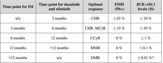
complete hematologic response (CHR) with major cytogenetic response (MCyR) after 3 months; complete cytogenetic response (CCyR) after 6 months; and major molecular response (MMR) after 12 months of therapy. Therefore, follow-up is crucial for disease monitoring. It is recommended that these patients perform a peripheral blood collection and analysis every 3 months after starting TKI treatment. Patients who do not achieve these milestones should be immediately considered for an alternative TKI/therapy (Tables 1.2 and 1.3).38,42,43
Table 1.2 Expected levels of CML patients response after TKI therapy initiation (ELN guidelines).38,42 Additional information on different TKIs can be accessed in section 1.1.2.3 –CML therapeutics.
Time point for IM Time point for dasatinib and nilotinib
Optimal response
FISH (Ph+)
BCR-ABL1 levels (IS)
n/a 3 months CHR ≤ 65 % ≤ 10 %
3 months 6 months CHR, MCyR ≤ 35 % ≤ 10 %
6 months 12 months CCyR 0 % ≤ 1 %
12 months >12 months MMR 0 % ≤ 0.1 %
>12 months n/a DMR 0 % ≤ 0.01 %*
*Expected result in two consecutive blood samples (assay sensitivity > 10-4)
10
CML molecular monitoring
BCR-ABL1 mRNA transcripts typically remain detectable after CCyR. Reverse transcriptase
polymerase chain reaction (RT-PCR) is thus far the only tool capable of monitoring responses after patients reach CCyR.43 This methodology has become an integral part of CML management since 1999 due to its sensitivity levels.44 The low levels of minimal residual disease (MRD) normally achieved during therapy call for a sensitive monitoring assay for reliable detection and quantitation.8,43 Currently, two PCR-based modalities can be performed to detect the BCR-ABL1 transcript in CML patient
samples: RT-nested PCR, a qualitative approach; or reverse transcriptase quantitative polymerase chain reaction (RT-qPCR), a quantifiable assay.
Nested PCR is a modification of PCR that was designed to improve its sensitivity and specificity. Basically, two pairs of primers target the same region: the first pair (outer primers) amplifies a fragment in a conventional PCR reaction; the second set (inner primers) anneals to sites within the first amplicon. In CML, RT-nested PCR provides characterization of the transcript allowing the identification of the more frequent transcripts: e13a2 or e14a2. This approach also allows for identification of one of the atypical fusion transcripts that are not amplified by the standard primer sets.44 As previously mentioned, there is a high controversy among the various published studies concerning the type of fusion transcript and its influence in CML phenotype and/or response to therapy.29,33–35 Recently, the ELN expert panel acknowledged that the transcript type (e13a2 vs e14a2 or other more atypical variants) may influence
response to therapy and disease outcome, which demonstrates the relevance of performing this molecular screening.38
RT-qPCR provides data on the actual percentage of BCR-ABL1 mRNA transcripts in peripheral
blood or bone marrow, and is capable to detect one CML cell in a background of 100 000 normal cells.43,45 It also provides a strong correlation between the results obtained from peripheral blood and bone marrow, allowing molecular monitoring without the necessity of obtaining bone marrow aspirations.38,43 RT-qPCR is definitely the current method of choice for CML molecular detection and quantification, and should be performed before initiation of specific TKI therapy to establish a baseline for BCR-ABL1 mRNA levels for each individual patient.38 As such, several efforts have been made to
optimize and standardize this methodology to reduce the heterogeneity of BCR-ABL1 expression results
amongst different clinical laboratories.42,46,47 According to guidelines from the National Comprehensive Cancer Network (NCCN) and the ELN, results are now conveyed as the ratio of BCR-ABL1 transcript
numbers to the number of control gene transcripts (typically, ABL1 gene).42 This value is then compared
to a standardized baseline representing 100 % IS (International Scale, defined by the median value of
BCR-ABL1 mRNA at the time of diagnosis in 30 patients with CML as established in the Study of
Interferon and STI571 - IRIS), that is independent of individual patient-specific BCR-ABL1 baseline
values.48
BCR-11
ABL1 transcripts from the baseline and is fixed at 0.1 % IS. These correlate to minimal risk of
transformation to advanced phases of disease with 100 % probability of remaining progression-free at 24 months. Generally, a 2-log reduction (BCR-ABL1 transcripts = 1 % IS) and 1-log reduction ( BCR-ABL1 transcripts = 10 % IS) correlate with CCyR (defined as absence of Ph+ chromosomes) and MCyR,
respectively (Table 1.2).38,42
Another integral part of molecular diagnosis and therapy management in CML is the assessment of the chimeric gene mutational status. Kinase domain mutation profile plays a minor role in selecting an initial TKI, but becomes highly relevant for patients where no ab initio response to the TKI is obtained,
or in relapsed cases. Indeed, BCR-ABL1 mutations range from 25 % to 30 % of early CP patients on first-line IM to approximately 70 % to 80 % of BC patients.38,49 Due to the clonal diversity of
BCR-ABL1-expressing cells, mutational screening is conventionally performed via Sanger sequencing that is
capable to detect a point mutation if the clone accounts for more than 15 % of all Ph+ cells.49
1.1.2.3
Therapeutics
Treatment depends on the type of leukemia, the disease stage, prior history of treatment, age and health of the patient, as well as the genetic profile. Most patients are treated with chemotherapy, while some may also undergo radiation therapy, stem cell transplantation or targeted therapy.37,39,40
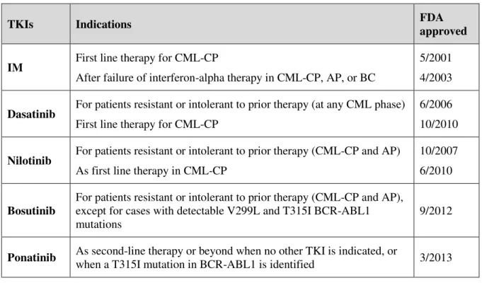
Up until the 1990s, the best nontransplant options for CML-CP management were nonspecific agents such as busulfan, hydroxyurea and interferon-alfa, which provide for temporary disease control. In fact, IFN-α is capable of reducing the proportion of the Ph chromosome but with multiple side effects.50,51 From the 1990s onwards, the treatment of choice resulting in curatives interventions, was the allogeneic stem cell transplantation (AlloSCT). However, AlloSCT not only carries high risks of morbidity and mortality, but also requires relatively young patients and an appropriate stem cell donor.52,53 CML treatment strategies and outcomes boosted with the design and development of small molecules capable of interfering and inhibiting the kinase activity of the BCR-ABL1 protein – TKIs.54– 57 TKI therapy is considered the standard first-line treatment for patients with newly diagnosed CML-CP. As shown in table 1.3, five TKIs are available and recommended in the management of CML patients: IM (first generation), dasatinib, nilotinib (second generation), bosutinib (second/third generation) and ponatinib (third generation).57–62
12
expressing leukemic cells.27, 57,66–68 Besides IM, dasatinib and nilotinib have indication for first-line therapy in CML. The choice of the most appropriate TKI is based on several factors such as blood counts at diagnosis, patient age, drug cost, comorbidities and drug toxicity profile. This ensures that the treatment is the most adequate, target-oriented and well tolerated by each patient, with minimal impact on quality of life.57,69,70 These molecules (dasatinib and nilotinib) are more potent inhibitors than IM (in
vitro) and have demonstrated the ability to induce more rapid and deeper responses in newly diagnosed
patients. However, their impact on long-term overall survival remains undetermined.71–74
Table 1.3 TKIs in CML management. Adapted from Leach, 2016.63
TKIs Indications FDA
approved
IM First line therapy for CML-CP
After failure of interferon-alpha therapy in CML-CP, AP, or BC
5/2001 4/2003
Dasatinib For patients resistant or intolerant to prior therapy (at any CML phase) First line therapy for CML-CP
6/2006 10/2010
Nilotinib For patients resistant or intolerant to prior therapy (CML-CP and AP) As first line therapy in CML-CP
10/2007 6/2010
Bosutinib
For patients resistant or intolerant to prior therapy (CML-CP and AP), except for cases with detectable V299L and T315I BCR-ABL1 mutations
9/2012
Ponatinib As second-line therapy or beyond when no other TKI is indicated, or
when a T315I mutation in BCR-ABL1 is identified 3/2013
AP, advanced phase; BC, blast crisis; CML, chronic myeloid leukemia; CP, chronic phase; IM, imatinib; TKI, tyrosine kinase inhibitor
Overcoming TKI resistance
It is known that 15 % to 40 % of CML patients will develop resistance or intolerance to first-line TKI treatment, with a need for a second-line TKI.57,75 This limited therapeutic potential is mainly due to TKIs resistance mechanisms that can be BCR-ABL1 dependent or independent. BCR-ABL1 dependent TKIs resistance mechanisms rely mostly on the overexpression of the BCR-ABL1 oncogene
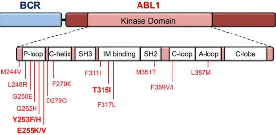
13 binding. The most recurrent alterations can occur at three different regions and seven specific residues: in the docking site for phosphate moieties of ATP named P-loop (M244V, G250E, Y253F/H and E255K/V); in the contact site related to selective BCR-ABL1 inhibition by IM (T315I); and in the catalytic domain (M351T and F359V).77,78
Generally, second generation TKIs are active against most of the IM-resistant mutations, however, CML patients harboring T315I, F317L or V299L mutations usually do not respond to dasatinib, whereas patients harboring the E255K/V, T315I, F359C/V or Y253H mutations have a lower response to nilotinib.79,80 More recently, bosutinib appeared as the newest second-generation TKI, and have been proved to be highly effective in CML patients with BCR-ABL1 kinase mutations that lead to IM failure (exceptions are the V299L and T315I mutations).81–85 The T315I mutation displays resistance to all current available TKIs, except for ponatinib, a third generation TKI that was recently approved for patients resistant or intolerant to prior TKI therapy (Table 1.3).83,86 Therefore, mutational screening could lead to a better and informed selection of second and third line TKI therapy to be applied in patients who failed first or second line treatment, providing clinical benefit for those involved.49, 85,87 Upon resistance to a first-line TKI, BCR-ABL1 mutation analysis is then recommended according to the NCCN and ELN guidelines.58 Patients that fail all current TKIs regimens are eligible for AlloSCT.38,88
BCR-ABL1 independent TKIs resistance mechanisms have been reported. These include patient compliance and drug metabolism.89,90 Other mechanisms are linked to drug transport and its intracellular influx. IM cell uptake is mediated by the human organic cation transporter 1 (OCT1). When its activity is high there is an improvement on overall survival and response rates 91. Conversely, patients with lower OCT1 activity are associated with sub-optimal responses and they are more likely to undergo leukemic transformation.91,92
IM exposure can also lead to a multidrug resistance (MDR) phenomenon, known as a recurrent cause of chemotherapy failure.93,94 This BCR-ABL1 independent resistance process is multifactorial and may comprise repair, drug detoxification and apoptotic resistance mechanisms. The MDR mechanism involving the expression and activity of ATP-binding-cassette (ABC) transporters, which modulates efflux transport, is the most studied one. These ABC transporters are transmembrane proteins that usually provide protections against xenobiotics. MDR1, for example, is overexpressed in
hematopoietic cells and encodes for the drug efflux pump P-glycoprotein, forcing substances out of cells and preventing drugs, such as TKIs, from achieving an effective intracellular concentration.95–99
1.1.2.4
Current trends
Diagnostics
14
probably the most sensitive technique available for detection of BCR-ABL1 transcripts. However,
detection of BCR-ABL1 through RT-qPCR strongly depends on the quality and efficiency of RNA
extraction and/or reverse transcription. False negatives may arise from RNA degradation or from repression of the BCR-ABL1 transcript. Specific concerns include optimization of RNA extraction and
real-time amplification protocols, to ensure the detection of minimal BCR-ABL1 copies and maximal
integrity of control gene; use of suitable control genes (e.g. ABL1, BCR and GUSB) for correct
normalization of results; control RT-qPCR fluctuations over time due to technical issues; and, the stochasticity of reverse transcription and PCR amplification processes, especially when analyzing samples with low copy number.42 Other downsides include complexity, sensitivity to contamination, lack of portability, limitations in terms of multiplexing and reporting standardization issues. These make it very difficult to shift CML molecular monitoring towards point-of-care (POC).100
The tight follow up of a CML patient after diagnosis increases the costs on the healthcare system, thus requiring the optimization of faster, cheaper and effective CML diagnostics methods. This is particularly important in countries in which molecular surveillance is not readily available to assist patient care. Note that the incidence of cancer is a worldwide issue, particularly impacting low-and middle-income countries, due to the increase of the population life expectancy and the lack of effective diagnostics and treatment capacity. In fact, the percentage of newly reported CML cases occurring in low-to-middle income countries has more than tripled over the past 40 years and only about 5 % of the global resources spent on cancer are deployed in these countries.101,102
Research on CML molecular diagnostics focuses on four key issues: i) increase detection sensitivity through robust nucleic acid amplification strategies; ii) increase the amount of information on leukemic cells status, such as type of fusion transcript, occurrence of mutations and kinase activity; iii) miniaturization towards decreasing sample volume requirements; and iv) fast and cost-effective methods for POC suitable for preliminary screening. Currently, CML diagnostics may be performed on a single microfluidic platform that uses a small volume of blood and incorporates RNA extraction, reverse transcription, qPCR and result calculation to detect the fusion transcript.103 Though this automated microfluidic device improves sensitivity, standardization and reduces the variability from test to test, a specialized instrument is required, as well as reference material calibrated to IS and validated by the WHO.104
As discussed previously, detection of the t(9;22) can be performed at chromosome level through karyotyping or FISH, or at the mRNA level through reverse transcriptase coupled to nested PCR and/or qPCR. Each one of these approaches has its advantages and disadvantages, and the safest option for CML monitoring is to integrate several strategies, at different levels (Figure 1.2; Table 1.4). Due to the reported persistence of leukemic DNA even with undetectable levels of chimeric transcript, detection of BCR-ABL1 genomic DNA using a DNA-based marker of the translocation will facilitate patient
15 events and consequent genetic heterogeneity. Compared with cytogenetic testing, PCR-based molecular monitoring offers an analytical sensitivity 100 to 1000 times greater than FISH or bone marrow cytogenetic analysis.43 However, nanoparticle (NP)-related assays offer unprecedent versatility for CML diagnosis, due to their size- and shape-dependent optical properties, and their ability to be loaded with a wide range of ligands at nanoscale. These features enabled the design of novel diagnostic systems that offer significant advantages in terms of sensitivity, selectivity, reliability and practicality, which will be further discussed on sections 1.2 (Nanomedicine) and 1.3 (Nanotechnology towards leukemia management).
For BCR-ABL1 kinase domain mutation analysis, the primary technique tool to monitor patients with TKI resistance, has been Sanger sequencing. However, the presence of mutations may be identified with more sensitive techniques, such as mass spectrometry or ultra-deep sequencing. Indeed, mass spectrometry can improve the sensitivity of mutation detection by 1-log compared to standard direct sequencing.106 A cohort study showed that next generation sequencing (NGS) detected emerging mutations, in both diagnostic and follow-up samples, earlier than Sanger sequencing in cases of therapy failure. Moreover, NGS enabled accurate identification of mutations in patients whose BCR-ABL1
transcript level fluctuated around 0.1 % IS (MMR) or deeper molecular response. However, further studies are needed to understand the benefit of ultra-sensitive mutation screening after MMR is achieved.107 NGS has also been applied to BCR-ABL1-independent gene mutations.108 The large panel of 25 genes that was analyzed combined with the high sensitivity of NGS based sequencing improved mutation detection and provided novel genetic information regarding CML biology, however routine NGS data in cancer genomics is still far from implementation in all clinical settings due to its cost and computational tools requirements.109
Though available, other techniques for the identification of mutations are still far from routine labs: pyrosequencing, denaturing high-performance liquid chromatography (D-HPLC), double-gradient denaturing electrophoresis, multiplex-PCR and allele-specific oligonucleotide PCR.42,110,111 These technologies may increase the level of detection for BCR-ABL1 subclones that might be underrepresented in the pool of blast cells, and consequently reduce the probability of unresponsiveness to therapy.112
16
there has been a continuous demand for methodologies capable to probe DNA, RNA and protein levels with enhanced sensitivity (Figure 1.2; Table 1.4).
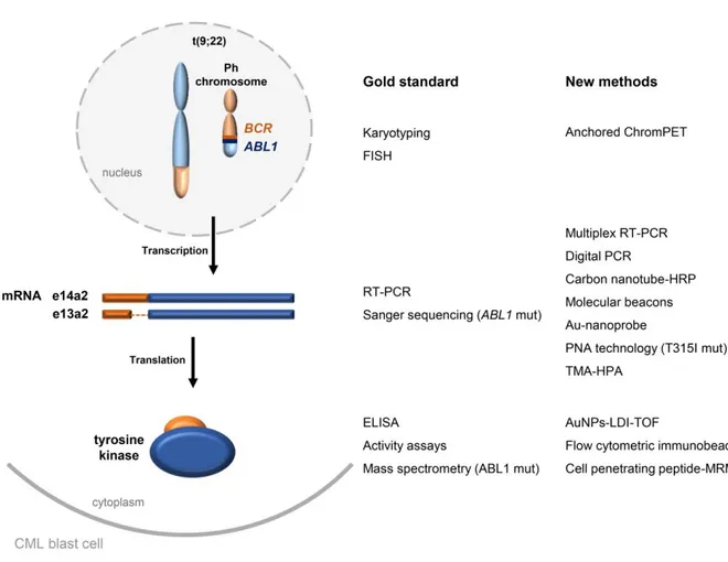
Figure 1.2 Conventional vs innovative methodologies for cytogenetic and molecular screening of CML,
via genomic DNA, mRNA and protein. The disease-causing translocation results in a shortened chromosome 9, the Philadelphia chromosome (Ph). The two most frequent BCR-ABL1 splicing variants
(accounting for 95 % of Ph+ cases) are represented, as well as the encoded enzyme. ChromPET, chromosomal paired-end tags; ELISA, enzyme-linked immunosorbent assay; FISH, fluorescence in situ
17 Table 1.4 Advantages and disadvantages of new approaches in CML molecular characterization.
Method Advantages Disadvantages Sensitivity Ref.
Anchored ChromPET
Analysis of multiple samples in a single assay Higher sensitivity than karyotyping, FISH or PCR
Does not detect splicing isoforms Complex implementation 10-4 (a) 114 Multiplex RT-nested PCR
Detection of multiple chromosomal
translocations/breakpoints in a single reaction Suitable for minimal residual disease
monitoring
Does not provide transcript quantification 10-4 (a)
115–118
Digital PCR Lower limit of detection than RT-PCR Less prone to erroneous quantification
(absolute copy number determination; no need for calibrators)
Expensive
Requires a rough estimate of target copy number before testing
10-6
(a) 119
Carbon nanotube-HRP
Does not require amplification step Cheaper than RT-PCR
May be extended to fluorescence/luminescent assays
Lack of standard protocol for high-purity carbon nanotubes in large scale applications
(b) 120
Molecular Beacon Does not require amplification step
May be used for sequence specific RT-PCR
Lower sensitivity than RT-PCR (b) 121,122
Au-nanoprobe colorimetric assay
Cheap, fast and single tube
Does not require amplification step Enables quantification of transcripts
Requires standardization protocol to be applied in clinical diagnostics
18
PNA-FISH Analyze clonal evolution Detection at single cell level
Less information than Sanger sequencing (probes designed for specific mutations)
2 % (c)
126
PNA directed PCR-clamping
Fast and cheap
Detection of mutations at single cell level Highly sensitive
Less information than Sanger sequencing (probes designed for specific mutation)
0.5 % (c)
126
TMA-HPA Single tube method Enables quantification
Reduces risk of carryover contamination and false positives
Complex design
Requires specialized equipment (luminometer)
10-4-10-5 (a)
127
AuNPs-LDI-TOF Does not require purification steps Low background signals
Time consuming
Requires internal standards
(b) 128
Immuno-beads Detection of all isoforms of chimeric protein Higher sensitivity than karyotyping or FISH. Does not require purification step
Lower sensitivity than PCR 10-3 (a)
30
Cell penetrating peptide-MRM
Allows determination of chimeric BCR-ABL1 activity
Requires concentration step (b) 129
ChromPET, chromosomal paired-end tags; FISH, fluorescence in situ hybridization; HRP, horseradish peroxidase; LDI-TOF,laser desorption ionization-time of flight; MRM,
multiple reaction monitoring; PNA, peptide nucleic acid; TMA-HPA, transcription-mediated amplification-hybridization protection assay
(a) Sensitivity values expressed as the number of BCR-ABL1 expressing cells in a background of BCR-ABL1 negative cells (e.g., successful detection of one K562 cell in a
background of 10 000 HL60 cells corresponds to a sensitivity value of 10-4). Sensitivity of gold standard methodologies: RT-nested PCR = 10-6; RTq-PCR = 10-5
(b) Information not available or different scoring units were used.
19 In myeloid neoplasms, conventional diagnostics consist of blood counts and biomarker evaluation on peripheral blood cells, complemented by morphologic examination of tumor tissues, which includes a collection of a substantial core biopsy of the bone marrow using a fine needle. Follow-up relies on peripheral blood assessment every 3 months for morphological, cytogenetic or molecular marker evaluation. Since there is a good correlation between blood and core biopsies in hematological disorders, a current debate focuses on whether bone marrow biopsies are still necessary.130,131
Liquid biopsies allow for non-invasive disease monitoring techniques and have been associated with circulating material originated from solid tumors, mainly circulation tumor cells (CTCs), circulating cell free DNA (cfDNA), microRNAs (miRNAs) and exosomes, that provide valuable information before and during treatment.132–134 However, this is even more so in liquid tumors, since a direct sample of tumor cells may be retrieved from a peripheral blood collection. As a result, besides the usual counting of leukocytes, hemoglobin, lymphocytes, and platelet levels, new prognostic markers should be addressed as far as disease outcome and clinical decision-making are concerned.135 For example, exosomes (vesicles of 30-100 nm that transport proteins, mRNAs and miRNAs) play an important role in cell communication, being able to induce some sort of transformation in receptor cells, including activation, proliferation, differentiation, or apoptosis 136,137. In CML-derived exosomes, a specific expression pattern of miRNAs was unveiled, which might be further explored for future use as disease biomarkers.138 In fact, higher concentrations of exosomes are found in sera of AML, CML and CLL patients with an abundant expression of surface proteins according to their cell of origin, which is rarely observed in exosomes of healthy individuals. Together, this supports the use of exosomes and miRNAs as leukemia biomarkers.
Therapeutics
Current treatment of CML patients has converted a fatal cancer into a manageable chronic disease. In fact, the 8-year survival rate of a CML patient is 87 % since 2001 and, since the advent of second-generation TKIs, the life expectancy of CML patients is now close to that observed for the general population.139,140 The majority of patients respond very well to chemotherapy, however it is still challenging for clinicians to know when it is safe to discontinue a successful treatment and why, in some patients, leukemia recurs.141 The recommendation is to continue medication permanently but this raises other problems, such as the development of resistance, non-compliance with treatment, fertility and pregnancy issues, and the economic burden of long-term medication. Hence, treatment cessation is currently one of the major discussion points regarding CML.106, 113,142
20
conjunction with the novel molecules targeting specific subtypes of CML causing aberrations, will enable personalization of CML management.
It has been reported that transfection of siRNA against BCR-ABL1 significantly reduced levels of
corresponding mRNA and oncoprotein, with induction of apoptotic cell death in human leukemia K562 cells.143 This approach was successfully applied using BCR-ABL1 siRNA in a female patient with recurrent Ph+ CML, resistant to IM (Y253F mutation) and to chemotherapy with AlloSCT.144
BCR-ABL1 mRNA levels decreased dramatically and led to the reduction of circulating CML cells without
side effects, suggesting that siRNA targeting of this gene has great potential for treatment of IM resistant CML.145 Kim et al developed a DNAzyme-based strategy to specifically target and cleave both the junction sequence and the site of the T315I point mutation that confers IM resistance. The designed molecules induced apoptosis and proliferation arrest in both wild-type and T315I-mutant CML cells, in a selective manner. Moreover, in vitro co-treatment with the DNAzyme targeting the T315I mutation
and IM resulted in enhanced inhibition of proliferation and induction of apoptosis in T315I leukemic cells as compared with IM alone, thereby abrogating IM resistance in CML cells. DNAzyme combined with IM treatment may be an alternative approach to overcome TKIs resistance in leukemic cells.146
Due to the genetic heterogeneity of BCR-ABL1 and sub-optimal response to chemotherapy by some