DOI: 10.1590/0004-282X20160120 ARTICLE
Higher positive identification of malignant
CSF cells using the cytocentrifuge than the
Suta chamber
A identificação de células neoplásicas no LCR foi maior com o uso da citocentrifuga do
que com câmara de Suta
Sérgio Monteiro de Almeida1,2, Indianara Rotta1,2, Arnaldo José de Conto1,2, Dario Antonelli Filho1,2, Carlos
Dabdoub Roda1,2, Edna Yoshiko Yamada1,2, Gisele M. B. Singer1,2
The most useful laboratory test for diagnosing neoplastic meningitis infiltration is cerebrospinal fluid (CSF) investigation. Accurate diagnosis is important for diagnostic, therapeutic, and prognostic consequences1.
Cerebrospinal fluid cytology is mandatory in all cases of known or strongly suspected malignancy. This is particu-larly true in cases of leukemia and lymphoma, in which the results of CSF cell counts and cytology are important fac-tors in determining and monitoring treatment2,3,4.
The two main methods of CSF cell concentration are cytocentrifugation and the gravitational facility sedi-mentation chamber. The literature provides no con-sensus on the optimal technique5,6, since both methods
have advantages and disadvantages. The aim of this pa-per was to compare two different methods of cell con-centration, cytosedimentation using a Suta chamber and cytocentrifugation.
1Universidade Federal do Paraná, Curitiba PR, Brasil;
2Faculdades Pequeno Príncipe, Instituto de Pesquisa Pelé Pequeno Príncipe, Curitiba PR, Brasil.
Correspondence: Sérgio Monteiro de Almeida; Hospital de Clínicas – UFPR, Seção de Virologia, Setor Análises Clínicas; Rua Padre Camargo, 280; 80060-240 Curitiba PR, Brasil; E-mail: [email protected]
Conflict of interest: There is no conlict of interest to declare.
Received 15 September 2015; Received in inal form 23 June 2016; Accepted 01 July 2016.
ABSTRACT
Objective: To deine how to best handle cerebrospinal luid (CSF) specimens to obtain the highest positivity rate for the diagnosis of malignancy, comparing two different methods of cell concentration, sedimentation and cytocentrifugation. Methods: A retrospective analysis of 411 CSF reports. Results: This is a descriptive comparative study. The positive identiication of malignant CSF cells was higher using the centrifuge than that using the Suta chamber (27.8% vs. 19.0%, respectively; p = 0.038). Centrifuge positively identiied higher numbers of malignant cells in samples with a normal concentration of white blood cells (WBCs) (< 5 cells/mm3) and with more than 200 cells/mm3, although this was not statistically signiicant. There was no lymphocyte loss using either method. Conclusions: Cytocentrifugation positively identiied a greater number of malignant cells in the CSF than cytosedimentation with the Suta chamber. However, there was no difference between the methods when the WBC counts were within the normal range.
Keywords: cerebrospinal luid; cytology, centrifugation, sedimentation.
RESUMO
Objetivo: Deinir qual a melhor forma de concentrar amostras de LCR para obter maior porcentagem de positividade para o diagnóstico de iniltração neoplásica. comparando dois métodos diferentes de concentração de células, sedimentação e citocentrifugação. Métodos:
Análise retrospectiva de 411 laudos de LCR. Resultados: Estudo comparativo descritivo. A identiicação de células neoplásicas no LCR foi mais elevada quando usada a citocentrífuga do que a câmara de Suta (28% vs 19,0%, respectivamente; p = 0,038). Centrifugação identiicou maior número de células neoplásicas em amostras com concentração de células < 5 células/mm3 e superior a 200 células/mm3, embora não signiicativo. Não houve perda de linfócitos usando qualquer um dos métodos. Conclusões: A citocentrifugação identiicou um número maior de células malignas no LCR do que a sedimentação com a câmara de Suta. No entanto, não houve diferença entre os métodos quando as contagens de leucócitos estavam dentro do intervalo normal.
METHOD
his study was approved by the HC-UFPR review board. A retrospective, longitudinal study was conducted utilizing CSF laboratory results from the data iles of the clinical pa -thology laboratory of the General Hospital – Universidade Federal do Paraná (UFPR). Samples of CSF were obtained from two populations of patients: adults and children who were referred to the laboratory with clinical suspicion of ma-lignant CNS iniltration and patients who underwent pro -phylactic intrathecal chemotherapy. Samples were referred to the laboratory from hematology, bone marrow transplan-tation, neurology, and neurosurgery services. All CSF sam-ples were obtained by lumbar puncture between March 1995 and December 2000. he CSF total cell count was assessed using a Fuchs Rosenthal chamber. he CSF was analyzed within a maximum of 30 minutes after arriving in the CSF section, and samples were maintained in room temperature.
Methods for cell concentration
For the diferential cell count and detection of malig -nant cells, CSF samples were concentrated using either a Suta chamber (1995–1998; 220 CSF samples) or a cytocen -trifuge (Cytopro 7620 Cytocen-trifuge, Wescor) (1998–2000; 191 CSF samples).
Suta chamber
he CSF volume applied was adjusted according to the CSF total cell count, as described: 0.1 to 9 cells/mm3- 2.0 mL; 10 to
50 cells/mm3- 1.5 to 2.0 mL; 50–100 cells/mm3- 1.2 to 1.8 mL;
100–200 cells/mm3- 1.0 to 1.5 mL; 200–500 cells/mm3- 0.8 to
1.0 mL; 500–1,000 cells/mm3- 0.5 to 0.8 mL; > 2,000 cells/mm3- 0.2 to
0.3 mL. he time of sedimentation was around 20 to 30 minutes7,8.
Cytocentrifuge
One mL of CSF was centrifuged during two minutes in a reg-ular centrifuge at 2,500 rpm; for CSF samples with total cell count ≥ 1,000 cells/mm3, CSF was diluted 1/20 (in this study there were
no samples with CSF total cell count ≥ 1,000 cells/mm3). From
the sediment 100 µL was transferred to the cytocentrifuge, cen-tifugated for two minutes at 1,200 rpm.
In both methods the CSF samples were protein-enriched with albumin, and slides were stained by the May Grünwald-Giemsa technique and observed by two trained researchers.
Malignant cells characteristics
Malignant cells were deined by the presence of one or more of the following characteristics: large size and/or nu-clei; an increase in the nucleus/cytoplasm size index in fa -vor of the nucleus; multiple nuclei; great, prominent, or mul -tiple nucleoli; variation in the size and format of the cells and nuclei; mitosis in groups of cells; frequent atypical mitosis; and irregular nuclear edges, hyperchromasia, and irregular grouping of the nuclear chromatin.
To calculate the positive rate of identiication of malignant cells in the CSF, we considered the irst sample (positive or neg -ative) of each patient and subsequent positive samples for a to-tal of 411 samples. he categorical variables were compared using either the Chi-square test (x2) or Fisher’s exact test, and
the continuous variables were compared using the Student’s t-test. A p-value ≤ 0.05 was considered signiicant. he results are presented as the mean ± standard deviation (SD).
Demographic and CSF characteristics of the groups studied
During the period of the study, 411 CSF samples were collected from 330 patients with possible malignant CNS iniltration. Of these patients, 180 (54.4%) were male and 150 (45.6%) were female. he mean ± SD age was 15.7 ± 15.8 years, and the median age was nine years. he par -ticipants in the group with the CSF samples prepared by the Suta sedimentation chamber (n = 220) and the group with the CSF samples prepared by the cytocentrifuge (n = 191) were well-matched with respect to age, gender, and main in-dication for CSF neoplastic cell search as well as basic CSF cell and biochemistry characteristics. he median inter -quartile range of age was 8 (4–23) years and 7 (4–23) years in the Suta chamber and cytocentrifuge groups, respectively (p = 0.82). A total of 57% (n = 125) and 47% (n = 90) of the CSF samples in the Suta chamber and cytocentrifuge groups, respectively, were from male patients (p = 0.08). he medi -an interquartile r-ange of white blood cell (WBC) count was 1.0 (0.3–3.0) and 1.0 (0.3–3.2) in the Suta chamber and cyto -centrifuge groups, respectively (p = 0.52). he red blood cell count was 1.2 (0–16) and 1.3 (0–17) in the Suta chamber and cytocentrifuge groups, respectively (p = 0.06). he glucose level was 46 (46–67) and 59 (50–69) in the Suta chamber and cytocentrifuge groups, respectively (p = 0.22). he total pro -tein was 23 (17–39) and 21 (14–38) in the Suta chamber and cytocentrifuge groups, respectively (p = 0.94).
Indication for neoplastic cell search in the CSF
RESULTS
Impact of CSF concentration methods on the WBC differential count
The differential characteristics of the WBCs in the CSF of both groups are indicated in Table 1. There was no statistical difference in the percentage of lymphocytes and neutrophils, suggesting no small cell loss in the Suta chamber. The percentage of monocytes was higher in Suta chamber preparations.
Rate of neoplastic cell detection by each concentration method
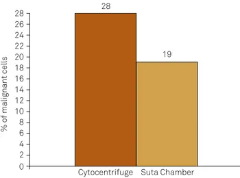
The cytocentrifuge detected malignancy in 27.8% (53/191) of the available samples. The Suta chamber de-tected malignancy in 19.0% (42/220) of the available sam -ples. The difference in detection rate was statistically significant (X² p = 0.038; OR = 1.6, 95%CI 1.0–2.6). There was 9% increase in the positive identification of neoplas -tic cells in the CSF using the cytocentrifuge (Figure 1). Although it was not statistically significant, there was an increased likelihood (1.6%) of identifying malignant CSF cells using the cytocentrifuge compared to the Suta cham-ber. There was no relationship between the positive iden-tification rate for a given method and the number of WBCs in the sample (Table 2).
Impact of the CSF WBC count on the rate of neoplastic cell detection in each cell concentration method
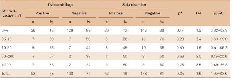
The majority of samples (74% and 76% in the Suta chamber and cytocentrifuge groups, respectively) had a normal number of WBCs in the CSF (p = 0.64 or = 1.12, 95%CI = 0.71–1.75) because the majority of samples (62%) in both groups were from patients with ALL. Patients with ALL receive prophylactic intrathecal che -motherapy; therefore, these samples were not necessari -ly from patients with CNS neoplastic involvement. There was no statistical difference between the two methods when analyzing CSF samples with a normal number of CSF WBCs (p = 0.17 or =1.5, 95%CI = 0.82–2.9; Table 2). The number of malignant CSF cells that were positively identified was higher in the samples with either a nor-mal range of WBCs (< 5 cells/mm3) or WBC count greater
than 200 cells/mm3, with a statistical trend observed in
the normal range (Figure 2, Table 2). If the WBC count was greater than 200 cells/mm3, then the percentage of
CSF neoplastic cells identified by the cytocentrifuge was 78% (7/9), whereas 50% (3/6) were identified by the Suta chamber. Thus, the probability of a clinical diagnosis of malignant CSF cells was 3.5-fold higher using the cyto-centrifuge than that using the Suta chamber, although this difference was not significant (Table 2).
Table 1. Cerebrospinal luid (CSF) white blood cells (WBCs) characteristics by cell concentration method.
n: the number of CSF samples in which the WBC type was identiied. The differential cell count was not performed in all samples. *Tcalculated (df) = Tcritical
Variable
Cytocentrifuge Suta chamber
Student’s t-test* P
n Mean + SD n Mean + SD
Lymphocytes (%) 44 63 + 27 42 56 + 30 1.05(84) = 1.98 > 0.05
Monocytes (%) 29 10 + 8.2 30 35 + 26 4.81(57) = 2.00 < 0.05
Neutrophils (%) 17 20 + 25 17 26 + 31 0.62(32) = 2.04 > 0.05
Table 2. Percentage of CSF malignant cells by CSF WBCs level.
CSF WBC (cells/mm3)
Cytocentrifuge Suta chamber
p* OR 95%CI
Positive Negative Positive Negative
n % n % n % n %
0–4 26 18 120 82 20 12 143 88 0.17 1.5 0.82–02.9
05-10 7 50 7 50 8 30 19 70 0.20 2.4 0.63–09.0
10-50 9 56 7 44 8 45 10 55 0.49 1.6 0.41–06.2
50–200 4 67 2 33 3 50 3 50 0.56 2.0 0.19–20.6
> 200 7 78 2 22 3 50 3 50 0.26 3.5 0.49–56.8
Total 53 28 138 72 42 19 178 81 0.04 1.6 1.00–02.6
Rate of positive identification of neoplastic cells in different neoplasm types by CSF concentration method
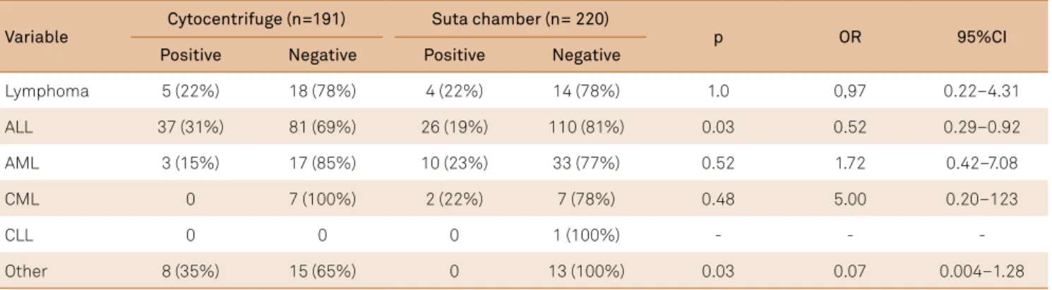
he main indication for neoplastic cell search in the CSF was ALL. Among these cases, the number of cells that were positively identiied as malignant was higher when the CSF
samples were prepared using cytocentrifuge (31%, n= 37) compared with when the samples were prepared using the Suta chamber (19%, n= 26). his diference was statistically signiicant (p = 0.03).
here was no diference in the number of cells that were positively identiied as malignant by both methods in lym -phoma cases. he detection of neoplastic cells in acute my -eloid leukemia and chronic my-eloid leukemia cases was higher when the CSF samples were prepared by the Suta sed-imentation chamber compared with the cytocentrifuge, but this diference was not signiicant (Table 3).
hese results suggest that the use of the cytocentrifuge increases the identiication of speciic types of malignant cells, such as those found in ALL. Among ALL cases, the probability of a clinical diagnosis of malignant CSF cells was 0.5 times higher using the cytocentrifuge compared with the Suta chamber to prepare the CSF samples.
DISCUSSION
he CSF cells must be concentrated before microscopic ex -amination for diferential WBC count or the identiication of
% of malignant cells
Cytocentrifuge 26
20
16
10
6
0 28
24
18
14
8
2 22
12
4
28
19
Suta Chamber
X² p = 0.04, OR = 1.6 (95%CI 1.0 to 2.6)
Figure 1. The percentage of positively identiied malignant
cells with respect to the cell concentration method used.
0-4 5-12 12-50 50-200 >200
WBC mm3 25
20
15
10
5
0 30
Cytocentrifuge
Suta N
Figure 2. The number of positively identiied malignant cells in the cerebrospinal luid (N) plotted against the number of white
malignant cells owing to their small number. Cellular components in the CSF can be concentrated by sedimentation, membrane il -tration, or centrifugation in a standard laboratory centrifuge or a cytocentrifuge. Although several studies have investigated9,10,11
which concentration method (sedimentation vs. cytocentrifuga-tion) is better for preparing CSF specimens for cytology diagno-sis mainly in suspected malignancy cases, two recent publications stated that the best method is not yet well established7,8.
In this study, we compared two diferent CSF prepara -tion methods that are widely used in routine CSF laborato-ries to diagnose malignant CNS iniltration. Although the methods were used at two separate periods in time, the groups were well-matched with respect to age, gender, neo-plastic cell search indication, and basic CSF cytology and biochemistry characteristics.
he rate of diagnosed malignant cells was higher when the CSF sample was concentrated by the cytocentrifuge than by the Suta chamber. In this study, the main indication for neoplastic cell search in the CSF was ALL, and the second most common indication was lymphoma. he other types of malignant diseases occurred at a low frequency, which limit-ed a deinitive conclusion. In contrast to acute leukemias, the involvement of the CNS in chronic leukemias such as chronic lymphocytic leukemia and chronic myeloid leukemia is not common. herefore, we cannot conclude which method is better for the identiication of speciic types of malignancy.
No diference in the lymphocytes and neutrophils was found between the two methods, which suggests that the CSF concentration method does not impact the WBC diferential count. he percentage of monocytes was lower among the CSF samples concentrated by the cytocentrifuge, which suggests a potential loss of these cells by the cytocentrifuge method. As an alternative interpretation, the lower number of monocytes de-tected using the cytocentrifuge may relect the relative enrich -ment of monocytes using the sedi-mentation method due to the loss of other cell types. Bots et al. concluded that sedimen-tation yielded a greater proportion of monocytes and eosino-phils12. Although monocytes are larger cells than lymphocytes
(a small lymphocyte is 8 to 10 µm and monocytes are 12 to
20 µm in diameter), the diference is negligible13. However, the
percentage of small cells that were lost was not signiicantly diferent between the methods, conirming Dyken’s inding14.
Concerning the impact of CSF WBC count on the rates of neoplastic cells detection by cell concentration methods, there was no diference in the rate of neoplastic cell detec -tion between the two methods with respect to the CSF WBC count. However, the percentage of positively identiied ma -lignant cells was higher by the cytocentrifuge method when the WBC count was normal and when it was greater than 200 cells/mm3.he majority of samples in this series had a
CSF WBC count within the normal range (WBC < 5 cells mm3).
Cerebrospinal luid samples with a normal WBC count range are regularly seen in cases with a suspected malignant CNS iniltration, particularly in cases of leukemias or lymphomas.
he most useful laboratory test and gold standard for di -agnosing neoplastic meningitis iniltration is CSF investiga -tion1. However, CSF cytology is highly disease speciic, with
a diagnostic sensitivity up to 45% when the patient presents with negative cytology on initial examination1,15. Sensitivity
increases to 80% with a second CSF examination, but can -not be enhanced signiicantly by further lumbar punctures16.
he preparation of the cell sediment is one of the more dif -icult technical components of the cell enrichment process, because it is subject to a number of potential disturbances. he literature is controversial regarding which technique is opti-mal for cellular identiication5. he cell sedimentation method
is considered the best method for the preservation of cellular structure and is superior, in this respect, to all methods involv-ing iltration or centrifugation7,9,17. Sedimentation also permits
the best cytological diferentiation of the CSF cells11.
Both sedimentation and cytocentrifugation have advan-tages and disadvanadvan-tages. Sedimentation methods can result in a 50% to 80% cell loss12,18, and it is not known whether one cell
type is afected more than another in this loss. While some stud -ies have shown that lymphocytes are disproportionately lost with the sedimentation method17, the morphology of the cells is
better preserved with this method. However, the centrifugation method has deleterious efects on more fragile cells14.
Table 3. Number of malignant cells detected in different neoplasm types by concentration method.
ALL: acute lymphocytic leukemia; AML: acute myeloid leukemia; CML: chronic myeloid leukemia; CLL: chronic lymphocytic leukemia.
Variable
Cytocentrifuge (n=191) Suta chamber (n= 220)
p OR 95%CI
Positive Negative Positive Negative
Lymphoma 5 (22%) 18 (78%) 4 (22%) 14 (78%) 1.0 0,97 0.22–4.31
ALL 37 (31%) 81 (69%) 26 (19%) 110 (81%) 0.03 0.52 0.29–0.92
AML 3 (15%) 17 (85%) 10 (23%) 33 (77%) 0.52 1.72 0.42–7.08
CML 0 7 (100%) 2 (22%) 7 (78%) 0.48 5.00 0.20–123
CLL 0 0 0 1 (100%) - - -
Centrifugation methods are subject to error due to the distortion and fragmentation of cells. Large numbers of leu-kocytes are destroyed by cytocentrifugation, limiting the ability to accurately identify malignant cells, because cellu-lar morphology is altered extensively. In this study, the main indication for neoplastic cell search was ALL. he results of our study suggest that the cytocentrifuge was a better con-centration method for CSF samples with this indication. he cytocentrifuge also is preferred in studying meninge -al leukemia because this method -allows for a better corre-lation between the cells found in the CSF and peripheral blood19,20. It should be noted that in the present study, the
preservation of cells was measured by the observation of cellular detail but was not reported herein.
he CSF volume needed for analysis in the Suta sedimen -tation chamber is between 0.5 and 1.5 mL and the prepara-tion time is 30 to 40 minutes. In contrast, cytocentrifugaprepara-tion requires only 200 µL of CSF for analysis, and the preparation time is four minutes. he volume of cells concentrated by cy -tocentrifugation is smaller than the volume concentrated by the Suta chamber, which decreases the time required to an-alyze the slide for malignant cells. In this study, we did not evaluate the time required to analyze the slides. With the in-creased number of WBCs, a smaller volume of CSF is neces-sary. An advantage of both methods is that each allows for hematological staining after concentrating the cells.
In addition to the concentration method, several pre-analytical steps are important to enhance the chances of tumor cell identiication. For example, large volumes of CSF samples should be used, and samples should be carried to the laboratory immediately after the lumbar puncture in or-der to minimize cell distortion or lysis17. It is preferable for
slides to be prepared within 30 minutes of CSF collection, as 1/3 of the cells, mainly neutrophils as well as malignant cells that have altered stability, disintegrate within 24 hours.
he rate of positive identiication of malignant cells in the CSF varies in the literature and is assumed to depend on sev-eral factors, including the method used to concentrate the CSF21. Other factors that inluence the ability to detect ma
-lignant CSF cells include the type of neoplasm, anatomic lo-cation of the neoplasm, presence of meningeal involvement and meningeal extension, and number of malignant cells in the CSF21,22. Primary cerebral tumors that exfoliate cells to
the CSF were all located adjacent to the ventricle. In contrast, cells from tumors deeply localized in cerebral parenchyma are more diicult to detect in the CSF23,24.
Currently, three methodologies prevail to generate data for counting and diferentiating cells in body luids. hese methods are manual microscopy, automated low cytometry, and automated impedance technology. Traditional manual microscopy is the gold standard. Automated cell analyzers have been able to generate automated counts of cells present in CSF samples in recent years, although most cannot pro-vide reliable counts of the low cell levels usually present in the CSF, including the normal WBC counts that are frequent in CNS malignant iniltration8,25. he disadvantages of this
method include high imprecision in low ranges (depending on the method) and interfering factors, which reinforces the importance of traditional methods and the necessity of es-tablishing the best concentration method3.
Flow cytometry used in combination with conventional cytology can lead to a signiicant increase in the detection rate of leptomeningeal iniltration of malignant cells and is there -fore of value in detecting these diseases4,7,26,27. he potential ap
-plication of low cytometry to the CSF study is more limited, however, because cell concentration is low in normal CSF, and the WBC concentration in particular is generally no more than 1/1000 that of blood. Ancillary techniques such as low cytom -etry are of increased importance but their use is restricted to speciic or large laboratories. Concentration methods remain important, and to deine how to best handle CSF specimens to obtain the highest sensitivity and speciicity for the diagnosis of malignancy remains an important issue.
he strength of this study is the substantial number of cases that were analyzed. he main limitation of this study is the lack of an optimal gold standard. Although only one method was used for each sample, the groups were compara-ble over time in several characteristics already reported. his is a descriptive comparative study, due to the small volume of CSF samples sent to the laboratory and because of this, there was no split of the samples for analysis. his could be a design bias. he sample size for non-hematologic cases is small. Furthermore, the preservation of cellular morphology was not reported in this study, and this is an important fea-ture when identifying malignant cells. Other issues not stud-ied were the analysis of diferences in the objective assess -ment of intra and inter-observer slides.
In conclusion, the positive rate of identiication of ma -lignant cells in CSF was slightly higher when the CSF sam-ple was concentrated by cytocentrifuge than Suta chamber. If the number of CSF WBCs was within the normal range, there was no diference between the methods.
References
1. Chamberlain MC: Lymphomatous meningitis in primary central nervous system lymphoma. Neurosurg Focus. 2006;21(5):E6. doi:10.3171/foc.2006.21.5.7
2. MacKenzie JM. Malignant meningitis: a rational approach to cerebrospinal luid cytology. J Clin Pathol. 1996;49(6):497-9. doi:10.1136/jcp.49.6.497
3. Fleming C, Russcher H, Lindemans J, Jonge R. Clinical relevance and contemporary methods for counting blood cells in body luids suspected of inlammatory disease. Clin Chem Lab Med. 2015;53(11):1689-706.
4. Broussalis E, Hutterer M, Oppermann K, Wipler P, Pilz G, Harrer A et al. Isolated leptomeningeal iniltration of a primary CNS B-cell lymphoma diagnosed by low cytometry and conirmed by necropsy. Acta Neurol Scand. 2012;126(3):e11-6. doi:10.1111/j.1600-0404.2011.01630.x
5. Whitmore EL, Hochberg F, Wolfson L, Royalty J, Taft PD. Quantitative cytocentrifugation in the evaluation of cerebrospinal luid. Acta Cytol. 1982;26(6):847-50.
6. Bigner SH, Johnston WW. The cytopathology of cerebrospinal luid. II. Metastatic cancer, meningeal carcinomatosis and primary central nervous system neoplasms. Acta Cytol. 1981;25(5):461-79.
7. Kluge H, Roskos M, Kluska MM. Cell preparation (sedimentation) and staining. In: Kluge H, Wieczorek V, Linke E, Zimmermann K, Isenmann S, Witte OW. Atlas of CSF cytology. Stuttgart: Thieme; 2007. p. 8-9.
8. Deisenhammer F, Sellebjerg F, Teunissen CE. Cerebrospinal luid in clinical neurology. New York: Springer; 2015.
9. Lemitz R, Kleine TO. Liquorzytologie: Ausbeute, verteilung und Darstellung von Leukozyten bei drei sedimenttionsverfahren im Vergleich zu drei zytozentrifugen-Modiikationen. Lab Med. 1994;18:91-9.
10. Seyfert S. An improved sedimentation technique for the cytologic preparation of cerebrospinal luid cells. Acta Neurol Scand. 1993;88(3):217-20. doi:10.1111/j.1600-0404.1993.tb04220.x
11. Wiethölter H, Oehmichen M, Sayer H. [Qualitative CSF cell diagnosis: methods and conclusiveness (author’s transl]. MMW Munch Med Wochenschr. 1979;121(18):631-66. German.
12. Bots GT, Went LM, Schaberg A. Results of a sedimentation technique for cytology of cerebrospinal luid. Acta Cytol. 1964;8(3):234-41.
13. Abbas K A, Lichtman AH, Pober JS. Cellular and molecular immunology. 7th ed. Philadelphia: WB Saunders; 2011.
14. Dyken PR. Cerebrospinal fuid cytology: practical clinical usefulness. Neurology. 1975;25(3):210-7. doi:10.1212/WNL.25.3.210
15. Glass JP, Melamed M, Chernik NL, Posner JB. Malignant cells in cerebrospinal luid (CSF): the meaning of a positive CSF cytology. Neurology. 1979;29(10):1369-75. doi:10.1212/WNL.29.10.1369
16. Wasserstrom WR, Glass JP, Posner JB: Diagnosis and treatment of leptomeningeal metastases from solid tumors: experience with 90 patients. Cancer. 1982;49(4):759-72. doi:10.1002/1097-0142 (19820215)49:4<759::AID-CNCR2820490427>3.0.CO;2-7
17. Oehmichen M. Cerebrospinal luid cytology: an introduction and atlas. Philadelphia: WB Saunders; 1976.
18. Woodruff KH. Cerebrospinal luid cytomorphology using cytocentrifugation. Am J Clin Pathol. 1973;60(5):621-7. doi:10.1093/ajcp/60.5.621
19. Davey DD, Foucar K, Giller R. Millipore ilter vs. cytocentrifuge for detection of childhood central nervous system leukemia. Arch Pathol Lab Med. 1986;110(8):705-8.
20. Ducos R, Donoso J, Weickhardt U, Vietti TJ. Sedimentation versus cytocentrifugation in the cytologic study of craniospinal luid. Cancer. 1979;43(4):1479-82. doi:10.1002/1097-0142 (197904)43:4<1479::AID-CNCR2820430439>3.0.CO;2-S
21. Glantz MJ, Cole BF, Glantz LK, Cobb J, Mills P, Lekos A et al. Cerebrospinal luid cytology in patients with cancer: minimizing false-negative results. Cancer. 1998;82(4):733-9. doi:10.1002/ (SICI)1097-0142(19980215)82:4<733::AID-CNCR17>3.0.CO;2-Z
22. Oostenbrugge RJ, Twijnstra A. Presenting features and value of diagnostic procedures in leptomeningeal metastases. Neurology. 1999;53(2):382-85. doi:10.1212/WNL.53.2.382
23. Watson CW, Hajdu SI. Cytology of primary neoplasm of the central nervous system. Acta Cytol. 1977;21(1):40-7.
24. Sá MJ, Vaz R, Cruz C. Cerebrospinal luid cytomorphologic indings in 41 intracranial tumors: a retrospective review. Arq Neuropsiquiatr. 1995;53(2):218-26. doi:10.1590/S0004-282X1995000200006
25. Liang X, Chen J, Xiao X, Yu Y, Li W, Zhang Z. Automated cell analysis of cerebrospinal luid with XE-5000. Clin Lab. 2014;60(11):1785-93.
26. Bromberg JE, Breems DA, Kraan J, Bikker G, Holt B, Smitt PS et al. CSF low cytometry greatly improves diagnostic accuracy in CNS hematologic malignancies. Neurology. 2007;68(20):1674-9. doi:10.1212/01.wnl.0000261909.28915.83