Mariana Antunes Coutinho
Licenciatura em Bioquímica
Development and Experimentation of
New Tools for Bioassays with Red
Blood Cells
Dissertação para obtenção do Grau de Mestre em
Biotecnologia
Orientador: Dr. Abel Oliva, Doutor, ITQB-UNL
Co-orientador: Dr. Hugo Águas, Doutor, FCT-UNL
Março 2016
Júri:Presidente: Prof. Doutora Cecília Roque, FCT-UNL
Arguentes: Prof. Doutor Ricardo Franco, FCT-UNL
Development and Experimentation of New Tools for Bioassays with Red Blood Cells:
Copyright © Mariana Antunes Coutinho, Faculdade de Ciências e Tecnologia, Universidade Nova de Lisboa.
Science never solves a problem without creating ten more.
Acknowledgments
This thesis would not have been possible without the guidance, support and help of certain people to whom I wish to express my heartfelt thanks, in particular:To Professor Abel Oliva, my supervisor, for allowing me to be part of this project, and for all his concern, patience and availability shown in the course of it.
To Professor Hugo Águas, my co-supervisor, for the opportunity, support and kind expression of sympathy.
To the institutions which provided me all the resources required for caring out this work, Instituto de Tecnologia Química e Biológica (ITQB) e CENIMAT.
To the entire Biomolecular Diagnostics group, who received me so well and provided me with an excellent work environment: to Carmo, my “second mother”, always so helpful, patient and worried. For all the personal and professional advices and all the transmitted knowledge which made the difference on my work. To Rita, “my partner in crime”, for the friendship, permanent availability and help, anytime without exception. To Mariana, for the fellowship and true friendship which has arisen and remained forever. To Catarina, which turned possible to conduct all the microfluidic assays. For all her dedication, commitment and availability to educate me with so much care. To Sofia, for all her attention and concern. To Ana Pepe and Kamila for the good moments of laughs. Finally, to the trainees, Antonio Claudio, Miguel, Nuno, Carolina and Patricia, for their sympathy and good environment provided at the laboratory.
To Mário, from Molecular Thermodynamics Laboratory, for his tireless help, permanent concern and helpful advices. To my “more than colleagues” from 4th floor of ITQB, Isabel, Anabela and Joana for their good willing and for making me smile every day. To Tiago, for his encouragement and support words and for distracting me when I needed.
To my best friends, for being the really best friends of the world: to Sara, Inês, Madalena, for being the other part of myself, not only during this phase, but for the last 11 years of my life. For the support and unconditional friendship. For making me always believe on my own skills and competences. Part of my success I owe to you. To my other but not less important friends, who have accompanied me not only on this but in all journeys throughout my life, Sara Silva, Mafalda, Ana, and to my colleagues and friends from the Faculty during the last six years.
Lastly, and most of all, I would like to gratefully thank the tireless and invaluable support of all my family: to my parents, which with so much sacrifice have made this dream come true, for suffering with my concerns and for rejoicing with my accomplishments.
Abstract
The aim of this work was to develop new tools for handling and studying of erythrocytes. In this context, biological nanoprobes were developed, combining Quantum Dots nanoparticles with specific antibodies to detect proteins in erythrocytes. Also, a microfluidic platform was designed and constructed to evaluate the cytotoxic effect in single-cell analysis of used QDs.To determine the experimental conditions for bioconjugation, preliminary studies were performed to analyse the electrophoretic behaviour of QDs and their pH stability. It was demonstrated that the mobility depends on the surface charge of QDs and it is not affected in pH range between 5 and 8.
To evaluate the cytotoxic effect of non-conjugated nanoparticles, QDs functionalized with three different groups were used: PDDA, PEG and COOH, at concentrations of 10, 50 and 100nM. The results showed that the interaction between QDs and cells induces a general oxidative stress, which increases with the concentration of QDs.
The conjugation reaction between COOH-QDs and monoclonal antibodies against antigenic protein AMA1 and transmembrane Glycophorin A protein was performed by EDC/NHS chemistry. The QDs-Ab complexes were characterized by agarose gel, comparing their molecular weight and electrophoretic mobility with non-conjugated nanoparticles. Using SDS-PAGE and Immunoblotting techniques, the samples were analysed in order to evaluate their ligation properties, as well as their biological activity. The final bioconjugates were then biological tested, namely in detection of GPA protein in erythrocytes and antigenic AMA1 protein in infected B.ovis erythrocytes, through the immunofluorescence assays on slide and in solution. Comparing the results obtained with QDs and the traditional assays with FITC conjugated antibodies, QDs-Ab complexes appear to be more resistant during large periods of excitation.
Resumo
O objetivo deste trabalho consistiu no desenvolvimento de novas ferramentas para o estudo e manipulação de eritrócitos. Neste contexto, foram criadas nanosondas biológicas através da conjugação de nanopartículas (Quantum Dots) com anticorpos específicos para a deteção de proteínas presentes nos eritrócitos. Foi também desenhada e construída uma plataforma de microfluídica para poder avaliar o efeito citotóxico dos QDs usados na bioconjugação.Para determinar as condições experimentais para a conjugação, foram elaborados estudos preliminares para analisar o comportamento electroforético dos QDs e a sua estabilidade em valores de pH diferentes. Foi demonstrado que a mobilidade depende da carga da superfície dos QDs e que não é afetada numa gama de pH entre 5 e 8.
Para avaliar o efeito citotóxico das nanopartículas não conjugadas, foram utilizados QDs com três funcionalizações diferentes: PDDA, PEG e COOH a 10, 50 e 100nM. Os resultados mostram que a interação dos QDs com as células induz um stress oxidativo generalizado, que aumenta com a concentração dos mesmos.
A reação de conjugação entre COOH-QDs e anticorpos monoclonais contra a proteína antigénica AMA1e contra a proteína transmembranar Glicoforina A foi realizada usando a via química EDC/NHS. Os complexos QDs-Ab foram posteriormente caracterizados através de géis de agarose, comparando o seu peso molecular e a sua mobilidade electroforética com nanopartículas não conjugadas. Através das técnicas SDS-PAGE e Immunoblotting, as amostras foram analisadas de forma a avaliar as propriedades de ligação, assim como a sua atividade biológica.
Os bioconjugados desenvolvidos foram depois testados a nível biológico, nomeadamente na deteção das proteínas GPA em eritrócitos e AMA1 em eritrócitos ovinos infetados com B.ovis, através de ensaios de imunofluorescência em lâmina e em solução. Comparando os resultados obtidos com QDs e os ensaios tradicionais usando anticorpos secundários conjugados com FITC, os complexos QDs-Ab aparentam ser mais resistentes em períodos de excitação maiores.
List of Abbreviations
Ab antibody
AMA-1 apical membrane antigen 1
BSA bovine serum albumin
COOH carboxylic acid
DAPI 4’, 6-diamidino-2-phenylindole dihydrochloride
EDC 1-ethyl-3-(3-dimethylaminopropyl)carbodiiamide
Fc fragment crystallisable
GPA glycophorin A
Hgb hemoglobin
H2DCFDA 2’, 7’-dichlorodihydrofluorescein diacetate
IgG Immunoglobulin G
IHC Immunohistochemistry
LEDs light emitting devices
LOC Lab-On-a-Chip
NaP sodium phosphate buffer
NHS N-hydroxysuccinimide
PDDA polydiallyldimethylammonium Chloride
PDMS polydimethylsiloxane
PEG polyethyleneglycol
PGMEA propylene glycol methyl ether acetate
PVDF polyvinylidene fluoride
QDs Quantum Dots
ROS reactive oxygen species
r.t. room temperature
Si silicon
SMCC 4-(N-maleimidomethyl) cyclohexanecarboxylic acid N-hydroxysuccinimide ester
Sulfo-SMCC 4-(N-maleimidomethyl) cyclohexane-1-carboxylic acid 3-sulfo-N-hydroxysuccinimide ester sodium
TBE tris borate EDTA
TBS tris buffered saline
TES N-[tris(hydroxymethyl)methyl]- 2-aminoethanesulfonic acid, 2-[(2-hydroxy-1, 1-bis(hydroxymethyl)ethyl)amino]ethanesulfonic acid
TP Transmembrane Protein
TTBS tris buffered saline with Tween
VyMs Veja y Martinez buffer
List of Contents
Acknowledgments... vii
Abstract………... ix
Resumo………..………... xi
List of Abbreviations………...……… xiii
List of Figures……….. xvii
List of Tables………..…………. xix
Chapter 1 | Introduction
1.1. Motivation………...……….. 11.2. Cell Analysis……….……… 2
1.2.1. Red Blood Cells………...………... 3
1.3. Quantum Dots………...……... 4
1.3.1. Properties………...……. 5
1.3.2. Biocompatibility, Functionalization and Bioconjugation………...……. 7
1.3.3. Applications………...…... 9
1.3.4. Cytotoxicity………...…… 10
1.4. Intra-erythrocytic Parasite Infections………..…… 11
1.4.1. Babesia ovis……….. 12
1.5. Membrane Proteins………... 14
1.5.1. Glycophorin A………... 15
1.6. Nanotoxicology………... 17
1.6.1. Reactive Oxygen Species………..…… 17
1.7. Microfluidic………...….. 18
1.7.1. Blood-on-a-chip……….….. 19
1.7.2. Microfabrication……….... 20
Chapter 2 | Materials and Methods
2.1. Blood collection………...…………..… 232.1.1. Uninfected ovine RBCs for subculturing………..…. 23
2.1.2. Human blood……….… 23
2.2. Quantum Dots………...………… 23
2.2.1. Characterization Electrophoretic Mobility and pH stability………...……….. 23 2.2.2. Bioconjugation of Antibodies to QDs……….……….... 24
2.2.2.1. Characterization Conjugated QDs……..……….. 25
- Agarose Gel Electrophoresis………...………...……… 25
- Western Blotting………...………. 25
2.3. Babesia ovis infected culture………....………….. 26
2.3.1. In vitro cultivation of Babesia ovis... 26
2.3.2. Immunofluorescence Assays……….………..……….. 28
- Indirect Immunofluorescence Assay………..……...……. 28
- Direct Immunofluorescence Assay using QDs-Ab conjugates……….. 29
2.4. Detection of Glycophorin A transmembrane Protein……….... 30
- Indirect Immunofluorescence Assay………..…… 30
- Direct Immunofluorescence Assay using QDs-Ab conjugates………... 30
2.5. Cytotoxic Assay: Oxidative Stress………..… 30
2.6. Microfluidic Platform……….… 30
2.6.1. Assembly for Biological Experiments with Microfluidic chips………..………... 32
Chapter 3 | Results and Discussion
3.1. Characterization of Quantum Dots………...…….……. 353.1.1. Electrophoretic Mobility and pH stability……… 35
3.2. Characterization of QDs-Ab conjugates………. 37
3.2.1. Analysis of Bioconjugation Protocol by Agarose Gel Electrophoresis……….……. 39
3.2.2. Characterization of Ab Conjugates to QDs by SDS-PAGE Electrophoresis…………...….... 40
3.2.3. Evaluation of immunogenicity of Ab-QDs complexes………...…….. 42
3.3. Detection of AMA1 Antigenic Protein in Infected Babesia ovis Erythrocytes………...…… 45
3.3.1. Babesia ovis in vitro culture………. 45
3.3.2. Detection of AMA1 Antigenic Protein………. 48
3.4. Detection of GPA Transmembrane Protein in Erythrocytes……….………... 51
3.5. Evaluation of ROS formation………...………… 53
3.5.1. Evaluation of ROS production on slide……….. 53
3.5.2. Design and Performance of Microfluidic chip in Oxidative Stress Assays…..………. 60
Conclusions and Future Perspectives
………
.
…….…...……. 67
List of Figures
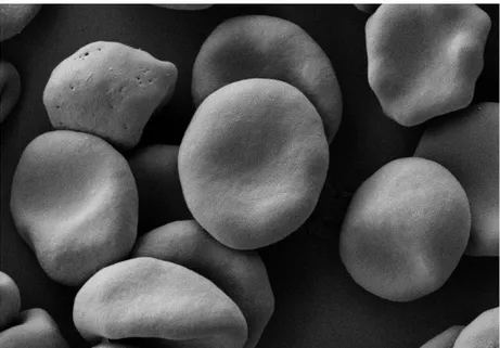
Figure 1.1 | Red blood cells by Scanning Electron Microscope………...……..… 1
Figure 1.2 | The components of blood……….………...… 4
Figure 1.3 | The structure of a typical CdSe/ZnS QD………...……… 5
Figure 1.4 | The QD size proportionally affects its emission wavelength……….. 6
Figure 1.5 | Absorption and emission spectra of four CdSe/ZnS QD examples……….. 6
Figure 1.6 | Many antibodies, proteins and other target or affinity molecules can be conjugated to QDs for biological applications……….… 7
Figure 1.7 | Structure of an IgG molecule………...………... 8
Figure 1.8 | General structures of five different classes of antibodies………...………… 8
Figure 1.9 | Schematic diagram showing a method for QD-antibody (QD-Ab) bioconjugation…………. 9
Figure 1.10 | Some applications of quantum dots as multimodal contrast agents in bioimaging……… 10
Figure 1.11 | Schematic representation of erythrocyte invasion by Babesia parasites……….… 13
Figure 1.12 | Schematic representation of a bovine RBCS with Babesia bovis parasite………..… 14
Figure 1.13 | Red blood cells interactions……….… 16
Figure 1.14 | Representative scheme of ROS generation through the reduction of molecular oxygen………...… 18
Figure 1.15 | Representation of a microfluidic device……….……… 19
Figure 1.16 | Representative scheme of PDMS microfludics fabrication……….… 21
Figure 1.17 | Representative scheme of photolithography and soft-Lithography process…………...…. 22
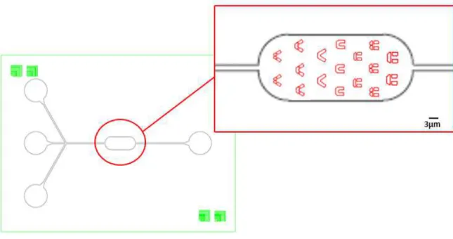
Figure 2.1 | Schematic diagram of the chip top view used for RBC trapping……….. 31
Figure 2.2 – Display of the complete assembly setup used during the microfluidic assays………….… 32
Figure 3.1 – Comparison of Electrophoretic Mobility and pH stability of three different QDs…...………...………... 36
Figure 3.2 – Conjugation of Antibodies to Carboxy-QDs using EDC or EDC/NHS………...…..….. 38
Figure 3.3 - Analysis of bioconjugation protocol by agarose gel electrophoresis…..………...….… 39
Figure 3.4 - Antibody reduction with DTT ………..……….. 40
Figure 3.5 - Separation of Ab-QDs complexes into fragments by SDS-PAGE electrophoresis……….. 41
Figure 3.6 - Separation and functional activity of Ab-QDs complexes……….... 43
Figure 3.7 – Visualisation of western membrane under UV-light…….……….………… 44
Figure 3.8 - Identification parasites in B.ovis infected cell of in vitro culture suspensions………... 46
Figure 3.9 - Schematic representation of direct and indirect immunofluorescence assay………...….... 49
Figure 3.10 - Immunofluorescence assay using anti-AMA1-QDs conjugates………...………….… 50
Figure 3.11 – Positive control by Indirect Immunofluorescence Assay.………...………... 50
Figure 3.12 – Detection of Glycophorin A transmembrane protein in human erythrocytes……….. 52
Figure 3.13 – Immunofluorescence assay using anti-GPA-QDs conjugates……….. 52
Figure 3.15 - Determination of cellular oxidative stress in erythrocytes cells incubated with
PDDA-QDs……… 55
Figure 3.16 – Determination of cellular oxidative stress in erythrocytes cells incubated with PEG-QDs ………...……… 56
Figure 3.17 - Determination of cellular oxidative stress in erythrocytes cells incubated with COOH-QDs ………..……….… 57
Figure 3.18 - Determination of cellular oxidative stress in erythrocytes cells induced by pH variation.………...…………..….…. 58
Figure 3.19 – Graphic representation of the fluorescence intensity of pictures from erythrocytes incubated with PEG and COOH-QDs and H2DCFDA.………...……...……….… 59
Figure 3.20 – Visualization of oxidative stress produced by trapped erythrocytes in microfluidic chip ……….………...………... 61
Figure 3.21 – Visualisation of a deformed trap in microfluidic chip……….……….… 62
Figure 3.22 - Different structures of designed traps…………..……….………. 62
Figure 3.23 – Visualisation of cell trapping ………...………...………... 63
Figure 3.24 – Sequence of images taken during the cell trapping.………...………... 63
Figure 3.25 – Example of microfluidic simulation performed by COMSOL software………. 64
List of Tables
Table 1.1 | RBC membrane glycophorins………...………. 15
Table 2.1 | Complete medium done with commercial serum……….…………. 27
Chapter 1 | Introduction
1.1.
Motivation
Blood is an important connective tissue and is vital to body’s survival due to its functions: transportation, regulation and protection. When some pathological condition occurs, blood is altered, affecting the entire health of the individual. Thus, monitoring blood can be an important source of clinical information related to the functioning of all tissues and organs of the body.
The tissue below (Figure 1.1) is composed by blood cells suspended in plasma. In particular, 96% of the total population of blood cells is made by red blood cells (RBCs) or erythrocytes (Figure 1.1). Erythrocytes are important for scientific and medical investigation given that some clinical conditions are directly associated with changes in their morphology or physiology (for example, anemia which is characterized by insufficient red cell mass, with values lower than 12.5g/dL). As a result, red blood cells are a convenient subject which should help investigators to know more about the blood disorders that may affect the individual’s health.
Figure 1.1 | Red blood cells by Scanning Electron Microscope: Red blood cells 1% suspension obtained from whole human blood. The cells were fixed with 0.075M of sodium calcodilate (CaCo), 2.25mM of MgCl2 and 1% of glutaraldehyde.
Thus, it is important to develop strategies and explore new approaches in order to deepen knowledge in this area.
assays that are cumbersome or even not possible with other techniques. Developing chips for analysis of individual cells is a challenge but can provide new options for biological and clinical research.
Also in the context of this work, and regarding the exploration of new approaches for cell studies, a type of nanoparticles has been experimentally tested as an alternative to traditional fluorophores, in particular, for cellular labeling. Quantum dots are fluorescent nanocrystals that have been attracting increased interest in bioresearch given their distinct characteristics. They present advantageous optical properties such as strong light absorbance, bright fluorescence and high photostability which make them ideal to be conjugated with biomolecules for application in life science research, diagnostic and therapeutics.
The aim of this work was focused on the development of new approaches to analyze blood. For this purpose, two tools were developed: the preparation of probes with Quantum Dots and specific antibodies to detect proteins related to pathological disorders and a microfluidic platform to capture and to analyze the oxidative stress produced by the interaction of QDs with cells. Within the aim of this work, the following goals were pursued:
To bioconjugate specific antibodies with Quantum Dots nanoparticles towards the identification of two transmembrane proteins:
- Glycophorin A, located on erythrocytes membrane
- Apical Membrane Antigen 1 which is expressed on the surface of erythrocytes invaded by Babesia ovis parasites.
To design, construct and optimize the setup of experimental microfluidic assays to evaluate the cytotoxic effect of Quantum dots in erythrocytes in oxidative stress, using a microfluidic platform through the single-cell approach.
1.2. Cell analysis
Cells are the essential units of biological processes (Lecault at al., 2012) and they are complex and dynamic entities. Cellular analysis is present in areas like life science, diagnostics and pharmaceutical industry (El-Ali et al., 2006; Dittrich et al., 2006). Part of research from basic cell biology and microbiology to applications in biotechnology is conducted through cell populations with high cell numbers (Schmid et al., 2010). However, interpretation of cell population data frequently implies the hypothesis that each cell in population is similar and sometimes, this is not true and can result in false interpratation (Yin and Marshall, 2012). For example, cell populations can be submitted to fast environmental changes, such as stress or perturbation and during such modifications, it can be observed that some cells deal better with the new conditions compared with other cells within the same population, even cells with identical functions usually respond differently.
correspond to the minimal functional unit of life. Nowadays, one of the main objectives is to comprehend the working mechanisms in this minimal unit (Schmid et al., 2010). These purpose can just be reached using highly sensitive methods with resolutions at the single cell and if possible, at subcellular level. So, to achieve a full single cell analysis, the development of high throughput systems competent of manipulation and analyzing individual cells is indispensable (Yin and Marshall, 2012). The challenge in single cell analysis is to reach this sensitivity precision, throughput and economy desired to detect and study complex subpopulation cells and during the same time (Lecault at al., 2012).
1.2.1. Red Blood Cells
Blood is often mentioned to as “liquid organ” and is a complex mixture of many types of components where each one has diverse properties. It is distributed via the vascular system throughout the entire body, making it indispensable for the existence of the organism. Blood contains innumerable information about the functioning of the body and it can be used to diagnose diseases.
Blood exhibits a wide variety of functions, such as supplying oxygen to tissues and nutrients, removing waste (such as carbon dioxide, urea and lactic acid), immunological functions (including circulation of white blood cells and detection of foreign material by antibodies), coagulation by the platelets (which is one part of the body’s self-repair mechanism), messenger functions (including the transport of hormones and the signaling of tissue damage), regulating body pH and temperature and finally, hydraulic functions (including the regulation of the colloidal osmotic pressure of blood).
cell mediated immunity) and natural killer cells (which are able to kill cells such as undetected virus cells or cells that have cancerous).
Figure 1.2 | The components of blood:It is composed by: erythrocytes (or red blood cells) that are cells without nucleus and they contain hemogloblin and carry oxygen. Leukocytes (or white blood cells) taht contain a nucleus and are present in lower number when compared with erythrocytes. They help to defend the body against infection and disease. At last, platelets which is
the component of blood responsible for initiating blood clotting. Like erythrocytes, they haven’t a nucleus (Adapted from:
https://www.urmc.rochester.edu).
Current methods for blood testing need large sample volume to be executed (3-5mL). A microfluidic approach is an attractive alternative for blood analysis due to its miniaturization benefits such as reduced reagent consumption, reduced sample requirement and decreased analysis time. Currently, several microfluidic systems are being developed to overtake a diversity of clinical problems for example, the detection of pathogens in vivo through the development of integrated microfluidic devices. These microfluidic devices are suitable candidates for point of care diagnostics due to small size and low cost production.
1.3. Quantum Dots
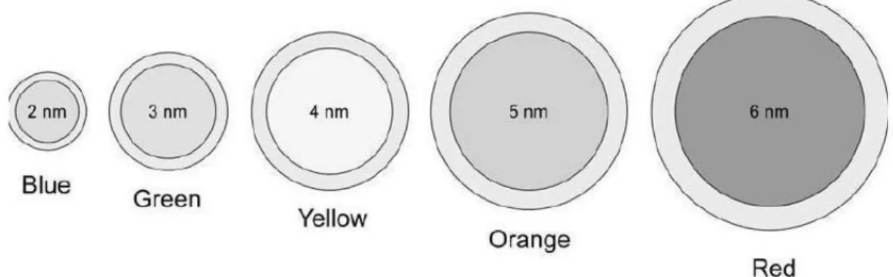
Quantum Dots are nanoparticles mainly made of a core composed by a semiconductor metal in a spherical crystalline form and this core is capped with a shell consisting of another semiconductor metal that has a larger spectral band gap (Figure 1.3) (Azzazy et al., 2007; Hermanson, 2008). QDs cores are usually composed by elements from groups II and VI (e.g. CdSe, CdTe, CdS and ZnSe) or groups III and V (e.g. InP and InAs). The commonly used QDs for biological applications are made of CdSe cores coated with a layer of ZnS because their chemistry is suitable for attaching biomolecules. The ZnS protects the core surface from oxidation, avoid leaching of the CdSe into the solution, promoting the minimal interaction with the surrounding environment, helping to their photostability (Bruchez, 2005) and finally, improves the photoluminescence yield (Kortan et al., 1990; Mews et al., 1994). The size of this core/shell structure is about 2-10 nm (usually less than 10 nm) but with functionalized amphiphilic molecules, the size of final nanoparticle can be larger than 10nm which allows an interaction with various type of biomolecules (Azzazy et al., 2007).
1.3.1. Properties
Quantum Dots are inorganic fluorophores that have size-tunable emission (i.e. there is an expected relationship between the size of the QDs and its emission wavelength), strong light absorbance, bright fluorescence, narrow symmetric emission bands and high photostability. They preserve excellent stability of optical properties upon conjugation to biomolecules and different quantum dots can be excited by a single light source at the same time (Alivisatos et al., 2005; Calpp et al., 2006; Salata, 2004). The color and emission wavelength of QDs are determined by their size and composition. As such, their emission spectra can be quite narrow, by varying the core diameter (Figure 1.4) (Azzazy et al., 2007; Hermanson, 2008). The emission spectra of QDs usually range between 450 nm and 850 nm and QD can be controlled to have the desired wavelength, whereas their excitation spectra is very broad (Figure 1.5) (Yezhelyev et al., 2006). Thus, the narrow emission spectrum reduces spectral overlap, which enhances the possibility to distinguish various fluorophores at the same time. The vast separation between their emission and excitation spectra (known as Stroke’s shift) makes the detection sensitivity be better as the whole emission spectra of QDs can be detected.
Figure 1.4 | The QD size proportionally affects its emission wavelength. By controlling the core size, it can develop a range of nanocrystals with distinct emission spectral characteristics. Therefore, larger nanocrystals absorb and emit in the red while smaller nanocrystals absorb and emit in the blue of the visible spectrum(adapted from Hermanson, 2008).
Figure 1.5 | Absorption and emission spectra of four CdSe/ZnS QD examples.The blue vertical line indicates
the 488-nm line of an argon-ion laser, which can be used to efficiently excite all four types of QD’s simultaneously
(adapted from Santos, 2009).
1.3.2. Biocompatibility, Functionalization and Bioconjugation
As described above, QDs can become hydrophilic introducing, for example, bifunctional ligands with an electrical charge. A common bifunctional ligand used for this purpose are mono or dithiols. They have a hydrophilic group, such as carboxylic acid, which binds to the metal ions present at the surface of QDs (Medintz et al., 2005). The introduction of functional ligands is fundamental for achieving solubility of the QDs but also for the bioconjugation with affinity molecules. Thus, QDs can be adapted to the specific application through the conjugation with biomolecules such as antibodies, peptides, oligonucleotides or aptamers, or by coating with streptavidin to be used for specific binding with biotin. (Figure 1.6) (Azzazy et al., 2007).
Figure 1.6 | Many antibodies, proteins and other target or affinity molecules can be conjugated to QDs for biological applications. For this purpose, the QDs must become biocompatible by coating with a hydrophilic layer that modifies the surface, preventing aggregation and non-specific binding (adapted from Azzazy et al., 2007).
There are several methods used for conjugation of QDs to the specific biomolecules, which include: electrostatic attraction, covalent linkage, adsorption and mercapto (-SH) exchange. The method to be used will depend on the features of the biomolecule of interest. Usually, the covalent binding of biomolecules to the functional groups on the surface of QDs is the most frequent used method because of its stability. For example, if the QD surface carries carboxyl groups, it can be conjugated to a biomolecule that has available amine groups via 1-ethyl-3-(3-dimethylaminopropyl)carbodiiamide (EDC) cross linker (Alivisatos et al., 2005; Lin et al., 2004).
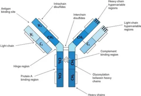
disulfide-bounded to the heavy chains in the CL and CH1 regions respectively as represented in figure 1.7. The heavy chains of each immunoglobulin molecular are similar.
Figure 1.7 | Structure of an IgG molecule (Adapted from Hermanson, 2008).
According to the class of immunoglobulin, the molecular weight for the heavy chains varies from 50 to a 75 kDa and approximately 25 kDa for the light chains. The whole molecular weight which corresponds to all subunits ranges from 150 to a 170 kDa. The light chains play an important role in establishing the specificity of antibodies. However, it is the antibody’s heavy chain variety that defines the individual class of immunoglobulin and all the master function of antibodies. The immunoglobulins can be divided into five main classes: IgA, IgD, IgE, IgG and IgM (Figure 1.8). Each class of antibodies has distinct sizes, structural constitution and different biological properties. For example, antibody classes IgD, IgE and IgG are constituted by elementary Ig monomeric structure consisting in two light and two heavy chains. In other hand, IgA molecules can present a basic Ig monomeric structure as a singlet, doublet or triplet while IgM molecules are large pentameric structure.
The heavy chains of immunoglobulin molecules are also glycosylated usually in CH2 domain inside the Fc fragment region (“Fragment crystallisable”) but also have two carbohydrates near the antigen binding sites. The Y-shaped unit is constituted by the two identical heavy chains and two identical light chains. The top of Y-unit contains the variable region also designated by antigen binding site. These sites have the suitable structural conformation to interact with complementary region of the antigen molecule. The binding site has affinity for a certain antigen because of 3D structural region of the molecule that complements the binding region of the antibody, involving the combination of van der Waals, ionic, hydrophobic and hydrogen bonding forces. Thus, antibodies possess functional groups in their structure that are appropriate for modification or conjugation processes (Hermanson, 2008).
For this purpose, effective approaches have been used to link biological molecules to QDs. There are several conjugation methods, like non-covalent conjugation of streptavidin coated QDs to biotinylated antibodies, covalent coupling between carboxylic acid coated QDs and primary amines of antibodies, site-directed conjugation via oxidized carbohydrates groups on the antibody Fc portion and conjugation to antibody fragments via disulphide reduction and sulfhydryl amine coupling (Xing et al., 2007).
The covalent strategy is reached through direct binding of the biomolecules to the QD surface coating with reactive groups such as a carboxylic acid or an amine or other cross-linker molecules. The procedures used consist on cross-linking reactions between carboxylic acid (-COOH) coated QDs and primary amines (-NH2) of antibodies which leads to a random bioconjugation, catalyzed by carbodiimide (Xing et al., 2007). A typical method to covalent conjugation uses the 1-ethyl-3-(3-dimethylaminopropyl)carbodiiamide (EDC) together with the presence of N-hydroxysuccinimide (NHS) to react the primary amino groups of biomolecules with carboxyl groups on the QD surface (Figure 1.9) (Pereira and Lai, 2008). One of the advantages of this method is related to the fact that proteins contain primary amines and do not need chemical modification (Xing et al., 2007). One limitation of the conjugation of proteins to QDs can be related to the functional sites accessible on the surface of a single QD. QDs can bind to the amine terminal groups in the antigen binding sites of the protein of interest and blocking them (Medintz et al., 2005).
Figure 1.9 | Schematic diagram showing a method for QD-antibody (QD-Ab) bioconjugation: covalent coupling between carboxylic acid (-COOH) coated QDs and primary amines (-NH2) on intact antibodies using EDC as a catalyst (adapted from Xing et al., 2007).
1.3.3. Applications
science applications like controlled drug delivery, imaging, cell labeling, biosensing, diagnosis, histochemistry and tissue engineered applications (Figure 1.10) (Chaudhuri and Paria, 2012).
Agrawal et al. have showed that QDs can be used for real-time detection of single molecules. They developed an immunoassay using antibodies conjugated to different QDs that would bind to different sites of the target biomolecules. If both QDs conjugated to antibodies bind at the same time, the signal is detected (Agraw et al., 2006).
Another important application is the molecular imaging in vitro and in vivo. Concerning the imaging, QDs are suitable for this due to their high photostability for extended periods of time and great resistance to phobleaching, without damaging the sample.
In cellular localization studies, QDs are used to clarify the dynamic properties of cell surface receptors (Dahan et al., 2003). Cellular labeling studies with QDs have been used as application in last years. It has been shown that cellular labeling with QDs take advantage to the standard fluorophores because it allows prolonged visualization of cells under continuous illumination just as multicolor imaging (Medintz et al., 2005).
Figure 1.10 | Some applications of quantum dots as multimodal contrast agents in bioimaging (adapted from Michalet et al., 2005).
1.3.4. Cytotoxicity
alloys with unknown toxicological characteristics (Hermanson, 2008). To evaluate the utility of these nanomaterials, it’s necessary to investigate their potential toxicity (Derfus et al., 2004).
QD toxicity depends on several factors that are related to their physicochemical properties and environmental conditions like size, charge, concentration, outer coating bioactivity (capping material, functional groups), oxidative state, photolytic and mechanical stability (Hardman, 2006; Male et al., 2008).
For QDs cores, the common used element is cadmium and studies have been done to quantify the amount of free cadmium ions (Cd2+) in different biological systems (Derfus et al., 2004; Chang et al., 2006). Studies with cells exposed to QDs have shown that the cytotoxic effects come from the intracellular release of free cadmium ions from the QDs caused by the oxidative degradation of QDs (Kirchner et al., 2005). In fact, the cadmium has been reported as the main cause of cytotoxicity but there are other possible factors that contribute to the toxicity such as the catalysis for the formation of reactive oxygen species (ROS). To overcome these obstacles, an alternative solution is the addition of surface coatings. For this strategy, it has been used different kinds of coating comprising a semiconductor shell such ZnS or small ligands (Smith et al., 2008). The right capping of CdSe/ZnS QDs with hydrophilic coating demonstrated no adverse effects on cells in proliferation studies (Alivisatos et al., 2005; Jaiswal and Simon, 2004; Michalet et al., 2005).
Chang et al relates that is important to differentiate the origin of cellular toxicity, if it appear from the interaction of QD surface molecules with cell membranes or from the intracellular uptake of endocytosed QDs. Maysinger et al suggest two probable processes to elucidate why QDs lead to cell damage or/and death: leaching of constituent metals of the QDs core with comprised integrity (ions of Cd, Se, Pb, etc.) and/or formation of reactive oxygen species (ROS) as a consequence of cell-QD interaction (Maysinger et al., 2007).
It’s obvious that QDs citotoxicity can be affected by several other factors but there are still others issues that need to be clarified and optimized, such as the diversity of surface coatings used, the differences in experimental conditions tested (like concentration used), the duration of QDs exposure to the cell or even the media chosen (Smith et al., 2008).
1.4. Intra-erythrocytic Parasite Infections
One area which is of clinical importance is the intra-erythrocytic infections and the way they affect red cells contents, function and membrane transport.
Parasites are microorganisms that live off other hosts to survive. They grow, reproduce and secrete toxins to the host that makes it sick. These parasitic infections are a big problem in tropical and subtropical regions. Malaria is an example of a deadly disease caused by an intra-erythrocytic parasite. Some of these infections propagated due to an insect which acts as a vector of the disease and transmits it while feeding the host (Cooke, B.M. et al., 2005).
to have a significant medical impact, so study towards diagnosis and treatment will be important to overtake that potential threat to the mammalians. In this disease the symptoms range from a silent infection until an acute, like malaria disease, resulting sometimes in death (Homer et al., 2000).
1.4.1.
Babesia ovis
Plasmodium and Babesia are apicomplexan haemoprotozan parasites that infect red blood cells and cause severe diseases, namely malaria and babesiosis, with significant medical and veterinary importance. Comparing the cellular and molecular mechanisms, the pathogenesis of the diseases caused by these two organisms is notably similar. However, they are phylogenetically different. In the research of Malaria during the last years, various studies were concentrated on the parasites to comprehend the mechanisms that cause the parasite induced changes in red blood cells. Now, it’s possible to apply this information to Babesia parasites. This relation can help to learn more about the biology of Babesia parasites, the way they cause infection and disease and how to develop novel therapeutic strategies or vaccines for these infections (Cooke, B.M. et al., 2005). Like Plasmodium,
Babesia parasites can also be cultured in vitro. This characteristic together with the moderately short intracellular growth cycle time, becomes them ideal models to discover parasite red blood cell interactions (Jackson et al., 2001).
Similarly to Malaria, Babesiosis is a parasitic infection caused by haemotropic protozoa of the genus
Babesia, family Babesiidae, order Piroplasmida, within the phylum Apicomplexa and is transmitted by the bite of an infected tick. These parasites infect the erythrocytes of an extensive diversity of wild and domestic animals. Along the years, Babesia has drawn attention as an emerging zoonotic problem because of the economic losses in cattle industry in various tropical and subtropical regions in the world (Silva et al., 2009; Horta et al., 2014). Normally, babesiosis reveals various symptoms such fever, extensive erythrocytic lysis leading to anemia, icterus, hemoglobinuria and death.
B. ovis is characterized by small round parasites, localized typically at the periphery of the red cell. This is transmitted by Rhipicephalus bursa. B. ovis is highly pathogen particularly in sheep. Mortality rates in vulnerable hosts vary from 30 to 50% in field infections (Aktas et al., 2005). This disease is considered economic important in terms of death cases, yield losses and the costs of treatment in the livestock industry (Sevinc et al., 2013). The RBC invasion is very similar to that for B. bigemina in which the parasite invades, grows and replicates asexually in erythrocytes of their vertebrate host, causing all the previously described symptoms.
Babesia parasites have a complex life cycle that can be divided into three main stages: gamogony, sporogony and merogony. When there is a bite from an infected tick, the sporozoites of Babesia can directly invade the host erythrocytes and suffer an asexual growth cycle in the erythrocytes stage as shown in figure 1.11 (Vial and Gorenflot, 2006; Yokoyama et al., 2006). Once already inside the erythrocyte of the host, the sporozoites divide into two or sometimes four merozoites which follow an asexual reproduction also known as merogony. Subsequently there is a rapid intracellular multiplication which leads to a destruction of the host erythrocytes with release of new parasites that will infect and lyse other erythrocytes (Vial and Gorenflot, 2006). However, the molecular interactions between
Babesia merozoites and host erythrocyte and their biological roles are not totally understood (Yokoyama, 2006).
Figure 1.11 | Schematic representation of erythrocyte invasion by Babesia parasites. The whole process
comprised essentially five steps: (1) Attachment of the parasite onto the host RBC. (2) Re-orientation of the parasite. (3) Formation of tight-junction between the the apical part of parasite and erythrocyte surface. (4) Invasion. (5) Internalization of the parasite within the infected RBC (adapted from Yokoyama et al., 2006).
Figure 1.12 | Schematic representation of a bovine RBCS with Babesia bovis parasite. Apical organelles and
different proteins within the parasite that are involved in erythrocyte invasion are also shown as well as the formation of ridges on the surface of the infected RBC (adapted from Gohil et al., 2010).
Micronemes are small, cigar-shaped organelles that cluster at the apical end of protozoan body and their number is different according to the species and the development stages (Figure 1.12). There are two invasion molecules considered in micronemes products (in B. bovis too merozoites): The apical membrane antigen 1 (AMA-1) and the other, thrombospondin-related anonymous protein (TRAP), with 82 and 75 kDa respectively. The AMA1 is expected to be implicated in RBC invasion and TRAP in recognition and possible attachment and invasion of host erythrocytes (Yokoyama et al., 2006; Gohil et al., 2010).
AMA1 is a type I transmembrane protein that is found on the surface of the invasive forms of parasites. It is an asexual blood-stage protein expressed in the invasive merozoite from Apicomplexan parasites (Triglia et al., 2000). The development of a vaccine is an urgent priority because is knwown that methods used to control the clinical signs, leave carcinogenic substances in sheep and goat milk (Carletti et al., 2016). The study of the protein molecules expressed on the surface of merozoites is relevant for the development of potential vaccine candidates. AMA1 might be a key vaccine candidate because it is expressed in the asexual life cycle of this parasite (Narum and Thomas, 1994) because it was observed that antibodies which target the extracellular domain of AMA1 block the invasion of erythrocytes (Thomas et al., 1984).). Due to the involvement of AMA1 in the invasion process by parasite, the study of this protein and the RBCs with microfluidic, open new possibilities to continue a detailed investigation in order to achieve the treatment of this disease.
1.5. Membrane Proteins
Membrane proteins are divided into peripheral proteins and integral proteins. Peripheral proteins are located on cytoplasmic surface of lipid bilayer and anchored via integral proteins. They are responsible for membrane elasticity and stability. Integral or transmembrane proteins (TP) are embedded in membrane via hydrophobic interactions with lipids. These proteins are involved in such important biological cell functions, like virus binding, cell surface antigenicity, cell-cell recognition and communication, cellular transformation, transport, energy transduction etc. In red blood cell membrane, there are three types of TPs: Band 3, Glycoproteins and Aquaporins.
Glycoproteins are proteins with covalently bounded carbohydrates. They were classified into sialoglycoproteins which are a combination of sialic acid and glycoprotein, i.e, a combination of sugar and protein. These sialoglycoproteins are rich in sialic acid which give the erythrocytes a very hydrophilic charged coat. Approximately 10% of erythrocyte membrane is covered by sialoglycoproteins and their sialic acid content gives negative charge on the cell surface, preventing adherence of RBCs to each other and to vessel walls (Viitala and Järnefelt, 1985).
Membrane cells are constituted by several target proteins of currently available drugs for treatment of diverse disorders. So, studying this cell component, is possible to develop new tools to label the known targets or even identify new ones.
1.5.1. Glycophorin A
Sialoglycoproteins are also known as Glycophorins (GP). They are composed of 60% carbohydrate including sialic acid and 40% protein. The existence of glycophorins in erythrocyte membrane was discovered by Fairbanks et al. There are four different types of glycophorins and they comprise approximately 2% of the total RBC membrane protein (Fairbanks et al., 1971). These include Glycophorin A (GPA), B (GPB), C (GPC) and D (GPD) which belong to a group of important transmembrane proteins that play an important role in cell-cell interactions (Tomita and Marchesi, 1975).
Table 1.1 | RBC membrane glycophorins (adapted from: Chasis and Mohandas, 1992).
Glycophorin A is the most abundant, constituting approximately 75% of the total sialoglycoproteins in erythrocyte membrane (Furthmayr et al., 1975) with 5 to 9 × 10⁵ copies per cell. GPA was the first membrane protein to be isolated and sequenced and has been used as a model for topology of receptors and other transmembrane glycoproteins in surface cells (Tomita and Marchesi, 1975) (Table 1.1). This glycophorin is exclusively expressed in erythroid cells (Gahmberg et al., 1978; Yurchenco and Furthmayr, 1980).
GPA GPB GPC GPD
Copies per cell (×103) 500-900 80-30 50-100 20
Molecular mass (kDa) 36 20 32 23
Initially, the hematologic interest in glycophorins was restricted to blood bank serologists and the characterization of blood group antigens situated on these type of proteins. Nevertheless, with more recent functional studies, it is become notorious that some glycophorins also play different but important roles in regulating RBC membrane mechanical properties and in maintaining RBC shape. As well as play a crucial role in modulating RBC-RBC interactions, they also modulate the RBC interactions with vascular endothelium and other circulating blood cell, avoiding the RBC interaction (Figure 1.13) (Dahr, 1986).
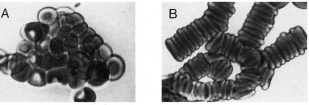
Figure 1.13 | Red blood cells interactions. (A) RBC aggregation due to strong associations, for example such as
those originated by Ig binding. This type of aggregation needs much bigger forces for cell dissociation. (B) Aggregation resulting from weak cell to cell interactions, which occurs physiologically and overcome the repulsive negative charge of the surface but are easily dissociated (adapted from: adapted from: Chasis and Mohandas, 1992).
RBC membrane have glycophorin variants and deficiencies that were initially discovered by serological and immunochemical assays and later, characterized at a molecular level. These different phenotypes have some particular interest due to the functional role of glycophorin in the membrane. In pathological genetic conditions where GPA is not present, cells appear to exhibit normal physiologic properties. As such, individuals with this different phenotype are clinically healthy showing no observable changes in erythrocyte morphology and do not present anemia (Chasis and Mohandas, 1992).
An interesting and relevant data is the discovery that GPA also works as a receptor for infective agents like P. falciparum and may function as a chaperone which is a protein that assists the assembly or disassembly of other macromolecular structures. This function enables the targeting of specific transmembrane proteins to the cell membrane. A protein from malarial parasite was isolated and it was verified that it combines in a specific way with glycophorin A, showing that this glycoprotein has a receptor function for malarial parasite and in this process the sialic acid content of glycophorin A are involved since this interaction is inhibited by sialic acid. Therefore, GPA is a likely RBC surface attachment site for P. falciparum (Vanderberg et al., 1985). Due to the presence of GPA in erythrocytes and its involvement in RBC invasion by malarial parasite which is notably identical to Babesiosis, the intra-erythrocytic disease under study, make this glycoprotein a good model for the following assays.
binding of some important metal ions for example, Mg2+ and Ca2+ to the red cell membrane. It’s also a receptor for enterotoxigenic E.coli (Tayyab and Qasin, 1988).
1.6. Nanotoxicology
An important area of the nanotechnology field of knowledge is the nanotoxicology. It refers to the study of the concentration of nanomaterials with biological entities. This study is centered on the relationship between the physical and chemical properties (e.g. size, shape, surface chemistry, compositions and aggregation) of nanostrcutures with the induction of toxic biological responses. This type of information is important to characterize the nanomaterial applications in areas like biotechnology, biomedical applications and toxicity screening.
Quantum dots are composed by metallic elements like cadmium and selenium and their impact in cells and tissues is subject of study. Due to their chemical composition, they rise concerns about their biological interaction. As a result, their cytotoxic effect through the induction of oxidative stress was evaluated (Santos, 2009; Qu et al., 2013).
1.6.1. Reactive Oxygen Species
The term ROS (Reactive Oxygen Species) is used to characterize forms of oxygen that are energetically more reactive than molecular oxygen. ROS are an essential component in stress responses and its level defines the type of response (Shao et al., 2008).
Cells are sensitive to ROS and when their production exceeds the antioxidant defense, cellular redox balance is shifted and the cells are then in a state of oxidative stress. This sensitivity to ROS is dependent on the cell type, the level and the duration of oxidant production, the species of ROS originated and the specific site where ROS are produced (Maysinger et al., 2007).
These species are constantly produced within red blood cells, as a consequence of their physiological role. The main function of erythrocytes is to transport large amount of oxygen (O2) and mediation of carbon dioxide (CO2) production during their lifetime, resulting in oxidative stress. Studies indicate that many physiological and pathological conditions such as inflammation or eryptosis for example, are developed through ROS action (Çimen, 2008).
Figure 1.14 | Representative scheme of ROS generation through the reduction of molecular oxygen (adapted from Gechev et al., 2006).
High levels of ROS can result in non-controlled oxidation of a variety of cellular structures, including membrane damage (Apel and Hirt, 2004) and can provoke apoptosis or necrosis according to the level of damage (Maysinger et al., 2006). RBCs are constantly exposed to both endogenous and exogenous source of reactive oxygen species (ROS) and studies have demonstrated that oxidative stress plays an important role in damaging the RBC membrane and improving its deformability (Mohanty, J.G. et al., 2013). So, this study can be used as a model for the oxidative damage of RBCs functions during their long journey in the blood stream.
RBCs were incubated with chosen QDs to evaluate their cytotoxic effect. The induction of oxidative stress is an important toxic feature of nanomaterials (Hoet and Boczkowsky, 2008). QDs are redox active nanoparticles (effective donors and acceptors) and can generate highly reactive free radicals with exposure to light. Since this technology is to be used in live cells, its potential cytotoxic effects is an essential factor to be considered because it may affect the cell physiology. The size, charge and concentration of the QDs, their outer shell bioactivity and oxidative, photolytic or mechanical stress are all factors that, collectively and individually, can determine their cellular toxicity. The literature on toxicity of QDs is a mix of reports of numerous type of QDs with widely varying physicochemical parameter, making comparisons a quite difficult. So it is imperative to test the used QDs in our cell model to evaluate its toxicity because the unknown interferences of these nanoparticles with physiological processes could lead to misinterpretation of the results, whatever the application for.
1.7. Microfluidic
enabling comprehensive genomic and proteomic analysis from small homogeneous subpopulations down to single cells (Toner and Irimia, 2005).
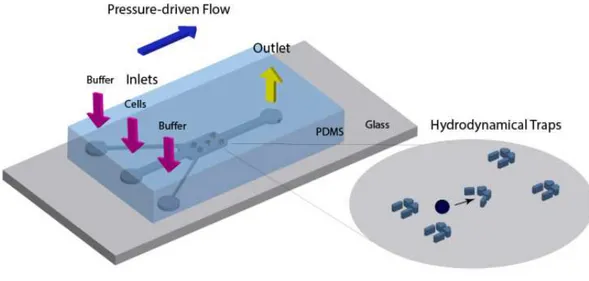
Figure 1.15 | Representation of a microfluidic device: a PDMS chip consisting of microchannels containing
arrays of V and U chapped hydrodynamical traps.
1.7.1. Blood-on-a-chip
Blood sampling and analysis are of great interest for medical and science applications and play a crucial role in diagnosis of various physiologic and pathologic conditions (Toner and Irimia, 2005). However, this requires a deep understanding of the biology involved and the use of appropriate techniques.
In the early steps of blood research, the observation of the cells was made by microscopes. Later, tissue-staining techniques were developed enabling the initial characterization of populations. Even nowadays, the analysis of cell morphology of the peripheral blood is done with Wright-Giemsa staining protocols and the full blood count are the most basic and used techniques in investigations in hematology (Bauer, 1999). Flow cytometry techniques used nowadays offer higher capability because of its advanced details and higher throughput, representing a major advance in cell identification and separation. Nevertheless, the complexity of this technique needs higher specialization of the operator. To reduce the possible errors, it is necessary to reduce the time from blood collection to analysis. The availability of analysis techniques at the site where blood is collected is considered a challenge that would need faster, cheaper and simpler approaches (Toner and Irimia, 2005). For these reasons, a microfluidic chip was developed to trap red blood cells.
1.7.2. Microfabrication
fabrication is based on photolithography. It is a modeling method that uses irradiation to transfer a pattern to a substrate. This technique allows the obtention of direct replica of a plastic, glass or silicon wafer. This replica can be repeated several times for obtaining a functional micro-device (Meira, 2015). The irradiation through a mask with photoresist layer allows the selective removal of resist in the development (Franssila, 2010). Photoresists are photosensitive polymeric materials that are used to create a patterned coating on a surface. These type of materials allowed the reduction of structures downwards to 100nm. Photoresists can be positive or negative, property which define their susceptibility to UV rays. In case of positive photoresists, the exposed zones become soluble in the developer and consequently, the unexposed areas remains insoluble. On the other hand, with negative photoresists, the exposed areas are insoluble to the developer and the unexposed become soluble (Franssila, 2010). Among the photoresist frequently used in microfabrication are negative SU-8 or P-50100. There is also a positive variant, the novolac resin SPR 220-7 (Meira, 2015).
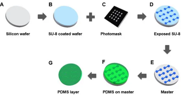
The crucial step in whole chip microfabrication is the production of the SU-8 mold to create PDMS replicas because PDMS replicates with very precision all the features of the master mold so any defect will affect the PDMS chip structure. PDMS (Polydimethylsiloxane) is a silicon based elastomer and it presents a hydrophobic surface. This is the most used material in microfluidics due to its transparency, elasticity, and low cost. In addition, it can be bonded without adhesives and it can be easily structured by molding with nano-precision but for this purpose, it requires a mold. For this, Si wafer is coating with SU-8 to originate a mold (Figure 1.16: A – E). A master consists of a positive relief of photoresist on a silicon (Si) wafer and serves as a mold for PDMS (Figure 1.16: E). The master is covered with this silane derivative to simply the peeling of the replica (F and G). SU-8 is a thick photoresist i.e. a polymer which can be patterned using light and it’s the most used material for high and narrow microstructures. This photoresist has excellent chemical, optical, mechanical and thermal properties. So, the SU-8 and PDMS is the most used combination in microfluidic.
The process of microfabrication of a microfluidic chip starts with the design of a system of channels in a CAD program. A commercial printer uses the CAD file to produce a high resolution transparency mask. This transparency is used as a photomask in contact photolithography to produce a master and here, the replica molding process starts.
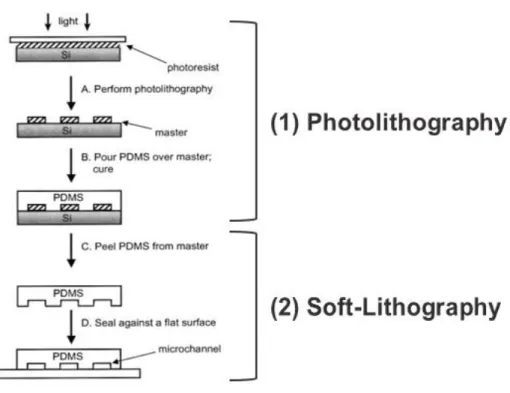
In turn, soft-lithography is a low cost production method based on elastomeric stamps, allowing the creation of three-dimensional patterns at room temperature. This process include some methods and one of the most common is the replica moulding (casting), i.e, the transfer of a pattern from a master into replica by solidifying a liquid precursor against the master (Elhadj et al. 2010). It is considered one of the main techniques in microfluidic production due to its lower coast (Figure 1.17).
Figure 1.17 | Representative scheme of photolithography and soft-Lithography process. Photolithography (1) followed by soft-lithography (2) allow to obtain a direct replica of the inverse mask (negative resist) (A), (B) and (C), after the UV exposure and development. At the right time, deposition and etching can be performed through the addition and subtraction to achieve the desired pattern (D) (adapted from Meira, 2015).
After crosslinking, the replica is peeled off the mould. Finished this step, the replica can be used directly to produce chips, or for the casting of subsequent chips, to prevent damage of the master. Here, it is common to use an epoxy mould from PDMS mould. The use of epoxy allows an increase of the number of replicas. Finally, the replica is cut into individual chips and access holes for inlet and outlet are made through a sharp tip. In the end of the process, chips are sealed to a glass slide, closing the cannels from the bottom.
Chapter 2 | Materials and Methods
2.1. Blood collection
For studies with intra-erythrocytic infections, in particular Babesia ovis culture in vitro and identification of AMA1 protein, ovine blood was collected and subsequently processed. In trials to identification of Glycophorin A protein and to investigate the oxidative stress produced by cells when incubated with QDs, human blood was used.
2.1.1. Uninfected ovine RBCs for subculturing
Desfibrinated ovine blood (Oxoid) was collected from healthy ovine donors. The blood was separated in two falcons of 50mL for packed RBCs and it was centrifuged during 20 min, 2500 rpm at 4⁰C. The supernatant was collected to another falcon and this is the serum. Then, the RBCs were washed in VyM (Vega et al., 1985) to reach the final volume of 50mL. Two more centrifugations were made and the final RBCs are stored at 4⁰C.
2.1.2. Human Blood
The human blood samples were obtained following the standard international rules of WMA Declaration of Helsinki – Ethical Principles of Research Involving Human Subjects. A few drops of blood was collected by pricking the finger of volunteers and then the sample was preserved in 200 μL of 2.1% sodium citrate to not coagulate. The blood was centrifuged (10 min, 2500 rpm at 4⁰C) and the supernatant was discard. Pellet was washed in 1x PBS. The final sample was stored at 4˚C.
2.2. Quantum Dots
The ability to bioconjugate QDs makes them suitable for applications as biological label, immunostaining or as live cells markers. Their chemical composition can be determinant for subsequent assays. Preliminary experiments are necessary to choose the appropriate type of these nanoparticles before their use with cells. As such, three types of water soluble CdSe/ZnS quantum dots were bought from Ocean Nanotech manufacturer in order to assess which ones were suitable for the cells being studied.
2.2.1. Characterization of Electrophoretic Mobility and pH stability
The QDs were tested at different pH conditions in agarose gel to evaluate changes in fluorescence intensity and to observe their migration profile. It is a technique suitable also for estimation of the nanoparticle sizeEDTA 0.5M) diluted from the 5x stock solution with bidistilled water. The 1% agarose gel was prepared by dissolving under heating 0.5g of agarose (Thermo Scientific) in 50mL of 1x TBE and 2.5μL of SYBR® safe DNA gel stain (Thermo Scientific) were added. 5μL of each QD solution previously prepared were mixed with 2μL of 6xDNA loading dye (Fermentas), before loading onto the gel. 4μL of 1kb and 5μL of 100bp DNA ladder (Fermentas) were loaded onto the gel for estimation of the size of each type of QDs. The gel ran at 100V during 90 minutes using a Mini-Sub Cell GT system (Biorad) and was visualized under blue sample tray (suitable for gels stained with SYBR® stain) using Gel Doc™ EZ Imager and Image LabTM software.
2.2.2. Bioconjuagtion of antibodies to QDs
The QD conjugation was performed by direct conjugation of antibodies to carboxy-QDs through amine-carboxylic acid coupling using EDC/NHS chemistry. This protocol is divided into three steps: the QDs activation with EDC and NHS solutions, the bioconjugation of activated QDs with antibodies and the purification and concentration of final bioconjugates.
the total removal of possible free antibody in solution. The final QD-antibody sample can be diluted to a desired concentration.
2.2.2.1. Characterization of conjugated QDs
Agarose Gel ElectrophoresisThe characterization of the QD-Ab conjugates was performed by agarose gel electrophoresis. Free COOH-QDs and free antibodies were used as control.
The procedure followed this protocol: 2μL of each bioconjugation resulting sample as well as 2μL of 50nM free COOH-QDs and 2μL of free antibody solutions were mixed with 2μL of 6xDNA loading dye, making a final volume of 10µL with 10 mM Na-P buffer. The final samples were loaded onto the gel. The 1% gel was previously prepared by heating 0.5 g of agarose in 50 mL of 1x TBE buffer, without adding SYBR® safe DNA gel stain. 6μL of 1kb DNA ladder was also loaded onto the gel to possible estimate the molecular weight. The electrophoresis was conducted at voltage ranging 45 to 90V during 2h15min because the high molecular weight of the final bioconjugates. The gel was visualized under UV sample tray (suitable for gels using ultraviolet illumination) using Gel Doc™ EZ Imager and Image LabTM software.
SDS-PAGE Electrophoresis
Samples of antibody-QD conjugates, free antibodies and free QDs were analyzed by SDS-PAGE electrophoresis (Bolt and Mahoney, 1997), in reduced and non-reduced conditions. In the non-reduced conditions, 5μL of each bioconjugate sample were used. Then, 5μL of Loading Dye non-reduced (10% v/v glycerol in water and bromophenol blue to the desired coloring) were mixed. 3µL of 50nM COOH-QDs control was mixed with 3µL of Loading-dye non-reduced while 2µL of each antibody control was mixed on the same proportion as the loading dye. For the reduced conditions, the same number of samples were prepared in the same way but mixed with Loading-dye mixed with Dithiothreitol (DTT, Sigma) at a concentration of 0.1M (1:1, 2.5µL of DTT+2.5µL of Loading dye). All the samples were loaded into the 4-15% gradient precast gel (Biorad) as well as the 5μL of protein ladder (MW 20-250 kDa, NZYTech) to estimate the protein molecular weight. The gel was run for 1h30 minutes at a constant voltage of 120V in Tris-Glycine 1x Running Buffer diluted from a 10x stock solution (250 mM Glycine, 1.92 mM Tris-base and 1% SDS) previously prepared. In the end, gel was visualized using a gel imaging instrument Gel DocTM EZ Imager with UV Tray.
Western Blotting
three filters and one filter paper as well as the gel and the membrane were placed in the semi-dry system (Xcell IITM Blot Module, Life Technologies). One more filter paper and three more filters were placed in the same semi-dry system in this order over the remaining pieces for membrane transfer. The transfer was performed for 1 hour at 30V at room temperature. After the transfer, the membrane was softly washed with approximately 20 mL of TBS 1x pH 7.6 (Tris-buffered saline: Tris-HCl 10mM and NaCl 150mM) with constant agitation at room temperature. Then, the membrane was blocked with 40mL of freshly prepared Blocking buffer (5% w/v non-fat milk and TBS 1x) overnight at 4⁰C with slowly agitation. The membrane was cut and incubated with secondary antibody anti-mouse IgG and anti-rabbit with HRP conjugate (R&D Systems), previously diluted in Blocking buffer (1:5000) for 2 hours at room temperature under shaking conditions. Then, it was washed twice with approximately 20mL of T-TBS (TBS 1x containing 0.05% Tween 20, Sigma) for 5 minutes each time with slowly agitation at room temperature, followed by one more wash with just 20mL of 1x TBS under the same conditions. To visualize the immunoreactive proteins, a detection reagent was used (Western Lightning ECL Pro, Perkin Elmer Life Sciences) and the images were obtained in a molecular imager ChemiDoc, using the Quantity One software, in Chemiluminescence mode and the bioconjugated QDs was visualized under the UV-Light in the same molecular imager.
2.3. Babesia ovis infected cultures
One goal of this work was to create a label tool to identify the AMA1 in the RBC membrane through the conjugation of QDs with a specific antibody for this transmembrane protein. As previously described, AMA1 is expressed on the surface of erythrocytes invaded by Babesia parasites. As such, it was necessary to use an in vitro culture in order to obtain infected erythrocytes to test with bioconjugates obtained by the process described in section 2.2.2.