UNIVERSIDADE DE LISBOA
FACULDADE DE CIÊNCIAS
DEPARTAMENTO DE BIOLOGIA VEGETAL
Characterization of the subtilisin-like
protease 3 in malaria parasites
Mariana Justino Almeida
Mestrado em Microbiologia Aplicada
2011
2
UNIVERSIDADE DE LISBOA
FACULDADE DE CIÊNCIAS
DEPARTAMENTO DE BIOLOGIA VEGETAL
Characterization of the
subtilisin-like protease 3 in
malaria parasites
Dissertação de Mestrado orientada pela Professora Doutora Photini Sinnis, New York University, e Professora Doutora Rita Zilhão, Faculdade de Ciências da Universidade de Lisboa.
Mariana Justino Almeida
Mestrado em Microbiologia Aplicada
2011
3
UNIVERSIDADE DE LISBOA
FACULDADE DE CIÊNCIAS
DEPARTAMENTO DE BIOLOGIA VEGETAL
Characterization of the
subtilisin-like protease 3 in
malaria parasites
The work presented in this thesis was developed at the Department of Parasitology in the New York University Langone Medical Center (NYUMC)
Mariana Justino Almeida
Mestrado em Microbiologia Aplicada
2011
4
This work was presented at the 22
ndMolecular Parasitology Meeting in Woods Hole,
5
ACKNOWLEDGMENTS
I am very grateful to Photini for accepting me in her lab during the past year. She was always a great mentor and it was a true pleasure to be under her supervision during this project.
I would like to thank Professor Rita Zilhão for being my internal supervisor and for being available.
I wish to acknowledge present and past members of the lab (Brandy, Francesco, Andy, Bhamini and Kelsey) that have helped me with suggestions and technical support, specially Brandy and Francesco, who were crucial for the existence and development of this project.
Thank you Sandra, Jean and Carolina for saving my life and always finding a way to give me mosquitoes to start cycles when I most needed.
Thank you to all the people in the department who are not individually mentioned here but that, in some way, made my job easier.
Não posso deixar de agradecer a todo o grupo de malária do IMM, FMUL, especialmente à Vanessa, que me ensinou TANTO durante o estágio. Sem esse conhecimento e experiência nunca poderia ter tido a independência que tive durante este ano. Obrigado. Obrigado. Obrigado.
Por último, e mais importante, obrigado à minha família: aos meus pais por me deixarem ter esta aventura e acima de tudo pela educação que me deram; ao gordo, por ser o gordo; aos meus queridos avózinhos por perceberem que tinha de fazer isto; aos primos, tias e queridos amigos – são demasiados nomes para mencionar (é ingrato mas é bom sinal!).
6 ABSTRACT
Malaria is a huge global health threat, causing thousands of deaths and suffering every year, worldwide. Although there are some effective drugs available at present, the parasite often acquires resistance in the face of drug pressure. In order to choose the most efficient strategy to fight against this disease, it is imperative that we understand the basic biology of its causative agent Plasmodium.
Plasmodium species express a number of proteases that have important roles in fundamental processes
and, therefore, are potential drug targets. A number of serine proteases belonging to the subtilisin-like family, also named subtilases, have been identified in apicomplexan parasites like Toxoplasma,
Neospora and Plasmodium. The Plasmodium genome has three subtilisin-like proteases. It has been
shown in Plasmodium falciparum that the subtilisin-like proteases 1 (PfSub1) and 2 (PfSub2) have important roles in the invasion and egress of parasites from erythrocytes. However, little is known about subtilisin-like protease 3 (Sub3) in Plasmodium species. In this study, we used gene targeting to disrupt the sub3 gene in the rodent malaria parasite Plasmodium berghei ANKA and analyzed the effect of the deletion in the life cycle of the mutant.
When mosquitoes were infected with the sub3 deletion mutants, we found no differences in the number of oocysts or salivary gland sporozoites compared to controls. We performed in vitro and in vivo experiments to further study the ability of the deletion mutants to establish infection in the mammalian host. Results show that recombinant parasites are able to normally infect and develop in the liver and blood. In consequence, deletion of sub3 gene did not impair or significantly affected parasite development in its natural hosts.
7 RESUMO
Estima-se que em 2009, metade da população mundial se encontrava em risco de contrair malária. Recentes estatísticas mostram que, no mesmo ano, aproximadamente 800 000 seres humanos morreram. Esta é uma doença que atinge maioritariamente os países em desenvolvimento do continente africano, asiático e sul-americano.
A malária, ou paludismo, é causada por parasitas do género Plasmodium, pertencente ao filo
Apicomplexa. A este filo pertencem vários parasitas eucariotas unicelulares com importante relevância
médica, como é o caso dos géneros Crisptosporidium e Toxoplasma.
As espécies de Plasmodium apresentam um ciclo de vida complexo que requer dois hospedeiros: o mamífero, onde apenas ocorre reprodução assexuada, e o vector invertebrado, onde adicionalmente há recombinação genética. O ciclo inicia-se quando o mosquito fêmea Anopheles tem uma refeição de sangue, ao picar a pele do hospedeiro, e inocula na derme parasitas sob a forma de esporozoítos. Seguidamente, os esporozoítos entram na corrente sanguínea, chegando ao fígado onde iniciam o seu desenvolvimento nos hepatócitos. Apesar de esta fase ser assintomática, os parasitas multiplicam-se assexuadamente originando milhares de parasitas denominados merozoítos que, quando libertados no sangue, são capazes de invadir eritrócitos. Nos eritrócitos o parasita inicia a fase seguinte do seu desenvolvimento, passando por sucessivos estádios de maturação que culminam na libertação de novos merozoítos. Ao fim de vários ciclos de multiplicação no sangue, alguns merozoítos diferenciam-se em formas sexuais – os gametócitos –, que são ingeridos durante a refeição de sangue do mosquito dando início à fase sexuada do ciclo de vida. Após recombinação no vector, o parasita começa uma nova fase de reprodução assexuada originando milhares de esporozoítos, que migram do intestino para as glândulas salivares do mosquito. Numa próxima refeição, os esporozoítos são inoculados na pele do hospedeito dando início a um novo ciclo.
A elevada morbilidade e mortalidade da malária torna a sua erradicação uma das principais prioridades da Organização Mundial de Saúde. Durante décadas, têm sido feitos inúmeros esforços com o intuito de erradicar a doença. Há cinquenta anos atrás, a malária foi eliminada em muitas áreas do planeta, através do uso de drogas como a cloroquina. No entanto, devido ao desenvolvimento de resistência por parte do parasita, a incidência da doença aumentou novamente. Recentemente têm sido utilizadas novas e eficientes drogas como a artemisina. Infelizmente, foram identificados recentemente casos de resistência à artemisina. Se este fenómeno se alastrar, as consequências podem ser devastadoras e, por essa razão, é extremamente importante que novos compostos e alvos sejam descobertos.
As proteases do parasita da malária apresentam um enorme potencial como alvos de novos compostos devido ao papel crucial que desempenham em várias fases fundamentais à continuidade do ciclo de
8 vida. As subtilases pertencem à grande família das proteases de serina e caracterizam-se pela ordem onde se localizam os resíduos que compõe o seu centro activo: Asp – His – Ser. Em P. falciparum são conhecidas três subtilases: subtilase 1, 2 e 3. Enquanto que Sub1 está envolvida na via proteolítica que induz a saída dos merozoítos dos eritrócitos, a Sub2 está implicada na invasão dos mesmos. O presente trabalho focou-se particularmente numa protease sobre a qual não existem quaisquer estudos funcionais – a subtilase 3 (Sub3).
Para melhor compreender o papel da Sub3 no parasita, o nosso trabalho foi dividido em duas etapas: o estudo do padrão de expressão do gene sub3 e acaracterização de um parasita com uma deleção no mesmo gene. Todas as experiências foram realizadas em P.berghei, um parasita de roedores reconhecido como um modelo válido para a investigação experimental em malária.
A expressão de sub3 foi analisada em esporozoítos no intestino e glândulas salivares do mosquito, bem como em parasitas presentes no sangue de ratinhos infectados. A presença de RNA mensageiro de
sub3 foi detectada em esporozoítos no intestino do mosquito, com resultados reprodutíveis em todas as
experiências realizadas. No entanto, os resultados obtidos nos restantes estádios analisados não foram igualmente consistentes.
O parasita mutante, gerado por recombinação homóloga, contém uma deleção no gene que codifica a subtilase 3. A região delectada no locus recombinante inclui o codão de iniciação da tradução, os resíduos de histidina e serina do centro activo e parte da região codificante.
O primeiro passo na análise da população de mutantes, também denominados parasitas sub3Δ, foi a infecção de mosquitos Anopheles fêmea. Um defeito nesta fase do ciclo pode significar um bloqueio na transmissão do parasita. Contudo, os vários ensaios experimentais mostraram que o desenvolvimento do parasita recombinante ocorre de forma normal no vector, culminando com a presença de esporozoítos nas glândulas salivares do mosquito. O sucesso da infecção no vector prova, ainda, que os gametócitos previamente ingeridos pelo mosquito estão funcionalmente activos.
Em seguida, avaliámos a mobilidade dos esporozoítos sub3Δ. Se esta propriedade for afectada, os parasitas serão menos eficientes a migrar do local de inoculação para os vasos sanguíneos originando, potencialmente, um parasita com menor probabilidade de continuar o seu percurso. Para analisar a capacidade de locomoção dos mutantes realizámos experiências in vitro e in vivo. Quando depositados numa superfície de vidro, os esporozoítos recombinantes revelam uma ligeira diminuição na sua mobilidade. Contudo, quando em contacto com uma monocamada de hepatócitos, os esporozoítos
sub3Δ atravessam as células com igual eficiência relativamente aos parasitas controlo. Um resultado
similar foi obtido in vivo: após inoculação na derme de ratinhos, os esporozoítos sub3Δ chegam ao fígado com sucesso e sem atraso relativamente aos controlos. Em conjunto, os resultados indicam que a via natural de infecção do parasita não foi afectada.
9 Posteriormente analisámos a fase hepática do ciclo de vida. Tal como foi descrito anteriormente para o estudo da locomoção, o desenvolvimento do parasita sub3Δ no fígado foi analisado in vitro e in vivo. Ambos os ensaios demonstram que a multiplicação de parasitas sub3Δ durante a fase hepática ocorre tal como é esperado em parasitas wild type.
Em suma, os resultados apresentados mostram que apesar de RNA mensageiro de sub3 ser detectado nos parasitas, a deleção de sub3 não afecta o progresso do parasita recombinante ao longo do ciclo de vida. Curiosamente, ao contrário do observado para as subtilases 1 e 2, não foram detectadas diferenças fenótipicas, nas fases do desenvolvimento analisadas, entre o parasita mutante gerado para estudar a subtilase 3 e os parasitas wild type, tornando difícil compreender o papel desta protease no parasita.
10 TABLE OF CONTENTS Abstract ... 6 Resumo ... 7 List of Abbreviations ... 12 Introduction Malaria – Prevalence of the disease and life cycle of its causative agent ... 14
Malaria – the need to discover novel drug targets ... 16
Plasmodium subtilisin-like proteases ... 17
Material and Methods Construction of sub3Δ plasmid ... 19
Generation of sub3Δ mutant parasites ... 20
Selection of recombinant parasites ... 21
Cloning recombinant parasites in mice ... 21
Integration of transfected plasmid in sub3Δ clone ... 22
Detection of parasites in the blood ... 22
Mosquito infection ... 22
Mosquito dissection ... 23
Gliding motility assay ... 23
in vitro sporozoite traversal assay ... 23
in vitro development assay ... 23
Determination of prepatent period ... 24
Quantification of parasite burden in the liver by qPCR ... 24
Detection of sub3 mRNA transcripts ... 24
Cells ... 25
Mice ... 25
Antibodies ... 25
11
Microscopy ... 25
Results Pyrimethamine-resistant parasites were selected after electroporation of schizonts with sub3Δ transfection plasmid ... 26
sub3Δ parasites were successfully cloned in mice ... 27
The transfected fragment was correctly integrated in the sub3Δ parasites ... 28
sub3 mRNA transcripts were detected in P.berghei ANKA midgut sporozoites ... 29
sub3Δ parasites developed in the mosquito vector ... 30
sub3Δ sporozoites glided in vitro ... 32
sub3Δ sporozoites were able to traverse hepatocytes in vitro ... 33
sub3Δ parasites invade and multiply inside hepatocytes in vitro ... 34
sub3Δ blood stages were detected after sporozoites were injected in mice ... 36
sub3Δ parasites were quantified in the liver upon intravenous and intradermal injection of sporozoites in mice ... 37
Discussion and Conclusions ... 39
Future Work ... 42
References ... 43
12 LIST OF ABBREVIATIONS
bp: base pair
cDNA: complementary DNA
DEPC-H2O: Diethylpyrocarbonate treated water DMEM: Dulbecco's Modified Eagle's Medium DNA: deoxyribonucleic acid
EDTA: Ethylene diamine Tetraacetic Acid EtBr: ethidium bromide
EEFs: exoerythrocytic forms
FITC: fluorescein isothiocyanate fluorescent label GFP: green fluorescent protein
h : hour
hDHFR: human dihydrofolate reductase i.d. : intradermaly
i.p. : intraperitoneal i.v. : intravenously
iRBC: infected red blood cell min : minute
mRNA: messenger RNA RBC : red blood cell RT : room temperature
PBS: phosphate buffered saline PCR: polimerase chain reaction
PfSub: Plasmodium falciparum subtilase p.i. : post infection
PFA: paraformaldehide PV: parasitophorous vacuole
qPCR: quantitative Polimerase Chain Reaction RNA: ribonucleic acid
13 RPMI: Roswell Park Memorial Institute medium
rRNA: ribosomal RNA SERA: serine-rich antigen
TAE: Tris base, acetic acid and EDTA Buffer UV: ultra violet
WHO: World Health Organization WT : wild type
14 INTRODUCTION
Malaria – Prevalence of the disease and Lifecycle of its causative agent
In 2009, about 3.3 billion people - half of the world's population - were at risk of malaria, making it the world's most deadly parasitic and vector borne disease. Every year, it leads to about 250 million known cases and, in 2009 alone, it was responsible for nearly 800 thousand deaths, killing more under-5-year-olds than any other disease (WHO, 2010). Malaria affects mainly regions of Africa, Asia and South America (Figure 1).
Figure 1. Global map of malaria endemic countries highlighted in dark grey (WHO, 2010).
Malaria is caused by parasites of the genus Plasmodium. In humans, it is caused mainly by four species:
P. malariae, P. ovale, P.vivax and P. falciparum, with the last being responsible for the most severe form
of the disease. Plasmodium species belong to the ubiquitous and highly successful phylum Apicomplex which contains other parasites of major medical importance, such as Toxoplasma and Cryptosporidium.
Plasmodium spp has evolved a complex life cycle with essential developmental stages in both the
anopheline mosquito vector and the vertebrate host (Figure 2). Infection is initiated when an infected mosquito containing Plasmodium sporozoites in its salivary glands bites a mammalian host while it probes for blood. During this process sporozoites are injected into the dermis, where most of them will move through until, eventually, contact blood vessels and enter the circulatory system (Amino et al., 2008).
15 Once in the blood, sporozoites are rapidly sequestered in the liver sinusoids which they have to cross, primarily through Kupffer cells, in order to reach and invade hepatocytes (Murray et al., 2005; Frevert et
al., 2005). Sporozoites do not invade the first hepatocyte they encounter. Instead, they migrate through
several hepatocytes before invading a final one in which a parasitophorous vacuole (PV) is formed (Mota
et al., 2001). Following productive invasion, each sporozoite differentiates into an exoerythrocytic form
(EEF) and multiplies by schizogony inside the hepatocyte, thereby generating thousands of new parasites that are now capable of infecting erythrocytes – the merozoites (Aly et al., 2009; Sturm et al., 2006). When EEF development is completed merozoite-filled vesicles, the merosomes, are formed and released into the liver sinusoid (Sturm et al., 2006). Merosomes will then enter the bloodstream and finally rupture and release the first generation of merozoites (Baer et al., 2007).
Figure 2. Life cycle of Plasmodium. Infected female Anopheles mosquito injects sporozoites while it takes a blood meal (1). Sporozoites reach the liver (2) and multiply asexually generating merozoites (3) which are then released in vesicles and reach the blood (4). In the bloodstream, the merozoites invade erythrocytes and multiply until they egress only to invade a new cell (5). Some merozoites develop into sexual forms, called gametocytes that circulate in the bloodstream (6). When a mosquito bites an infected human, it ingests gametocytes which further develop into mature sex cells named gametes (7). A diploid zygote is formed by fusion of a female gamete with a male gamete. The zygote transforms in to a motile ookinete that burrow through the mosquito's midgut wall and form oocysts (8). Inside the oocysts, thousands of active sporozoites develop eventually bursting it and releasing all the parasites in the hemolymph (9). Eventually sporozoites migrate to the salivary glands and a new cycle begins when the mosquito bites the next host (10). Adapted from Baer et al., PloS Pathogen 2007 (11)e171.
16
Plasmodium, as other related apicomplexan parasites such as Babesia, have the unusual capacity to
rapidly invade red blood cells (RBC) – erythrocyte invasion by merozoites occurs within seconds and by simultaneous formation of a PV. Once invasion is completed the merozoite transforms into a ring stage, which is characterized by a large digestive vacuole, where hemoglobin digestion results into the formation of the malaria pigment, hemozoin. The ring stage develops into a trophozoite that multiplies by schizogony originating an erythrocyte schizont, which ultimately generates newly formed merozoites that, when fully developed, egress from the RBC and initiate a new cycle (reviewed in Silvie et al., 2008). After several cycles of multiplication in the blood, some merozoites will differentiate in gametocytes. The factors that initiate and regulate the formation of gametocytes are not yet well understood.
Ingestion of gametocytes by a mosquito during a blood meal initiates the cycle in the invertebrate host where both asexual and sexual reproduction take place. Ingestion of gametocytes activates the formation of gametes – gametogenesis - in the mosquito midgut lumen (Bilker et al., 1997, 1998). This process generates the female and male gametes that will fuse to form the zygote, the only diploid form in the life cycle of Plasmodium. Soon after zygote formation, meiose takes place and genetic recombination occurs. The spherical zygote transforms into an ookinete, an elongated motile cell that uses motility to penetrate and later traverse the mosquito midgut epithelium. The migration through epithelial cells activates the ookinete to switch to a sessile mode and initiate its transformation into an oocyst, a process that takes approximately 10-12 days. In the oocyst, sporoblasts are formed within which, mitotic divisions take place and, consequently, sporozoite formation begins. The budding of sporozoites from the sporoblasts is asynchronous and happens prior to sporozoite egress from the oocyst, culminating in release of parasites into the mosquito circulatory system – the hemolymph. Parasite ligands recognize specific host receptors that allow the sporozoite to adhere to the basal lamina of the salivary gland but not to any other mosquito tissue (reviewed in Aly et al., 2009). Parasites will then invade the salivary glands and complete their development into sporozoites that are fully capable of starting a new cycle when injected in the mammalian host upon a mosquito blood meal.
Malaria – The need to discover novel drug targets
When merozoites egress from the erythrocyte, malaria symptoms begin: the paroxysms, consisting of chills, fever and malarial rigors. It may remain relatively mild or progress to severe attacks with hours of sweating, chills, persistent high fever and exhaustion (Murray et al., 2005). If not treated, P. falciparum malaria can evolve in to a severe and, maybe fatal, disease called cerebral malaria. The fact that
17 infection can cause serious complications in humans together with the high prevalence of malaria, makes its eradication one of today's top priorities of the World Health Organization.
Fifty years ago, malaria had been eliminated from many areas of the world through treatment with the fast-acting and inexpensive drugs chloroquine and pyrimethamine-sulphadoxine (Greenwood et al., 2008). Unfortunately, the global spread of drug resistance resulted, by the 1980's, in a resurgence of disease incidence and mortality. The introduction of new drugs, notably the artemisinin-based combination therapies, reversed that trend (Eastman and Fidock, 2009). However, recent reports suggest that resistance to derivatives of artemisinin is now emerging (Dondorp et al., 2009; Noedl et al., 2008; White 2008). These observations have triggered a concerted search for new drugs that could be deployed if artemisinin resistance were to spread.
Besides screening for new drugs that will interfere with transmission or the onset of the disease, we must expand our understanding of the basic biology of Plasmodium species. An intrinsic knowledge of parasite-specific processes can be exploited to discover new drug targets or adopt different intervention strategies. With this intention, we focused our efforts on the study and characterization of parasite proteases, namely, the subtilisin-like protease 3.
Plasmodium subtilisin-like proteases
Subtilisin-like proteases, or simply subtilases, are an ancient group of serine proteases that are widely dispersed throughout evolution. They are functionally diverse but, in most cases, are secreted to function either within the secretory system or extracellularly. Subtilases are distinguished from other serine proteases by the order in which their catalytic triad residues are arranged: Asp – His – Ser. (Siezen and Leunissen, 1987; Rawlings et al., 2002).
Subtilases have been characterized in several Apicomplexan parasites like Toxoplasma, Neospora and
Plasmodium (Miller et al., 2001, 2003; Louie and Conrad, 1999). Currently, there are three different
subtilases known to be present in P. falciparum: Pf subtilase (PfSub) 1, 2 and 3. Orthologues of all three genes are evident in the available genomic sequence data from a number of other Plasmodium species. However, all experimental work to date on Plasmodium subtilases has focused on the P.falciparum and
P.berghei enzymes. P.berghei is a specie that infects rodents and is often used as an animal model to
study experimental malaria. In this section, we briefly describe the current knowledge on each of the
Plasmodium subtilases.
Work in P. falciparum revealed details of a proteolytic pathway in which egress of merozoites from the erythrocyte is triggered by the discharge of PfSub1 into the PV lumen (Yeoh et al., 2007; Blackman et al.,
18 1998). There, PfSub1 directly mediates the proteolytic maturation of a family of abundant, soluble, papain-like putative proteases called the serine-rich antigen (SERA) family, previously implicated in egress (Aly et al., 2005; Pang et al., 1999). It has been subsequently shown that PfSub1 is also required for merozoite maturation. Upon its release into the PV just before egress, PfSub1 additionally carries out the well described primary proteolytic processing of the most abundant merozoite surface component, an essential, glycolipid-anchored, heteromultimeric protein complex called MSP1/6/7 (Koussis et al., 2009). This complex seems to be essential for an initial low-afinity binding of the merozoite to the surface of erythrocytes prior to invasion. These findings identify PfSub1 as a multifunctional enzyme that plays a major role both in the egress and invasion of erythrocytes.
In 2005, Harris and colleagues discovered that PfSub2, is responsible for the shedding of MSP1 and AMA-1. As previously mentioned MSP1 complex is required for initial binding to the erythrocyte. AMA-1 is thought to be involved in the subsequent interactions between host cell surface and merozoite, mediating re-orientation and junction formation. PfSub2 is stored until rupture of the schizont and it is successively released onto the parasite surface where it proteolitically cleaves the MSP1 complex and AMA1. The proteolytic maturation of these proteins are conserved among Plasmodium species and essential for RBC invasion (Blackman et al., 1991a; 1996; Howell et al., 2001). Therefore, Subtilase 2 is implicated in the success of erythrocyte invasion by merozoites.
The presence of a P. falciparum gene encoding a third subtilase was only revealed during the acquisition of the genome sequence. The predicted PfSub3 primary sequence features a most unusual HGHGTFIAG motif flanking the predicted active site His residue. Microarray studies have indicated that the
P.falciparum gene is transcribed in blood stages and sporozoites (Le Roch et al., 2003). However, mass
spectrometry studies only detected expression in the sporozoite (Florens et al., 2005) There are no published reports on functional characterization of Sub3 from any Plasmodium species. However, if this subtilase is indeed expressed as an active enzyme, the unusual motif may play a role in substrate recognition and it is precisely this kind of unusual sequence signature that may provide distinctive specificity characteristics that can be exploited in the design of selective drugs.
The main goal of this study was to get a better understanding of subtilase 3. In order to do this we used gene targeting to disrupt subtilisin-like protease 3 gene in the rodent malaria parasite P.berghei and characterized the mutant parasites.
19 MATERIALS AND METHODS
Construction of plasmid sub3Δ Sequences of homology
A 726bp fragment of sub3 (accession number PBANKA_110680) 5’ untranslated region (UTR) sequence was amplified using primer set 1 as HindIII/PstI fragment from genomic DNA of P.
berghei ANKA strain. This sequence is located 96 bp upstream the start codon of sub3 coding
sequence. A second fragment of 668bp KpnI/EcoRI was amplified using primer set 2. This sequence is located within the coding sequence 26bp upstream the stop codon.
Polimerase Chain Reaction (PCR)
Fragments used to make the transfection plasmid were amplified from 100ng of genomic DNA of
P.berghei ANKA strain by PCR using Pfu DNA polymerase. Primer sequences and their targets are
represented schematically in (Annex IV and V). All PCR products mentioned in this thesis were run in an agarose gel in TAE 1X, stained in ethidium bromide (EtBr), destained in TAE 1X and visualized on a UV Transiluminator. The size of amplified products was determined by comparison with a DNA ruler (HiLo DNA Marker, Bionexus).
Cloning and purification of PCR Products
Each amplified fragment was cloned in pCR4-TOPO vector (Invitrogen) and E.coli TOP-10 competent cells were transformed with the plasmid. DNA was extracted from 3mL of transformed bacteria grown overnight in Luria-Bertani medium with 100µg\mL of ampicilin. Presence of fragments with the correct size was tested by double digestion of the DNA. DNA extracted from bacteria transformed with the upstream fragment was digested with HindIII and PstI (New England BioLabs). DNA from bacteria transformed with the downstream fragment was digested with KpnI and EcoRI (New England BioLabs). Enzymatic digestions were performed for 2h at 37ºC using 1U of each enzyme in appropriate provided buffers. The digested fragments were separated in a 1.5% agarose and extracted using Qiagen gel extraction kit.
Insertion of fragments in the pDEF-hDHFR plasmid
pDEF-hDHFR (Annex I) was digested with HindIII as previously described and dephosphorilated with CIP phosphatase for 1h at 37ºC. Following this the digested upstream fragment was inserted upstream the hDHFR cassette through ligation with T4 DNA Ligase (Fermentas) performed overnight at room temperature (RT) using a proportion of vector:insert of 1:4. After transformation of E.coli XL-10 Gold competent cells and confirmation of insertion of the fragment by double digestion, the resultant plasmid was digested with KpnI and the digested downstream
20 fragment was inserted downstream the hDHFR cassette using the same methodology described above. The correct generation of the plasmid sub3Δ (Annex II) was confirmed by sequencing.
Generation of sub3Δ mutant parasites Preparation of the sub3Δ plasmid
The transfection plasmid was isolated from E. coli XL-10®Gold ultracompetent cells using Qiagen Maxiprep (Qiagen), starting from 250mL of bacteria grown overnight in standard Luria-Bertani medium with 100µg\mL ampicilin. The DNA pellet was resuspended in 350 µl of 10mM Tris–1 mM EDTA (pH 8.0) at a final concentration of 1 µg/µl. In order to excise the fragment for transfection, 78µg of the final construct was digested for 4h at 37ºC with restriction enzymes
HindIII and EcoRI (New England BioLabs) in a final volume of 100 µl. To guarantee complete
digestion and prevent the presence of circular autonomously replicating plasmids, 2 µl of each sample was analyzed by 0.7% agarose gel electrophoresis after 2h of digestion. The 3029bp linearized fragment was purified by ethanol precipitation and stored at -20ºC.
Preparation of recipient parasites
One male Wistar rat (Taconic farms Inc) was injected intraperitonally (i.p.). with infected frozen stocks of P. berghei strain ANKA GFP 507 clone 1, which expresses GFP under the control of the ef1a promoter. When parasitemia reached 3% on day 3 post infection (p.i.), blood was collected by cardiac puncture and cultured in vitro with 141.25mL of RPMI (Sigma-Aldrich), 35mL of Fetal Calf Serum (Thermo Fisher Scientific), 1.25mL of Gentamycin (Sigma-Aldrich) for each culture. The cultures were incubated at 37ºC, 5%CO2, for 20h at 77 rpm.
Electroporation of schizonts with linearized DNA
Parasite schizonts were collected and purified by gradient centrifugation using a 55% Nycodenz\1XPBS solution. After centrifugation the brown layer of schizonts was collected and the number of schizonts was determined using a hemocytometer. Recombinant parasites were generated using the program U33 of the Amaxa Nucleofector device (Lonza Walkersville Inc). 107 schizonts were electroporated with 12µg of the linearized fragment (Janse et al., 2006). Immediately after transfection, schizonts were injected intravenously (tail vein) in a female Swiss Webster mice. Each set of electroporated schizonts was injected intravenously (i.v.) in one female naïve mouse.
21 Selection of recombinant parasites
Drug Selection
The selective cassette hDHFR confers resistance to the drug pyrimethamine. 24h after transfection parasites were put under pyrimethamine pressure by providing the drug to the infected mice in their drinking water. Pyrimethamine was first dissolved in DMSO (7 mg/mL) and subsequently diluted 100X in drinking water at a 4.0 pH. During treatment parasitemia was monitored daily and after 5 days of treatment pyrimethamine pressure was removed. When parasitemia reached 4-5% blood was collected by performing cardiac puncture on anesthetized infected mice.
Extracting parasite genomic DNA from infected whole blood
Approximately 1.3mL of whole blood collected from each mouse was purified in order to obtain RBCs only. A two-step purification protocol was performed by using a column filled with glass beads (Sigma) followed by purification through a column filled with cellulose (Whatman). Columns were washed with 1X PBS until 25mL were collected from the second column. After centrifugation at 3000rpm for 7minutes (min) a pellet of RBCs was obtained and lysed by adding 25mL of 0.2% saponin (Sigma) in 1XPBS and incubated at RT for 5min. After centrifugation at 3000 rpm for 15min parasite pellet was collected and genomic DNA extracted using QIAamp DNA blood mini kit (Qiagen).
Presence of recombinant parasites
Presence of mutants was confirmed by detection of the hDHFR cassette. PCR was performed using DNA polymerase and primer set 3 in order to amplify a 482bp fragment within the hDHFR cassette. In each PCR reaction 100ng of parasite genomic DNA previously extracted was used. PCR product was run in a 2.5% agarose gel.
Cloning recombinant parasites in mice
Proportion of mutants present in the blood of the two mice injected with electroporated schizonts was determined by PCR using primer set 4. PCR product was run in a 0.7% agarose gel. Frozen blood from the mouse infected with the transfected population of parasites was inoculated i.v. in a naïve Swiss Webster donor mouse. At 0.93% parasitemia infected blood was collected by performing cardiac puncture in the infected mouse. The concentration of parasites was determined using a hemocytometer. The blood was serial diluted in order to inject 2 parasites in 200µL of DMEM in each mouse (Menard and Janse, 1997). Mice were monitored daily by blood smear and on day 10 p.i. positive mice were sacrificed and parasite genomic DNA was extracted as previously described. The presence of clones was tested by PCR amplification of a hDHFR fragment using primer set 3. The presence of WT parasites was tested by
22 PCR using primer set 5 which amplified a 623bp fragment in the 5' end of the sub3 coding sequence. PCR product obtained using primer set 5 was run in a 2.5% agarose gel.
Integration of transfected fragment in sub3Δ originated clones
Integration of the transfected DNA at the correct location was verified by PCR using 100ng of parasite genomic DNA. 5' integration was tested using primer set 6 which amplifies a 1965bp fragment. Primer set 7 tested for 3' integration and it amplifies a 982bp fragment. Primer set 8 was used to test the presence of episomal plasmid and it amplified a 1225bp. All PCR products were run in a 1.5% agarose gel..
Detection of parasites in the blood
A drop of blood was collected from the tail of a mouse and placed on one end of a microscope slide. A blood film was made by using a spreader slide to disperse the blood evenly over the slide's length. The slide was left to air dry for 2min, after which the blood was fixed by immersing it briefly in 100% Methanol (Sigma). After fixation the slide was stained for 10 min in 1:5 Giemsa solution in water. The slide was observed under a light microscope at a 100X magnitude. Parasitemia was determined by counting the number of infected RBCs (iRBCs) in a minimum total of 1000 RBCs.
Mosquito infection
Preparation of mice
A frozen vial of infected blood was inoculated by i.p. injection in one Swiss Webster naïve donor mouse. Parasitemia was monitored daily and at 4-5% parasitemia blood was collected from the mouse. The infected blood was diluted in DMEM to 1% parasitemia and 200µL of this solution was injected i.p. in each Swiss Webster mouse. A group of five mice were injected per donor mouse. The injected animals were monitored daily and on day 3-4 p.i. mice showed a high percentage of mature gametocytes and, therefore, were considered ready for mosquito feeding.
Mosquitoes Infection
Three- to five-day old Anopheles stephensi mosquitoes were starved for a minimum period of two hours prior to feeding. After starving, mosquitoes fed on anaesthetized infected Swiss Webster mice for 15 min. 24h later after the first feeding mosquitoes had a similar second blood meal.
23 Mosquito dissection
Midguts
The average number of oocysts per midgut was determined by dissection of 10-15 female mosquitoes on day 10 p.i.. Harvested midguts were observed under a fluorescence microscope at 10x magnification. The average number of midgut sporozoites was determined by dissection of 10-15 female mosquitoes on day 12-15p.i.. Collected midguts were pooled and homogenized. Number of sporozoites was determined using a Neubauer chamber (average of sporozoites per quadrant x 104 x dilution factor), that was previously maintained in a moist chamber for 10min.
Salivary glands
On day 18-21 p.i. salivary glands of mosquitoes were dissected, pooled and homogenized. Released sporozoites were counted as described above
.
Gliding motility assay
Glass eight-chambered Lab-Tek wells were coated with 10 μg/mL of monoclonal antibody 3D11 in 1x PBS overnight at 4ºC. After washing wells 3 times with PBS, 2 x 10⁴ sporozoites were added to each well in DMEM–3%BSA medium and incubated for 1h at 37ºC. The medium was then removed and the wells were fixed with 4% PFA for 30min at 37ºC. After fixation, wells were washed and blocked with PBS–1% BSA for 1h at 37ºC. To visualize the trails, the wells were incubated with 10 μg/mL biotinylated mAb 3D11 followed by streptavidin-fluorescein isothiocyanate (1:100 dilution; Amersham Pharmacia, GE Healthcare, Piscataway, NJ). Trails were observed under a fluorescence microscope at 40x magnification.
In vitro sporozoite traversal assay
3 x 10⁵ HepG2 cells were plated on previously collagen type I coated coverslips. 16-18h later 2,5 x10⁴ sporozoites dissected in 1%BSA\DMEM were added to the cells in the presence of 1mg\mL TOTO-1 (Invitrogen) for 1h at 37ºC. Cells were washed with DMEM and fixed with 4%PFA for 30min at 37ºC. The number of TOTO-1 positive cells in 20 fields was counted under a fluorescence microscope at 40x magnification.
In vitro development assay
HepG2 cells were seeded on previously collagen type I coated coverslips and allowed to grow until sub-confluent (12-16h). Salivary glands were dissected in 10X Pen\Strep DMEM and 5 x 10⁴ sporozoites were added to each coverslip. After an incubation period of 24h at 37ºC and 5%CO2, cells were fixed with 4% PFA for 1 hour at 37ºC. Parasites were stained with mAb 2E6, 10ug\mL, followed by anti-Mouse
24 IgG conjugated to FITC (1:100). Incubation with antibodies was performed at 37ºC for 1h. DNA was stained by incubation of cells with Hoechst 33258 2ug\mL for 3 min. The number of stained EEFs per coverslip was counted under a fluorescence microscope at 40x magnification.
Determination of prepatent period
After salivary gland dissection C57BL/6 mice were injected i.v. with the indicated number of sporozoites and the onset of blood stage infection was determined by daily observation of Giemsa-stained blood smears, beginning on day 3p.i. until day 7p.i.
Quantification of parasite burden in the liver by qPCR
Each Swiss Webster and C57BL/6 mouse was injected i.d. or i.v., respectively, with 104 sporozoites dissected from salivary glands of infected female mosquitoes. 38 h.p.i. mice were sacrificed and livers were harvested, washed in 1X PBS and weighted. 1mL of TriReagent® was added per 10g of tissue and livers were homogeneized. After incubation at RT for 15min, 1mL of the homogenate was used to extract total RNA following the product protocol. Reverse transcription was performed with 2µg of RNA using the Applied Biosystems high capacity RNA to cDNA kit. Obtained cDNA was used to determine liver parasite burden which was quantified by qPCR as previously outlined (Bruña-Romero et al., 2001) using primer set 9 which recognizes P.berghei-specific sequences within 18S rRNA (Kumar et al., 2004). 10-fold dilutions of a plasmid construct containing the P.berghei 18S rRNA gene were used to create a standard curve.
Detection of sub3 mRNA transcripts Sample collection
2.5 x 10⁶ P.berghei ANKA sporozoites were collected from midguts harvested from a minimum number of 50 infected mosquitoes on day 12 p.i.; 1.5x10⁶ P.berghei ANKA sporozoites were collected from dissected salivary glands of a minimum number of 100 infected mosquitoes on day 18 p.i.; P.berghei ANKA mixed blood stages were collected after lysis of RBCs collected and purified from infected whole blood harvested from two infected Swiss Webster mice when parasitemia was above 6%.
RNA extraction and transcripts detection
Collected samples were incubated for 5min at 37ºC in pre-warmed TriReagent®. After RNA extraction and precipitation following the product protocol, the RNA was treated with DNaseI (Invitrogen) for 15min at RT. Enzyme was inactivated with 25mM EDTA and RNA was precipitated
25 with 3M NaOAc and 100% EtOH. cDNA was obtained by reverse transcription (Applied Biosystems high capacity RNA to cDNA kit) and used to perform PCRs. Primer set 5 was used to detect sub3 transcripts and primer set 10 was used to amplify a 260bp fragment in the hsp70 coding sequence. PCR products were run in a 2.5% agarose gel.
Cells
HepG2 cells (American Type Culture Collection), a human hepatoma cell line, were maintained in DMEM (Gibco) supplemented with 10% fetal calf serum and 1mM glutamine at 37ºC in 5%CO2.
Mice
Four to five-week old female Swiss Webster Mice and C57BL/6 mice (Taconic) were used in the described assays. All experiments using animals were approved by the Institutional Animal Care and Committee at the New York University School of Medicine.
Antibodies
mAb 3D11 was used for motility assays and it is directed against the repeat region of P. berghei CSP (Yoshida et al.,1980). For the development assays we used mAb 2E6, which is directed against
Plasmodium Hsp70 (Tsuji et al., 1994).
Nucleic Acids
RNA and DNA extracted from samples were dissolved in DEPC-H2O. Concentration was determined by spectrophotometry using the NanoDrop spectrophotometer (Thermo Scientific).
Microscopy
26 RESULTS
Pyrimethamine-resistant parasites were selected after electroporation of schizonts with linearized sub3Δ plasmid
To get a better understanding of the subtilase 3 in malaria parasites we generated a 1111bp deletion in the sub3 gene coding sequence of the rodent malaria parasite P. berghei ANKA (Annex III). The generated sub3Δ mutants were obtained by electroporation of schizonts with linearized DNA containing regions of homology to the sub3 locus, followed by a double crossover event (Figure 3A). Electroporated schizonts were injected and allowed to grow in mice 1 and 2. sub3Δ parasites contain only one of the residues that make up the catalytic triad, the aspartic acid. Besides lacking the two other residues, a histidine and a serine, the mutants are also missing the start codon and the oxyanion hole structure, which contributes to the binding affinity of the enzyme.
A hDHFR selection cassette and its upstream and downstream regulatory elements are present in the recombinant locus and confer the sub3Δ parasites resistance to pyrimethamine, an antimalarial drug. Uptake and correct integration of the fragment in the genome is a rare event and for this reason the proportion of mutants in the population of parasites is extremely low. In order to select the mutant parasites, infected mice 1 and 2 were treated with pyrimethamine from day 1p.i. to day 5p.i and parasitemia was monitored (Figure 3B). When drug pressure was introduced on day 1p.i. parasitemia decreased from 0.5% to a level that it was undetectable by blood smear until day 5 p.i. when pyrimethamine treatment was removed. In the absence of drug pressure parasitemia increased over time due to the multiplication of parasites that had survived the treatment.
27
Figure 3. Generation and selection of sub3Δ mutants. (A) Schematic representation of the sub3 WT and sub3Δ
locus. A hDHFR selection cassette (green) confers resistance to pyrimethamine and it is flanked by regions of
homology (grey) to the sub3 locus. Upstream the hDHFR there is a 726bp fragment of the 5'UTR and downstream a 668bp fragment of the 3'end of the sub3 coding sequence. The recombinant locus has a 1111bp deletion in the
sub3 gene. (B) Parasitemia in transfected mice 1 and 2 was monitored daily for 10 days. A pyrimethamine pressure
period (green box) began on day 1p.i. and it was kept until day 5p.i.
sub3Δ parasites were successfully cloned in mice
Parasites detected in mice 1 and 2 survived and multiplied in their host after a 5 days-period of selective pressure. The presence of pyrimethamine-resistant parasites can be observed due to the generation of
sub3Δ mutants and\or presence of WT parasites that were able to survive the treatment. The existence
of recombinant parasites within these populations was confirmed by the presence of a 482bp fragment amplified by PCR from a region in the hDHFR cassette (Figure 4A).
To characterize the sub3Δ mutants a population of 100% recombinant parasites must be obtained. This is accomplished by injecting mice with a low number of parasites. The number of parasites and mice to be used in the cloning experiment were decided based on the proportion of mutants in the total parasite population in infected mice 1 (transfected population 1) and 2 (transfected population 2). The enlargement of the recombinant locus due to integration of the hDHFR cassette and presence of 668bp of the sub3 coding sequence enables to discriminate the recombinants from the mutants by PCR. Primers annealing to the 5' and 3' UTRs of the sub3 locus amplify a fragment of 2563bp in the WT and a larger fragment of 3013bp in the mutant. In the transfected population 1 only recombinant DNA was detected whereas in transfected population 2 both recombinant and WT DNA was detected (Figure 4B). The cloning experiment was performed with blood from mouse 1 by injection of 2 parasites of the transfected population 1 per naïve mouse.
On day 10p.i. 5 of the 10 injected mice were positive for parasites in the blood. To distinguish between pure populations - only WT or only mutant - and mixed populations (WT and mutant) we performed PCRs to amplify either the hDHFR cassette or a 626bp fragment of the deleted sub3 coding sequence. Amplification of the hDHFR fragment and absence of the sub3 fragment in all populations suggested that only recombinant parasites were present in the blood of the infected mice (Figure 4C and 4D).
To characterize the sub3Δ mutant one clone was chosen and used during the course of all experiments. Since we were not able to obtain a WT pure population from the cloning experiment, the parasite strain
P. berghei ANKA used to generate the mutants was included as the control in all experiments performed
28
Figure 4. Selection of recombinant sub3Δ parasites. (A) Presence of mutants in the transfected populations. Amplification of 482bp fragment in the hDHFR cassette in the transfected populations. Genomic DNA from a mutant parasite containing the hDHFR cassette integrated in its genome was used as a positive control (first lane); WT genomic DNA was used as a negative control (second lane); Water sample - blank (third lane). Amplification of the fragment in transfected populations 1 and 2 (fourth and fifth lane). (B) Proportion of mutants in the transfected populations. A 2563bp band is amplified from the WT locus in the WT population used as positive control (first lane) and in the transfected population 2 (third lane). A 3013bp fragment amplified from the recombinant locus is detected in both transfected populations 1 (second lane) and 2 (third lane). (C) Presence of mutants in all the cloned positive populations. Detection of a 482bp fragment amplified from the hDHFR cassette in a successfully generated mutant containing the hDHFR cassette integrated in its genome (first lane) and in all the cloned positive populations (third to seventh lane). Absence of a fragment in the WT population used as a negative control (second lane). (D) Absence of WT parasites in all the positive cloned population. A 626bp fragment amplified from the deleted sub3 sequence is amplified in the WT population used as a control (first lane) but not in the positive populations (second to sixth lane). T.P.1 – transfected population 1; T.P.2 – transfected population 2
The transfected fragment was correctly integrated in the sub3Δ parasites
Corrected integration of the transfected DNA was tested and confirmed by performing three diagnostic PCRs. 5' integration was confirmed by detection of a 1965bp product amplified from the 5'UTR upstream the UTR region used for the recombination event and part of the hDHFR cassette. Similarly, 3' integration was confirmed by detection of a 982bp product that includes part of the hDHFR cassette and 3'UTR downstream the region used for the homologous recombination (Figure 5A). Finally, we confirmed that
29 and to the pDEF-hDHFR transfection plasmid backbone were used (Figure 5B). Together these results show that a sub3Δ mutant was successfully generated.
Figure 5. Correct integration of the transfected DNA in the sub3Δ clone (A) 5' and 3' Integration. Amplification of a 1965bp fragment (second lane) and a 982bp fragment (fourth lane) in the recombinant clone (fourth lane) was detected. A WT population was used as a negative control (first and third lane). (B) Absence of amplification of the plasmid backbone discards the hypothesis of episomal presence of the plasmid in the recombinant clone (third lane). pDEF-hDHFR plasmid was used as a positive control (first lane) and WT as a negative control (second lane). 5'INT – 5' integration; 3'INT – 3' integration; Δ – recombinant clone.
sub3 mRNA transcripts were detected in P.berghei ANKA midgut sporozoites
Knowing the expression profile of a gene is extremely important to speculate on its function. Unfortunately, the data available on this matter is not consistent among the different sources. A study detected expression both in the sporozoite and blood stages while another study only detected it in the sporozoite stage. Here, we report the attempt made to clarify in which stages of P. berghei ANKA WT parasites subtilase 3 is expressed.
We extracted and purified RNA from midguts and salivary glands harvested from infected mosquitoes and mixed blood stages purified from whole blood collected from infected mice. The presence of messenger RNA (mRNA) obtained by reverse transcription was tested by PCR. In all experiments detection of hsp70 transcripts was used as a control since hsp70 gene codes for a heat shock protein which is consistently expressed and therefore its transcript must be observed in the analyzed stages. There are no introns in sub3 and therefore a control for genomic DNA contamination in the RNA samples had to be included in this assay. Only the presence of a PCR product with the expected size amplified from the samples of cDNA and its absence in the RNA sample confirms the detection of mRNA of either
30 Data obtained showed that mRNA was detected in the midgut of infected female mosquitoes. A faint band can also be seen in infected salivary glands and blood (Figure 6). However, despite the fact that this assay was performed multiple times, in salivary glands and blood stages the results were not consistent. Infected livers were also examined but hsp70 transcripts were never detected by this methodology (not shown). For this reason, we can only affirm with confidence that sub3 transcripts were detected in midgut sporozoites.
Figure 6. Detection of sub3 transcripts. Expression assays were performed on P.berghei ANKA infected midguts (top panels), infected salivary glands (middle panels) and mixed blood stages purified from whole blood of infected mice (bottom panels). Primers were used to amplify hsp70 (left panels) or sub3 fragments (right panels). Each panel shows a product resulted from amplification of WT genomic DNA used as a PCR positive control (first lane), a sample which had water instead of DNA – blank sample (second lane), a product amplified using cDNA made from extracted RNA (third lane) and a reaction using only RNA (fourth lane). RT+ - sample of cDNA obtained by reverse transcription (RT) ; RT- - sample of the extracted RNA.
sub3Δ parasites developed in the mosquito vector
The part of the life cycle of Plasmodium parasites that takes place in the mosquito is extremely important as this is when sexual recombination occurs. Additionally, if multiplication and maturation of sporozoites inside the vector is altered it will influence the efficiency of transmission to the mammalian host. To characterize the development of the recombinant parasites in this stage Anopheles stephensi mosquitoes fed on blood from either sub3Δ or WT infected mice. A total of 5 independent cycles were
31 done and on day 12 post feeding of each cycle we dissected and examined the midguts of female mosquitoes (Figure 7A). The use of a GFP expressing strain facilitated the count of the number of oocysts per midgut which revealed no difference between the mutants and the controls (Figure 7B).
On day 15 post feeding of each cycle midguts from female mosquitoes were dissected to determine the average number of sporozoites per midgut (Figure 7C). A high variation in this number was observed between cycles - varying from 15000 to 78000 in the controls and from 10000 to 66000 in the mutants. However, comparison between the two parasites within the same cycle reveals that, except on cycle 3, the number of sporozoites per midgut in the WT and in the sub3Δ parasites are similar (Figure 7C). The presence of sporozoites in the salivary glands was examined from day 18 to day 21 post feeding after dissection of a minimum of 20 female mosquitoes. Although the number of sporozoites was variable its presence was always observed (Figure 7D). These results showed that parasite development in the vector is not impaired by the deletion in the mutants. Therefore, the data indicate that subtilisin-like protease 3 is not required in the gametogenesis process, zygote formation, ookinete development or in migration of sporozoites to the salivary glands of the mosquito.
32
Figure 7. sub3Δ sporozoite development in the mosquito. (A) Picture of a midgut dissected from a WT infected female mosquito (left panel) and a sub3Δ infected female mosquito (right panel). Because the parasite expresses GFP, oocysts are visible in green. Pictures were taken under a fluorescence microscope at 10x magnitude (B) Average number of oocysts per midgut in WT infected female mosquitoes (black bar) and sub3Δ infected mosquitoes (grey bar) on day 12 post feeding. (C) Average number of sporozoites in the midgut of WT infected mosquitoes (black line) and sub3Δ infected mosquitoes (grey line) on day 14 post feeding of each cycle. (D) Table of the average number of sporozoites per mosquito present in the salivary glands of WT and sub3Δ infected female mosquitoes from day 18 to day 21 post feeding. ND - not determined.
sub3Δ sporozoites glided in vitro
Sporozoites are highly motile stages that rely on gliding motility to navigate inside of the host. As parasites glide they leave a trail of proteins behind, with circumsporozoite protein (CSP) being the most abundant. By using antibodies against this protein we were able to associate a sporozoite to its trail and consequently quantify motility of the parasites (Figure 8A). Two independent experiments were performed in triplicates.The percentage of gliding was determined by counting the sporozoites that were associated with at least one trail in a minimum total of 500 parasites. Only a completed circle is considered as one trail. In both experiments sub3Δ parasites glide, in average, 13% less than the control (Figure 8B). Moreover, we examined the number of trails that were left by motile sporozoites characterizing them in three categories: only 1 trail, 2 to 10 trails and more than 10 trails. The distribution of the sporozoites amongst the different categories was similar in both the WT and the recombinant parasites, with the majority of the gliding sporozoites being associated with more than 10 trails in both cases - 60% and 65% respectively. The lowest number of sporozoites belongs to the first class (only 1 trail) representing 10% of the parasites in both cases (Figure 8C). Combined, these data suggest that gliding motility is not compromised by the mutation in the sub3 locus.
33
Figure 8. Gliding motility of sub3Δ sporozoites in vitro. (A) Image of WT (left panel) and sub3Δ (right panel) sporozoites and trails after incubation with antibody directed to CSP, the most abundant protein in the trail. Trais were visualized under a fluorescent microscope at a 40x magnification. (B) Percentage of WT (black bar) and
sub3Δ (grey bar) sporozoites that are associated with at least one trail. (C) Percentage of WT and sub3Δ sporozoites
that are associated with 1 trail (black bar), 2-10 trails (dark grey bar) and more than 10 trails (light grey bar).
sub3Δ sporozoites were able to traverse hepatocytes in vitro
As previously mentioned sporozoites migrate through cells before invading. During this process, the parasites go through the plasma membrane of the host cell, without forming a parasitophorous vacuole. The disruption of the host cell membrane is followed by death or survival of the cell, depending on the successful resealing of the membrane. To analyze the capacity of sporozoites to migrate through cells we used TOTO-1, a high-affinity nucleic acid stain based upon a symmetric cyanine dye dimer that exhibits excitation/emission maxima ~514/533 nm when bound to nucleic acids. Therefore, only the cells that
34 had their membrane disrupted, allowing the access of the dye to the cell, and then resealed, will be positive.
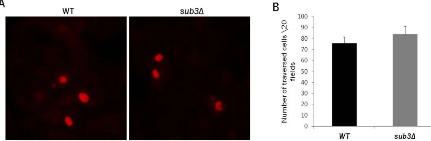
The traversal capacity of sub3Δ sporozoites in vitro was assessed by incubating sporozoites with a monolayer of hepatocytes in the presence of TOTO-1. Cells in which the nucleus was stained and, consequently, showed fluorescence in red were considered positive traversed cells (Figure 9A). This experiment was performed three times in triplicates. Number of traversed cells per 20 fields was counted in each triplicate for both the controls and the deletion mutants. The average number of traversed cells in the mutants (76) was similar to the controls (84). This result not only supports the motility data previously obtained, it also complements it by showing that, when in contact with a monolayer of cells, the mutant was able to traverse cells normally (Figure 9B).
Figure 9. Migration of sporozoites through hepatocytes. (A) Microscopy images of cells with stained nucleus in red after addition of WT sporozoites (left panel) and sub3Δ sporozoites (right panel) in the presence of TOTO-1. Cells were visualized under a fluorescence microscope at 40x magnitude (B) Average number of traversed positive cells per 20 fields by WT (black bar) and sub3Δ (grey bar) sporozoites.
sub3Δ parasites invade and multiply inside hepatocytes in vitro
Besides examining the capacity of sporozoites to glide we were also interested in studying the ability of the mutant parasites to infect and multiply inside the host cell. In order to do this we carried out an in
vitro development assay in which either WT or sub3Δ sporozoites were added to a monolayer of human
hepatoma cells and allowed to multiply for 24h. After that period of time, samples were fixed and GFP signal was amplified by immunofluorescence using a monoclonal antibody that recognizes Hsp70. This extra step facilitates observation of nuclei and counting of EEFs (Figure 10A). Two independent experiments in triplicate were performed and the total number of EEFs per coverslip was counted. On
35 average, 3300 WT and 3100 sub3Δ parasites successfully developed inside the hepatocyte, revealing that there was no significant difference between the controls and the recombinant parasites (Figure 10B). As a consequence of the data presented we can say that neither invasion of cells by sporozoites nor their development inside the host cell in vitro was affected by the deletion in the sub3 gene.
Figure 10. Parasite development in vitro. (A) Microscopy images of EEFs 24h post infection. WT EEFs are shown on the top and sub3Δ EEFs on the bottom. HepG2 nuclei were stained with Hoechst (top and bottom left panels), EEFs' GFP signal was amplified by immunofluorescene (top and bottom middle panels) and images of nuclei and EEFs were merged (top and bottom right panels). Cells were visualized under a fluorescence microscope at 40X magnification (B) Average number of WT (black bar) and sub3Δ (grey bar) EEFs per coverslip 24h post infection of a monolayer HepG2 cells with sporozoites.
36 sub3Δ blood stages were detected after sporozoites were injected in mice
Apart from scrutinizing the properties of sub3Δ parasites in vitro, we examined and characterized the capacity of salivary gland sporozoites to infect in vivo. Since P. berghei is used as an experimental mouse model it permits the use of mice to conduct in vivo studies. As a first hint to understand if the recombinant parasites have their capacity of infecting a host altered, we infected susceptible naïve C57BL\6 mice and determined the prepatent period – the number of days that took the parasites to become detectable in the blood. A one day difference in the prepatent period indicates an approximately 10 fold increase or decrease in the infectivity of the parasite depending on if it takes less or more time to be detected, respectively.
Four groups of five mice were infected with a certain number of parasites per mouse: 100 WT sporozoites (WT100), 100 mutant sporozoites (Δ100), 1000 WT sporozoites (WT1000) and 1000 mutant sporozoites (Δ1000). Results demonstrated that, as expected, a 10 fold difference in the number of sporozoites injected culminated in a one day difference in the prepatent period. In order to be considered positive at least one parasite had to be observed when searching the smear. The data collected from the observation of blood smears starting on day 3p.i. showed that in both groups infected with 100 sporozoites, parasites were first detected on day 4p.i., although 2 mice in the WT group did not becomepositive after observation of smears until day 10p.i.. In addition, in both groups of mice infected with 1000 sporozoites the parasites became detectable one day earlier, on day 3p.i., than the mice infected with 100 sporozoites (Figure 11). Collectively, these data gives evidence that there is no delay in the onset of the pathology in the sub3Δ recombinant parasites.
37
Figure 11. Prepatent Period. The day when parasites became detectable in the blood of each mouse was determined by blood smear. Four groups of five mice each were respectively injected with 100 WT (WT100) or 1000 WT sporozoites (WT1000) , 100 (Δ100) or 1000 mutant sporozoites (Δ1000). The presence (+) or absence (-) of parasites was recorded.
sub3Δ parasites were quantified in the liver upon intravenous and intradermal injection of sporozoites in
mice
Although the prepatent period in the mutants and the controls is the same, if the difference in infectivity between mutant and control was less than 10-fold, it would not be detectable in the previous set of experiments. In an attempt to find if that was the case, we conducted a more sensitive experiment. Two groups of 5-weeks old female C57BL\6 mice were injected intravenously with WT or sub3Δ sporozoites. After 38h.p.i. the parasite burden in the liver was quantified by qPCR. Parasite burden in the liver is quantified by the number of copies of parasite 18S ribosomal RNA (rRNA). We observed a similar
38 average number of 18S rRNA copies in both groups of mice, showing that when injected directly in the blood, sporozoites reach the liver and develop normally in vivo (Figure 12A).
The following in vivo experiment explored the ability of parasites to reach the liver by their natural route of infection, that is, beginning in the dermis. Two groups of five week-old female Swiss Webstar mice were infected by i.d. injection with WT or sub3Δ salivary gland sporozoites in the ear. This experiment provides further information on the capacity of these parasites to glide in vivo since they must migrate from the skin to the liver. I.d. injection of sporozoites contributed to a delay in reaching the liver as well as a decrease in the number of parasites that successfully reached the blood. The fact that we used a less susceptible host, compared to C57BL\6 mice, contributed, together with the two previous observations, to a lower number of copies detected. This experiment was done three times and, along with what was previously seen in other stages, recombinant parasites were not affected in their ability to reach and develop inside the liver of its host.
Figure 12. Quantification of parasite burden in the liver. (A) Average number of copies of P.berghei 18s risbosomal RNA in livers harvested 38h.p.i. from mice infected intravenously with either WT (black bar) or sub3Δ (grey bar) salivary gland sporozoites. (B) Average number of copies of P.berghei 18s risbosomal RNA in livers harvested 38h.p.i. from mice infected intradermaly with either WT (black bar) or sub3Δ (grey bar) salivary gland sporozoites.