1. Department of Pulmonology, Hospital Egas Moniz - Centro Hospitalar Universitário de Lisboa Ocidental, Lisboa, Portugal; 2. Department of Pulmonology, Centro Hospitalar Universitário de São João, Porto, Portugal;
3. Department of Pathology, Centro Hospitalar Universitário de São João, Porto, Portugal;
4. Department of Radiology, Centro Hospitalar Universitário de São João, Porto, Portugal;
5. Department of Rheumatology/Centro Hospitalar Universitário de São João, Porto, Portugal;
IntroductIon
Pleuroparenchymal fibroelastosis (PPFE) is a rare clini -cal–pathological entity1. It was officially described in
2004 by Frankel et al.2and is considered a specific
en-tity within the group of interstitial lung diseases (ILD)5.
The aetiology, pathophysiology and natural course of PPFE are unclear. A few associated conditions have been described1, including connective tissue diseases
(CTD) such as rheumatoid arthritis (RA)1and
cuta-neous systemic sclerosis (SSc)3. Although the
associa-tion with RA has been pointed in some reports, they all refer to the same article from 1980 with description of progressive upper lobe fibrosis in patients with RA, whose histological features were similar to those of PPFE4. ILD is common in SSc and PPFE might be a rare
additional pattern of ILD associated with this disease3.
cAse 1
59-year-old woman, non-smoker, with a 20-year diagno sis of RA. She was treated with Methotrexate for the first 5 years after diagnosis, which was switched to Adalimumab because of uncontrolled disease with a subsequent good response. She had no significant co-morbidities and denied relevant occupational or envi-ronmental exposures.
In the beginning of 2014 she complained of dry cough and exertional dyspnoea, without arthralgias or other relevant symptoms. Laboratorial analysis showed an erythrocyte sedimentation rate of 97mm/h. High Resolution Computed Tomography (HRCT) of the chest revealed interlobular septal thickening in both lungs, with predominance in the periphery of the lo wer lobes, compatible with a Nonspecific Interstitial Pneu-monia (NSIP) pattern. In addition, an irregular pleural and subpleural interstitial thickening in the upper lobes was also observed, predominantly in the apical se -gments (Figure 1-A). Lung function tests showed a re-ACTA REUMATOL PORT. 2019;44:264-269
Pleuroparenchymal fibroelastosis in
association with connective tissue disease:
a new interstitial pneumonia to be aware of
Carvalho J1, Vieira AC1, Ferra J1, Novais e Bastos H2, Caetano Mota P2,
Melo N2, Guimarães S3, Pereira JM4, Bernardes M5, Morais A2
AbstrAct
Pleuroparenchymal fibroelastosis (PPFE) is a rare and recently described interstitial pneumonia. It consists of progressive fibrosis involving the pleura and subpleu-ral lung parenchyma, predominantly in the upper lobes, with defined and reproducible clinical, radio-logical and histopathoradio-logical criteria. No effective treat-ment has yet been shown to modify the natural course of the disease, which vary greatly in the literature. Se -veral conditions have been associated with PPFE, in-cluding connective tissue diseases (CTD). The authors present two cases of female patients with a CTD (rheumatoid arthritis and limited cutaneous systemic sclerosis, respectively) who presented with typical bi-lateral upper lobe thickening in chest High Resolution Computed Tomography (HRCT). In the first case, di-agnosis was based on “definite” radiological and histopathological criteria for PPFE, while in the second case diagnosis was established on clinical grounds after discussion in a multidisciplinary team meeting. The authors present these cases of CTD-associated PPFE in order to raise awareness of this entity among clinicians.
Keywords: Interstitial lung disease; Connective tissue
duction in lung diffusing capacity for carbonmono -xide (DLCO, 68%), without other abnormalities. Blood gases were in the normal range. Flexible bron-choscopy with bronchoalveolar lavage (BAL) was per-formed and a high lymphocytosis of 41.6% with CD4+ predominance was noticed in the total and differential cell count. She started oral prednisolone 40 mg/day. After 1 month of follow-up, a favourable clinical and analytical evolution was already evident and steroids were gradually tapered off and stopped.
In 2015 Adalimumab was stopped due to neutrope-nia and she was again given prednisolone for arthral-gias. She remained without any relevant respiratory symptoms and had a stable lung function. After a rigor-ous haematologic evaluation, a diagnosis of medullary hypoplasia secondary to the rheumatic di sease was es-tablished. Adalimumab was restarted in 2016. A follow-up chest HRCT performed at that time revealed wors-ening of subpleural and septal interstitial thickwors-ening, especially in both upper lobes (Figure 1-B). Due to per-sistent polyarticular flare, treatment was switched from Adalimumab to Rituximab.
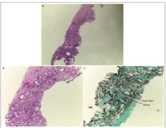
A follow-up chest HRCT in 2018 showed similar imaging features, namely those previously described in the upper lobes. The analysis of the serial chest HRCT findings raised the suspicion of PPFE and the patient underwent percutaneous transthoracic lung biopsy, which revealed pleural fibrosis and prominent sub-pleural and parenchymal fibroelastosis (Figure 2). She
is currently monitored in an ILD specialized centre and remains clinically and functionally stable, with no changes to prior medications.
cAse 2
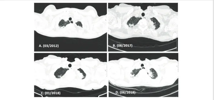
60yearold woman, nonsmoker, diagnosed with limi -ted cutaneous systemic sclerosis since she was 22 years old comprising Raynaud’s phenomenon, skin thick-FIGure 1.(A,B,C) – Evolution of High Resolution Computed Tomography (HRCT) thoracic imaging over time (axial images). Upper lobes pleural and subpleural parenchymal thickening representing fibrotic lesions are shown. Progression of fibrosis is seen over time.
FIGure 2.Section of upper lobe biopsy showing: (A, B) visceral pleura fibrosis and homogeneous dense subpleural fibrosis with prominent elastosis (C). Staining: A, B) haematoxylin and eosin; C) orcein staining. Magnification: A) 40×; B, C) 100×.
ening, digital ulcers, positive antinuclear antibodies at a 1:1000 titre with a centromere pattern and positive anti-centromere antibodies. She never developed any other specific organ involvement, had no significant comorbidities and denied relevant occupational or en-vironmental exposures. Her usual treatment included only nifedipin 30 mg daily. A chest HRCT performed in 2012 had no other significant features than appa rent residual upper lobes pleural and subpleural thicke -ning (Figure 3-A). Serial transthoracic echocardiogra-phy showed no evidence suggestive of pulmonary artery hypertension. SSc remained stable over the years, without any signs of activity and no treatment adjust-ment was required.
In 2016 she developed persistent dry cough, with mild exertional dyspnoea and weight loss of 5kg over 6 months. She denied other constitutional symptoms. Laboratory blood tests were unremarkable. A new chest HRCT (01/2017) revealed stable pleural and subpleu-ral thickening in the upper lobes and new patchy pe-ripheral consolidations in the upper and middle lobes and scattered solid micronodules (Figure 3-B). Lung function tests showed small airway obstruction and de-creased DLCO (62%); blood gases analysis was within the normal range. The BAL had no malignant cells or pathogenic microorganisms and total and differential cellular count showed mild neutrophilia (8,4%) and eosinophilia (2,0%). A percutaneous transthoracic lung
biopsy was subsequently performed, with histologic findings of lymphoid interstitial pneumonia (LIP), without evidence of additional lesions. Serial chest HRCT showed discrete progression of the upper lobes pleural and subpleural thicke ning (Figure 3-C, D). The case was reviewed and discussed in an ILD multidisci-plinary diagnosis meeting and a definitive diagnosis of LIP and PPFE in the SSc context was established. Since the patient remained clinically and functionally stable and the imaging extension was below 20%, no target-ed therapy was pres cribtarget-ed. She is currently monitortarget-ed in an ILD speciali zed centre.
dIscussIon
PPFE is a rare and recently described interstitial pneu-monia2,5-8. It consists of progressive fibrosis involving
the pleura and subpleural lung parenchyma, predomi -nantly in the upper lobes5,8, with a particular his to
-pathologic pattern of visceral pleural thickening with collagenous fibrosis, subpleural elastosis and intra-alve-olar collagenous fibrosis2, 9.
PPFE occurs mainly among non-smokers and has no gender predominance, with a median age of pre-sentation of 46 to 57 years, according to different se-ries1,6,9,10. In the depicted cases both patients were ne
-ver smokers with an age of presentation within the
FIGure 3.Evolution of thoracic imaging by High Resolution Computed Tomography (HRCT) over time (axial images). Upper lobes pleural and subpleural parenchymal thickening representing fibrotic lesions are shown. A slow progression of the fibrosis is seen over time.
range seen in literature.
Similarly to the reported cases, clinical presentation is not specific, with the most common symptoms con-sisting of exertional dyspnoea of insidious onset, dry cough and weight loss1,8,9,10,11. Chronic dull pleuritic
pain1,8,9,10, recurrent lung infections8 and platythorax
due to upper lobes fibrosis1,9,10,12 may also occur.
Spon-taneous pneumothorax is a characteristic complication in the natural history of PPFE, being present in ~30% of patients1,6,10, frequently with persistent air leak and
poor reexpansion of the underlying lung6, 10.
Lung function tests may present a restrictive venti-latory impairment. An increased residual volume /to-tal lung capacity ratio (RV/TLC) may be found due to compensatory hyperinflation of the lower lobes in sponse to upper lobe collapse. DLCO is typically re-duced and DLCO/VA can be either normal or slightly reduced. Arterial hypoxaemia and hypercapnia may arise with disease progression1, 9, 10. In both cases
pa-tients had reduced DLCO with normal DLCO/VA but presented minor ventilatory changes. This may explain their pauci-symptomatic clinical picture.
Diagnostic criteria for PPFE were proposed in 2012 by Reddy et al.8HRCT imaging criteria for PPFE were
divided in “definite” or “consistent with”. “Definite” cri-teria consist of pleural thickening and subpleural fibrosis concentrated in the upper lobes, while the lo -wer lobe involvement is less marked or absent8, 9.
Histological criteria for PPFE were also divided in “defi -nite” and “consistent with”. “Defi-nite” criteria include upper zone pleural fibrosis with subjacent intra-alveo-lar fibrosis, accompanied by alveointra-alveo-lar septal elastosis8,9
(Table I).
Despite that a definitive diagnosis of PPFE tradi-tionally requires histological examination11, a signifi-cant proportion of patients do not undergo lung biop-sy due to reasons such as very advanced disease, risk of iatrogenic pneumothorax or the fact that the disease can be strongly suspected based on clinical and radio-logical features10. For those cases where no biopsy is
available, a label of ‘‘consistent with PPFE’’ has been suggested6,8,10 and a multidisciplinary diagnostic
meet-ing is crucial to establish the diagnosis8. Recently
Enomoto and colleagues13proposed a clinical
diagno-sis of PPFE based on a combination of radiological find-ings of “definite PPFE”, radiologic confirmation of disea se progression and exclusion of other lung dis-eases with identifiable aetiologies, with results sup-porting accuracy and confidence in clinical diagnosis. The second reported case is an example of this
situa-tion. The diagnosis of PPFE was established in a mul-tidisciplinary diagnostic meeting, based only on clini-cal and radiologiclini-cal findings, with no need to pursue histologi cal confirmation.
In addition to genetic predisposition1,3,5,6,8,9,14-16, se
-veral conditions have been associated with PPFE, in-cluding previous lung and bone marrow transplanta-tion, chemotherapy, recurrent infections and connective tissue diseases (CTD), such as rheumatoid arthritis or systemic sclerosis1,3,5,6,8,9,15, as depicted above
(Table II). Radiological and histological features like those of PPFE have been reported in CTD patients with apical fibrosis in earlier case reports, prior to the recog-nition of this entity4, 6. These are the only data
sup-tAbLe I. crIterIA For the dIAGnosIs oF PLeuroPArenchymAL FIbroeLAstosIs HRCT Imaging Criteria of PPFE
“Definite”
• Upper lobe pleural thickening and subpleural fibrosis, and
• Lower lobe involvement less marked or absent “Consistent with”
• Upper lobe pleural thickening and subpleural fibrosis, but
– Distribution of changes not concentrated in upper lobes, or
– Presence of features of coexistent disease elsewhere
Histological Criteria of PPFE
“Definite”
• Upper zone fibrosis of the visceral pleura, and • Prominent, homogenous, subpleural intra-alveolar
fibrosis with alveolar septal elastosis, and
• Sparing of the parenchyma distant from the pleura, and
• At most mild, patchy lymphoplasmocytic infiltrates, and
• At most small numbers of fibroblastic foci present “Consistent with”
• Intra-alveolar fibrosis as above, but
– Not associated with significant pleural fibrosis, or – Not predominantly beneath the pleura, or – Not in an upper lobe biopsy
Adapted from: Thusen J, Pleuroparenchymal Fibroelastosis: Its Pathological Characteristics6
porting a definitive diagnosis of PPFE in patients with RA found by the authors4. In a recent study of patients
with CTD associated with ILD17, radiologic PPFE
le-sions were detected in 19% of patients. From these, 43% had SSc, 29% primary Sjogren syndrome, 11% poly/dermatomyositis, 6% RA and 28% overlapping CTDs. In this study the presence of PPFE lesions was identified as an independent risk factor of poor prog-nosis17. An idiopathic form of PPFE has also been
re-ported and has been included in the latest international
classification of idiopathic interstitial pneumo-nias1,3,5,6,8,9.
An aetiological theory developed by Thusen in 20136proposes that fibrosis with a PPFE pattern may
be the common final pathway shared by any form of lung injury leading to an intraalveolar fibrinous res -ponse.
Although PPFE may occur isolated within the lung, there is a high prevalence of coexistent ILD in patients with PPFE of the upper lobes8,12,15,17 reaching 75% in
one series12, mainly UIP pattern8,9,20. In these cases
PPFE may either represent or not the predominant abnormality (on HRCT and/or histology). It is essential to determine the predominant abnormality because it may have different therapeutic and prognostic impli-cations. In the presented cases, both patients with PPFE had a coexisting ILD: NSIP in the first case and LIP in the se cond case.
No treatment has yet been shown to modify the natu ral course of PPFE1, 7 and management of this disea
-se is ba-sed on limited evidence, mainly from the expe-rience of specialized centres7,18. A treatment approach
proposed by Brompton’s interstitial lung disease unit18
suggests introduction of a macrolide (azithromycin or clarithromycin 3 times/week) in patients presenting with recurrent infections due to its immunomodulato-ry effects. Patients with progressive or severe disease should be given corticosteroids at a moderate or low dose, with or without hydroxychloroquine18. If there
is no improvement, immunosuppressants such as azathioprine or mycophenolate mofetil may be consi -dered. Nevertheless, immunosuppressive drugs should be started with caution, since previous studies showed worse outcomes in idiopathic disease with intense im-munosuppression18. In advanced stages lung
trans-plantation should be considered in suitable pa-tients1,7,8,9,18,19.
Since parenchymal fibrosis is an important histo-logical feature of PPFE, some case reports evaluated the potential efficacy of antifibrotic agents in preventing lung function decline, with promising results15,19,20.
The clinical course of PPFE vary greatly in the litera -ture: some describe a slowly progressive disease over 10-20 years after presentation, and it may take years before patients become symptomatic; while others des -cribe a rapid clinical deterioration despite
treat-ment1,8,12. Once PPFE becomes symptomatic, patients
may remain stable for a long period of time or progress inexorably to hypercapnic respiratory failure, with a 40–66% mortality rate in a few years with or without tAbLe II. underLyInG dIseAses or condItIons
thAt mAy be AssocIAted wIth
PLeuroPArenchymAL FIbroeLAstosIs (PPFe) underLyInG dIseAses or condItIons thAt mAy be AssocIAted wIth PPFe
Underlying diseases or conditions that may be associated with PPFE
• Idiopathic PPFE • Hereditary PPFE
– Family history of PPFE
– Observed association with mutations in telomere-related genes (TERT, TERC, RTEL1) • Bone marrow or stem-cell transplantation • Lung Transplantation
• Chemotherapy (alkylating agents) • Radiotherapy
• Respiratory infections – Recurrent bronchitis – Aspergillus
– Mycobacterium avium intracellulare
• Autoimmune diseases – Systemic sclerosis – Rheumatoid arthritis – Psoriasis – Ankylosing spondylitis – Ulcerative colitis
– Primary Sjögren syndrome – Poly/dermatomyositis
• Hypersensitivity pneumonitis • Occupational dust exposure
– Asbestos – Aluminium
Portillo K et al, Pleuroparenchymal Fibroelastosis: Is it Also an Idiopathic Entity?1; Watanabe K, Pleuroparenchymal Fibroelastosis: Its Clinical Characteristics9; Newton CH et al,
Pleuroparenchymal Fibroelastosis Associated with TERT Mutations14; Newton CA et al, Telomere-related lung fibrosis is
an identifiable abrupt exacerbation6,8,10.
In conclusion, the authors present two cases of CTD-associated PPFE to raise awareness of this entity among clinicians. Imaging features of PPFE are very sugges-tive, if not pathognomonic10and a clinical diagnosis is
possible with multidisciplinary diagnostic meetings. Histopathological confirmation is often unnecessary and is reserved for cases of ILD with uncertain diag-nosis or suspicion of underlying neoplasm. Despite only recently recognized, case reports with radiologi-cal and histologiradiologi-cal features of PPFE in patients with CTD from as early as the 1960s12. This entity is
well-documented among patients with CTD, occurring alone or in association with other ILDs and the evi-dence suggests that the presence of PPFE harbours a worse prognosis (the words “in these patients” were eliminated). It may also require a specific treatment and affect the choice of immunosuppressive therapy in a particular patient since one needs to be cautious with immunosuppression in PPFE.
corresPondence to Joana Carvalho
Hospital Egas Moniz - Serviço de Pneumologia Rua da Junqueira nº 126, 1349-019 Lisboa E-mail: [email protected]
reFerences
1. Portillo K, Guasch Arriaga I, Ruiz-Manzano J. Pleuroparenchy-mal Fibroelastosis: Is it Also an Idiopathic Entity?. Arch Bron-coneumol. 2015;51:509–514.
2. Frankel SK, Cool CD, Lynch DA, Brown KK. Idiopathic pleu-roparenchymal fibroelastosis. Description of a novel clinico-pathologic entity. Chest.2004;126:2007–2013.
3. assoun D, Dirou S, Arrigoni PP, et al. Radiological pleuro-parenchymal fibroelastosis associated to limited cutaneous sys-temic sclerosis: a case report. BMC Pulm Med Published Online First: 18 May 2018. doi:10.1186/s12890-018-0641-5 4. Petrie GR, Bloomfield P, Grant IW, Crompton GK. Upper lobe
fibrosis and cavitation in rheumatoid disease. Br J Dis Chest. 1980;74:263–267.
5. Travis W, Costabel U, Hansell D, et al. An Official American Thoracic Society/European Respiratory Society Statement: Up-date of the International Multidisciplinary Classification of the Idiopathic Interstitial Pneumonias. Am J Respir Crit Care Med 2013; 188:733–748.
6. Thusen JH. Pleuroparenchymal Fibroelastosis: Its Pathological Characteristics. Current Respiratory Medicine Reviews 2013; 9: 238-247.
7. Bonifazi M, Montero MA, Renzoni EA. Idiopathic Pleuro-parenchymal Fibroelastosis. Curr Pulmonol Rep 2017; 6:9–15. 8. Teddy TL, Tominaga M, Hansell DM, et al. Pleuroparenchymal Fibroelastosis: a spectrum of histopathological and imaging phe-notypes. Eur Respir J 2012; 40: 377–385.
9. Watanabe K. Pleuroparenchymal Fibroelastosis: Its Clinical Characteristics. Current Respiratory Medicine Reviews 2013; 9: 229-237.
10. Camus P, Thusen J, Hansell D, Colby T. Pleuroparenchymal fi-broelastosis: one more walk on the wild side of drugs?. Eur Respir J 2014; 44: 289–296.
11. Redondo MT, Melo N, Mota PC, et al. Idiopathic pleuro-parenchymal fibroelastosis: A rare but increasingly recognized entity. Rev Port Pneumol 2015; 21(1):41-44.
12. Nakatani T, Arai T, Kitaichi M, et al. Pleuroparenchymal fi-broelastosis from a consecutive database: a rare disease entity?. Eur Respir J. 2015; 45:1183–1186.
13. Enomoto Y, Nakamura Y, Satake Y, et al. Clinical diagnosis of pleuroparenchymal fibroelastosis: A retrospective multicenter study. Respiratory Medicine 2017; 133:1–5.
14. Newton CH, Batra K, Torrealba J, Meyer K, Raghu G, Garcia CK. Pleuroparenchymal Fibroelastosis Associated with TERT Mutations. Eur Respir J 2017; 49: 1700696. doi:10.1183/ 13993003.00696-2017.
15. Boerner EB, Costabel U, Wessendorf TE, Theegarten D, Bonel-la F. Idiopathic pleuroparenchymal fibroeBonel-lastosis (PPFE) – A case study of a rare entity. Rev Port Pneumol 2017; 23(6):352--355.
16. Newton CA, Batra K, Torrealba J, et al. Telomere-related lung fi-brosis is diagnostically heterogeneous but uniformly progres-sive. Eur Respir J 2016; 48:1710–1720.
17. Enomoto Y, Nakamura Y, Colby TV, et al. Radiologic pleuro-parenchymal fibroelastosis-like lesion in connective tissue dis-ease-related interstitial lung disease. PLoS ONE 12(6): e0180283.
18. Pleuroparenchymal fibroelastosis. Royal Brompton Hospital in-terstitial lung disease unit. http://www.rbht.nhs.uk/patients/ condition/pleuroparenchymal-fibroelastosis/. Accessed in Oc-tober 28th 2018.
19. Sato S, Hanibuchi M, Takahashi M, et al. A patient with idio-pathic pleuroparenchymal fibroelastosis showing a sustained pulmonary function due to treatment with pirfenidone. Intern Med. 2016; 55(5): 497–501.
20. Nasser M, Chebib N, Philit F, et al. Treatment with nintedanib in patients with pleuroparenchymal fibroelastosis. Eur Respir J 2017; 50 (suppl 61): PA4876