A R T I G O O R I G I N A L
C E N T R A L A U D I T O R Y P R O C E S S I N G I N P A T I E N T S
W I T H S Y S T E M I C L U P U S E R Y T H E M A T O S U S
Ana Paula Bruner,
*Emilia Inoue Sato,
**Liliane Desgualdo Pereira
****Audiology Division Otolaryngology Department, Santa Casa School of Medicine of São Paulo
**Full Professor Rheumatology Division, Department of Medicine, Federal University of São Paulo
***Full Professor Speech Pathology and Audiology Department, Federal University of São Paulo
Introduction
Neuropsychiatric (NP) disorders are frequent in pa-tients with Systemic Lupus Erythematosus (SLE)1-3 and may result from a number of different patho-genic mechanisms and events occurring in the cen-tral and peripheral nervous systems.4A number of disorders have been reported associated to SLE, from mild mental dysfunctions to seizures, psycho-sis, headaches, depression, cognitive dysfunction and anxiety disorders.5-9 Attention span disorders, impaired processing of short and long term me-mory, and verbal and visual-spatial information have also been associated to SLE.10
Central Auditory Processing is a complex ana-lysis performed by the auditory system from the outer ear to the interpretation of the auditory infor-mation by structures in the central nervous system (CNS) and in the brain. The following processes and mechanisms are involved: selective attention, sound detection, sensation and localization, dis-crimination of isolated and sequential sounds, as well as speech recognition, comprehension and memory. All these processes present both neu-rophysiologic and functional correspondents.11-13
Temporal processing is one of the functions per-formed by the auditory system. It involves a num-ber of temporal skills such as resolution, masking, integration and ordering, which allow a listener to hear acoustic variations in a certain time inter-val.12-14These skills are essential for the processing of the sounds of speech.14-16Temporal processing tests offer a comprehension of the way in which acoustic events occurring over time are perceived by the listener and how his/her work memory functions. The primary auditory cortex and the as-sociation cortex are involved in gap-detection tasks during auditory stimulation, where the same group of neurons is activated before and after a silent in-terval.17,18Thus, the temporal resolution skill de-pends on the integrity of central neurological struc-tures, especially the auditory cortex.
Auditory processing impairment results from
Abstract
Systemic Lupus Erythematosus (SLE) patients can also present neuropsychiatric disorders, such as impaired memory and attention. Central auditory processing depends on a great number of skills con-trolled by the central nervous system.
Objective: To characterize the performance of SLE
patients, with and without neuropsychiatric disor-ders (NP), in central auditory processing tests.
Patients and Methods: A prospective controlled
study of Dichotic Speech and Temporal Processing Tests was carried out at a tertiary teaching Institu-tion. Three groups were formed with 20 individuals each, totalizing sixty women with age varying from 18 to 48 years: Group I – SLE patients without neu-ropsychiatric disorders; Group II – SLE patients with neuropsychiatric disorders as well; and Group III - age and gender matched healthy controls.
Results: Dichotic Speech Test showed similar
re-sults for patients in Group I and for healthy con-trols; however, most of the patients in Group II pre-sented impaired performance with great variability of response. SLE patients (Groups I and II) presen-ted a significantly lower performance in Temporal Processing tests than controls.
Conclusion: SLE patients especially those with
neuropsychiatric disorders presented impaired central auditory processing, which may contribu-te to the memory and atcontribu-tention impairment.
Keywords: Audiometry; Audiology; Central
A N A PA U L A B R U N E R E C O L.
functional abnormalities in one or more of these auditory processes, and may be caused by neuro-logical problems associated to demyelination, brain atrophy, focal brain lesions, and others.13 Pa-tients with others systemic diseases and altered cognitive and psycho-emotional functions may also present central auditory processing impair-ment.19
Many studies have shown the association between poor central auditory processing and cen-tral nervous system abnormality in various disea-ses.13,18,19However, auditory processing evaluation is limited in SLE. Auditory processing disorders could impair the cognitive-communicative and/or language related functions in lupus patients.
The aim of the study was to evaluate the central auditory processing skills in SLE patients, with and without neuropsychiatric disorders, using dicho-tic listening and temporal processing tests.
Patients and Methods
A total of 40 adult women with SLE, according to ACR classification criteria20from a tertiary Rheu-matology out-patient clinic and twenty healthy vo-lunteers were studied. The participants were sub-divided into three groups: 20 SLE patients without NP involvement (Group I), 20 SLE patients with NP involvement (Group II), and 20 age-matched healthy women with no systemic disease, auditory or neurological impairment (Group III). Patients’ age ranged from 18 to 48 years. All participants were literate and signed a post informed consent form approved by the Institutional Ethics Com-mittee.
Group I included patients without neu-ropsychiatric feature. Patients with memory or at-tention impairment or any other cognitive impair-ment complaints were excluded. Group II NP ma-nifestations were: lupus headache (10%), panic syndrome (10%), strokes (15%), anxiety disorder (15%), major depression (30%), psychosis (40%) and/or seizures (65%). Seventy-five percent of pa-tients presented more than one manifestation.
All the participants had normal21hearing thres-holds and normal mobility of the tympanic- ossi-cular system assessed by tonal threshold audio-metry, speech reception testing, percentage index of speech recognition, and acoustic immitance. None of the participants presented mechanical or neurosensorial hearing loss, which could interfe-re with the tests interpinterfe-retation.
Auditory processing was evaluated through three specific tests performed in the following or-der: Binaural integration task of the Dichotic Digit Test (DDT) Portuguese version22for assessment of recognition of overlapped verbal sounds in dicho-tic hearing (different auditory stimuli presented to each ear simultaneously) and hearing capability of binaural integration; Random Gap Detection Test – RGDT (Auditec, Saint Louis, MO)23for iden-tification and quaniden-tification of the subject’s reso-lution capacity; Pure tone Duration Pattern Test (DPT)24,25to assess sound discrimination abilities related to duration and temporal ordering and is also known as temporal processing. Responses were expressed in percentages of correct answers for the DDT and DPT, and in milliseconds (ms) for the RGDT.
The DDT was comprised of 80 words presented in pairs, a different pair of digits in each ear pre-sented at the same time (dichotic hearing). There were 20 pairs of dissyllabic words (digits) which were randomly repeated twice in each ear.
In RGDT the subject was expected to identify the presence of two short consecutive sounds; the gap detection was expressed as the shortest inter-val (in milliseconds) of detection of both sounds. The test includes stimuli at four frequencies (0,5k, 1k, 2k, 4kHz), presented to both ears at the same time.
In DPT the subject was expected to identify a se-ries of three sounds by verbalizing if the sound was short or long (V_DPT) or by imitating or humming the sound (H_DPT). Stimuli were presented sepa-rately to the right and left ears.
Hearing tests were performed at 50dBNS in a sound treated booth using TDH-49 supra-aural he-adphones (Telephonics, Denmark). A two channel Orbiter-Madsen 922 audiometer (GN Otometrics,
Denmark) calibrated according to ANSI S 3:6 – 1989 guidelines was used. The audiometer was connec-ted to a CD recorder/player (Panasonic Inc., USA)
for digital recording of the tests.
Statistical analysis: Descriptive analysis was per-formed using mean and 95% of confidence inter-val. Patients in Groups I and II were compared to the control (Group III). In DDT, V_DPT and H_DPT tests responses were considered abnormal when the average number of correct answers was below the minimum average of the control group. RGDT interpretation was performed by averaging the gap detection threshold for all tonal stimuli (0,5k, 1k, 2k, 4kHz). The results were considered abnormal
C E N T R A L A U D I T O R Y P R O C E S S I N G I N S L E
when they exceeded the maximum threshold ob-served in the control group. The correlation betwe-en results and duration of the disease was carried out using Spearman’s correlation test (r). Inferen-tial analysis of auditory processing tests was based on the results obtained from each ear for DDT and DPT, or the frequency tested in RGDT. The compa-rison of abnormal results in Group I and Group II was performed using Tukey test. Fisher’s exact test was used to compare the results obtained in each separate ear in the SLE patients who responded below the average of the control group.26P value ≤ 0,05 was considered significant.
Results
The mean disease duration of Group I and II was 7.7±4.71 years and 7.9±5.19 years, respectively. The mean age of group I, II and III was 29± 4.64, 31±7.82 and 32±8.47 years, respectively.
Dichotic Digit Test (DDT)
Group I and III presented similar responses on DDT. However, most Group II patients had poor DDT responses with great variability. The mean percentage of correct responses on DDT of Group I, II and III was respectively 99% (CI 98.17–99.83%), 93.4% (CI 90.3–96.5%) and 99.6% (CI 99.2–100.0%); p=0.027 for right ear, and 98.6% (CI 97.65–99.35%), 91.5% (CI 88.8–94.92%) and 99.4% (CI 98.91–99.89%); p=0.027 for left ear.
No significant difference on DDT results was ob-served in any of the three groups when comparing right and left ear (p=0.054). Groups I and III pre-sented similar average performance on DDT (p=0.860), whereas Group II had poorer results when compared to the other two groups (p<0.001). Random Gap Detection Test (RGDT)
RGDT results were poor in Group I and worse in Group II, whereas healthy controls presented the best responses. Only one patient of Group II sented outlier responses (Figure 1). Group III pre-sented gap detection thresholds ≤ 10 ms on all sound frequencies of RGDT [mean 6.6 ms (CI 5.92–7.28 ms)]. Groups I and II presented a mean of 16 ms (CI 11.95–20.05 ms) and 46.1 ms (CI 31.24–60.96 ms), respectively, on all frequencies.
There was no significant difference on RGDT re-sults obtained by different sound frequencies in any of the three groups (p=0.065). When
compa-ring the three groups, Group III presented the best results, i.e., the lowest average gap detection thres-holds on RGDT, and Group II presented the hig-hest average gap detection thresholds on RGDT when compared to Group I and Group III (p<0.001 for all the comparisons).
Duration Pattern Test (DPT)
A significant number of Group I patients presen-ted poor DPT results (V_DPT and H_DPT, for both right and left ear stimuli), when compared to Group III. The worst DPT responses were found in Group II (Figure 2).
A similar response on the right and left ears on DPT tests was found in Group II and III. The per-centage of correct responses in V_DPT and H_DPT tests was similar for all three groups. Only Group I presented a significant difference between DPT scores in each ear, the left ear presenting a lower average response than the right ear (p<0.001). The averages of the three groups were distinct when compared two by two in each ear. A higher avera-ge of correct responses was found in Group III, fol-lowed by Group I and finally by Group II (p<0.001). Only Group II presented a significant correlation between DDT and V_DPT results (p=0.016). No cor-relation was found between the performance on any of the auditory processing tests in patients from Group I and healthy controls.
There was no correlation between the perfor-mance on DDT or RGDT and the disease duration. A weak negative correlation was found between DPT responses and the disease duration in Group I (V_DPT in the right, r= -0.49; p= 0,029, and left ears, r= -0.44; p= 0,05, and H_DPT in the right ear, r= -0.60; p= 0,005, and r = -34; p= 0.144 in the left ear), suggesting that the percentage of correct answers decreases as the disease progresses in the-se patients (Figures 3 and 4). No correlation was observed in Group II.
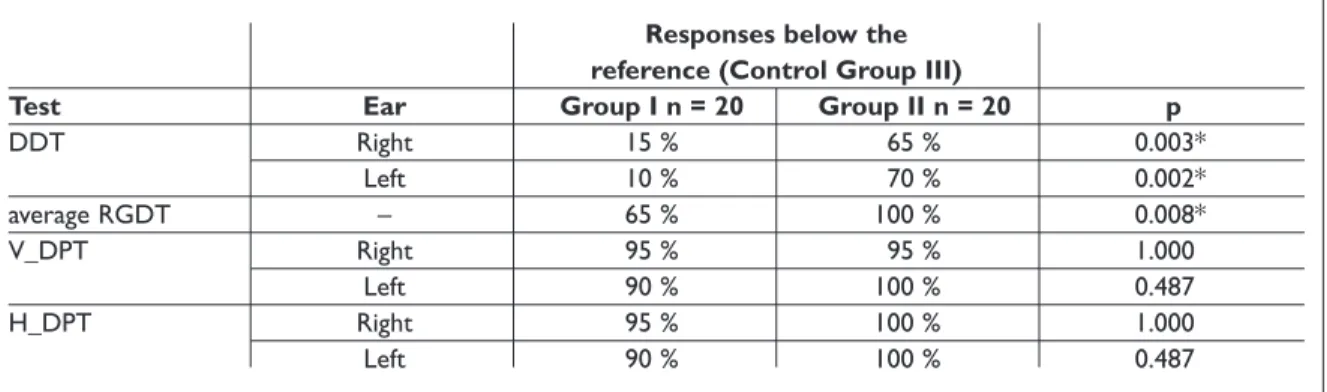
Table I depicts the abnormal test results in Group I and II, using the healthy controls as refe-rence. A great occurrence of abnormal RGDT and DPT results (both V_DPT and H_DPT) was found in both groups.
Discussion
Our study showed that SLE patients present impai-red auditory processing skills, mainly those with neuropsychiatric disorders. Most abnormal results
A N A PA U L A B R U N E R E C O L.
were found in tests of temporal processing. The studied sample was comprised solely of fe-males, which is in accordance with the prevalence of SLE in this gender.27The neuropsychiatric symp-toms associated to SLE varied greatly and were si-milar to those previously reported in literature.1,28,29 SLE patients with NP disorders had poorer DDT responses with greatest standard deviation, when compared to the other two groups. The same alte-ration was also observed in patients with epilepsy in another study.30No significant differences were found between the responses of the right and left ears in any of the groups, differently from what was reported by Kimura.31This difference may be ex-plained by the characteristics of the verbal stimu-li used in the test. Although both the Engstimu-lish and the Portuguese language versions use representa-tions of numbers, in the former language the words were monosyllables and in Portuguese, the words were disyllables.
Concerning RGDT, only Group III presented re-sults similar to or better than those found in cur-rently available normative data.23,32,33It is expected that normal individuals respond correctly to 70% or more of the stimuli in DPT.24In the current study none of the SLE patients presented a percentage of correct responses as high as the healthy controls. SLE patients with NP involvement presented the worst responses (for both V_DPT and H_DPT,
re-gardless of the tested ear).
Temporal processing involves the right and left hemispheres. The right hemisphere recognizes the whole and provides ordering and the corpus callo-sum transfers the information to the left
hemisphe-re which will label it linguistically after sequencing the message, regardless of the ear tested.34,35Thus, in SLE patients with corpus callosum involvement,
DPT would become a valuable tool by differentia-ting verbal and humming responses.2,36
The normal interval for detection and ordering of two distinct acoustic stimuli is at least 20 ms.17 In the current study, SLE patients with NP involve-ment presented a higher interval, suggesting that the relatively short duration of the acoustic stimu-li in DPT was insufficient to allow ordering of the distinct sounds by these patients.
SLE patients with NP involvement presented the greatest variability of responses in all the tests and were characterized as the most heterogeneous group. This finding may be related to structural neurological deficits, since performance variability has been suggested as a predictive factor of neu-rological impairment.37However, a number of pa-tients in Group II did not present neurological di-sorders likely to cause structural neurological se-quelae. It is also important to note that the current study did not consider the possibility of
perma-Figure 1. Box-plots for RGDT results by sound frequency (Hz) of groups I, II and III.
RGDT Random Gap Detection Test; Group I: patients with
systemic lupus erythematosus and no neuropsychiatric involvement; Group II: patients with systemic lupus erythematosus and neuropsychiatric involvement;
Group III: control group. * aberrant responses.
Confidence intervals (averaging the gap detection for all stimuli) Group I: 11.95 – 20.05 ms, Group II: 31.24 – – 60.96 ms, Group III: 5.92 – 7.28 ms.
Figure 2. Box-plots for V_DPT results of groups I, II and III for the right and left ear.
V_DPT Duration Pattern Test by verbalizing; Group I:
patients with systemic lupus erythematosus and no neuropsychiatric involvement; Group II: patients with systemic lupus erythematosus and neuropsychiatric involvement; Group III: control group;
Confidence intervals for right ear Group I: 60.4 – – 68.6%, Group II: 40.3 – 52.3%, Group III: 81.4 – 87.6%; and for left ear Group I: 53,3 – 63.7%, Group II: 39.4 – – 49.6%, Group III: 81.5 – 87.5%.
C E N T R A L A U D I T O R Y P R O C E S S I N G I N S L E
nent neurological damage as a variable in the eva-luation of auditory processing, since both anato-mical and functional abnormalities of the CNS may be involved in abnormal central auditory proces-sing.38
The disease duration did not seem to interfere in the test results of Group II patients. This finding may be explained by the great variability of res-ponses and neuropsychiatric symptoms presen-ted by these patients. Patients without NP involve-ment were not influenced by the duration of the di-sease in DDT and RGDT, but presented worse res-ponses in V_DPT in both ears and H_DPT in the right ear as the duration of the disease increased.
It has been reported that performance in these tests may be intrinsically correlated to the work memory, and the current study agrees with pre-vious reports.39 Other studies of specific neu-ropsychiatric disorders have also not found a cor-relation between the duration of the disease and attention deficits40or cognitive/psychological im-pairment41in SLE patients.
Patients in Group I and healthy controls showed no correlation of the performance between speci-fic auditory processing tests. This is comprehensi-ble, considering that each test involves distinct physiological mechanisms. However, SLE patients with NP involvement presented significantly lower performance on DDT and DPT, suggesting
Tabela I. Percentage of SLE patients with responses below those of the reference group (healthy controls).
Responses below the reference (Control Group III)
Test Ear Group I n = 20 Group II n = 20 p
DDT Right 15 % 65 % 0.003* Left 10 % 70 % 0.002* average RGDT – 65 % 100 % 0.008* V_DPT Right 95 % 95 % 1.000 Left 90 % 100 % 0.487 H_DPT Right 95 % 100 % 1.000 Left 90 % 100 % 0.487
DDT Dichotic Digit Test; RGDT Random Gap Detection Test; V_DPT Duration Pattern Test by verbalizing; H_DPT Duration Pattern Test by humming; Group I:
pa-tients with systemic lupus erythematosus and no neuropsychiatric involvement; Group II: papa-tients with systemic lupus erythematosus and neuropsychiatric in-volvement; * significant p values (<0,05).
Figure 3. Box-plots for V_DPT and duration of the disease of Groups I and II for the right and left ear.
V_DPT Duration Pattern Test by verbalizing; Group I:
patients with systemic lupus erythematosus and no neuropsychiatric involvement; Group II: patients with systemic lupus erythematosus and neuropsychiatric involvement.
Figure 4. Box-plots for H_DPT and disease duration of Groups I and II for the right and left ear.
H_DPT Duration Pattern Test by humming; Group I:
patients with systemic lupus erythematosus and no neuropsychiatric involvement.
A N A PA U L A B R U N E R E C O L.
these tests are highly sensitive to CNS disor-ders.23,30,41-43 The majority of SLE patients without NP involvement performed well on DDT, unlike patients with SLE with NP disorders who presen-ted a higher and significant percentage of poor res-ponses, once again showing that these individuals present greater difficulty in recognizing familiar linguistic sounds in dichotic hearing.
As to RGDT, the results showed a significant im-pairment in temporal resolution, especially in pa-tients with NP involvement, which adds to the dif-ficulty in acoustic perception of speech sounds.
Most SLE patients and all those with NP invol-vement responded poorly to DPT for stimuli pre-sented to at least one of the two tested ears. A cou-ple of possible reasons may explain why SLE pati-ents without NP involvement also presented poor DPT responses: a higher demand of attention ca-pabilities40or the presence of mild undetectable CNS dysfunctions most likely associated to micro-vascular abnormalities or anti-CNS neuron auto-antibodies.4,8,28,44,45These discrete abnormalities may still be asymptomatic and clinically undetec-table,1,10but could determine the poorer responses. Temporal processing and Dichotic digit results have shown to be very sensitive at detecting neu-romorphological lesions such as cranial trauma, epileptic seizures, etc.24,46,47 48Tests that assess tem-poral resolution have been found to be more sen-sitive to cortical than to brain stem lesion.47
Conclusions
Auditory processing tests contributed to the iden-tification of abnormal physiological auditory me-chanisms in SLE patients. The current findings sug-gest SLE may be associated to difficulties in cen-tral auditory processing, impairing dichotic liste-ning and temporal processing, especially in patients with neuropsychiatric involvement.
Correspondence to
Ms. Ana Paula Bruner
Departamento de Fonoaudiologia UNIFESP. Rua Botucatu, 802, São Paulo,
SP, Brazil / CEP: 04023-900. Phone/Fax: +55-11-5576-4531 E-mail: [email protected]
Acknowledgments
Our deepest gratitude to the physicians from the Rheumatology Division of the Internal Medicine
Department of the Federal University of São Paulo School of Medicine for their essential collaboration.
References
1. Cotton F, Bouffard-Vercelli J, Hermier M, et al. Ap-port de lÍRM cérébrale dans une série de 58 cas de malade lupique avec ou sans manifestations neu-ropsychiatriques. [MRI of central nervous system in a serie of 58 systemic lupus erythematosus pa-tients with or without overt neuropsychiatric mani-festations]. Rev Med Interne 2004; 25: 8-15.
2. Appenzeller S, Rondina JM, Li LM, Costallat LTL, Cendes F. Cerebral and corpus callosum atrophy in systemic lupus erythematosus. Arthritis Rheum 2005; 52:2783-2789.
3. Shimojima Y, Matsuda M, Gono T, Ishii W, Ikeda S. Relationship between clinical factors and neu-ropsychiatric manifestations in systemic lupus erythematosus. Clin Rheumatol 2005; 24:469-475. 4. Sanna G, Bertolaccini ML, Mathieu A. Central
ner-vous system lupus: a clinical approach to therapy. Lupus 2003; 12:935-942.
5. Appenzeller S, Kobayashi E, Costallat LTL, et al. Magnetic resonance imaging in the evaluation of patients with aseptic meningoencephalitis and connective tissue disorders. Arq Neuropsiquiatr 2000; 58:45-51.
6. Koseda-Dragan M, Hebanowski M, Badzio-Jagiello H, et al. New diagnostic criteria for the neu-ropsychiatric form of systemic lupus erythemato-sus. Psychiatr Pol 2000; 34:993-1005.
7. Weiner SM, Otte A, Schumacher M, et al. Diagnosis and monitoring of central nervous system involve-ment in systemic lupus erythematosus: value of F-18 fluorodeoxyglucose PET. Ann Rheum Dis 2000; 59:377-385.
8. Csépány T, Bereczki D, Kollár J, Sikula J, Kiss E, Csi-ba L. MRI findings in central nervous system syste-mic lupus erythematosus are associated with im-munoserological parameters and hypertension. J Neurol 2003; 250:1348-1354.
9. Takahashi T, Kokubun Y, Okuhata Y, Sawada S, Mi-zutani T. A central nervous system lupus showing peculiar findings on cranial magnetic resonance imaging (MRI). Rinsho Shinkeigaku 2003; 43:409--416.
10. Sabaddini MG, Manfredi AA, Bozzolo E, et al. Cen-tral nervous system involvement in systemic lupus erythematosus patients without overt neuropsy-chiatric manifestations. Lupus 1999; 8:11-9. 11. Phillips DP. Central auditory processing: a view
from auditory neuroscience. Am J Otol 1995; 16:338-352.
12. American Speech-Language-Hearing Association (ASHA). Central auditory processing: current status of research and implications for clinical practice. Am J Audiol 1996; 5:41-54.
C E N T R A L A U D I T O R Y P R O C E S S I N G I N S L E
13. Chermak GD. Deciphering auditory processing di-sorders in children. Otolaryngol Clin North Am 2002; 35:733-749.
14. Phillips SL, Gordon-Salant S, Fitzgibbons PJ, Yeni-Komshian G. Frequency and temporal resolution in elderly listeners with good and poor word recogni-tion. J Speech Lang Hear Res 2000; 43:217-228. 15. Emmanuel DC. The auditory processing battery:
survey of common practices. J Am Acad Audiol 2002;13:93-117.
16. Purcell DW, John SM, Schneider BA, Picton TW. Hu-man temporal auditory acuity as assessed by enve-lope responses. J Acoust Soc Am 2004;116:3581--3593.
17. Bellis TJ. Mechanisms of selected auditory and re-lated processes. In: Bellis TJ. Assesment and mana-gement of central auditory processing disorders in the educational setting: from science to practice. 2nd ed. New York: Delmar Learning, 2003: 51-102. 18. Sulakhe N, Elias LJ, Lejbak L. Hemispheric
asymmetries for gap detection depends on noise type. Brain Cogn 2003; 53:372-375.
19. Musiek FE, Baran JA, Pinheiro ML. Neuroaudio-logy: case studies. San Diego: Singular Publishing Group; 1994: 279p.
20. The 1997 Update of the 1982 American College of Rheumatology Revised Criteria for Classification of Systemic Lupus Erythematosus, 1997. http://www. rheumatology.org/publications/classificati- on/SLE/1997UpdateOf1982RevisedCriteriaClassifi-cationSLE.asp
21. Davis, H. Abnormal hearing and deafness. In: Davis H, Silverman SR, ed. Hearing and deafness. New York: Holt, Rinehart and Winston, 1970: 83-139. 22. Pereira LD, Schochat E. Processamento auditivo
central: manual de avaliação. São Paulo: Lovise; 1997: 147-149.
23. Keith RW. Random gap detection test. Auditec of Saint Louis. Missouri: 2000.
24. Musiek FE, Baran JA, Pinheiro ML. Duration pat-tern recognition in normal subjects and patients with cerebral and cochlear lesions. Audiology 1990; 29:304-313.
25. Musiek FE. Frequency (pitch) and duration pattern tests. J Am Acad Audiol 1994; 5:265-268.
26. Fisher LD, Van Belle G, ed. Biostatistics: A metho-dology for the health sciences. 1993. New York: John Wiley & Sons Inc, 1993: 858p.
27. Vilar MJP, Sato EI. Estimating the incidence of systemic lupus erythematosus in a tropical region (Natal, Brazil). Lupus 2002;11:528-532.
28. Bluestein HG. Neuropsychiatric manifestations of systemic lupus erythematosus. N Engl J Med 1987; 317:309-311.
29. Miguel EC, Pereira RM, Pereira CA, et al. Psychiatric manifestations of systemic lupus erythematosus: clinical features, symptoms and signs of central
nervous system activity in 43 patients. Medicine (Baltimore) 1994;73:224-232.
30. Kimura D. A note on cerebral dominance in hea-ring. Acta Otolaryngol 1963; 56:617-618.
31. Kimura D. Left-right differences in the perception of melodies. Q J Exp Psychol 1964; 16:355-358. 32. Snell KB. Age-related changes in temporal gap
de-tection. J Acoust Soc Am 1997; 101:2214-2220. 33. Schneider BA, Pichora-Fuller MK. Age-Related
Changes in Temporal Processing: Implications for Speech Perception. Semin Hear 2001; 22: 227-240. 34. Musiek FE, Pinheiro ML, Wilson DH. Auditory
pat-tern perception in 'split brain' patients. Arch Oto-laryngol 1980; 106:610-612.
35. Musiek FE, Pinheiro ML. Frequency patterns in cochlear, brainstem and cerebral lesions. Audiology 1987; 26:79-88.
36. Appenzeller S, Faria A, Marini R, Costallat LT, Cen-des F. Focal transient lesions of the corpus callo-sum in systemic lupus erythematosus. Clin Rheu-matol 2006; 25:568-571.
37. Paulus MP, Geyer MA, Braff DL. Use of methods from chaos theory to quantify a fundamental dysfunction in the behavioral organization of schi-zophrenic patients. Am J Psychiatry 1996; 153:714--717.
38. Gladman DD, Goldsmith CH, Urowitz MB, et al. The systemic lupus international collaborating cli-nics/American College of Rheumatology (SLICC/ ACR) damage index for systemic lupus erythemato-sus international comparison. J Rheumatol 2000; 27:373-376.
39. Schucard JL, Parrish J, Shucard DW, McCabe DC, Benedict RH, Ambrus J. Working memory and pro-cessing speed deficits in systemic lupus erythema-tosus as measured by the paced auditory serial ad-dition test. J Int Neuropsychol Soc 2004; 10:35-45. 40. Kerr EN. Attentional capacity in patients with
systemic lupus erythematosus. Diss Abstr Int [B] 1996; 56:5771.
41. Kozora E, Thompson LL, West SG, Kotzin BL. Analy-sis of cognitive and psychological deficits in syste-mic lupus erythematosus patients without overt central nervous system disease. Arthritis Rheum 1996; 39:2035-2045.
42. Strouse AL, Hall JW. Test-retest reliability of a di-chotic test for assessing central auditory function in Alzheimer's disease. Audiology 1995; 34:85-90. 43. Olivares-García MR, Peñaloza-López YR,
García-Pe-droza F, et al. Identificación de la lateralidad auditi-va mediante una prueba dicótica nueauditi-va con dígitos en español, y de la lateralidad corporal y orientaci-ón espacial en niños con dislexia y en controles. [Identification of auditory laterality by means of a new dichotic digit test in Spanish, and body latera-lity and spatial orientation in children with dyslexia and in controls]. Rev Neurol 2005; 41:198-205.
A N A PA U L A B R U N E R E C O L.
44. Futrell N, Schultz LR, Millikan C. Central nervous system disease in patients with systemic lupus erythematosus. Neurology 1992; 42:1649-1657. 45. Muñoz-Málaga A, Anglada JC, Paez M, Girón JM,
Barrera A. Psicosis como manifestación de lupus eritematoso sistémico: valor de prueba de la banda lúpica frente a los anticuerpos antirribosomales. [Psychosis as the initial manifestation of systemic lupus erythematous: the role of lupus band test and antiribosomal antibodies.] Rev Neurol. 1999; 28:779-781.
46. Shinn JB. Temporal processing and temporal pat-terning tests. In: Musiek FE, Chermak GD. Handbo-ok of (central) auditory processing disorder: audi-tory neuroscience and diagnosis. San Diego: Plural Publishing, 2007: 231-256.
47. Musiek FE, Shinn J, Jirsa R, Bamiou D, Baran JA, Zaidan E. The GIN® (Gaps-in-Noise) Test perfor-mance in subjects with confirmed central auditory nervous system involvement. Ear Hear 2005;26:608-618.
48. Musiek FE, Wilson DH. SSW and dichotic digit sults pré- and post-commissurectomy: a case re-port. J Speech Hear Disord 1979;44:528-533.