Universidade de Lisboa
Faculdade de Medicina
Development of regulatory T cells
in the human thymus:
one step beyond
Helena I. M. Nunes Cabaço
Doutoramento em Ciências Biomédicas
Especialidade de Ciências Biopatológicas
Faculdade de Medicina
Development of regulatory T cells
in the human thymus:
one step beyond
Desenvolvimento de células T reguladoras
no timo humano
Helena I. M. Nunes Cabaço
Tese orientada pela Prof. Doutora Ana Espada de Sousa
Doutoramento em Ciências Biomédicas
Especialidade de Ciências Biopatológicas
Cover and Back Images: Foxp3 expression in the human thymus.
Foxp3-expressing cells are mostly observed in the medulla, which can be identified by the “dark wholes” (Hassall’s corpuscles) and by the scarcity of thymocytes as compared to the cortical area, where cells are densely packed. Some Foxp3+ cells can also be found in this region, where the more immature CD4+CD8+ double positive thymocytes (yellow) prevail.
Frozen sections of a human pediatric thymus were stained with antibodies to CD8 (green), CD4 (red), Foxp3 (white in cover figure; blue in figure in the back) and, in the cover figure, DAPI (blue; nucleic acid counterstain). Image was obtained using a Zeiss LSM510 META Laser Scanning Confocal Microscope (at Instituto de Medicina Molecular).
A impressão desta dissertação foi aprovada pela
Comissão Coordenadora do Conselho Científico
da Faculdade de Medicina de Lisboa
em reunião de 28 de Setembro de 2010.
“As opiniões expressas nesta publicação
são da exclusiva responsabilidade do seu autor.”
Dissertação apresentada à
Faculdade de Medicina da Universidade de Lisboa,
para obtenção do grau de Doutor em Ciências Biomédicas.
A presente dissertação foi realizada na
Unidade de Imunologia Clínica
do Instituto de Medicina Molecular,
Faculdade de Medicina da Universidade de Lisboa.
O trabalho aqui apresentado foi co-financiado pelo POCI 2010 e o FSE.
Bolsa de Doutoramento da Fundação para a Ciência e a Tecnologia
(Referência: POCTI/SFRH/BD/12617/2003)
Ao Rodrigo
Ao Miguel
Aos meus Pais
Acknowledgements ... i
Abbreviations ... iii
Resumo... vii
Summary ... xi
Chapter I: Introduction ... 1
A. Human T cell development and insights from mouse models ... 3
1. The thymus: brief historical perspective ... 3
2. Phylogeny, ontogeny and structure of the thymus ... 3
3. Composition of the thymic stroma ... 5
4. Overview of T cell development ... 6
5. T cell commitment and early T cell development... 7
6. Early TCR rearrangements and β-selection of developing thymocytes ... 9
7. TCRα rearrangements in immature thymocytes ... 11
8. TCRαβ T cell development: insights from mouse models ... 12
a. Positive and negative selection of developing thymocytes ... 12
b. CD4 and CD8 lineage decision of developing TCRαβ T cells ... 14
9. Development of human TCRαβ T cells ... 16
B. Development of regulatory T cells ... 19
1. Regulatory T cells: identification and roles in health and disease ... 19
2. Foxp3 as a molecular Treg marker... 20
3. Thymic Treg development: insights from mouse models ... 24
4. Thymic development of human Tregs ... 26
Chapter III: Foxp3 expression in pre-DP human and murine thymocytes ... 55
Abstract ... 55
Introduction ... 56
Results ... 57
Foxp3 expression in early human T cell development ... 57
Foxp3 expression in early mouse T cell development ... 59
Requirement for TCRα expression in Foxp3 induction ... 60
Discussion ... 63
Methods ... 65
Acknowledgements ... 66
References ... 67
Supplementary Figures ... 71
Chapter IV: Differentiation of human Foxp3+ DP thymocytes ... 75
Abstract ... 75
Introduction ... 76
Results ... 78
1. Human Foxp3+ double positive thymocytes comprise a significant proportion of cells uncommitted to the CD4 or CD8 lineage ... 78
2. Foxp3+ cells at the DP CD3high stage display an early mature phenotype and a diverse TCR Vβ repertoire ... 79
3. Foxp3+ DP CD3high cells express other Treg-associated markers and include a unique activated subpopulation. ... 81
4. Foxp3+ DP CD3high cells express functional IL-7Rα receptor ... 83
5. CD103 is already present in Foxp3+ DP CD3high cells and it is expressed in most Foxp3+ CD8SP cells ... 85
6. Multiple regression analysis supports a precursor-progeny relationship between Foxp3+ DP CD3high and Foxp3+ SP thymocytes ... 87
Discussion ... 91
References ... 98
Supplementary Figures ... 106
Chapter V: Conclusions and Future Perspectives... 115
Chapter I : Introduction
Figure 1: Structure of the human thymus ... 4
Figure 2: Thymic T cell development... 6
Figure 3: Stages of T cell development in human and mouse thymus and occurrence of TCR rearrangements. ... 7
Figure 4: Generation of sjTRECs and cjTRECs. ... 11
Figure 5: The affinity model of thymocyte selection. ... 12
Figure 6: Kinetic signaling model of CD4/CD8 lineage choice in T cell development. ... 15
Figure 7: Stages of human T cell development. ... 16
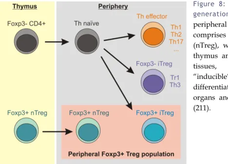
Figure 8: Thymic and peripheral generation of Foxp3+ Tregs. ... 23
Figure 9: Differentiation of thymic Tregs. ... 25
Chapter III: Foxp3 expression in pre-DP human and murine thymocytes Fig. 1. Foxp3 expression in the early stages of human thymocyte development. ... 58
Fig. 2. Murine Foxp3 is expressed in pre-DP thymocytes. ... 60
Fig. 3. TCRα expression is required for Foxp3 induction in pre-DP thymocytes. ... 62
Supplemental Fig. 1. Comparison of three different clones of anti-human Foxp3 mAb. ... 71
Supplemental Fig. 2. Intracellular protein staining for Foxp3 in thymocyte subsets of C57Bl/6 (wild type, WT), TCRα-deficient (TCRαº), MHC class II-deficient (MHC class IIº) or Foxp3-GFP mice. ... 72
Chapter IV: Differentiation of human Foxp3+ DP thymocytes Figure 1: Foxp3 expression in human DP thymocytes. ... 79
Figure 2: Foxp3+ cells present an early mature phenotype and a diverse repertoire at the DP CD3high stage. ... 80
Figure 4: Foxp3+ DP cells in the human thymus express functional IL-7 receptor. ... 83 Figure 5: Up-regulation of Foxp3 and/or CD25 on Foxp3+ DP thymocytes upon short-term
culture with IL-2 or IL-7. ... 85 Figure 6: CD103 is expressed in Foxp3+ DP CD3high and in the majority of Foxp3+ CD8SP
human thymocytes. ... 86 Figure 7: Associations between DP CD3high and SP stages of Foxp3+ and Foxp3- human
thymocytes. ... 89 Supplemental Figure 1: Analysis of the co-expression of CD39, CD73, CD25 and HLA-DR within
CD3high DP, CD4SP and CD8SP subsets in the human thymus. ... 106 Supplemental Figure 2: Analysis of the expression of P-STAT5 and CD25+ within human DP
thymocytes upon IL-7 stimulation. ... 107 Supplemental Figure 3: Loss of the activated phenotype of Foxp3+ DP thymocytes in culture and
up-regulation of Foxp3 on SP thymocytes upon short-term culture with IL-2 or IL-7. ... 108 Supplemental Figure 4: Comparison of the TCR Vβ family distribution within CD8SP subsets
defined according to Foxp3 and CD103 expression. ... 109 Supplemental Figure 5: Relationship between the DP, CD4SP and CD8SP subsets in the human
thymus according to Foxp3 expression. ... 110 Supplemental Figure 6: Multiple regression analysis relating the cell numbers of Foxp3+CD4SP to those of Foxp3-CD4SP thymocytes. ... 111
Chapter V: Conclusions and Future Perspectives
Chapter I : Introduction
Table 1: Regulation of CD8α and CD8β expression during human T cell development. ... 17 Table 2: Treg cell markers. ... 24
Chapter IV: Differentiation of human Foxp3+ DP thymocytes
Table I: Canonical correlation analysis. ... 87 Table II: Multiple regression analysis. ... 88 Supplemental Table I: Multiple regression analysis. ... 112
ACKNOWLEDGEMENTS
I ought to thank the many people whose contribution made this work possible over these past 5 years.
I sincerely thank my supervisor, Dr. Ana Espada de Sousa, for her wise guidance and input, for the availability for discussion of the work and for continuous help in building this project. I also thank all the personal support and comprehension along the way. I wish to thank the medical and surgical team at Hospital de Santa Cruz that made this work possible, namely Dr. Miguel Abecasis and Dr. Rui Anjos. A special thank you to the staff, particularly to the secretary, D. Helena Duarte, and also to Sr. João Jorge, Sr. Augusto Calvário and Sr. Eduardo Lopes, whom I directly contacted weekly, for their kindness and availability at all times.
I also wish to thank the families of the children undergoing surgery, and most specially the children, for their invaluable contribution. I had each and every one of you in my mind, wishing you all the best in the future and hoping that this much needed surgery would bring nothing but health and joy to your lives.
I thank the more recent but very important contribution of Dr. Íris Caramalho, namely her valuable input, help and discussions. I also thank all my colleagues at Unidade de Imunologia Clínica of Instituto de Medicina Molecular, particularly Rui Soares for guidance in the real-time PCR world and for extensive over-lunch discussions, and Dr. Russell Foxall for discussion and critical review of the manuscripts. I further thank Dr. Rui Victorino for valuable input and discussion.
I want to thank Dr. João Barata for making this work possible and for valuable discussion and input. I also thank Bruno Cardoso and Leila Martins, from Dr. João Barata’s group, for technical advice and for sharing the numerous trips to Hospital de Santa Cruz so that material would be available and this work would be possible.
I wish to thank all the people who collaborated in the first part of this work, which represented an important and gratifying learning experience. Particularly, I want to thank Dr. Bruno Silva Santos for the very pleasant co-mentoring of the project, Dr. Julie Ribot for most mouse experiments and discussion, Ana Luísa Caetano for technical
assistance and Dr. Íris Caramalho for valuable help and input. I also want to thank Dr. Marta Monteiro, from Dr. Luís Graça’s group, for providing mice; Dr. Isabel Alcobia and Ricardo Laranjeiro, from Dr. Leonor Parreira’s group, for providing OP9 cell lines and advising on technical details; people at Instituto Gulbenkian de Ciência from Dr. Jocelyne Demengeot’s group, namely Dr. Louise Bergman for murine intrathymic injections and Dr. Andreia Lino for providing mice.
I thank the members of the Thesis Committee, Dr. António Jacinto, Dr. Rémi Cheynier and Dr. Bruno Silva Santos, for critical discussion of the work.
I thank Dr. Nuno Sepúlveda for performing the statistical analyses presented in the second part of the work.
I also wish to thank Dr. António José Cidadão for carefully listening to my questions in histology and for always trying to help, either by discussing and sharing his knowledge or by providing reagents.
I also thank all the other people that contributed with questions, comments and discussion in meetings (Immunology Club at IMM, IGC/IMM Immunology Meeting, UIC Retreat, …).
A special thank you to Dr. Jocelyne Demengeot, whose words at the right place and time made it possible for me to be presenting this work at this moment.
I also must thank Fundação para a Ciência e a Tecnologia for finantial support and for always dealing with every issue rapidly and competently.
Miguel, muito obrigada pelo apoio incondicional. Obrigada pela paciência e sensatez com que sempre lidaste com os bons e menos bons momentos e com o meu (difícil) feitio. Rodrigo, obrigada por todos os momentos de afecto e brincadeira e por seres um filho tão compreensivo. Muito obrigada Mãe e Pai por estarem sempre presentes e me apoiarem em todos os momentos. Obrigada Nuno e João Luís pelo apoio e pelos momentos de brincadeira com o vosso sobrinho. Obrigada à minha restante família pelo apoio. Obrigada.
ABBREVIATIONS
Abbreviation Description
AIRE Autoimmune regulator
APC Antigen presenting cell
Bcl-2 B-cell lymphoma 2
CCR Chemokine receptor
CD Cluster of differentiation
cjTRECs Coding-joint TRECs
CMJ Corticomedullary junction
cTEC Cortical thymic epithelial cell
CTLA-4 Cytotoxic T-lymphocyte antigen 4
DAPT N-[N-(3,5-difluorophenyl)-L-alanyl]-sphenylglycine t-butyl ester
DC Dendritic cell
DL1 Delta-like 1
DN Double negative
DNA Deoxyribonucleic acid
DP Double positive
EDP Early double positive
Foxp3 Forkhead box P3
FTOC Fetal thymus organ culture
γc Common γ chain
GALT Gut-associated lymphoid tissues
GAPDH Glyceraldehyde-3-phosphate dehydrogenase
GARP Glycoprotein A repetitions predominant
GATA3 GATA-binding protein
GFP Green fluorescent protein
GITR Glucocorticoid-induced tumor necrosis factor (TNF) receptor family-related gene/protein
GW Gestational week
hmFTOC Human/mouse hybrid FTOC
ICOS Inducible T cell co-stimulator
IL Interleukin
IPEX Immune iysregulation, polyendocrinopathy, enteropathy, X-linked syndrome ISP Immature single positive
Abbreviation Description
Jak Janus kinase
LAP Latency-associated peptide
mAb Monoclonal antibody
MHC Major histocompatibility comlex
mTEC Medullary thymic epithelial cell
NK Natural killer
nTreg “Natural” Treg
PCR Polymerase chain reaction
PD-1 Programmed cell death 1
PDC Plasmacytoid dendritic cell
PECAM-1 Platelet endothelial cell adhesion molecule 1 (CD31)
pTα Pre-TCRα
RAG Recombinase activating gene
RNA Ribonucleic acid
RTE Recent thymic emigrant
RTOC Reaggregate thymic organ culture
RT-PCR Reverse transcription PCR
RUNX Runt-related transcription factor
SCID Severe combined immune deficiency
SEM Standard error of mean
sjTRECs Signal-joint TRECs
SOCS1 Suppressor of cytokine signaling 1
SP Single positive
STAT5 Signal transducer and activator of transcription 5
TCR T cell receptor
TEA T-early α
TEC Thymic epithelial cell
TGF-β Transforming growth factor β
Th T-helper
Th-POK T-helper inducing POZ/Kruppel-like factor
TN Triple negative
TNF Tumor necrosis factor
Abbreviation Description TOX Thymus high mobility group (HMG) box protein
Tr1 Type 1 regulatory T cells
TRA Tissue restricted antigens
TREC TCR rearrangement excision circles
Treg Regulatory T cell
RESUMO
O timo é um órgão linfóide primário essencial para o desenvolvimento das células T, sendo responsável pela formação de um repertório capaz de combater possíveis patogéneos estranhos ao organismo sem comprometer a tolerância ao próprio. Esta tolerência é maioritariamente garantida pelo timo, quer pela eliminação de linfócitos T potencialmente auto-reactivos, quer pela produção de uma população de células T, denominadas células T regulatoras (Treg), devotada ao controlo da resposta imune, particularmente no contexto de processos autoimunes e inflamatórios.
A diferenciação no timo de progenitores provenientes da medula óssea em células T CD4 ou CD8 maduras ocorre através de uma sequência de estadios de desenvolvimento que podem ser definidos com base na expressão das moléculas CD3, CD4 e CD8. Em humanos, células CD4- CD8- CD3- triplamente negativas (TN) adquirem inicialmente CD4 (estadio CD4 monopositivo imaturo, CD4ISP) e posteriormente CD8, tornando-se células CD4+ CD8+ duplamente positivas (DP). Na sequência de rearranjos genéticos do receptor de células T (TCR), este é expresso à superfície e é testada a sua capacidade de reconhecer o complexo de histocompatibilidade major (MHC), fundamental para a apresentação de antigénios. Células cujo TCR não reconheça MHC morrem por apoptose (“morte por negligência”). Apenas células apresentando TCRs que reconhecem MHC do próprio recebem sinais de sobrevivência (“selecção positiva”) e de diferenciação em células T CD4 ou CD8 maturas. Estudos no ratinho mostram que a intensidade e duração da sinalização pelo TCR durante a diferenciação tímica são determinantes na decisão de uma célula se tornar uma célula T CD4 ou CD8. Uma sinalização excessivamente intensa pelo TCR induz “selecção negativa”, resultando na apoptose de linfócitos potencialmente autoreactivos. A sinalização pelo TCR é também essencial para o desenvolvimento tímico de Tregs. Pensa-se que a sua selecção ocorre num intervalo muito restrito de afinidade das ligações TCR-ligando, entre a selecção positiva de células T convencionais e a selecção negativa de linfócitos T autoreactivos com TCR de alta afinidade. Neste sentido, tem sido atribuída às Tregs a característica peculiar de possuírem uma autoreactividade aumentada.
Actualmente, o factor de transcrição Foxp3 (forkhead box P3) é considerado o marcador mais adequado para a identificação de Tregs. O seu papel essencial no desenvolvimento e função de Tregs é suportado pela associação de mutações de Foxp3
com o síndrome fatal IPEX (immune dysregulation, polyendocrinopathy, enteropathy, X-linked
syndrome, síndrome auto-imune linfoproliferativo severo) e também pelo síndrome
autoimune fatal observado em ratinhos scurfy, “naturalmente” deficientes em Foxp3. Outras moléculas cuja expressão tem também sido associada a Tregs incluem a cadeia α do receptor de interleucina 2 (IL-2Rα, CD25), CTLA-4 (cytotoxic T-lymphocyte antigen 4), o ectoapirase CD39 e baixos níveis da cadeia α do receptor de IL-7 (IL-7Rα, CD127).
Neste estudo investigámos o desenvolvimento de Tregs, identificadas pela expressão de Foxp3 e de outros marcadores associados, em timos de indivíduos em idade pediátrica. Em particular, perguntámos quando ocorre o comprometimento dos timócitos para a linhagem de Tregs durante o desenvolvimento, uma vez que esta questão é relevante para o repertório das Tregs. Perguntámos ainda como se compara o desenvolvimento das Tregs com o de linfócitos T convencionais.
Na primeira parte do trabalho analisámos o padrão de expressão de Foxp3 em timócitos humanos e murinos. Os nossos resultados revelam a expressão de Foxp3 em fases anteriores ao estadio DP, nomeadamente em timócitos no estadio CD4ISP em humanos e no estadio CD4- CD8- duplamente negativo 4 (DN4) em ratinhos. Mostram ainda que, em ratinhos, células Foxp3+ DN4 possuem rearranjos genéticos da cadeia α do TCR (TCRα). Recorrendo ao ratinho deficiente em TCRα como uma ferramenta genética mostramos que, na ausência de TCRα, a expressão de Foxp3 no estadio DN4 é abolida. Colectivamente, os nossos dados revelam a expressão de Foxp3 em fases pré-DP em timócitos humanos e murinos e confirmam que a expressão precoce de Foxp3 é dependente do TCR.
Na segunda parte do trabalho estudámos a indução de Foxp3 no estadio DP e a possível diferenciação de timócitos Foxp3+ DP no estadio mais maturo Foxp3+ SP. Usando um ensaio de reexpressão, que consiste na clivagem de moléculas de superfície usando pronase e na síntese e reexpressão em cultura de moléculas transcripcionalmente activas, mostramos que a expressão de Foxp3 na fase DP ocorre em “verdadeiras” células DP. Mostramos ainda que os timócitos Foxp3+ DP apresentam um fenótipo maturo e uma distribuição das famílias TCR Vβ que se assemelha à dos timócitos DP que não expressam Foxp3. Identificamos também uma população de timócitos Foxp3+ DP que apresenta um aumento do programa transcripcional associado com o fenótipo activado/supressor na periferia, expressando elevados níveis de marcadores associados com Tregs, tais como CD25, CTLA-4, CD39 e MHC classe II (HLA-DR). Esta população está também presente em timócitos Foxp3+ CD4SP, nos quais a intensidade de expressão
dos marcadores supramencionados parece decair antes da exportação das células do timo para a periferia. Os timócitos Foxp3+ CD8SP não apresentam a referida população, mas expressam a molécula CD103 (αE), constituinte da integrina αEβ7. A expressão de
CD103 está associada com a migração para a mucosa, o que pode explicar os níveis reduzidos de Tregs CD8 circulantes. Mostramos ainda que a molécula CD103 está já presente em células Foxp3+ no estadio DP, o que sugere uma possível relação precursor-progenia entre os timócitos Foxp3+ CD103+ DP e CD8SP. A possível diferenciação de timócitos Foxp3+ DP em Foxp3+ SP foi analisada com base num modelo estatístico de análise de regressão múltipla. Este modelo suporta um contributo significativo das células Foxp3+ DP para o pool Foxp3+ SP. Em resumo, os nossos dados indicam que Foxp3 é induzido na fase DP e que uma parte significativa dos timócitos Foxp3+ DP se diferencienciam em células Foxp3+ SP, que irão incorporar o pool de Tregs na periferia.
Em contraste com o que é observado na periferia, mostramos que uma fracção significativa dos timócitos Foxp3+ DP expressa níveis consideráveis do receptor de IL-7. A funcionalidade deste receptor à superfície dos timócitos Foxp3+ DP foi confirmada através do estado da fosforilação da molécula STAT5 (signal transducer and activator of
transcription 5), associada à acção da IL-7, após estimulação de timócitos com esta
citoquina. Mostramos ainda que IL-7 induz o aumento de intensidade de CD25 nos timócitos Foxp3+ DP em cultura. Estes resultados sugerem que a IL-7 poderá ter um papel significativo no desenvolvimento timico de Tregs em humanos. Esta observação pode ser de importância particularmente significativa em situações onde há alterações dos níveis de IL-7, tal como no contexto de situações clínicas de linfopénia como observado, por exemplo, na patologia do HIV.
Em conclusão, os nossos dados suportam que a expressão de Foxp3 em timócitos humanos requer sinalização pelo TCR e ocorre antes da decisão CD4 vs. CD8. Mostramos que os timócitos Foxp3+ DP expressam um fenótipo activado semelhante ao apresentado em Tregs activadas na periferia mas associado à expressão de receptor de IL-7 funcional, o que naquela fase possivelmente protege os timócitos de apoptose/selecção negativa. Identificámos ainda uma subpopulação das células Foxp3+ DP que expressa CD103 e que está provavelmente na origem das células Foxp3+ CD8SP, com migração preferencial para a mucosa. Finalmente, modelos de análise de regressão múltipla suportam que um comprometimento significativo para a linhagem Treg ocorre na fase DP, com possíveis implicações para o repertório e função da população de Treg.
Palavras-chave: Estudos humanos, Timo, Desenvolvimento de Células T, Células T
SUMMARY
The thymus is a primary lymphoid organ crucially involved in the development of T cells. While producing a self-tolerant T cell repertoire that warrants surveillance to foreign pathogenic threats, the thymus also produces a lineage of regulatory T cells (Tregs) that are deeply involved in the control of immune responses, particularly in the context of inflammatory and autoimmune processes.
Differentiation of blood-derived T cell progenitors to mature CD4 or CD8 T cells in the thymus is accomplished by sequential development through a series of stages that can be defined by the expression of CD3, CD4 and CD8. In humans, CD4-CD8-CD3- triple negative (TN) cells first acquire CD4 (CD4 immature single positive stage, CD4ISP stage) and then CD8 to become CD4+CD8+ (double positive stage, DP stage) cells. Upon T cell receptor (TCR) gene rearrangements, DP cells “test” their TCR for recognition of the antigen-presenting molecules named major histocompatibility complex (MHC). While cells that are unable to recognize self-MHC undergo “death by neglect”, DP thymocytes bearing TCRαβ complexes that can engage self-MHC molecules are signaled to survive, a process designated as “positive selection”, and to differentiate into functionally mature CD4 single positive (CD4+CD8-CD3high, CD4SP) or CD8 single positive (CD4-CD8+CD3high, CD8SP) T cells. Data from mouse models suggest that the strength and duration of TCR signaling is determinant in CD4 vs. CD8 lineage decision during thymic differentiation. Strong TCR signaling induces “negative selection”, resulting in programmed cell death of potentially auto-reactive cells. TCR signaling has been shown to be essential for Treg differentiation in the thymus. It has been proposed that Treg selection may occur within a very narrow window of affinity of TCR-ligand interactions, between positive selection of conventional CD4 T cells and negative selection of high-affinity self-reactive T cells. In agreement, increased self- reactivity is considered a unique feature of Tregs.
At present, the forkhead winged-helix transcription factor Foxp3 is considered the best available marker to identify Tregs. The essential role of Foxp3 in Treg development and function is supported by the association of Foxp3 defects with the fatal condition IPEX (immune dysregulation, polyendocrinopathy, enteropathy, X-linked syndrome) and also by the lethal autoimmune syndrome observed in scurfy and Foxp3 knock-out mice. Other molecules whose expression has also been associated with Treg
function include the interleukin 2 receptor α chain (IL-2Rα, CD25), the cytotoxic T-lymphocyte antigen 4 (CTLA-4), the ectoapyrase CD39 and low levels of the IL-7 receptor α chain (IL-7Rα, CD127).
We have investigated the development of human Tregs, as identified by the expression of Foxp3, as well as of other Treg-associated markers, in human pediatric thymuses. Specifically, we asked when commitment to the Treg lineage occurred during T cell development, since this is particularly relevant for the generation of the Treg repertoire. In addition, we assessed the developmental pathway of Tregs in the thymus. In particular, we asked whether Treg development occurred similarly to what is observed in conventional, Foxp3- T cells.
In the first part of the work we dissected the early expression pattern of Foxp3 in human and murine thymocytes and showed that Foxp3 expression can be detected in pre-double positive (pre-DP) subsets, particularly in CD4ISP thymocytes in humans and in double negative stage 4 (DN4) cells in mice. We further showed that Foxp3-expressing murine thymocytes at the DN4 stage possess productive TCRα gene rearrangements. In addition, using the TCRα-deficient mouse model as a genetic tool we have found that Foxp3 expression was abolished in TCRα-deficient DN thymocytes. Collectively, our data have identified the pre-DP expression of Foxp3 in human and murine thymocytes and confirmed the TCR-dependency of the early Foxp3 expression in pre-DP thymocytes.
In the second part of the work we studied the induction of Foxp3 at the DP stage and the possible differentiation of Foxp3+ DP thymocytes into the SP stages. Using a re-expression assay, where surface molecules were cleaved using pronase and transcriptionally active molecules were allowed to be synthesized and re-expressed, we were able to show that Foxp3 expression at the DP stage occurs in bona fide DP thymocytes. We also showed that Foxp3+ DP thymocytes have a mature phenotype and a TCR Vβ family distribution that closely resembled that of Foxp3 negative DP thymocytes. We further found that a population within Foxp3-expressing DP thymocytes presented an up-regulation of the transcriptional program associated with an activated/suppressor phenotype in the periphery and expressed high levels of the Treg-associated markers CD25, CTLA-4, CD39 and MHC class II (HLA-DR). This population was also found within Foxp3+ CD4SP thymocytes, in which the expression of those markers appeared to decline prior to thymic egress. While Foxp3+ CD8SP
thymocytes were devoid of high expression of those markers, they homogeneously expressed CD103 (αE), which is part of the αEβ7 integrin. CD103 expression is associated
with migration to the mucosa, which might explain the very low levels of circulating CD8 Tregs. In addition, we found that Foxp3+ cells expressing CD103 were already present at the DP stage, indicating possible precursor-progeny relationship between Foxp3+CD103+ DP and CD8SP thymocytes. In order to further investigate a possible direct precursor-progeny association between Foxp3-expressing DP and SP cells we performed statistical analysis of our data using multiple regression analysis. This model supported the significant weight of Foxp3+ DP cells to the SP pool. Overall, our data thus indicate that considerable Foxp3 induction occurs at the DP stage and that a significant part of Foxp3+ DP thymocytes probably progresses to the SP stages, and will incorporate the Treg pool upon thymus egress.
In addition, and in clear contrast to what is observed in the periphery, we found that a large proportion of Foxp3+ DP thymocytes expressed considerable levels of IL-7 receptor (IL-7R). We assessed the functionality of IL-7R at the surface of Foxp3+ DP cells and showed that phosphorylation of STAT5 (“signal transducer and activator of transcription 5”), a downstream target of IL-7, was readily observed upon exposure to that cytokine. Furthermore, short-term culture with IL-7 induced the upregulation of CD25 on Foxp3+ DP thymocytes. Together, these results suggest that IL-7 may play a role in the development of human Tregs. Although requiring further investigation, this finding may be of particular significance in the context of situations where IL-7 levels are altered, such as lymphopenia, as observed in HIV pathology.
In conclusion, our data support that the expression of Foxp3 in human thymocytes requires TCR signaling and occurs prior to CD4 or CD8 lineage commitment. Foxp3+ DP thymocytes were shown to express an activated phenotype that is reminiscent of that found in peripheral activated Tregs but is associated with the expression of a functional IL-7 receptor, which may protect them from cell death/negative selection. A subpopulation of Foxp3+ DP expressing CD103 is likely to give rise to a population of Foxp3 CD8 T cells with preferential homing to the mucosa. Finally, multiple regression models support that a significant commitment to the Treg lineage occurs at the DP stage, with possible implications for Treg repertoire and function.
Chapter I
CHAPTER I: INTRODUCTION
A. HUMAN T CELL DEVELOPMENT AND INSIGHTS FROM MOUSE MODELS
1. THE THYMUS: BRIEF HISTORICAL PERSPECTIVE
The word “thymus” is thought to come from the Latin derivation of the greek θυμός (“thymos”), meaning “warty excrescence”, but also “soul” or “spirit”, and the ancient Greeks believed it to be the seat of the soul. Later, it was also considered to be the organ of purification of the nervous system, a protective thoracic cushion and a center of regulation of fetal and neonatal respiration (1).
Originally described by Claudius Galen of Pergamum (130-200 AD) during his precise descriptions and studies of neurological functions and anatomy, the thymus was considered an “organ of mystery” for almost 2000 years. Galen observed that the thymus was proportionally largest in size during infancy and that it shrunk with aging (1). Studies on the evolution of thymic size during fetal and infant life were also performed by William Hewson in 1777. He further noted that the thymus was filled with ‘particles’ resembling those in lymph and blood, and believed that “the thymus exists during the early periods of life only when those particles seem to be the most wanted” (2).
By the mid 50’s the lymphopoietic function of the thymus was established. However, immunologists still did not believe it to have an immunological function and considered the thymus an evolutionary redundant “lymphocyte graveyard” (2). In the early 1960’s, Jacques Miller’s work on the role of the thymus in mouse lymphocytic leukemia and the impact of neonatal thymectomy in tolerance (3-5) lead the way for the recognition of the thymus as a non-redundant specialized organ of the immune system with a unique function in the establishment and maintenance of the T cell pool.
2. PHYLOGENY, ONTOGENY AND STRUCTURE OF THE THYMUS
The thymic structure has not been identified in species more primitive than jawed vertebrates and is a common feature to all of them (6). Its precise embryological origin, the number of organs per animal and the final anatomical positions all vary markedly in different species. In spite of this, they share their origin from pharyngeal pouches, which arise as specialized pockets of the foregut endodermal tube and harbor primordia for organs and tissues later found in chest, neck, or head regions, including the thymus and the parathyroid gland (7).
The human thymus is located anatomically in the anterosuperior mediastinum, in front of the heart and behind the sternum. It achieves the maximum absolute size during the first year of life (8). The thymus undergoes a process named “involution”, whereby it starts reducing its mass and changing its architecture with age, and extensively fills with adipose tissue after puberty (1, 8).
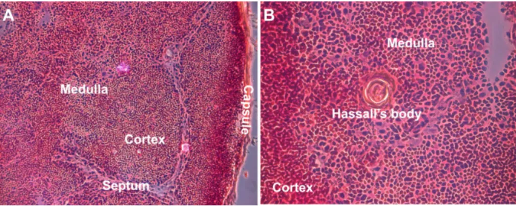
The thymus consists of two distinct lobes joined by a connective tissue isthmus. A thin connective tissue capsule surrounds each lobe and, in most species (although not in mouse), gives rise to septa that partially subdivide the thymus into interconnecting lobules of variable size and orientation. It is morphologically similar across species, being divided into cortical and medullary regions separated by a vascular corticomedullary zone (9, 10). Those areas can be distinguished histologically (Figure 1). While the cortex contains densely packed immature thymocytes within a sparse thymic epithelial framework, the medulla is less dense and contains prominent epithelial cells and Hassall's bodies (or Hassall's corpuscle). First described by the British physician Arthur Hill Hassall in 1849, Hassall's bodies are concentric structures composed of stratified keratinizing epithelium that are rare in rodents but rather frequently found in humans and other primates (9-11). They were suggested to represent the “graveyard” for dead thymocytes or the “privileged” area for the maturation of medullary thymocytes, and also to be active in cell signaling, transcription and metabolism mediated by cytokines or growth factor receptors (11-17).
Figure 1: Structure of the human thymus. Hematoxylin/eosin staining of a section of a
pediatric thymus (7 year old boy). The cortex is composed of densely packed thymocytes (A, B) while in the medulla thymocyte density is lower and contains Hassall’s bodies (B). Magnification: 10x (A), 40x (B).
The epithelial thymic primordium begins its development at the end of the 4th
gestational week (GW) and it is present by the 7th GW, with a distinct organization into a
peripheral and central cell layer by the 8th GW (18). Seeding of the thymus is a very early
event, with the first lymphoid progenitor cells arriving by the 8th GW via the peripheral
blood (18-20). At this time progenitor cells that seed the thymus originate from the fetal liver (19, 20). Colonization of the thymic primordium by T cell progenitors is necessary for further development of the thymic microenvironment, including the compartmentalization into cortex and medulla and the development of Hassall's corpuscles (21, 22). Mature T cells are not detected in the thymus before 12 to 13 GW and progressive colonization of the periphery occurs between 13 and 14 GW (23-26). After the 16th GW fetal liver precursor cells colonize the bone marrow, which becomes the
main source of thymic progenitors from the 22nd GW onwards throughout adult life (20).
The thymus has no afferent lymphatics, and thymic progenitors are thought to enter the thymus through the large venules at the corticomedullary junction (CMJ). T cells re-enter the circulation through the vascular lining of post-capillary venules at the CMJ, where efferent lymphatics drain into an adjacent pair of lymph nodes (10).
3. COMPOSITION OF THE THYMIC STROMA
The unique capacity of the thymus to efficiently promote T cell differentiation and repertoire selection is mediated by thymic epithelial cells (TECs), which are the major constituents of the thymic stroma. This epithelial nature of the thymus makes it a unique lymphoid organ (10). Cortical and medullary TECs are currently thought to be mainly derived from a bipotential cTEC-mTEC progenitor population present in the thymus anlage and possibly persisting beyond the embryonic period (27-29). Nonetheless, epithelial cells of the thymic microenvironment are very heterogeneous, and distinct cell types occur in the subcapsular zone, cortex, medulla and Hassall's corpuscles (18). They differ in their ultrasctructural characteristics, antigenic expression and their capacity to synthesize thymic hormones, which are important for intrathymic maturation and modulation of lymphocyte responsiveness (10).
The thymic stroma also comprises several non-epithelial cell types, such as endothelial cells, which contribute to the vasculature, and mesenchymal cells, that influence TECs and T cell progenitors (29). Bone-marrow derived cells are also present, including dendritic cells (DC), B cells and macrophages, involved in the shaping of the T cell repertoire and the elimination of apoptotic thymocytes (9, 29). Macrophages are
found throughout the thymus but occur at higher density in the cortex, while DCs are frequently present along the thymic CMJ and medulla (18) and B cells are mostly found in the medulla (30, 31).
4. OVERVIEW OF T CELL DEVELOPMENT
The highly ordered thymic architecture plays a crucial role in normal thymic function. Phenotypically distinct thymocyte populations are present in specific thymic areas (Figure 2). Progenitor cells lacking CD4 or CD8 expression (double negative cells, DN) enter the thymus at the CMJ and move through the cortex to the subcapsular zone, where they undergo early events. Large, mitotically active lymphoblasts can be found in the subcapsular zone (10, 29). The thymic cortex contains smaller CD4+CD8+ double positive (DP) thymocytes, while in the medulla single positive CD4+ CD8- (CD4SP) and CD4- CD8+ (CD8SP) T cells are screened for self reactivity before leaving the thymus through the CMJ (9, 29).
Figure 2: Thymic T cell development. T cell progenitors enter the thymus through the blood vessels at the CMJ. Development of CD4-CD8- double negative (DN) cells is accompanied by an outward movement towards the subcapsular zone. CD4+CD8+ double positive (DP) cells scan the cortex for positively selecting ligands. After positive selection and lineage commitment, single positive (SP) cells move to the medulla, where they scan medullary antigen presenting cells, mainly dendritic cells (DCs) and medullary thymic epithelial cells (mTECs), before they leave the thymus. cTEC: cortical TEC. Adapted from (32).
5. T CELL COMMITMENT AND EARLY T CELL DEVELOPMENT
Hematopoietic precursors in humans are present within a population of cells that express CD34 (33). CD34 is expressed on pluripotent stem cells, as well as on progenitor cells that are already committed to particular hematopoietic lineages, but not on their differentiated progeny (34-36). In the thymus, CD34+CD38lo cells include the most immature population, based on TCR rearrangement status (Figure 3) (37) and on their T, natural killer (NK) and DC precursor potential (36, 38). The presence of multipotential precursors in the human thymus indicates that T cell commitment takes place within the thymus itself (39). This is also supported by the fact that TCR gene rearrangements, which are the most definite marker for T cell commitment, were not found in fetal liver or cord blood CD34+ cells (40, 41). Neonatal cord blood CD34+ cells were also found not to express recombinase activating gene (RAG-1) or pre-TCRα mRNA (41). Thus, at least a proportion of the precursors that seed the human thymus are multipotential and commit to the T cell lineage in the thymus (20).
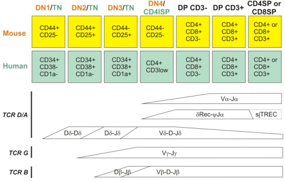
Figure 3: Stages of T cell development in human and mouse thymus and occurrence of TCR rearrangements. Parallel between the stages of T cell development in human and mouse and timing of TCR rearrangements. TN, triple negative (CD4-CD8-CD3-); CD4ISP, CD4 immature single positive (CD4+CD8-CD3low). Adapted from (37).
Cells expressing the highest levels of CD34 lack CD1a, a molecule that is widely expressed in cortical thymocytes (42, 43). Appearance of CD1a on CD34+ thymocytes marks an important checkpoint in early T cell development, since it is associated with the induction of T cell commitment, as CD34+CD1a+ thymocytes largely lose the ability to develop into non-T cells (36). CD34+CD1a+ cells, in contrast to CD34+CD1a- precursors, have strong T cell, but little NK-cell, and no DC or plasmacytoid DC (PDC) precursor activity (36). CD34 is downregulated in the subsequent population, which expresses CD4 but not CD8 or surface CD3, and is known as CD4 immature single positive (CD4ISP) (42, 44). CD4ISP cells are fully committed to the T cell lineage, since they lack NK or DC precursor activity but can develop into T cells, as demonstrated using a hybrid human-mouse fetal thymic organ culture (hmFTOC) system in which human thymocytes are cultured in murine fetal thymic lobes (45).
Human T cell development is critically dependent on the cytokine interleukin 7 (IL-7). The IL-7 receptor consists of two chains, an α chain (IL-7Rα) and a common γ chain (γc), shared with the receptors for IL-2, IL-4, IL-9, IL-15 and IL-21. Genetic defects in genes encoding for γc (46, 47), IL-7Rα (48, 49) or the Janus kinase Jak3, a component of the IL-7-induced signaling transduction pathway (50, 51), are associated with profound T cell deficiency and account for most severe combined immune deficiencies (SCID). The precise function of IL-7 on human T cell development is not completely understood. CD34+ thymic T cell progenitors proliferate in response to IL-7, and development from CD34+ precursors in a fetal thymic organ culture (FTOC) in the presence of inhibition of IL-7R signaling almost completely blocks the transition of CD34+CD1a+ cells into CD4ISP (52). Furthermore, IL-7 may be directly involved in the induction of TCRβ V-D-J rearrangements, although this is yet to be confirmed (53). In support of this, IL-7 addition enhances the levels of episomal DNA resulting from TCR gene rearrangements, known as TCR rearrangement excision circles (TREC; see section 7), in human thymic organ cultures (TOCs) (54).
Notch proteins constitute a family of highly conserved transmembrane receptors whose receptor-ligand interactions have also been shown to play a crucial role in T cell development (55-58). This is supported by studies where cord-blood and bone marrow CD34+ cells co-cultured with the murine bone marrow stromal cell line OP9 expressing the Notch ligand DL1 (delta-like 1) were shown to undergo full T cell development (58, 59). On the other hand, the γ-secretase inhibitor DAPT
(N-[N-(3,5-difluorophenyl)-L-alanyl]-Sphenylglycine t-butyl ester), which inhibits Notch signaling, strongly impaired T cell development in both the FTOC (60) and OP9-DL1 (61) systems. However, inhibition of TCRαβ development by DAPT was rescued by forced expression of intracellular Notch1 by retrovirus-mediated gene transfer (61). Furthermore, TCRβ V-DJ but not D-Jβ rearrangements were strongly inhibited by DAPT (60). These findings indicate that Notch is required for induction and maintenance of T cell specification.
6. EARLY TCR REARRANGEMENTS AND β-SELECTION OF DEVELOPING THYMOCYTES
The TCR expressed by the majority of T cells is a heterodimer composed of α and β chains. TCR diversity is generated by random rearrangement of multiple germline-encoded variable (42 Vα and 47 Vβ segments), diversity (2 Dβ segments), and joining (61 Jα and 13 Jβ segments) gene segments, non-germline-encoded N region insertions, and α and β chain pairing (62, 63). In humans, an estimated >1018 different TCR can be
produced and the diversity of TCR in a human at any given time has been estimated to be > 2x10⁷ (63).
During the early stages of T cell development, the TCR loci undergo rearrangement in the sequence TCR D>G>B>A (37, 64, 65). The current model of TCR rearrangements in early T cell development, as described by Dik et al. (37), is depicted in Figure 3. However, there is some controversy with respect to the cell phenotype in which each rearrangement of particular loci occurs, probably due to the sensitivity of the methods used to analyze TCR rearrangements.
Studies in the mouse have shown that productive rearrangement of TCRB genes leads to the transport to the cell surface of a pre-TCR complex consisting of TCRβ protein associated with an invariant pre-TCRα (pTα) chain and the CD3 complex (66-69), and the presence and association of those molecules has also been shown to occur in human thymocytes (70, 71). Signaling through the pre-TCR induces survival, proliferation, differentiation and initiation of TCRA gene rearrangements (69, 71). This process, known as “β-selection”, represents the first checkpoint of T cell development, as cells that fail to generate a TCRβ chain do not proceed along the αβ lineage differentiation pathway (69). In the mouse, this checkpoint mainly occurs at the DN3 stage and has been associated with the expression of CD27 (72, 73)
In humans, occurrence of TCRβ rearrangements and ensuing β-selection has generated some debate. While a PCR-based and GeneScalling analysis found some productive TCRβ V-DJ rearrangements within CD34+CD1a+ cells (37), a less sensitive southern blot analysis detected the first TCRβ V-DJ rearrangements in CD4ISP cells (65). Intracellular TCRβ protein can be detected in CD4ISP cells and at higher frequency at the subsequent CD4+CD8α+CD8β- stage (early double positive stage, EDP) (65, 74). However, intracellular TCRβ negative populations have been found in CD4+CD8α+β- DP cells (65, 75), indicating that not all populations downstream of CD34+CD1a+CD4- cells are post-β selection. A study by the Toribio group (75) has located the β-selection checkpoint later in development, at the transition from the CD4+CD8α+CD8β- to the CD4+CD8α+CD8β+ stage, since the later subset expresses surface TCRβ and pTα. However, in this study no surface pre-TCR was found in EDP cells, while a low level of CD3 was detected on a small proportion of EDP in the study of Blom et al. (65). Furthermore, pTα mRNA is mainly present at the CD4ISP stage (70, 71, 76) and is downregulated but still expressed in EDP and TCRαβ- DP cells (65, 70, 71), while it is absent at other stages of development (70, 71), supporting the possibility that β-selection occurs at early stages of human T cell development.
Recently, CD28 expression at the CD4ISP stage has been described as a marker of cells that have passed the β-selection checkpoint (76). CD4ISP CD28+ thymocytes express intracellular TCRβ and differentiate into CD3+ TCRαβ+ cells in the OP9 system, in the absence of Notch signaling (76). In this study pTα mRNA was shown to be highly expressed in CD4ISP CD28- cells and almost completely downregulated in CD4ISP CD28+ thymocytes. In contrast, expression of TCR Cα, which marks opening of the TCRα locus (73), was strongly and specifically induced in CD4ISP CD28+ thymocytes (76). CD4ISP TCRβ+ cells had been previously suggested to be in cycle, to express elevated levels of CD1a, CD4, CD28, CD45RO and CD71 and to differentiate in FTOC into TCRαβ+ T cells with faster kinetics than CD4ISP TCRβ- cells (39). These data further indicate that β-selection can occur within the CD4ISP population.
Overall, it is considered that β-selection in human thymocytes may not be directly associated with a particular stage. It may begin in a small subset of CD34+CD1a+CD4- cells, while a larger proportion occurs in cells that have up-regulated the expression of CD4, and subsequently CD8α or both CD8α and CD8β.
7. TCRα REARRANGEMENTS IN IMMATURE THYMOCYTES
Rearrangements at the TCRA locus are initiated after TCRβ-expressing cells have expanded, as a result of β-selection, and returned to slow cycle conditions (53, 71).
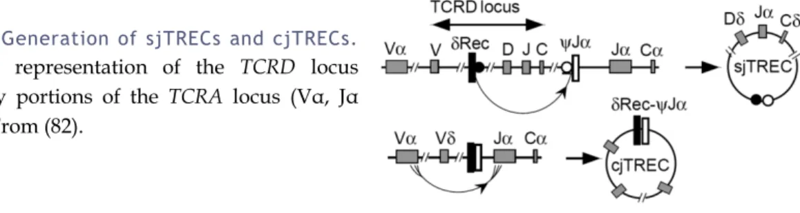
The human TCRA and TCRD gene segments are interspersed on chromosome 14q11, with the TCRD locus being located between the Vα and Jα gene segments (Figure 4) (77). Therefore, deletion of the TCRD gene plays an important role in the differentiation of TCRαβ cells. Excision of the δ locus during TCRα gene rearrangements results in the generation of DNA circles that persist episomally, producing two types of TRECs that are identical in ~70% of T cells (78). Two non-functional TCRδ-deleting gene segments, δRec and ψJα, have been identified that flank the major part of the TCRδ gene (79). The high frequency of δRec-ψJα rearrangements in human thymocytes suggests that a major fraction of the thymocytes use this rearrangement to delete their TCRδ genes (79). Rearrangement of δRec to ψJα produces signal-joint (sj) TRECs. Subsequent Vα-Jα rearrangement deletes the remaining Vδ gene segment and produces coding-joint (cj) TRECs. Since the α locus is not subject to allelic exclusion (80, 81), a maximum of two sjTRECs and two cjTRECs may be present per cell (53).
Figure 4: Generation of sjTRECs and cjTRECs.
Simplified representation of the TCRD locus flanked by portions of the TCRA locus (Vα, Jα and Cα). From (82).
TCRA rearrangements were proposed to occur mainly in the small CD3- DP
population (64, 71). In the mouse, productive TCRA rearrangements and transcripts have been shown to already be present at the DN stage (83), despite being generally associated with the wave of expression of RAG-1/RAG-2 at the DP stage (84). In agreement, Dik et al. recently showed, using the more sensitive real time quantitative PCR technique, that δRec-ψJα rearrangements in humans can already be detected at the CD4ISP stage (37). Furthermore, analysis of T-early α (TEA), whose transcription initiates TCRA recombination by opening the 5’ site of the Jα cluster, showed that TEA-Cα transcripts are already detected at the CD34+CD38+CD1a+ stage (37).
8. TCRαβ T CELL DEVELOPMENT: INSIGHTS FROM MOUSE MODELS a. Positive and negative selection of developing thymocytes
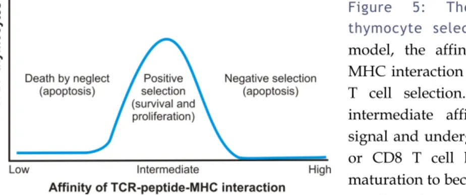
Studies in the mouse have shown that thymocytes expressing surface TCRαβ will only survive if their TCR forms an MHC-restricted receptor that is signaled by low affinity self-peptides, a process called “positive selection”. Thymocytes that fail to signal through their TCR undergo apoptosis, which is termed “death by neglect”, while high-affinity contacts will also result in programmed cell death, and this is known as “negative selection” or “clonal deletion” (Figure 5) (53). Experimental affinity measurements thus support the idea that TCR affinity for positive selection ligands is lower than that for negative selection ligands (85-87). However, the “selection paradox” that recognition of self is essential for thymocyte survival but may also induce cell death remains an incompletely resolved issue (32).
Figure 5: The affinity model of thymocyte selection. According to this
model, the affinity of the TCR-peptide– MHC interaction is the key determinant of T cell selection. Only thymocytes with intermediate affinity receive a survival signal and undergo positive selection, CD4 or CD8 T cell lineage commitment and maturation to become part of the peripheral T cell pool. Low affinity for self-peptide– MHC complexes results in death by neglect, which is thought to account for 80–90% of the loss of thymocytes during thymic selection, while high-affinity binding induces cell death by apoptosis (negative selection or clonal deletion). DP: double positive. Adapted from (32).
Despite an inherent propensity of the TCRαβ receptor to bind to MHC, probably enforced by the expression of CD4 and CD8, the generation of MHC-restricted receptors from random α/β pairs is thought to be relatively infrequent (88-91). However, the α locus is structured such that multiple V/J recombination events can occur on the same allele, thus allowing several productive TCRα gene rearrangements to be tested per cell (92).
Efficient positive selection of developing thymocytes requires interactions with self-peptide-MHC complexes displayed by cTECs (93-96), and some studies have also assigned a role for cTECs in negative selection (29, 97). cTECs provide specialized
accessory interactions that MHC+ epithelial cells from other tissues do not (98). These accessory molecules are poorly defined, although CD83 has been shown to be involved in CD4 T cell development (29, 99). Despite studies demonstrating that intrathymic expression of MHC molecules by non-TECs, including thymocytes themselves, can support positive selection, the relative efficiency of this process and the nature of the TCR repertoire that is selected are unclear (100-104).
The physiological peptide-MHC repertoire expressed by cTECs in vivo, including the number and nature of the self-peptides involved in positive selection, and the way that these complexes might shape the polyclonal αβ T cell repertoire are largely unknown (32, 105). Positive selection of functional T cells apparently requires peptides with antagonist or partial agonist properties (106) that are structurally related to ligands that can fully activate mature T cells (32), thus emphasizing the importance of the quality (affinity) rather than the quantity (avidity) of the TCR–peptide–MHC interaction (32, 85). Furthermore, the generation of a fully diverse T cell repertoire in vivo has been shown to be positively correlated with the complexity of selecting ligands (105).
Evidence suggests that the TCR must remain engaged and sustain signaling for the duration of positive selection (107). In a study where Bousso et al. used two-photon microscopy and reaggregate thymic organ cultures (RTOCs), a system that allows the organotypic reaggregation of discrete thymocyte populations with stromal cells, to study thymocyte-stromal cell interactions these were shown to last on average 6 to 12h when MHC recognition occurred (108). Those interactions took place both as long-lived cellular associations displaying stable cell-cell contacts and as shorter, highly dynamic contacts (108).
Positive selection induces a signal that results in RAG gene repression, long-term survival, migration into the medulla and differentiation into mature T cells (105). The quality control of developing T cells through interactions with peptide-MHC complexes on medullary APCs is indispensible for central tolerance (32), as revealed by the severe manifestations of systemic autoimmunity in murine models where there is inhibition of positively thymocytes to enter the medulla or premature egress of thymocytes (109, 110), disorganization of the medullary architecture (111) or disrupted development of mTECs (112-116).
mTECs are unique in their “promiscuous” gene expression ability (117-119). A subset of mTEC selectively expresses the gene that encodes the autoimmune regulator AIRE, a transcriptional regulator of target genes that greatly controls the ectopic transcription of
numerous tissue restricted antigens (TRAs) in the thymus and is essential for T cell tolerance (119). AIRE is defective in the human autoimmune condition APECED (polyendocrinopathy-candidiasis-ectodermal dystrophy) (120, 121), and its deficiency in mice results in a reduction in the expression of TRAs in mTECs, defective tolerance induction and organ-specific autoimmunity (118, 122, 123). AIRE expression in mTECs enables intrathymic self-antigen presentation, thereby aiding in the establishment of thymocyte tolerance to peripheral antigens (29).
Promiscuous gene expression is thought to be a stochastic process, with only 1 to 5% of mTECs expressing a particular TRA (117, 124). However, it was recently found that mature mTECs are replaced every 2 to 3 weeks and are thus continuously altering the topology of antigen expression within the medulla (125, 126). Tolerance induction towards TRAs expressed in few dispersed mTECs may result from the numerous encounters with APCs in the 4 to 5 days that thymocytes spend in the medulla (127, 128) and/or from the transfer of mTEC-derived TRAs to neighbouring DCs (129, 130), which are known to have a central role as APCs in negative selection in the medulla and also in the cortex (100, 127, 131), thus increasing the probability that such antigens are encountered by developing thymocytes.
b. CD4 and CD8 lineage decision of developing TCRαβ T cells
Positively selected DP thymocytes ultimately develop into CD4SP or CD8SP T cells, depending on whether they received signals through MCH class II or MHC class I, respectively (132). Classical models considered that CD4/CD8 lineage choice resulted in the transcriptional termination of one or the other coreceptor gene as a consequence of the same TCR signaling event that mediated positive selection, with the random or instructed nature of the termination of transcription underlying the “stochastic” (133-138) or “instructive” (139-142) models. Currently, CD4/CD8 lineage choice seems to be best explained by the kinetic signaling model (Figure 6), which proposes that lineage choice is determined by TCR-signal duration and that cytokines, such as IL-7, serve as “sensors” that detect that duration (143-145).
Figure 6: Kinetic signaling model of CD4/CD8 lineage choice in T cell development. According to this model,
positively-selecting TCR signals induce DP thymocytes to downregulate Cd8 transcription and thus become CD4+ CD8low. TCR-signal duration and cytokine signaling then determine whether a given thymocyte commits to the CD4 or the CD8 lineage. Adapted from (142).
In order to identify potentially useful TCRs, unselected DP thymocytes are unresponsive to other survival signals, such as IL-7, both through the downregulation of surface cytokine receptors and the expression of suppressor of cytokine signaling 1 (SOCS1), which is a potent intracellular suppressor of cytokine signal transduction (146, 147).
According to the kinetic model, TCR-signaled DP thymocytes first terminate their
Cd8 gene transcription and then assess the effect of the absence of CD8 on TCR signaling
(142, 143). Whether TCR-mediated positive selection signals persist or cease in the absence of CD8 determines if the cell becomes a CD4 or CD8 T cell, respectively. This implies that positive selection and lineage choice are sequential and not simultaneous events, and that TCR-mediated positive selection signals terminate Cd8 gene transcription and convert DP thymocytes into intermediate lineage-uncommitted
Cd4+Cd8- cells, with lineage decision at this stage depending on whether TCR signaling
persists or ceases (142). Since the progressive decrease in cell-surface CD8 eventually disrupts CD8-dependent signaling by MHC-class I-restricted TCRs, IL-7 or other γ chain cytokines are important for cell survival in the absence of TCR signaling (142, 148). Furthermore, IL-7 and other γ chain cytokines have been shown in vitro to promote “co-receptor reversal” by enhancing Cd4 silencing and promoting reinitiation of Cd8 gene transcription (143, 148).
Several transcription and nuclear factors have been identified that are involved in the regulation of Cd4 and Cd8 gene transcription. Th-POK (T-helper inducing POZ/Kruppel-like factor) seems to be both necessary and sufficient for CD4-lineage specification, probably acting as a master regulator of CD4 T cell differentiation (149,
150), while RUNX (runt-related transcription factor) proteins, in particular RUNX3, seem to promote CD8 T cell differentiation (151-153). Th-POK and RUNX3 negatively regulate each other’s expression and thus reinforce lineage choices (154-158). TOX (thymus high mobility group (HMG) box protein) (159) and GATA3 (GATA-binding protein) (160) also appear to play a role in lineage commitment, particularly in CD4 lineage choice (142).
9. DEVELOPMENT OF HUMAN TCRαβ T CELLS
The transitional stages that thymocytes undergo during their differentiation and maturation in the human thymus have mainly been studied using hmFTOCs and SCID-hu mice, a model that consists in transplanting small pieces of SCID-human fetal liver and thymus under the kidney capsule of SCID mice (Figure 7) (161-164).
Figure 7: Stages of human T cell development. Delineation of human T cell development
stages, as defined by the studies of the Vandekerckhove group in the 1990’s using hmFTOCs, SCID-hu mice and pronase re-expression assays (161-164). Adapted from (53).
CD3-TCR Low Low/+ Low/+ + + + +
CD1a + + + + + - - CD69 - + + + + + Low CD27 - - - + + + + CD45RA - - - + CD45RO + + + + + + - RAG transcripts ++ + + - - - - Development in hmFTOC CD4SP and CD8SP None CD4SP and CD8SP CD8SP CD4SP CD8SP or CD4SP respectively CD8SP or CD4SP respectively
Expression of CD69 on human thymocytes is induced at the CD3low to CD3high transition and has been linked to positive selection (162). In support of this, CD69- CD3+ thymocytes were found to be corticosteroid-sensitive, whereas CD69+ CD3+ cells were resistant (162), and these populations express high and low levels of RAG-1 mRNA, respectively (163). Post-selection CD69+ thymocytes, which are initially CD27- CD1a+ CD45RA-, sequentially up-regulate CD27, down-regulate CD1a and eventually acquire CD45RA upon maturation (161-164). The identification of an early postselection CD69+CD27−CD4SP population that gives rise to both CD4SP and CD8SP cells when cultured in hmFTOC has been suggested to indicate that, similarly to what has been described in the mouse model, human DP thymocytes down-regulate CD8 expression soon after positive selection (162, 163). Cells committed to the CD4 lineage will then permanently stop the production of CD8, while CD8 lineage-committed cells will terminate the production of CD4 and start re-expressing CD8 (163).
Down-regulation of CD8β during positive selection appears to parallel CD69 up-regulation (163). The expression of CD8α and CD8β chains is differentially regulated on human thymocytes (Table 1) (74). On cells differentiating into CD4SP the expression of CD8β chains is more rapidly lost, whereas CD8αα chains may remain expressed well after the acquisition of CD27 (162).
TABLE 1:REGULATION OF CD8α AND CD8β EXPRESSION DURING HUMAN T CELL DEVELOPMENT.
Antigen TN CD4ISP EDP DP CD4SP CD8SP
CD4 - + + + + -
CD8α - - + + - +
CD8β - - - + - +
TN: triple negative; ISP: immature single positive; DP: double positive; EDP: early DP; SP: single positive.
Interestingly, one study has suggested that CD69 expression might be maintained by the thymic microenvironment and only lost during or after emigration from the thymus (162). In the SCID-hu model, where virtually all human peripheral cells can be considered to be recent thymic emigrants (RTEs) due to their short half-life (<24h), RTEs were found to resemble that of cord blood T cells, being mostly CD3highCD69-CD27+CD1a-CD45RA+ (162). However, around 10 to 30% of the emigrant T cells have a CD45RA-/dullCD45RO+ phenotype, indicating that both CD45RA+CD45ROdull and CD45RA-/dullCD45RO+ thymocytes emigrate from the thymus (162). Accordingly, it has been shown that a minor fraction of cord blood T cells are CD45RA-/dull, and that
these cells are functionally immature, likely corresponding to recently emigrated thymocytes (165).
TRECs (see section 7) have been proposed by Douek et al. to represent a molecular marker of recent thymic emigrants in humans, as had been previously proposed in chickens (166), allowing the assessment of thymic output in the periphery (82). Studies in the mouse have shown that TRECs are stable (167) and are not duplicated during mitosis, and are thus diluted out with cell proliferation (168). sjTREC levels were found to be predominantly present in CD45RA+ naïve but not in CD45RO+ memory peripheral blood, reflecting the enrichment in RTEs and/or the lack of extensive cell division of the former population (82). Moreover, they were lower in thymectomized individuals as compared to age-matched controls, further supporting the thymic origin of sjTRECs (82). Although sjTRECs do decline with age, they are still found in 80 year old individuals, indicating that thymic function persists throughout life (82).
Other markers that have been described to be preferentially, but not exclusively, expressed on RTEs are the cell-surface molecules CD31 (platelet endothelial cell adhesion molecule 1, PECAM-1) (169) and CD103 (αE component of the αEβ7 integrin)
(170) on naïve CD4 and CD8 T cells, respectively, which identify populations enriched in TRECs.
B. DEVELOPMENT OF REGULATORY T CELLS
1. REGULATORY T CELLS: IDENTIFICATION AND ROLES IN HEALTH AND DISEASE
Although the existence of T-cell mediated immune suppression was highly controversial several decades ago and the suppressor-T-cell concept was abandoned in the 1980’s due to its elusive nature, studies in 1995 by Sakaguchi et al. hinted at a rebirth of the idea of a population of cells specialized in immune suppression (171). Classical experiments showed that thymectomy of mice 3 days after birth prevented the export of Tregs from the thymus into the periphery and predisposed the mice to systemic autoimmune diseases, which could be ameliorated through the transfer of thymocytes or splenocytes from adult euthymic mice (11, 172-175). Sakaguchi and colleagues described a population of IL-2 receptor α chain (CD25)-expressing CD4+ T cells that were capable of suppressing immune responses in a variety of experimental models, including neonatal thymectomy (171). Several mechanisms of Treg-mediated suppression have been described, mediated by secreted soluble factors, including the production of anti-inflammatory cytokines, by direct cell-cell contact or by modulating the activation state and function of antigen presenting cells (176). Noteworthy, once activated by a particular antigen, Tregs can suppress responder T cells irrespective of whether they share antigen specificity with the Treg (177).
The presence in humans of CD4+CD25+ T cells that exhibited similar features to the homologous mouse CD4+CD25+ regulatory T cell subset has also been demonstrated (178-182). However, CD25 expression in human peripheral blood CD4 T cells does not define a distinct population, as it does in mouse. Furthermore, while up to 30% of human CD4 T cells express low levels of CD25, only the 2–4% expressing the highest levels of CD25 have been shown to be functionally suppressive and are considered Tregs (183). The majority of CD4+ CD25high cells express the memory marker CD45RO, CD62L (L-selectin) and the IL-2Rβ chain (CD122) (182, 183). Within this population, HLA-DR expressing cells have been shown to have the highest suppressive capacity (184). The in vitro characteristics of the human CD4+CD25high T cells were similar to those observed in the murine population. For instance, Tregs did not proliferate when stimulated through their TCR in the absence of exogenous IL-2 and were able to suppress the activation of other T cells in a cell-cell contact dependent manner (183). Furthermore, Tregs in both humans and mice could suppress both proliferation and