Histochemical and immunohistochemical evaluation of
angiogenesis in rabbits (
Oryctolagus cuniculus
)
submitted
to skin grafts associated with platelet-rich plasma
1Josiane M. Pazzini2*,Eduardo L. Serafim2, Fátima Gärtner3, Irina Amorim3, Fátima Faria3, Alexandra Rêma3, Paola C. Moraes2 and Andrigo B. De Nardi2
ABSTRACT.- Pazzini J.M., Serafim E.L., Gärtner F., Amorim I., Faria F., Rêma A., Moraes P.C. & De Nardi A.B. 2017. Histochemical and immunohistochemical evaluation of angio-genesis in rabbits (Oryctolagus cuniculus) submitted to skin grafts associated with platelet-rich plasma. Pesquisa Veterinária Brasileira 37(12):1519-1525. Departamento de
Clínica e Cirurgia Veterinária, Faculdade de Ciências Agrárias e Veterinárias, Universidade
Estadual Paulista, Campus Jaboticabal, Via de Acesso Paulo Donatto Castellane s/n, Jaboti cabal, SP 14884900, Brazil. Email: [email protected]
Histochemical staining consists of a set of specific chemical reactions of structures or tissueendogenous substances. Immunohistochemistry allows verification of proteins in tissues related to biological and pathological factors. The standardization of methods to assess angiogenesis resulting from formation of new blood vessels in procedures with sti mulants is important to facilitate the implementation of research as well as to assist inter pretation of data. In rabbits some markers of angiogenesis antibodies in the skin are not standardized because of crossreactions that may occur because the antibodies are made from such animals.The aim of this study was to analyze the immunohistochemical metho ds through dyes and immunohistochemical markers angiogenesis in rabbits (Oryctolagus cuniculus) having undergone reconstructive surgery with skin grafts associated with plas
ma angiogenesis stimulator rich in platelets, in order to evaluate which method would be better to visualize the vessels, as well as to evaluate which antibody would promote better immunostaining, and find the differences between the methods and to standardize the me thodology to be applied in experiments using rabbits. Sixteen rabbits were used, split into two groups of eight animals: Gprp (plasma rich in platelets) and Gc (control, saline solution, 9%). The same technique of reconstructive surgery using graft mesh was performed on each rabbit. The groups differed only in the application of plateletrich plasma before the surgical wound synthesis. Samples for evaluation of angiogenesis were collected 15 days after the surgical procedure. The dyes Hematoxylin & Eosin and Masson’s Trichrome were used in the histochemical study to evaluate vascular proliferation. Markers CD31, CD34 and Caveolin1 was used for the immunohistochemical study. The evaluation between the groups (Gprp and Gc) in regard to the categorical variable (vascular proliferation intensity) used the KruskalWallis test with p values equal to or less than 0.05 being considered sig nificant. The immunohistochemistry was subjected to analysis of variance for a completely randomized design, with two groups and five repetitions (medium) and 5% significance
1 Received on November 2, 2016.
Accepted for publication on March 21, 2017.
2 Departamento de Clínica e Cirurgia Veterinária, Faculdade de Ci
ências Agrárias e Veterinárias (FCAV), Universidade Estadual Paulista (Unesp), Via de Acesso Professor Paulo Donato Castellane s/n, Jabo ticabal, SP 14884900, Brasil. Pesquisa de doutorado com apoio da Capes, Programa de Doutorado Sanduíche no exterior e Cnpq (PDSE, Processo 147728/20169). Emails: [email protected], eduar
[email protected], [email protected], andrigo [email protected]; *Autora para correspondência: josipazzini@ hotmail.com
3 Departamento de Patologia Veterinária, Instituto de Ciências Biomé
RESUMO.- [Histoquímica e imunohistoquíca na avalia-ção da angiogênese em coelhos submetidos ao empre-go de enxertos cutâneos associado com plasma rico em plaquetas.] Colorações histoquímicas consistem de
um conjunto de reações químicas específicas das estru turas ou substâncias endógenas do tecido. Logo a Imuno histoquímica permite observar proteínas presentes nos tecidos relacionadas com fatores determinantes do com portamento biológico e patológico. A padronização dos métodos que avaliam a angiogênese decorrente de pro cedimentos que utilizam substâncias estimulantes à for mação de novos vasos são importantes, a fim de facilitar a execução das pesquisas, bem como auxiliar na interpre tação dos dados, visto que em coelhos alguns anticorpos marcadores de angiogênese na pele ainda não são padro nizadas em virtude das reações cruzadas que podem ocor rer devido aos anticorpos serem confeccionados a partir de tais animais. Objetivouse analisar os métodos histo químicos por meio das colorações e imunohistoquímicas com marcadores de angiogênese em coelhos (Oryctolagus cuniculus) submetidos ao emprego de enxertos cutâneos
associado com estimulador de angiogênese plasma rico em plaquetas, a fim de avaliar qual método seria melhor para visualização dos vasos, bem como avaliar qual anti corpo promoveria melhor imunomarcação, buscandose assim encontrar a diferenças entre os métodos e padroni zar a metodologia a ser aplicada em experimentos que uti lizem coelhos. Utilizouse 16 coelhos, separados em dois grupos com oito animais, compreendendo os grupos Gprp (plasma rico em plaquetas) e Gc (controle, solução fisio lógica 0,9%). Em todos os animais foi realizada a mesma técnica de cirurgia reconstrutiva de enxertia do tipo ma lha, os grupos diferiram apenas a aplicação do plasma rico em plaquetas antes da síntese da ferida cirúrgica. As amos tras para avaliação da angiogênese foram coletadas após 15 dias do procedimento cirúrgico. Utilizouse no estudo histoquímico as colorações Hematoxilina & Eosina e Tri crômico de Masson para avaliação da proliferação vascu lar, e os anticorpos CD31 e CD34 e Caveolina 1 para ava liação imunohistoquímica. A comparação entre os grupos (Gprp e Gc) em relação à variável categórica (intensidade
de proliferação vascular) foi utilizado o teste de Kruskal Wallis, com valores de p iguais ou inferiores a 0,05 foram considerados significativos. Os dados imunohistoquímico foram submetidos à análise de variância para um delinea mento inteiramente ao acaso, com 2 grupos e 5 repetições (médias) e nível de significância de 5%. Nas comparações múltiplas das médias dos grupos, utilizouse o teste de Tukey (a = 0,05). A intensidade de proliferação vascular
avaliada pelo método histoquímico HE e Tricômico de Masson encontrouse que tal variável foi significativa no Gprp, quando comparado com o Gc. Avaliando os métodos utilizados não houve diferença significativa. A contagem microvascular (MVC) realizada com os diferentes mar cadores (Caveolina1, CD31 e CD34) foi significativa no Gprp. Correlacionando a contagem microvascular dos três marcadores utilizados não houve diferença significativa, no entanto observouse marcação mais intensa dos vasos utilizando o anticorpo Caveolina1, sendo intensa a marca ção dos capilares, vasos de pequeno calibre, bem como em vasos maiores. Nas avaliações de CD31 e CD34 observou que houve imunomarcação dos vasos, porém não foi inten sa como a Caveolina1, alguns casos apresentaram fundo, bem como marcação discreta. Os resultados encontrados neste estudo evidenciaram os métodos histoquímicos são eficazes para avaliação semiquantitativa da angiogênese. A comparação imunohistoquímicas da Caveolina1, CD31 e CD34 como marcadores de angiogênese em coelhos evi denciaram que ambos os anticorpos são capazes de imu nomarcar os vasos neoformados, porém a Caveolina1 apresentou melhor imunomarcação de vasos de pequeno e médio calibre, bem como menor presença de fundo, em bora não seja um marcador específico para angiogênese pode ser utilizada como marcador imunohistoquímico de endotélio vascular em coelhos.
TERMOS DE INDEXAÇÃO: Angiogênese, enxertos cutâneos, imu nohistoquímica, histoquímica, coelho.
INTRODUCTION
According to Pazzini et al. (2016), the use of gel with pla teletrich plasma in the subcutaneous tissue of the wound recipient bed provides excellent results when compared level. Multiple comparison of groups resulted in the Tukey test (p=0.05) used. The amount
of vascular proliferation assessed by histochemical method HE and Masson’s Trichrome was found to be a significant variable in Gprp when compared with group Gc. When eva luating the methods used, there was no significant difference. There was no difference in the three markers which were used for correlating microvessels; however, there was more intense staining of vessels when Caveolin1 Antibody was used. This caused intense ma rking of the capillaries and small vessels, as well as of larger vessels. When using CD31 and CD34, the same was observed, but it was not as intense as with Caveolin1; though some cases showed sincere and discreet marking. The results of this study demonstrated that the histochemical methods performed are effective for semiquantitative assessment of angio genesis. The immunohistochemical comparison of Caveolin1, CD31, and CD34 as markers of angiogenesis in rabbits showed that both antibodies could immunostain the newly for med vessels; but the Caveolin1 showed better immunostaining in small and mediumsized vessels, as well as a minor presence in the background. Although not specific markers for angiogenesis, they can be used as immunohistochemical markers of vascular endothelium in rabbits.
with saline, indicating that the intense vascular prolife ration is related to the use of PRP, suggesting the ability of this compound to promote angiogenesis at the applica tion site Skin grafts are thoroughly studied to be presently effective in correcting wounds on members;however, this method is seldom used due to the time it takes for the for mation of granulation tissue (Pazzini & Moraes 2015), by a segment of the epidermis and dermis being completely re moved from the donor region and transferred to the recep tor site without the presence of vascular pedicle (Hedlund 2008) These cases are more prone to complications, such as necrosis after surgery.
However, problems encountered related to neovascu larization in surgical procedures are targets of studies in various medical areas, and the plateletrich plasma (PRP) has led to many studies reducind complications caused by the lack of angiogenesis (Vendramin et al. 2006).
Thus, the resources used to assess and quantify the angiogenesis are by histochemical and immunohistoche mical methods, which are also employed in situations as prognostic markers as well as for research purposes (Weidner et al. 1991).
Histochemical stains consist of a set of specific chemi cal reactions of endogenous substances or structures of the tissue. Through this, viewing microorganisms, carbohydra tes, connective tissue, amyloid materials and lipids as well as pigments is made possible.
In case, Immunohistochemistry allows for the obser vation ofproteins in related tissues with determinants of biological and pathological behavior. Through specific re actions between antigen and antibody, the presence of flu orescence or color in the microscopic analysis (Bancroft et al. 2012) can also be noted.
Thus, to evaluate tissue angiogenesis it is possible to use stains such as HE (Hematoxylin and Eosin) and Masson’s Trichrome, but they are not specific stains for vessel iden tification. There are also immunohistochemical markers of vascular endothelium that are specific for angiogenesis evaluation, being the most used for this purpose Factor VIII, CD31, CD34, Vegf and CD105 (Bancroft et al. 2012).
That said, in the evaluation of angiogenesis in tissues, the primary dyes used are HE (hematoxylin & eosin) and Masson’s trichromic. The immunohistochemical markers most commonly used vascular endothelium for this pur pose such as factor VIII, CD 31, CD34, VEGF and CD 105 (Bancroft et al. 2012), but other endothelial markers for quantification of vessels, such as Caveolin1 have been used. Nevertheless, an antibody used to identify the cyto plasm, membrane and endothelium presents success ful reports of its use for assessing angiogenesis. Serafim (2016) indicates its use and success as an angiogenesis marker in skin fragments of rabbits undergoing recons tructive surgery.
However, there still is not a particular standard method for assessing angiogenesis, especially in laboratory ani mals, which are used as an experimental model in veterina ry medicine as well as in human medicine. Thus, research such as this is necessary to evaluate methods of identifying the formation of new vessels resulting from procedures
using stimulants angiogenesis to facilitate the implementa tion of the investigation, as well as aid in the interpretation of the data.
The purpose of this study was to analyze the immuno histochemical methods through dyes and immunohisto chemical markers of angiogenesis in rabbits (Oryctolagus cuniculus) having undergone skin grafts associated with plateletrich plasma, an angiogenesis stimulator. In order to evaluate which method would be better to visualize the vessels, as well as to evaluate which antibody would pro mote better immunostaining, in order to find the differen ces between the methods and to standardize the methodo logy to be applied in experiments using rabbits
MATERIALS AND METHODS
Ethics statement. This study was approved by the Ethics
Committee on Animal Use (CEUA) of the Universidade Esta dual Paulista (UNESP), Jaboticabal Campus (Protocol number 11767/14).
Patient selection. Surgical procedures of this study were per
formed in the Veterinary Hospital “Governador Report Natel” of the Faculty of Agricultural and Veterinary Sciences (FCAV), Uni versidade Estadual Paulista (UNESP), Jaboticabal Campus. The slides destined to histological analysis were made in Veterinary Pathology Laboratory of the University of Porto, Portugal.
The experimental model used to perform the reconstructive surgery was the rabbit (Oryctolagus cuniculus). 16 rabbits White New Zealand were used, with about 60 days of age, females and an average weight of 3 kg from producer specialized in the creation of the species. The rabbits were kept at the biotery Postgraduate Course in Veterinary Surgery as recommended by the National Council for Animal Experimentation Control (CONCEA).
The animals were separated into two groups of eight animals: the GPRP (rich plasma gel platelets) group and Gc (saline solution, 9%)group. Overall, the same technique of reconstructive surgery
grafting mesh type was performed on the animals. The groups di ffered only in the application of plateletrich plasma before the surgical wound synthesis.
Specimens for evaluation of angiogenesis were collected 15 days after the surgical procedure. The animals were euthanized with propofol at a dose of 10mg/kg, administered intravenously to deepen the anesthesia, and the infusion of potassium chloride was undertaken immediately according to the ethical principles of animal testing recommended by the Council National Animal Experimentation Control (CONCEA).
Then, the wounds were excised with a margin of 1 cm intact skin around the 16 cm² lesion. The fascia was at the depth limit. Each fragment was individually identified as fixed on the paper card and placed in a 10% formalin solution4. After 48 hours, the
solution was replaced by 70% alcohol5. Then, the material was re
ferred to the Veterinary Pathology Laboratory at the University Porto, Portugal, for making the slides for microscopic evaluation.
The samples were processed in a conventional histological processing routine, embedded in paraffin blocks, and done with a realization of histological sections. These were cut into micro tome6 with a thickness of 2µm.
Hematoxylin & Eosin were used to evaluate the vascular pro liferation in the Histocheminal study. For the immunohistochem
4 Formol a 10% tamponado, Indalabor, Dores do Indaiá, Minas Gerais,
Brasil.
5 Alcool 70%, Tupi, Ibaté, São Paulo, Brasil.
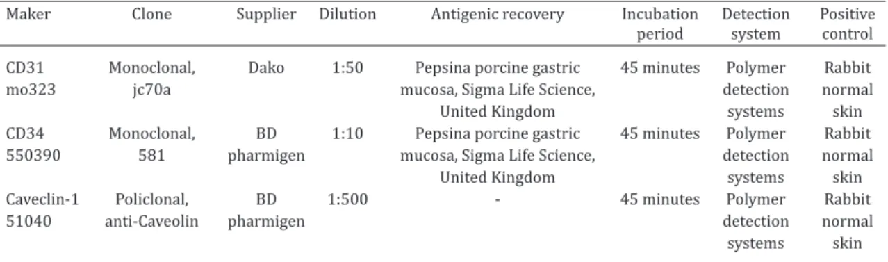
ical study, a panel of antibodies specific for several antigens was applied (Table 1). The glass slides was previously cleaned and degreased, and prepared with prepared with organosilanebased adhesive (3aminopropyl triethoxysilane, C.O. Sigma Chemical, USA). Novolinktm Polymer Detection Systems was used according
to the manufacturer’s instructions.
Methods of data interpretation. The analysis of histological
sections was performed by the same pathologist without prior knowledge of the identification of the groups (FG). Histological evaluation of tissue was examined under an optical microscope7
at 400x magnification. The photomicrographs were held on the NIS program, Nikon Imaging Software Elements version 30.4 NIS program, Nikon Imaging Software Elements version 4.30.
The data obtained using Hematoxylin & Eosin, as well as us ing Masson’s trichrome were determined by the semiquantitative and quantitative method. The semiquantitative technique of vas cular proliferation was determined according to the intensity of the vessels were found. The findings were translated into quan titative variables by index assignment for histological findings, as recommended by Garros et al. (2006) and recorded on individual records for each animal.
The angiogenic indexes for Hematoxylin & Eosin, Masson’s trichrome, Caveolina, CD31 and CD34 were determined by mi crovessel counting technique (MVC) as done under Maeda et al. (1995). The areas were analyzed with the largest number of ves sels in the depth of the lesion. Any endothelial cell or cell group stained positively separate from adjacent microvessel, healthy cells, and other connective tissue elements, were regarded as a unitary vessel, and also, the vessels containing lumen. The ves sels containing lumen were considered the same way. Microves sel counting was carried out in five fields, previously selected at a high vascular density, with the microscope set to a magnification of 400x. The microvessel count (MVC) was determined twice by a single evaluator at two different times and was expressed as a mean number of vessels in each case studied.
Statistical analysis. Comparison between groups (Gprp and
Gc) from the categorical variable (vascular proliferation intensi ty) was used the KruskalWallis test with p values equal to or less than 0.05 (p<0.05) were considered significant. For the analysis was used Npar1way procedure computer program SAS (SAS 9.1, SAS Institute, Cary. NC, USA), was used for the anaylsis as well as the GraphPad Prism program, Version 4.00.
The immunohistochemical data were submitted to analysis of variance for a completely randomized design, with two groups with five repetitions (medium) at a 5% significance level. Multiple com parisons of group called for our using the Tukey test (α=0.05). For this analysis, we used the General Linear Models Procedure (GLM) of the SAS computer software (SAS 9.1, SAS Institute, Cary. NC, USA).
RESULTS AND DISCUSSION
The proliferation of vascular were determined by the se miquantitative and quantitative method.by usingHE and Masson’s trichrome has been found to be a significant va riable in group GPRP (p=0.01) (Fig.1A) when compared with the control group (Fig.1B). The evaluation of the me thods used concluded that there was no significant diffe rence (p>0.05).
Low cost and ability to demonstrate various structures are assigned to features in HE staining, which is frequen tly used in routine laboratories (Bancroft et al. 2012). Masson’s trichrome is also used, but to a lesser extent be cause of its longer preparation time and how it requires di fferent reagents compared to HE; its purpose is to highlight the types of surrounding tissue and to make it possible to distinguish blood vessels from other structures (Bancroft
et al. 2012).
The characteristics of the HE and Masson’s trichrome were observed during microscopic analysis in this study as described in the literature (Bancroft et al. 2012). One can distinguish the préblood vessels apparently formed using Masson’s Trichrome, as is evidenced from other structures whenfacilitating the analysis; however, HE has also been observed in the same structures, only requiring further attention during the review because the vessels are not evidencing much compared with Masson’s Tri chrome.
For semiquantitative evaluation of the vessels, the HE and Masson trichrome staining were chosen to be used be cause of there being described few methods for assessing angiogenesis in the skin of rabbits. HE staining alone is commonly used for this purpose. Thus, the immunohisto chemical methods and analyses performed in this study are in agreement with Reis (2015), and Serafim (2016), who used such dyes in a semiquantitative evaluation of blood vessels in rabbits fragments undergoing reconstructive surgery since they obtained good results. However, in this study, the quantitative technique for vessel counting using HE and masson trichrome was also performed, in order to evaluate the difference between the methods (p=0,05). Al though there was no significant difference between the two methods of analysis, it is recommended to use the quantita tive technique because of its accuracy.
Microvessel count (MVC) was performed with different markers (Caveolin1, CD31, and CD34) and was significant
7 Microscópio Nikon eclipse E200.
Table 1. Antibodies used for immunohistochemistry cutaneous tissues of rabbits conducted at Veterinary Pathology Laboratory, University of Porto, Portugal, 2016
Maker Clone Supplier Dilution Antigenic recovery Incubation Detection Positive
period system control
CD31 Monoclonal, Dako 1:50 Pepsina porcine gastric 45 minutes Polymer Rabbit
mo323 jc70a mucosa, Sigma Life Science, detection normal
United Kingdom systems skin
CD34 Monoclonal, BD 1:10 Pepsina porcine gastric 45 minutes Polymer Rabbit
550390 581 pharmigen mucosa, Sigma Life Science, detection normal
United Kingdom systems skin
Caveclin1 Policlonal, BD 1:500 45 minutes Polymer Rabbit
51040 antiCaveolin pharmigen detection normal
at GPRP (p=0.01) (Fig.2A) compared with the control group (Fig.2B), averaging vessels 56.5 and 20.9 respectively appe aring in groups. When correlating microvessel count of the three markers there was no significant difference (p>0.05). However, there was more intense staining of vessels. When using Caveolin1 Antibody the marking of the capillaries, small vessels were intense, as well as inlarger vessels. When assessing the CD31 and CD34, marking of vessels were ob served, but was not as intense as with Caveolin1. Some cases showed background and discreet marking. The results of this study are similar to Serafim (2016), which reports success in using Caveolin1 as a marker of angiogenesis in the skin fragements of rabbits undergoing reconstructive surgery.
According to Abbas et al. (2013), Caveolin1 is an an tibody used for identifying cytoplasm membrane and en dothelium; it’s not specific for angiogenesis. The results of this test were contrary to that as described in the literature (Abbas et al. 2013.) because of Caveolin 1 showingintense immunolabeling of neovascularization, proving it to be an excellent marker of angiogenesis in the skin of rabbits.
Besides other specific markers being used to evaluate the angiogenic index as VEGF, CD 31, CD 34, CD105 and Fac tor VIII (Bancroft et al. 2012), in rabbits there is a restric
tion of some of these markers due to polyclonal antibodies being obtained from lab animals, especially rabbits and mice (Ball et al. 1990, Hay et al. 2002).
The specific markers of vascular endothelial (VEGF, CD 31, CD 34 and Factor VIII) obtained from rabbits are more susceptible to submit changes, such as cross reactions and false negatives, which can compromise the result. Changes often result in intense staining of various structures, inten se presence of background, or no marking (Bancroft et al. 2012). The results of this study are in agreement with the literature, since when using specific markers for angioge nesis (Bancroft et al. 2012) CD34 and CD31 the presence of background in some samples was observed, and in indi vidual cases occurred lighter markup of the vessel. Althou gh it has not presented significant difference between the assessment of markers, the microvascular count of CD 31 and CD 34 required greater attention in order to avoid fai lure due to the fund’s presence and the discrete markings. Although Caveolin1 is not highly specific for vascular en dothelium, the labeling in endothelial rabbit tissue was sa tisfactory when compared with other antibodies employed in this work, making it suitable for microvessel count in rabbits. These results are consistent with Serafim (2016).
CONCLUSIONS
The results of this study demonstrated the histoche mistry methods performed with Hematoxylin and eosin as well as with Masson’s Trichrome staining being useful for semiquantitative assessment of angiogenesis, although there was not specific demand greater attention from the pathologist at the time of the analysis in order to avoid fai lures in counting; however we recommend using Masson’s trichrome for histochemical evaluation of angiogenesis as it shows the vessels.
The immunohistochemical comparison of Caveolin1, CD31, and CD34 as markers of angiogenesis in rabbits sho wed that both antibodies could immunolabel newly formed vessels, but the Caveolin1 showed better immunolabeling
of small and medium sized vessels and lower presence of background, being considered as immunohistochemical marker of vascular endothelium in rabbits.
Acknowledgements.- The authors are grateful to the Coordination of Improvement of Higher Education Personnel (CAPES) for the scholarship given to Josiane Morais Pazzini; Faculty of Agricultural and Veterinary Sciences of Jaboticabal (FCAV), Universidade Estadual Paulista (Unesp), for the structure and laboratory support; CAPES for the scholarship accor ded in Brazil and Portugal (PDSE); the laboratory team at the University of Porto for supporting activities conducted.
REFERENCES
Abbas A.K., Kumar V., Fausto N. & Aster J.C. 2013. Robbins & Cotran Pato logia, Bases Patológicas das Doenças: inflamação e reparo. Vol.2. 9a ed.
Elsevier, Rio de Janeiro, p.2974.
Fig.2. Histologic aspect of microvessel count (MVC) in rabbits (Oryctolagus cuniculus). Dermal photomicrography of rabbits submitted
to the reconstructive surgery procedure at the Veterinary Hospital of the Faculty of Agricultural and Veterinary Sciences (FCAV) of Unesp, Jaboticabal Campus, 2015. (A) Presence accentuated neovascularization in circumscribed area in GPRP, noting the presence
of background (arrow). (B) Discreet presence of neovascularization in circumscribed area in Gc, noting the presence of background
(arrow). (C) Discreet presence of neovascularization in circumscribed area in Gc, noting the presence of background (arrow). Marker
Bancroft’s J.D., Layton C. & Suvarna S.K. 2012. Theory and Practice of Histological Tetchniques.7th ed. Elsevier, Churchill Livingstone, p.381 423.
Reis N.P. 2015. Epitelização de enxertos cutâneos em feridas recentes de coelhos tratados com membrana amniótica canina e/ou laserterapia. Master Dissertation, Universidade Estadual Paulista Júlio de Mesquita Filho, Jaboticabal, São Paulo. 48p.
Garros I.C., Campos A.C.L., Tâmbara E.M., Tenório S.B., Torres O.J.M., Agu lham M.A., Araújo A.C.F., SantisIsolan P.M.B., Oliveira R.M. & Arruda E.C.M. 2006. Extrato de Passiflora edulis na cicatrização de feridas cutâ neas abertas em ratos: estudo morfológico e histológico. Acta Cir. Bras. 21(3):5565.
Hay F.C., Westwood O.M.R., Nelson P.N. & Hudson L. 2002. Practical Immu nology. Blackwell Science, Malden, MA.
Hedlund C.S. 2008. Cirurgia de Pequenos Animais.3ª ed. Elsevier, São Pau lo. 1390p.
Maeda K., Chung Y.S., Takatsuka S., Ogawa Y., Onoda N., Sawada T., Kato Y., Nitta A., Arimoto Y., Kondo Y. & Sowa M. 1995. Tumor angiogenesis and
tumour cell proliferation as prognostic indicador in gastric carcinoma. Brit. J. Cancer 72(2):319323.
Pazzini J.M. & Moraes P.C. 2015. Princípios e Técnicas de Cirurgias Recons trutivas da Pele de Cães e Gatos: atlas colorido. Medvep, Curitiba, p.95 102.
Pazzini J.M., De Nardi A.B., Huppes R.R., Gering A.P., Ferreira M.G.P.A., Silvei ra C.B.P., Luzzi M.C & Santos R. 2016. Method to obtain platelet rich plas ma from rabbits (Oryctolagus cuniculus). Pesq. Vet. Bras. 36(1):3944.
Serafim E.L. 2016. Fechamento de defeitos em padrão de figura geométri ca associado ao emprego de anestesia por tumescência com lidocaína em coelhos (Oryctolagus cuniculus).Master Dissertation, Universidade Estadual Paulista Júlio de Mesquita Filho, Jaboticabal, SP. 68p.
Vendramin F.S., Franco D., Nogueira C.M., Pereira M.S. & Franco T.R. 2006. Plasma rico em plaquetas e fatores de crescimento: técnica de preparo e utilização em cirurgia plástica. Revta Col. Bras. Cir. 33:2428.