Assessment of the efects of bismuth subgallate on proliferation
of myoibroblasts: an experimental study in rats
Avaliação dos efeitos do subgalato de bismuto na proliferação de miofibroblastos
Rubianne Ligório de Lima1
*
, Cláudia Paraguaçu Sampaio1, Karin Caroline Seidel1, Melina Branco1, Rafaela Mabile Sobreiro1
Abstract
Background: Bismuth subgallate is an insoluble heavy metal that is used for its astringent and hemostatic properties.
Objective: To evaluate the efects of bismuth subgallate on the healing process by observation of myoibroblasts in the skin of rats. Methods: A sample of 60 Wistar rats was used. Each rat was subjected to a dorsal skin wound and allocated to one of two groups: a control group, in which 0.9% sodium chloride was administered daily, or an experimental group, in which 0.5 mg of bismuth subgallate was administered daily. Each of these groups was further subdivided into three subsets, which were reoperated after 3, 7 and 14 days respectively for excision and collection of the skin wound specimens. Samples were treated with hematoxylin eosin, picrosirius, and immunohistochemical staining to enable assessment of myoibroblast counts, inlammatory response phase, and collagen synthesis. Results: No inlammatory process diferences were detected between the control and experimental groups at 3 days (p = 1), 7 days (p = 0.474), or 14 days (p = 303). Evaluation of types I and III collagen in the control group did not demonstrate healing beneits at 3 days (p = 0.436), 7 days (p = 0.853), or 14 days (p = 0.436); whereas in the experimental group there were increases in types I and III collagen at 3 and 14 days (p = 0.005). Immunohistochemical analysis conirmed the results of hematoxylin eosin staining, since there were no diferences between subsets in terms of area of myoibroblasts, in the experimental (p = 0.4) or the control (p = 0.336) groups. Conclusions: Administration of bismuth subgallate to skin wounds in rats did not result in any evidence of beneits to healing, i.e., no diference in ibroplasia was detected when experimental and control groups were compared.
Keywords: wound healing; myoibroblasts; otolaryngology.
Resumo
Contexto: O subgalato de bismuto é um metal pesado e insolúvel, utilizado por suas propriedades adstringentes e hemostáticas. Objetivo: Avaliar os efeitos do subgalato de bismuto na cicatrização mediante observação de mioibroblastos em pele de ratos. Métodos: Foram utilizados 60 ratos da linhagem Wistar, que receberam uma ferida no dorso da pele. Os animais foram divididos em dois grupos: controle (aplicação diária de cloreto de sódio a 0,9%) e experimental (aplicação diária de 0,5 mg de subgalato de bismuto). Cada grupo foi subdividido em três subgrupos, que foram reoperados para retirada da ferida em 3, 7 e 14 dias. Foi realizada coloração de hematoxilina eosina, picrosirius e imuno-histoquímica para avaliar contagem de mioibroblastos, resposta inlamatória e síntese de colágeno. Resultados: Não foi encontrada diferença entre os grupos controle e experimento com relação ao processo inlamatório – subgrupos 3 dias (p = 1), 7 dias (p = 0,474) e 14 dias (p = 303). A avaliação dos colágenos tipo I e III no grupo-controle não demonstrou benefícios de cicatrização – 3 dias (p = 0,436), 7 dias (p = 0,853) e 14 dias (p = 0,436); já no grupo experimental, houve aumento dos colágenos tipos I e III nos subgrupos 3 e 14 dias (p = 0,005). A imuno-histoquímica conirmou os resultados encontrados na coloração hematoxilina eosina, na qual a área de mioibroblastos entre os subgrupos, nos grupos experimental (p = 0,4) e controle (p = 0,336), foi indiferente. Conclusão: A utilização do subgalato de bismuto em ferida de pele de ratos não evidenciou benefícios na cicatrização, ou seja, não houve diferença na ibroplasia quando comparados os grupos experimental e controle.
Palavras-chave: cicatrização; mioibroblastos; otolaringologia.
1Pontifícia Universidade Católica do Paraná – PUC-PR, Faculdade de Medicina, Curitiba, PR, Brazil.
Financial support: None.
Conlicts of interest: No conlicts of interest declared concerning the publication of this article. Submitted: March 28, 2016. Accepted: July 28, 2016.
INTRODUCTION
Bismuth subgallate is a yellowish substance that is presented in the form of an odorless powder and which undergoes discoloration in the presence of sunlight.1
It is increasingly being used by professionals working in otorhinolaryngology and dentistry because of its astringent and hemostatic properties. Applications include topical treatment of open wounds, treatment of gastroduodenal ulcers, as an antidiarrheal agent, to control colostomy odor, during dental surgery, for management of epistaxis and, empirically, in adenotonsillectomies.1-3
When performing an amygdalectomy, the otorhinolaryngologist’s primary concerns are to reduce transoperative bleeding, reduce duration of surgery, and avoid postoperative complications; in other words, to achieve a safe procedure.1-3
Bismuth subgallate is a heavy metal that is relatively insoluble in water and has astringent properties (via activation of factor XII of the coagulation cascade), accelerates formation of blood clots, and improves hemostasis.4 In view of its increasing use, there is
a need for controlled and randomized studies with acceptable methodology to provide better support for its use.
Some otorhinolaryngology services do not employ bismuth subgallate, claiming that its administration during the immediate postoperative period is not
beneicial, whereas others describe it as very useful
for controlling bleeding. In general, studies have focused on its use to control bleeding during the immediate postoperative period.
There is no objective answer in the literature with
relation to the beneits of subgallate for hemostasis or
for healing and studies are contradictory. Additionally,
its possible beneicial effects on healing have not been
investigated and there is room for improvement in terms of use of comparison or control groups.
Fibroplasia is an important phase in healing and if
bismuth subgallate does induce increased ibroplastic
activity, it is to be expected that the number of
myoibroblasts will be increases, leading to an early
contraction process, optimizing healing.
Fibroblasts are responsible for synthesis, remodeling, and deposition of the matrix and also interact with the matrix. The structural molecules that make up the new extracellular matrix contribute to formation of granulation tissue, which in turn provides a foundation for cell migration. Formation of granulation tissue begins around the third day.5
Fibroblasts secrete a monomer called procollagen.
The reticular ibers of type III collagen are narrower
than those of type I collagen and have larger quantities of carbohydrates. Wound healing initiates with type III collagen, which will later be substituted by the more resistant type I collagen.5
The overall objective of this article was to evaluate whether bismuth subgallate has effects on some of the phases of healing by histological and immunohistochemical analysis and by observation
of myoibroblast development in dorsal skin wounds.
Since it is a material that is easy to handle (in powder form) and is inexpensive, its use in healing could
beneit the population, if studies indicate that such a
correlation does exist.
METHODS
After approval was granted by the local Animal Research Ethics Committee (protocol 780), and in accordance with Brazilian College of Animal Experimentation (COBEA) recommendations, the experiments were conducted at the Experimental Surgery and Operating Techniques Laboratory at the Pontifícia Universidade Católica do Paraná (PUC-PR) during July of 2013.
A sample of 60 male Wistar rats (Rattus norvegicus
albinus, Rodentia mammalia), all young adults aged
110 days and with mean weight from 250 g to 300 g, were obtained from the University’s Central Animal House. These animals were provided with food and water ad libitum. The same sample of rats was also used in a project entitled “Evaluation of the effects of bismuth subgallate on angiogenesis: experimental study with rats”.
At the Experimental Surgery and Operating Techniques Laboratory, the animals were anesthetized with 0.1 ml/100 g of body weight with a mixture of 1 ml of ketamine (50 mg) with 1 ml of 2% xylazine (20 mg) administered intramuscularly to the posterior thigh. They were then positioned in decubitus ventral on a wooden support to which front and hind legs
were ixed. An area of 24 cm2 (6 centimeters long by
4 centimeters wide) was shaved on the dorsal region of each animal, located with relation to an imaginary line between the front limbs and extending 6 centimeters in the caudal direction. Shortly afterwards, antisepsis was performed with povidone iodine and the operating
are was delimited with a fenestrated sterile ield.
was taken to a depth suficient to expose the dorsal
muscle fascia.
After the operation, each animal was given intramuscular potassium diclofenac at a dosage of 10 mg/kg with the objective of achieving analgesia
and preventing inlammation. The animals were
transported back to the Central Animal House. The animals were marked and separated at random to form three experimental groups and three control groups of 10 animals each. Animals in experimental groups were administered 0.5 mg of bismuth subgallate to their wounds daily. The wounds of control animals were treated daily with 0.9% sodium chloride solution, as recommended in the literature. One control group and one experimental group each underwent euthanasia on day 3, day 7, and day 14.
The animals were reoperated to remove the wound specimens on the days scheduled for each group. Each operation was performed after anesthesia of the animal (as described above). The specimen removed included a margin of 1 cm of intact skin around each surgical wound and was to the depth of the dorsal musculature.
Immediately after reoperation, each animal was euthanized with a lethal dose of intraperitonial sodium thiopental (120 mg/kg), which is the euthanasia method for rodents and other small mammals recommended in Federal Veterinary Medical Council Resolution 714 of June 20, 2002.
None of the animals or specimens were lost during the study.
After removal from the rats, the histology specimens were laid out on labeled cards. They were then immersed in a receptacle containing 10% formol for 24 hours and were then cut and placed in cassettes for
mounting of the parafin blocks. Slides were stained
with hematoxylin eosin (HE) and picrosirius and analyzed under an optical microscope.
Monomorphonuclear and polymorphonuclear cells and vessels were counted on the HE slides to
characterize the inlammatory process, which was
scored as shown in Table 1 and then classiied as shown in Table 2.6
Initially, inlammatory process phases were compared
between the two groups, control and experimental, for each of the three assessment points (3 days, 7 days, and 14 days). Next, within each group, the assessment points were compared with each other, two by two. The null hypothesis that the distribution
of inlammatory process phases was equal at the two
different assessment times was tested against the alternative hypothesis that the distributions were different.
Picrosirius staining was used to assess collagen
by means of the birefringence speciic to each type
of collagen, using a polarized light microscope. The Mann-Whitney nonparametric test was used to compare the percentage collagen area in each group at each of the assessment times (3 days, 7 days, and 14 days). Intra-group comparisons of different assessment times against each other were performed using the Kruskal-Wallis nonparametric test. Fisher’s exact test was used for the comparative analyses of groups and
assessment times in terms of inlammatory process
phases. P values < 0.05 were considered indicative
of statistical signiicance. Data were analyzed with
IBM SPSS Statistics v.20.
Specimens were also sent for immunohistochemical processing, with the intention of testing for alpha-SMA,
Factor 8 and CD34. However, readings were only
conducted for alpha-SMA, because the smooth
muscle stain analyzed provided very little speciicity for Factor 8 and CD34.
After histological and immunohistochemical analysis, statistical analysis was conducted using parametric and nonparametric methods, before compilation of
the inal documentation and writing of the article
for publication.
Analysis of immunohistochemical results was based on the area stained a chestnut color, which
indicates the area of myoibroblast expression, and
on counting vessels to evaluate angiogenesis.
RESULTS
Evaluation of the inflammatory process by monomorphonuclear and polymorphonuclear cell counts
The two initial analyses of the inflammatory process revealed no differences in healing between the respective groups at 3 days (p = 1), 7 days (p = 0.474), or 14 days (p = 0.303) (Figures 1 and 2).
Table 1. Scores for inlammatory process cell counts.
Number of cells
Polymorphonuclear Monomorphonuclear
Up to 50 –1 1
50-100 –2 2
> 100 –3 3
Table 2. Classiication of inlammatory process phases by inal
score for each group.
Inflammatory process score Final classification score
Acute –9 to –3
Subacute –2.9 to 3
Evaluation of collagen by picrosirius staining
Type I collagen was compared between experimental and control groups and intra-group analyses were conducted comparing the three different days against each other. Production of type I collagen in the experimental and control groups was similar, i.e. there was no quantitative difference in type I collagen between the groups (p = 0.330). In the experimental group, there was a considerable increase in type I collagen from 3 to 14 days, which was not observed in the control group (p = 0.024) (Figure 3).
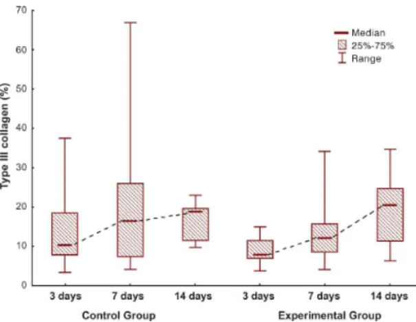
Analysis of the results for type III collagen was performed in the same manner as analysis of type I collagen results and revealed the same results (control group: p = 0.330; and experimental group: p = 0.024) (Figures 4-6).
Immunohistochemical processing with alpha-SMA analysis
The analysis of myoibroblasts did not reveal any
differences in terms of healing phase and analysis of vessels revealed no difference in angiogenesis (control group: p = 0.336; and experimental group: p = 0.400) (Figures 7 and 8).
DISCUSSION
Methods are currently being investigated that are intended to reduce and avoid transoperative and postoperative hemorrhages in adenotonsillectomies and several different approaches have been compared. Methods based on cryosurgery are considered
unfeasible because of the high cost and the dificulties
with handling and storage of liquid nitrogen. In turn, methods employing lasers demand the correct power beam, in addition to precise exposure time and focus angles if a technique is to be considered promising, and there is the possibility of damage to adjacent structures because of the high temperatures of the beam (750 to 900 °C).7
These problems related to other methods have resulted in worldwide acceptance and adoption of bismuth subgallate as a hemostatic agent for tonsil surgery, even in the absence of studies demonstrating
its true eficacy.
The literature does not cover the possible beneits
for healing of bismuth subgallate. With the intention of contributing to better options for hemostasis, we
Figure 1. Hematoxylin eosin staining for control group/14 day
subset.
Figure 2. Hematoxylin eosin staining for experimental group/14 day subset.
attempted to increase understanding of this substance’s healing properties.
Other authors reported excellent hemostatic results in the area of bleeding left by tonsillectomy using a gauze dressing soaked in a 100% solution of bismuth subgallate. The method was also considered low
cost and easily employed, and it is rare that suture or ligature of the vessels in the region is needed.8
When we compared the degree of inlammation
between the experimental and control groups, we noted
that there was no signiicant difference. The same was observed in a study that investigated the inlammatory
process with use of bismuth subgallate in partial hepatectomies in rats.9
Another study assessed healing in the dorsal area of Wistar rats, but using different periods for the subsets (1, 4, 7, 11, and 18 days), and also concluded that bismuth subgallate did not impact on the quality of the healing process and was biocompatible with the tissues. That study observed a larger area of granulation tissue, due to the physical presence of the material, concluding that it can be prescribed as a hemostatic
with no signiicant effect on the healing process, which demonstrates that this inding is independent
of the duration of administration of the substance.10
In the present study, no beneficial effect on skin healing from use of the bismuth subgallate
Figure 4. Comparison of groups for immature collagen.
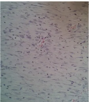
Figure 5. Picrosirius staining for control group/14 day subset.
Figure 6. Picrosirius staining for experimental group/14 day subset.
Figure 7. Immunohistochemical staining for control group/14
day subset.
Figure 8. Immunohistochemical staining for experimental
solution was observed and there was also no effect
on ibroplasia. A different study observed delayed healing and ibroplasia in mucosas, even though the
dilution and quantity of the substance administered were the same in both studies.11
One possible reason for the difference in the behavior of bismuth subgallate in mucosas and in skin is the individual properties of each of these tissue types. Compared with skin, oral mucosa exhibits smaller quantities of scar tissue, both clinically and
histologically, because it has fewer ibers. In terms
of formation of scar tissue, the differences between mucosa and skin are advantageous, both from esthetic and functional perspectives. Researchers believe that these differences are related to the embryonic origins of
the ibroblasts in the two types of tissue. The ibroblasts
in skin are derived from the mesoderm, and those in oral mucosa from cells of the neural crest.12
The smaller quantity of ibers originating from the
neural crest may enable a higher degree of absorption of the subgallate, which may have had toxic effects because of the greater quantity absorbed, altering
ibroplasia and, consequently, healing in general. In the skin, the larger quantity of ibers originating
from the mesoderm may have created a barrier, impeding greater absorption and resulting in reduced
toxicity. There was no effect on ibroplasia, and
healing was unaltered.
The picrosirius staining revealed a considerable increase in production of type I collagen from 3 to 14 days in the experimental group, which was not observed in the control group (p = 0.024). The immunohistochemical analysis did not detect this result. One possible explanation for this fact calls into question the effectiveness of the marker employed,
since it is not speciic for rats. In this case it would be
interesting to conduct the test again with a different
marker in order to conirm the immunohistochemical results, but, because of the costs and dificulties
involved, the decision was taken not to use a different
marker at this point and the parafin blocks have been
stored for future studies.
CONCLUSIONS
In rats, treating skin wounds with bismuth subgallate
did not exhibit beneits in terms of myoibroblast
proliferation when experimental and control groups were compared.
REFERENCES
1. Brasileiro HM, Lee IW, Rapoport A. Uso do subgalato de bismuto como agente hemostático em tonsiléctomia palatina: estudo de 201 casos. São Paulo: Atha; 2006. p. 181-5. ACTA ORL/Técnicas em Otorrinolaringologia.
2. Kim SH, Grein RL, Tramontina VA. Aplicação do subgalato de bismuto em cirurgia periodontal. JBC J Bras Odontol Clin. 1997;1(1):31-4.
3. Pillar RS, Brito EO. Aplicação do subnitrato de bismuto e do subgalato nas amigdaléctomias. Arq Otorrinolaringol. 2003;7(1):28-31.
4. Padilla RM, Valdés CM, Asato JR, et al. Eficacia del subgalato de bismuto, como agente hemostático tópico, en el sangrado transoperatorio de la adenoamigdalectomía o amigdalectomía. An Orl Mex. 2012;57(2):65-8.
5. Simões MJ, Cabral AC, Boyaciyan K, Kulay L Jr, Sasso WS. Aspectos ultra-estruturais dos fibroblastos e dos macrófagos durante o processo de reparação da pele de ratos. Rev Paul Med. 1986;104(3):132-5. PMid:3563265.
6. Vizzotto AO Jr, Noronha L, Scheffel DL, Campos AC. Influência da cisplatina administrada no pré e pós-operatório sobre a cicatrização de anastomoses colônicas em ratos. J Bras Patol Med Lab. 2003;39(2):143-9. http://dx.doi.org/10.1590/S1676-24442003000200002003;39(2):143-9.
7. Hernández-Paz SH, Ortiz-Reyes A, García-Guzmán CM. Estudio comparativo de dos agentes hemostáticos adicionados con epinefrina en la adenoamigdalectomía. Rev Esp Med Quir. 2012;17(1):3-7.
8. Brasileiro HM, Lee IW, Rapoport A. Relação entre as complicações hemorrágicas pós cirúrgicas e o volume das tonsilas palatinas. Rev Bras Otorrinolaringol. 2004;70(1):58-61. http://dx.doi.org/10.1590/ S0034-72992004000100010.
9. Arroyo PC Jr, Silva RCMA, Santi D No, Santana D Jr, Ferreira FD, Silva RF. Uso do subgalato de bismuto para hemostasia local em hepatectomias parciais em ratos. Rev Col Bras Cir. 2004;31(3):165-71. http://dx.doi.org/10.1590/S0100-69912004000300005.
10. Tramontina VA. Efeito do subgalato de bismuto no processo de reparação de feridas em dorso de rato. Estudo experimental histológico, histométrico e fotográfico [dissertação]. São Paulo: Universidade Estadual de Campinas; 1997 [citado em 2016 mar 28]. Disponível em: http://www.bibliotecadigital.unicamp.br/ document/?code=vtls000118398
11. Seidel K, Sampaio CPP, Maeda CRS, et al. Avaliação dos efeitos do subgalato de bismuto na cicatrização: avaliação do processo inflamatório e da proliferação dos miofibroblástos. In: Anais do 43º Congresso Brasileiro de Otorrinolaringologia; 2013; São Paulo. São Paulo: Elsevier; 2013. p. 111.
*
Correspondence
Rubianne Ligório de Lima Rua Alberto Potier, 50/33 - Boa Vista CEP 82560-480 - Curitiba (PR), Brazil Tel.: +55 (41) 9941-6723 E-mail: [email protected]
Author information
RLL - MD from Pontifícia Universidade Católica do Paraná (PUC-PR). CPS - DDS from Universidade Estadual de Ponta Grossa (UEPG); MD from Pontifícia Universidade Católica do Paraná (PUC-PR); MSc in Surgery from Pontifícia Universidade Católica do Paraná (PUC-PR). KCS - MD from Pontifícia Universidade Católica do Paraná
(PUC-PR); Resident of Otorhinolaryngology at Hospital Santa Casa de Misericórdia de Curitiba. MB - Pontifícia Universidade Católica do Paraná (PUC-PR). RMS - MD from Pontifícia Universidade Católica do Paraná (PUC-PR); Board certiication student of Otorhinolaryngology at Hospital da Cruz Vermelha de Curitiba.
Author contributions