3 7 5 3 7 5 3 7 5 3 7 5
3 7 5 VOL. 36(3) 2006: 375 - 380
Marlon Aguiar MELO¹, Fernando ABRUNHOSA², Iracilda SAMPAIO²
ABSTRACT
Previous study on the resistance of larvae of Sesarma curacaoense submitted to starvation has revealed a facultative lecithotrophy during zoeal stages, but megalopa and first juvenile stages are exclusively feeding stages. In the present study, the gross morphology and fine structure of the foregut of S. curacaoense were investigated during larval, megalopa and first juvenile stages. The foregut of the zoea I show specific setae and a filter press apparently functional. The foregut undergoes changes in the zoea II (last larval stage) with increment of setae number, mainly on the cardiopyloric valve and complexity of the filter press. After metamorphosis to megalopa stage the foregut become rather complex, with a gastric mill supporting a medial and two lateral teeth well-developed. The foregut of the first juvenile is more specialized compared to the previous stage, showing similar characteristics of the decapod adults. These results provide further evidence of facultative lecithotrophic development in the larvae of S. curacaoense.
KEYWORDS
Morphology, lecithotrophy, foregut, larval development, grapsid crab.
A morfologia do estômago de larvas e pós-larvas de
Sesarma curacaoense
De Man, 1892: uma espécie com desenvolvimento larval lecitotrófico
facultativo
RESUMO
Estudo prévio sobre o efeito da inanição em larvas de Sesarma curacaoense propôs que estas larvas apresentam comportamento lecitotrófico facultativo. No presente trabalho a morfologia do estômago de S. curacaoense foi estudada durante os estágios larvais, megalopa e juvenil I. A estrutura do estômago da zoea I possui cerdas específicas e com filtro pilórico aparentemente funcional. Especialização no estômago do zoea II (último estágio larval) foi evidenciada pelo incremento do número de cerdas na válvula cárdio-pilórica e pela complexidade do filtro pilórico. Após a metamorfose para o estágio megalopa, o estômago ficou consideravelmente complexo, com o aparecimento de um moinho gástrico contendo um medial e dois laterais dentes bem desenvolvidos. O estômago do juvenil I mostrou-se ainda mais especializado que no estágio anterior, exibindo características morfológicas similares àquelas descritas para decápodes adultos. Estes resultados corroboram o proposto em trabalhos anteriores, nos quais é indicado que S. curacaoense possui um desenvolvimento larval lecitotrófico facultativo.
PALAVRAS-CHAVE
Morfologia, lecitotrofia, estômago, desenvolvimento larval, caranguejo grapsídeo.
¹ Universidade Federal do Ceará, Mestrado em Engenharia de Pesca, Campus do Pici, Bairro: Pici, 60.356-000. Fortaleza – CE – Brasil, e-mail – [email protected]
3 7 6 3 7 6 3 7 6 3 7 6
3 7 6 VOL. 36(3) 2006: 375 - 380 MELO et al.
INTRODUCTION
The semi terrestrial grapsid crab Sesarma curacaoense De Man, 1892, has been confused in previous reports under the name S. crassipes (Melo, 1996; Coelho and Ramos, 1972). The morphological characteristics of these two species are similar, but recent research has shown that the Brazilian species is S. curacaoense (Prado, unpublished).
Experimental studies on the larviculture of S. curacaoense, reared in the laboratory, demonstrated that this species has a reduced larval cycle, consisting of two zoeal stages and one megalopa (Anger et al. 1995). These authors reported that the thoracic appendages of the zoea I and II have advanced degrees of development. In fact, all feeding appendages and pereiopods are functional since zoea I (except third maxilliped). In other species these appendages are functional normally only in later larval stages.
On the other hand, an important investigation on the starvation resistance of larvae of S. curacaoense has revealed that their larvae possess high endotrophic potential (Anger, 1995), and that they are able to develop from embryo to the megalopa in complete absence of food. However, the feeding behavior for these larvae is facultative, once the zoea II of S. curacaoense have shown a delayed development under continued starvation periods. Further, the megalopa reach the juvenile stage without food only if the preceding zoeal stages had been fed continuously. Thus, S. curacaoense has facultative lecithotrophic zoeae and feeding megalopa. These facts suggest a more detailed study of the digestive system of the larvae from this species is needed, in order to understand their feeding biology.
Information on the functional morphology of the external feeding appendages and foregut may contribute directly to understand the feeding behavior of decapod species. Evidence of atrophy or absence of setae, filter press and gastric mill indicates no functionality of the foregut in pueruli of spiny lobster (Nishida et al., 1990; Wolfe and Felgenhauer, 1991; Lemmens and Knott, 1994) and glaucothoe of anomuran crabs (Abrunhosa and Kittaka, 1997a). Experiments of larviculture have confirmed that food isn’t required during pueruli and glaucothoe stages from the above species (Kittaka, 1994; Kittaka, 1988; Nishida et al., 1995, Abrunhosa and Kittaka, 1997a, b). In the present work, the detailed morphology of the foregut of S. curacaoense is described and compared during zoeal, megalopa and first juvenile stages. The functional significance of the foregut is also discussed.
MATERIAL AND METHODS
Two ovigerous S. curacaoense females were collected in Bragança City, northeastern Pará State in March 2004. The females were transported to laboratory and placed individually into 10-liter aquariums with aeration. After hatching, the females
were preserved in 70% ethanol and deposited in the Crustacean Collection of Bragança Campus-UFPA.
The larvae were collected and transferred to three transparent recipients (300 mL cap.) containing seawater. The water temperature was maintained at 28ºC, pH 8.3 and salinity 38‰. The zoeae were separated (30 larvae per container) and fed with Artemia nauplii. The water was changed every three days. The larval development consisted of 2 zoeal stages and a megalopa before molting into the first juvenile stage.
The larvae and postlarvae were fixed in 10% formalin. Later, 10 individuals of each stage were immersed in 20mL of 5% aqueous solution of KOH heated to 80°C for about, 30 minutes for the zoeae and 1 hour for the megalopae and juvenile stages. Then, the samples were washed and immersed in glycerol + ethyl alcohol 70% (1:1). The foreguts were dissected and stained with 1% methylene blue. The terminology used in the description followed Meiss and Norman (1977), Nishida et al. (1990), Abrunhosa and Kittaka (1997a) and Abrunhosa et al. (2003).
RESULTS
MORPHOLOGY OF THE FOREGUT
Foreguts of the zoea I and II (last stage) with cardiopyloric valve, dividing the cardiac chamber of the pyloric chamber, and the filter press, which become more complex after molt to second zoea (Fig. 1a, b). After metamorphosis to megalopa stage occurs specialization of the foregut, with appearance of the gastric mill ossicles (Fig. 2a). The ossicles are more distinct in the newly moulted juvenile than in the previous stage (Fig. 2b). Foreguts have a median horizontal line with setae in both chambers.
1. Zoea I (Fig. 1a)
Foregut length = 0.15mm. Foregut simple, chitinous and unarmed of hard structures.
Cardiac chamber: similar to pyloric chamber in length; with numerous fine setae in the floor; wall almost lacking setae; cardiopyloric valve enlarged and robust, strong cylindrical setae in the posterior portion.
Pyloric chamber: with fine setae on the posterior portion of the roof; filter press seems beehive in shape filling all inferior portion; ampullary net partially developed; interampullary ridge with row of elongated setae (dorsal brush).
2. Zoea II (Fig. 1b)
Foregut length = 0.25mm. Foregut more complex than that of the first stage and lacking hard structures.
Cardiac chamber: floor with row of elongated and fine setae; wall with few setae in anterior portion; cardiopyloric valve enlarged and armed with strong cylindrical setae on the posterior portion.
3 7 7 3 7 7 3 7 7 3 7 7
3 7 7 VOL. 36(3) 2006: 375 - 380 MELO et al.
fine setae on the posterior portion of the roof; filter press large, filling all inferior portion; ampullary net visually developed and functional; interampullary ridge with row of elongated setae.
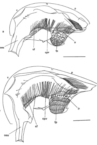
3. Megalopa (Fig. 2a)
Length=0.38mm. Substantial morphological change occurs at the megalopa foregut, with appearance of the gastric mill, oesophageal valve and others ossicles of the foregut.
Cardiac chamber: enlarged dorsolaterally by the ossicles of the gastric mill; the cardiac floor lies vertically; all ossicles of gastric mill are present, such as: zygocardiac (bearing lateral teeth), urocardiac (supporting the medial tooth), mesocardiac, pterocardiac, prepectinal, pectinal, postpectinal, subdentate, inferior lateral cardiac, lateral cardiopyloric, propyloric, exopyloric and pyloric; cardiopyloric valve strong and specialized having many short setae on the superior portion.
Pyloric chamber: compressed laterally; with fine setae on the posterior portion of the roof below the uropyloric ossicle; important ossicles present, i.e., anterior pleuropyloric, posterior pleuropyloric; median pleuropyloric; anterior inferior pyloric, preampullary, inferior ampullary and posterior inferior pyloric; filter press specialized with ampullary net well-developed and functional; interampullary ridge with row of elongated setae.
4. First juvenile (Fig. 2b)
Foregut length = 0.60mm. Foregut more specialized than that of megalopa stage; gastric mill, oesophageal valve and other ossicles of the foregut present.
Cardiac chamber: similar in shape to the previous stage (megalopa); gastric mill fully developed with articulated ossicles, such as: zygocardiac (bearing lateral teeth), urocardiac (supporting the medial tooth), mesocardiac, pterocardiac, prepectinal, pectinal, postpectinal, subdentate, inferior lateral cardiac, lateral cardiopyloric, propyloric, exopyloric and pyloric; cardiopyloric valve strong and specialized, having many short setae on the superior portion.
Pyloric chamber: similar to previous stage; with more fine setae on the posterior portion of the roof below uropyloric ossicle; anterior pleuropyloric, posterior pleuropyloric and median pleuropyloric ossicles more distinct; the ossicles anterior supra-ampullary, anterior inferior pyloric, presupra-ampullary, inferior ampullary and posterior inferior pyloric present but not well developed; filter press with ampullary net developed and functional; row of elongated setae in the interampullary ridge.
DISCUSSION
The foreguts of the larvae of decapod crustaceans have the function of mixing soft and fine organic particles with digestive enzymes (Factor, 1989; Nishida et al., 1995). The morphology observed at foregut of the zoea I of S. curacaoense is simple (Fig. 1a), but it shows specific setae and a filter press apparently
functional. According to Anger et al. (1995), these larvae also have developed and functional feeding appendages (except in the non-developed 3rd maxilliped). This suggests that the first larva is adapted to eat short and soft food particles such as freshly hatched Artemia nauplii, rotifers or microalgae. On the other hand, the zoea I of S. curacaoense is noted to reach the second stage without eating. This fact may be explained due to large quantities of yolk reserve, which are sufficient to supply the energy needed during first larval stage (Anger, 1995).
The foregut of the zoea II (last stage) doesn’t have a gastric mill (Fig. 1b). However, the degree of specialization is observed by increments of setae, mainly on the cardiopyloric valve, and in the complexity of the filter press. Thus, strongly suggesting that the zoeal foregut is well adapted for mixing food function. Similar results were obtained for larvae of Palinuridae and other decapods (Factor, 1989; Nishida et al., 1990; Mikami and Takashima, 1993, 1994; Abrunhosa and Kittaka, 1997 a, b; Abrunhosa and Melo, 2002; Abrunhosa et al., 2003). Therefore, hard particles of food are not recommended for larval culture of S.
3 7 8 3 7 8 3 7 8 3 7 8
3 7 8 VOL. 36(3) 2006: 375 - 380 MELO et al.
curacaoense.
The functional foregut found in the zoea I and II reveals that our investigations complement the observations by Anger (1995), since the larvae of S. curacaoense have facultative lecithotrophic behavior. Thus, when food is available, the larvae should accumulate additional energy reserves.
After metamorphosis to megalopa stage, significant changes do not occur in the morphology of the feeding appendages (Anger et al., 1995), although our observations showed that the morphology of the foregut become substantially complex, with the gastric mill well-developed with a medial and two lateral teeth (Fig. 2a). The presence of such structures indicates that the megalopa stage may eat hard particles of food. Similar observation was reported for brachyuran crabs (Factor, 1982; Minagawa and Takashima, 1994; Abrunhosa et al., 2003).
On the other hand, evidences of non-feeding stages have been reported in the transitional pueruli of spiny lobster of genus Panulirus and Jasus. These species show reduced and uncalcified mandibles, reduction in number of setae in the mouthpart
appendages and poorly developed foregut (Kittaka, 1988; Nishida et al., 1990; Kittaka, 1994; Lemmens and Knott, 1994; Nishida et al., 1995). These characteristics were also reported for
transitional glaucothoe of king crabs genus Paralithodes
(Abrunhosa and Kittaka, 1997a, b).
The foregut of the first juvenile stage (Fig. 2b) has a medial and two lateral teeth still more specialized than that of the previous stage and is similar to that of adults of other decapods described in literature (Meiss and Norman, 1977; Kunze and Anderson, 1979; Suthers and Anderson, 1981; Mikami and Takashima, 1994; Brösing et al., 2002). This suggests that the first juveniles of S. curacaoense are able to processes a large quantity of soft and solid food, which they encounter in the benthic environment.
LITERATURE CITED
Abrunhosa, F.A.; Kittaka, J. 1997a. Functional morphology of mouthparts and foregut of the last zoea, glaucothoe and first juvenile of the king crabs Paralithodes camtschaticus, P. brevipes and P. platypus. Fisheries Science, 63(6): 923-930.
Abrunhosa, F.A.; Kittaka, J. 1997 b. Morphological changes in the midgut, midgut gland and hindgut during the larval and postlarval development of the red king crab Paralithodes camtschaticus. Fisheries Science, 65(5): 746-754.
Abrunhosa, F.A.; Melo, M.A. 2002. Morfologia comparativa do primeiro e último estágios zoea e juvenil de Macrobrachium rosenbergii (De Man, 1879) (Decapoda: Palaemonidae). Revista Ciência Agronômica, 33(2): 65-68.
Abrunhosa, F.A.; Melo, M.A.; Abrunhosa, J.P. 2003. Development and functional morphology of the foregut of larvae and postlarva of Ucides cordatus (Decapoda, Ocypodidae). Nauplius, 11(1): 37-43.
Anger, K. 1995. Starvation resistance in larvae of a semiterrestrial crab, Sesarma curacaoense (Decapoda: Grapsidae). Journal of Experimental Marine Biology and Ecology, 187: 161-174. Anger, K.; Schreiber, D.; Montú, M. 1995. Abbreviated larval
development of Sesarma curacaoense (Rathbun, 1897) (Decapoda: Grapsidae) reared in the laboratory. Nauplius, 3: 127-154. Brösing, A.; Richter, S.; Scholtz, G. 2002. The foregut-ossicle system
of Dromia wilsoni, Dromia personata and Lauridromia intermedia (Decapoda, Brachyura, Dromiidae), studied with a new staining method. Arthropod Structure & Development, 30: 329-338. Coelho, P.A.; Ramos, M.A. 1972. A constituição e a distribuição da
fauna de decápodos do litoral leste da América do Sul, entre as latitudes de 5°N e 39°S. Trabalhos Oceanográficos da Universidade Federal de Pernambuco, 13: 133-236.
Factor, J.R. 1982. Development and metamorphosis of the feeding apparatus of the stone crab, Menippe mercenaria (Brachyura, Xanthidae). Journal of Morphology, 172: 299-312.
Factor, J.R. 1989. Development of the feeding apparatus in decapod crustaceans. In: Felgenhauer, B.E.; Watling, L.; Thistle, A.B. (Eds.). Crustacean issues.Vol. 6. p.185-203.
Kittaka, J. 1988. Culture of the palinurid Jasus lalandii from egg to puerulus. Nippon Suisan Gakkaishi, 54(1): 87-93.
Figure 2 - Foreguts of the megalopa and juvenile of Sesarma curacaoense
3 7 9 3 7 9 3 7 9 3 7 9
3 7 9 VOL. 36(3) 2006: 375 - 380 MELO et al.
Kittaka, J. 1994. Larval rearing. In: Phillips, B.F.; Cobb, J.S.; Kittaka, J. (Eds.). Spiny lobster management. Oxford: Fishing news book. p. 402-423.
Kunze, J.; Anderson, D.T. 1979. Functional morphology of the mouthparts and gastric mill in the hermit crabs Clibanarius taeniatus (Milne Edwards), Clibanarius virescens (Krauss), Paguristes squamosus McCulloch and Dardanus setifer (Milne Edwards) (Anomura: Paguridae). Australian Journal of Marine Freshwater Research, 30(5): 683-722.
Lemmens, J.W.T.J.; Knott, B. 1994. Morphological changes in external and internal feeding structures during the transition phyllosoma-puerulus-juvenile in the western rock lobster (Panulirus cygnus: Decapoda: Palinuridae). Journal of Morphology, 220(3): 271-280.
Meiss, D.E.; Norman, R.S. 1977. Comparative study of stomatogastric system of several decapod Crustacea. I. Skeleton. Journal of Morphology, 152(1): 21-53.
Melo, G.A.S. 1996. Manual de identificação dos Brachyura (Caranguejos e Siris) do litoral brasileiro. Ed. Plêiade/ FAPESP.604p.
Mikami, S.; Takashima, F. 1993. Development of the proventriculus in larvae of slipper lobster, Ibacus ciliatus (Decapoda: Scyllaridae). Aquaculture, 116: 199-217.
Mikami, S.; Takashima, F. 1994. Functional morphology of the digestive system. In: Phillips, B.F.; Cobb, J.S.; Kittaka, J. (Eds.). Spiny lobster management. Oxford: Fishing news book. p. 473-482.
Minagawa, M.; Takashima, F. 1994. Developmental changes in larval mouthparts and foregut in the red frog crab, Ranina ranina (Decapoda: Raninidae). Aquaculture, 126: 61-71.
Nishida, S.; Quigley, B.D.; Booth, J.D.; Nemoto, T.; Kittaka, J. 1990. Comparative morphology of the mouthparts and foregut of the final-stage phyllosoma, puerulus, and postpuerulus of the rock lobster Jasus edwardsii (Decapoda: Palinuridae). Journal of Crustacean Biology, 10(2): 293-305.
Nishida, S.; Takahashi, Y.; Kittaka, J. 1995. Structural changes in the hepatopancreas of the rock lobster Jasus edwardsii (Crustacea: Palinuridae) during development from the puerulus to post-puerulus. Marine Biology, 123(4): 837-844.
Suthers, I.M.; Anderson, D.T. 1981. Functional morphology of mouthparts and gastric mill of Ibacus peronii (Leach) (Palinura: Scyllaridae). Australian Journal of Marine Freshwater Research, 32: 931-944.
Wolfe, S.H.; Felgenhauer, B.E. 1991. Mouthpart and foregut ontogeny in larval, postlarval, and juvenile spiny lobster, Panulirus argus Latreille (Decapoda: Palinuridae). Zoologica Scripta, 20: 57-75.