High frequency of visceral leishmaniasis in dogs under veterinary clinical
care in an intense transmission area in the state of Tocantins, Brazil
Alta frequência de Leishmaniose visceral em cães submetidos a atendimento clínico-veterinário em área de transmissão intensa no estado do Tocantins, Brasil Helcileia Dias Santos1* Samara Rocha Galvão1 Francisca Elda Ferreira Dias1
Taiã Mairon Peixoto Ribeiro1 Osmar Negreiros Filho2 Sebastiana Adriana Pereira Sousa3 Silvia Minharro1
ISSNe 1678-4596
INTRODUCTION
Visceral leishmaniasis (LV) is a disease in the Americas that is caused by the protozoa Leishmania (Leishmania) infantum
(Sin. L. (L.) chagasi). This disease affects domestic and wild animals, and humans. Dogs (Canis lupus familiaris) are considered to be the main domestic reservoir. The main vector,
Lutzomyia longipalpis, and more rarely, L. cruzi, feed on dogs and subsequently transmit the disease to humans (DANTAS TORRES et al., 2009). A disease incidence rate is an estimate of the risk of developing a disease in an exposed population. A high incidence
rate emphasizes the need to implement robust control measures. A highest incidence rate (34.98 per 100,000) of VL has been reported in Tocantins, Brazil in 2011 (DATASUS, 2015). This State has been considered by the Pan American Health Organization as high transmission area (OPAS, 2014). Araguaína – a city located in the extreme north of Tocantins – is a municipality with the highest number of reported cases. According to the Araguaína’s Center for Zoonosis Control, 1,114 cases were reported between 2001 and 2013.
Measures adopted in Brazil to control VL involve treating infected persons, control-ling vectors, and diagnosing and removing
in-1Escola de Medicina Veterinária e Zootecnia, Universidade Federal do Tocantins (UFT), Campus Araguaína, BR 153, Km 112, 77804-970,
Araguaína, TO, Brasil. E-mail: hdsantos@uft.edu.br. *Corresponding author. 2Secretaria Estadual de Saúde, Tocantins, TO, Brasil.
3Curso de Pós-graduação em Ciência Animal,Universidade Federal de Goiás (UFG), Goiânia, GO, Brasil.
ABSTRACT: A direct search for parasites were used as the diagnostic test to determine the frequency of Leishmania spp. infection in dogs
(Canis lupus familiaris) under veterinary clinical care in the city of Araguaína, Tocantins, Brazil. For this approach, lymph node cell samples
were collected using needle aspiration from 649 dogs of different breeds and ages. Two hundred and sixty four (40.7%) dogs tested positive for amastigote forms of Leishmania spp. Furthermore, 202 (76.5%) dogs that tested positive showed some clinical sign of disease, while 62 (28.4%) dogs were asymptomatic. Dogs <2 years old or those that lived alongside poultry species in peri-domicile areas had a greater chance of infection (P<0.05). Our results revealed the importance of frequently monitoring leishmaniasis in dogs, and the need to train veterinary professionals who work in high-transmission areas on the clinical diagnosis of canine visceral leishmaniasis.
Key words: visceral leishmaniasis, cytodiagnosis, risk factors, prevention and control.
RESUMO: O objetivo deste estudo foi determinar a frequência de infecção por Leishmania spp. em cães (Canis lupus familiaris) da cidade de Araguaína, Tocantins, submetidos à atendimento clínico-veterinário, utilizando a pesquisa direta do parasito como forma de diagnóstico. A população estudada foi de 649 cães, de diferentes raças e idades, dos quais foi coletada uma amostra de células de linfonodo através de punção aspirativa. Entre os animais com exame positivo 202 (76,5%) apresentaram algum sinal clínico da doença e 62 (28,4%) animais animais assintomáticos apresentaram exames positivos. Animais com até dois anos de idade e que conviviam com galináceos no peridomicílio apresentaram maior chance de infecção (P<0,05). Os resultados demonstram a necessidade de vigilância constante dos animais em relação
a leishmaniose e denota a importância do aperfeiçoamento dos profissionais veterinários, que atuam em áreas de transmissão intensa, para o
diagnóstico clínico da leishmaniose visceral canina.
Palavras-chaves: Leishmaniose visceral, citodiagnóstico, fatores de risco, prevenção e controle.
fected dogs. Leishmania diagnosis in humans and animals should be based on observing the clinical signs and symptoms that are sugges-tive of the disease followed by a laboratory test that can confirm the presence of parasites in tissues, or the presence of anti-leishmania
antibodies in serum (COURA-VITAL et al., 2011; BRASIL, 2014). Canine visceral leish-maniasis (CVL) has signs that are similar to other diseases, which is challenging for clini-cal diagnosis (BANETH, et al., 2008). There-fore, it is important that other methods com-plement diagnosis.
A parasitology examination that detects amastigote forms of Leishmaniaspp. is a direct method that can be used to diagnose CVL. Tissue fragments can be used from the spleen or liver, or the material from lymph node or bone marrow needle aspirations can be used (BABIKER et al., 2007). The sensitivity of this method depends on factors such as parasite density, type of material collected, and time for which the microscopic analysis was performed. Sensitivity may also differ depending on the organ under analysis. Nevertheless, a positive result confirms infection (BABIKER et al., 2007; BRASIL, 2014).
The present study aimed to determine the frequency of CVL in Araguaína, Tocantins, Brazil using a direct search for parasites as the diagnostic test; this study also assessed the individual factors and clinical characteristics that are associated with the occurrence of CVL.
MATERIALS AND METHODS
The present study was performed in the urban area of the Araguaína municipality located in north Tocantins, Brazil at 7º11’28” latitude, 48º12’26” longitude, with an average altitude of 277m. The municipality covers a 4,000-km2 area, with a resident population in the urban area of 142,925 inhabitants (IBGE, 2010).
The samples analyzed were collected from 649 dogs under veterinary care at five Araguaína city clinics from 2007 to 2014. Lymph node needle aspirations were collected by the veterinarians at these clinics and sent to the Parasitology Laboratory of the School of Veterinary Medicine and Animal Science at the Universidade Federal do Tocantins for leishmaniasis diagnosis using a direct parasitic search. Samples were stained using the Rapid
Panoptic Staining Kit (Instant Prov, New Prov, Brazil), and the slides were analyzed within 20 minutes using an optical microscope with an immersion objective. Samples were considered positive when at least one complete amastigote form of Leishmania spp. (nucleus, kinetoplast, and cytoplasmic limit) was observed.
Dogs were classified as either asymptomatic (no signs suggestive of disease) or symptomatic (the presence of at least one sign of VL infection such as dermatologic and ophthalmic disorders, lymphadenomegaly, splenomegaly, onychogryphosis, apathy, or severe loss of weight) (COURA-VITAL et al., 2011; ALMEIDA et al., 2013). Additional data such as sex, age, breed, presence of other animals in the domicile (dogs, cats and poultry), access to the outdoors (either free or controlled), permanent site of the animal defined by a period of 18 to 22 hours (inside or outside the domicile), and presence or absence of organic matter in the peri-domicile were voluntarily provided by the owners. This data was registered by the veterinarian in a standardized form provided by the laboratory for monitoring the samples. The data were tabulated and descriptive analysis was performed using Epi Info 3.5.4. A chi-square test with Yates correction was calculated with a 95% confidence interval.
RESULTS AND DISCUSSION
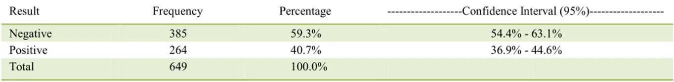
Amastigote forms of Leishmania spp. were observed in 264 (40.7%) samples (Table 1). The prevalence of Leishmania in our sample population was greater than that observed in other studies where the same technique was used (ALMEIDA et al., 2010), and similar to the results observed in seroprevalence studies performed in other areas of Brazil where the infection is endemic (AMÓRA et al., 2006; FRAGA et al., 2012). In our study, the method used had a low sensitivity, which may indicate that the true prevalence may be detected by using more sensitive diagnostic methods (GOMES et al., 2008; SRIVASTAVA et al., 2011).
2007). The CVL frequency was significantly greater
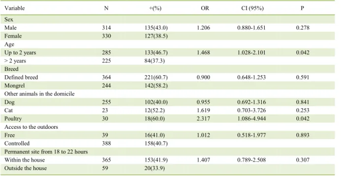
in dogs <2 years old (P=0.042), which differed from the observations of BARBOSA et al. (2010), but similar to the results of DANTAS-TORRES et al. (2006) and FIGUEIREDO et al. (2014). The higher frequency in young dogs may be related to a lower immune maturity of these animals, whereas the adaptive immune system is not fully mature at birth. Only after weeks to months do most mammals become immunologically mature, which predisposes young animals to an increased risk of developing some diseases (DAY, 2007).
When stratified by smaller age bands (<1 year, 1-6 years, and >6 years), we observed a significantly lower CVL frequency (P=0.002) in dogs >6 years (23.5%). No difference was observed for the remaining groups (46.7%, <1 year; 43.3%, 1-6 years). These findings differed from the results observed in other areas of Brazil where the infection is endemic, where the frequency of leishmaniasis was higher in dogs that were 1-6 years old (RONDON et al., 2008). These results suggest that in areas of high incidence, the Leishmaniasis infection risk may be related to environmental rather than individual factors of the host, as also suggested by ALMEIDA et al. (2010).
We did not observe any significant differences with regard to a dog belonging to a specific
breed or not (39.3% and 41.8%, respectively). This
lack of significance may be explained by the diversity
of the breeds examined, considering that there is evidence of differences in genetic susceptibility between some breeds (BANETH et al., 2008). Among
the dogs with a specific breed, the frequency of
Leishmaniaspp. was greater in dogs belonging to the Boxer (62.5%), Dachshund (47.1%), and Rottweiler (45.2%) breeds. Similar observations were reported by other studies that showed that the Leishmania
frequency was higher in dogs with shorter hair and
larger bodies (MOREIRA Jr. et al., 2003; JULIÃO et al., 2007; RONDON et al., 2008).
The presence of other animals in the domicile, such as other dogs and cats, were not positively associated with CVL. However, in houses where the owner reported the presence of poultry in the peri-domicile area, a significant increase in CVL (OR 2.31; P=0.042) was observed. The presence of poultry as a risk factor for CVL has also been observed by MOREIRA Jr et al. (2003) and OLIVEIRA et al. (2010). These studies reported that this association may occur because chicken blood may provide food for the vector or that the organic matter produced may favor vector development. However, it should be noted that in other studies conducted in Brazil, the presence of poultry near dogs could offer protection against CVL (AZEVEDO et al., 2008; BELO et al., 2013). Therefore, this variable should be further studied to gain insight on its role in the epidemiology of CVL.
In 90% of 427 dogs, the owners reported that the dogs had restricted access to the outdoors. This variable did not show any association with CVL, which is in agreement with the observations of MOREIRA JR. et al. (2003) and COURA-VITAL et al. (2011). These results indicated that animal infection may have occurred within the
domicile. Furthermore, no significant association
was observed between CVL and the presence of organic matter in the peri-domicile area (leaves and rubbish) or the permanent site of the animal
defined by the consecutive period of stay from 6
PM to 10 PM.
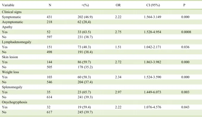
CVL clinical signs were observed in 76.5% of dogs that tested positive for amastigote forms of Leishmania spp. while 28.4% of dogs were asymptomatic (Table 2). This finding is in agreement with the findings of ALMEIDA et al. (2010) and FIGUEIREDO Table 1 -Result of direct parasitological exam through needle aspiration of lymph node cells for screening Leishmania spp. in dogs from the
urban area at the municipality of Araguaína - TO.
Result Frequency Percentage ---Confidence Interval (95%)---
Negative 385 59.3% 54.4% - 63.1%
Positive 264 40.7% 36.9% - 44.6%
et al. (2014), and in contrast with the findings of DANTAS-TORRES et al. (2006) and SILVA et al. (2010), who reported a higher parasite frequency in asymptomatic animals. The animals analyzed in this study were those under veterinary care, and this condition may have contributed to the observation of a high frequency of symptomatic animals.
Dermatosis (32.6%), lymphadenomegaly (27.7%), weight loss (22.7%), apathy (12.5%), splenomegaly (8.7%), and onychogryphosis (7.2%) were the most frequently observed clinical signs associated with CVL (Table 3). These signs were also the most frequently reported for CVL from other locations (RONDON et al., 2008; ALMEIDA et al., 2010). The other signs observed were eye disease (3.8%), limb edema (2.3%), diarrhea (1.5%), motor incoordination (1.1%), hepatomegaly (0.8%), and epistaxis (0.4%).
The observations from this study indicated that in areas with a high prevalence of
Leishmania spp., owners should monitor their dogs for CVL frequently. When a veterinarian detects a sign that is suggestive of CVL, they should perform complementary exams to confirm infection. The parasitology exam,
despite having a low sensitivity, is low-cost, easy, and quick to perform in the veterinary practice. The exam may also help with the early implementation of control measures for the domestic reservoir, thereby reducing human risk of infection.
CONCLUSION
The results reported in our study indicated that there is a high CVL frequency in Araguaína, Tocantins. Animals <2 years old and living alongside poultry in the peri-domicile areas are at the greatest risk of infection. Dermatosis was the most frequently observed clinical sign. In high-incidence areas, diagnosis through testing of samples of lymph node needle aspirations for parasites may be a faster and safer method to monitor dogs. However, this diagnostic approach does not exclude the importance of using serological or molecular methods for the diagnosis of visceral leishmaniasis, especially in asymptomatic dogs that have negative results from a direct examination. Therefore, it is important to train veterinarians in the clinical and laboratory diagnosis of CVL.
Table 2 - Variables related to the individual characteristics and permanent site of dogs in the city of Araguaína that are associated with positivity for Leishmaniaspp. in needle aspirations of lymph node cells.
Variable N +(%) OR CI (95%) P
Sex
Male 314 135(43.0) 1.206 0.880-1.651 0.278
Female 330 127(38.5)
Age
Up to 2 years 285 133(46.7) 1.468 1.028-2.101 0.042
> 2 years 225 84(37.3)
Breed
Defined breed 364 221(60.7) 0.900 0.648-1.253 0.591
Mongrel 244 142(58.2)
Other animals in the domicile
Dog 255 102(40.0) 0.955 0.692-1.316 0.841
Cat 23 12(52.2) 1.619 0.703-3.726 0.253
Poultry 30 18(60.0) 2.317 1.086-4.944 0.042
Access to the outdoors
Free 39 16(41.0) 1.012 0.518-1.977 0.893
Controlled 388 158(40.7)
Permanent site from 18 to 22 hours
Within the house 365 153(41.9) 1.407 0.789-2.508 0.307
ACKNOWLEDGEMENTS
We thank the Brazilian Conselho Nacional de
Desenvolvimento Científico e Tecnológico (CNPq) and the
Departamento de Ciência e Tecnologia do Estado de Tocantins for supporting our research and Universidade Federal do Tocantins (UFT)/Pró-Reitoria de Pesquisa e Pós-graduação (Propesq) for the research grant.
BIOETHICS AND BIOSSECURITY COMMITTEE APPROVAL
This study was approved by the Research Ethics Committee of the Fundação de Medicina Tropical do Tocantins, under number 32/2010.
REFERENCES
AMÓRA, S. S. A. et al. Factors related to positive testing of dogs for visceral leishmaniasis in endemic area in the state of Rio Grande do Norte, Brazil. Ciência Rural,v.36, n.6, p.1854-1859, 2006. Avail-able from: <http://www.scielo.br/pdf/cr/v36n6/a29v36n6.pdf>. Ac-cessed: Mar. 03, 2016. doi: 10.1590/S0103-84782006000600029.
ALMEIDA, A. B. P. F. et al. Prevalência e epidemiologia da leishma-niose visceral em cães e humanos, na cidade de Cuiabá, Mato Gros-so, Brasil. Ciência Rural, v.40, n.7, p.1610-1615, 2010. Available from: <http://www.scielo.br/pdf/cr/v40n7/a637cr2726.pdf>. Acces-sed: Mar. 06, 2016. doi: 10.1590/S0103-84782010005000102.
ALMEIDA, A. B. P. F. et al. Canine visceral leishmaniasis: diagnostic approaches based on polymerase chain reaction employing different biological samples. Diagnostic Microbiology and Infectious Disease, v.76, p.321-324, 2013. Available from: <http://dx.doi. org/10.1016/j.diagmicrobio.2013.03.017>. Accessed: Dec. 20, 2013. doi: 10.1016/j.diagmicrobio.2013.03.017.
AZEVEDO M. A. A. et al. Avaliação da leishmaniose visceral cani-na em Poxoréo, Estado do Mato Grosso, Brasil. Revista Brasileira de Parasitologia Veterinária, v.17, n.3, p.123-127, 2008. Available from: <http://www.scielo.br/pdf/rbpv/v17n3/a01v17n3.pdf>. Ac-cessed: Nov. 22, 2015. doi: 10.1590/S1984-29612008000300001.
BABIKER, Z. O. et al. Utility of lymph node aspiration in the diagnosis of visceral leishmaniasis in Sudan. American Journal of Tropical Medicine and Hygiene, v.76, n.4, p.689-693, 2007. Available from: <http://www.ajtmh.org/content/76/4/689.long>. Accessed: Mar. 04, 2016.
BANETH, G. et al. Canine leishmaniosis – new concepts and insights on an expanding zoonosis: part one. Trends in Parasitology, v.24, n.7, p.324-330, 2008. Available from: <http://www.sciencedirect.com/ science/article/pii/S1471492208001323>. Accessed: Aug. 07, 2016. doi:10.1016/j.pt.2008.04.001.
BARBOSA, D. S. et al. Seroprevalence and associated epidemio-logic variables with canine visceral leishmaniasis in endemic area, são luis, Maranhão state, Brazil. Ciência Animal Brasileira, v.11, n.3, p.653-659, 2010. Available from: <http://www.producao.usp. br/handle/BDPI/43966>. Accessed: Mar. 08, 2016. doi: 10.5216/ cab.v11i3.5933.
Table 3 - Variables related to the clinical characteristics of the dogs from the city of Araguaína that are associated with positivity for
Leishmania spp. in needle aspirations of lymph node cells.
Variable N +(%) OR CI (95%) P
Clinical signs
Symptomatic 431 202 (46.9) 2.22 1.564-3.149 0.000
Asymptomatic 218 62 (28,4)
Apathy
Yes 52 33 (63.5) 2.75 1.528-4.954 0.0008
No 597 231 (38.7)
Lymphadenomegaly
Yes 151 73 (48.3) 1.51 1.042-2.171 0.036
No 498 191 (38.4)
Skin lesion
Yes 144 86 (59.7) 2.72 1.863-3.982 0.000
No 505 178 (35.2)
Weight loss
Yes 103 60 (58.3) 2.34 1.524-3.590 0.000
No 546 204 (37.4)
Splenomegaly
Yes 35 23 (65.7) 2.97 1.449-6.073 0.003
No 614 241 (39.3)
Onychogryphosis
Yes 32 19 (59.4) 2.22 1.076-4.576 0.043
BELO, V. S. et al. A systematic review and meta-analysis of the factors associated with Leishmania infantum infection in dogs in Brazil. Veter-inary Parasitology, v.195, n.1-2, p.1-13, 2013. Available from: <http:// www.sciencedirect.com/science/article/pii/S0304401713001556>. Ac-cessed: Mar. 08, 2016. doi: 10.1016/j.vetpar.2013.03.010.
BRASIL. Ministério da Saúde. Secretaria de Vigilância em Saúde. Guia de vigilância em saúde. Brasília: Ministério da Saúde, 2014. Available from: <http://portalsaude.saude.gov.br/images/pdf/2016/ janeiro/15/guia-vigilancia-saude-atualizado-05-02-15-LV.pdf>. Accessed: Mar. 08, 2016.
COURA-VITAL, W. et al. Prevalence and factors associated with
Leishmania infantum infection of dogs from an urban area of Brazil
as identified by molecular methods. PLoS Neglected Tropical Diseases, v.5, n.9, p.e1291, 2011. Available from: <http://journals. plos.org/plosntds/article?id=10.1371/journal.pntd.0001291>. Accessed: Sept. 19, 2013. doi: 10.1371/journal.pntd.0001291.
DATASUS. Ministério de Saúde. Secretaria Executiva. Leishmaniose
Visceral - Casos confirmados notificados no sistema de informação de agravos de notificação - Sinan Net. Available from: <http://www. datasus.gov.br>. Accessed: Mar. 03, 2015.
DANTAS-TORRES, F. et al. Seroepidemiological survey on canine leishmaniasis among dogs from an urban area of Brazil. Veterinary Parasitology, v.140, p.54-60, 2006. Available from: <http://www. sciencedirect.com/science/article/pii/S0304401706001956>. Accessed: Feb. 27, 2013. doi: 10.1016/j.vetpar.2006.03.008.
DANTAS-TORRES, F. Canine leishmaniosis in South America. Parasite & Vector, v.2, (Supp.1), S1, p.1-8, 2009. Available from: <http://www.parasitesandvectors.com/content/2/S1/S1>. Accessed: Feb. 08, 2016. doi: 10.1186/1756-3305-2-S1-S1.
DAY, M. J. Immune system development in the dog and cat. Journal of Comparative Pathology, v.137, suppl.1, p.S10-S15, 2007. Available from: <http://dx.doi.org.ez6.periodicos.capes.gov.br/10.1016/j.jcpa.2007.04.005>. Accessed: Aug. 07, 2016. doi: 10.1016/j.jcpa.2007.04.005.
FRAGA, D. B. M. et al. Temporal distribution of positive results of tests for detecting Leishmania infection in stray dogs of an endemic area of visceral leishmaniasis in the Brazilian tropics: A 13 years survey and association with human disease. Veterinary Parasitology, v.190, n.3-4, p.591-594, 2012. Available from: <http:// www.sciencedirect.com/science/article/pii/S0304401712003226>. Accessed: Mar. 05, 2016. doi: 10.1016/j.vetpar.2012.06.025.
FIGUEIREDO, M. J. F. M. et al. Risk factors and clinical classification associated with seropositivity for canine visceral leishmaniasis. Ciencia Animal Brasileira, v.15, n.1, p.102-106, 2014. Available from: <http://www.scielo.br/pdf/cab/v15n1/13.pdf>. Accessed: Mar. 05, 2016. doi: 10.5216/cab.v15i1.25097.
GOMES, Y. M. et al. Diagnosis of canine visceral leishmaniasis: biotechnological advances. Veterinary Journal, v.175, p.45-52, 2008. Available from: <http://www-sciencedirect-com.ez6. periodicos.capes.gov.br/science/article/pii/S1090023306002358>. Accessed: Aug. 05, 2016. doi: 10.1016/j.tvjl.2006.10.019.
IBGE (INSTITUTO BRASILEIRO DE GEOGRAFIA E ESTATÍSTICA). IBGE-Cidades. Available from: <http://www. ibge.gov.br/cidadesat/topwindow.htm?1>. Accessed: Jan. 10, 2015.
JULIÃO, F. S. et al. Investigation of risk areas as complemental methodology for the control of canine visceral. Pesquisa Veterinária Brasileira, v.27, n.8, p.319-324, 2007. Available from: <http://www.scielo.br/scielo.php?script=sci_arttext&pid=S0100-736X2007000800001>. Accessed: Mar. 04, 2016. doi: 10.1590/ S0100-736X2007000800001.
MOREIRA JR, E. D. et al. Peridomestic risk factors for canine
leishmaniasis in urban dwellings: new findings from a prospective
study in Brazil. American Journal of Tropical Medicine and Hygiene, v.69, n.4, p.393-397, 2003. Available from: <http://www. ajtmh.org/content/69/4/393.long>. Accessed: Mar. 02, 2008.
OLIVEIRA, L. C. P. et al. Seroprevalence and risk factors for canine visceral leishmaniasis in the endemic area of Dias D’Avila, State of Bahia, Brazil. Revista da Sociedade Brasileira de Medicina Tro-pical, v.43, n.4, p.400-404, 2010. Available from: <http://dx.doi. org/10.1590/S0037-86822010000400013>. Accessed: Mar. 08, 2016. doi: 10.1590/S0037-86822010000400013.
OPAS (ORGANIZACION PANAMERICANA DE LA SALUD). Leishmaniasis. Epidemiological Report of the Americas, n.2, June 2014. Available from: <http://www.paho.org/hq/index. php?option=com_topics&view=readall&cid=6721&Itemid=4075 4&lang=pt>. Accessed: Mar. 05, 2016.
RONDON, F. C. et al. Cross-sectional serological study of canine
Leishmania infection in Fortaleza, Ceará state, Brazil. Veterinary
Parasitology, v.155, n.1-2, p.24-31, 2008. Available from: <http:// www.sciencedirect.com/science/article/pii/S0304401708002379>. Accessed: Mar. 08, 2016. doi: 10.1016/j.vetpar.2008.04.014.
SILVA, F. T. S. et al. Clinical aspects of canine visceral leishmaniasis in the district of Monte Gordo, Camaçari (BA). Revista Baiana de Saúde Pública, v.34, n.4, p.783-795, 2010. Available from: <http://inseer.ibict.br/rbsp/index.php/rbsp/article/viewFile/71/78>. Accessed: Mar. 06, 2016.