INTRODUCTION
Address to: Dr. Edelberto Santos Dias. Laboratório de Leishmanioses/CPqRR. Av. Augusto de Lima 1715, 30190-002 Belo Horizonte, MG, Brasil.
Phone: 55 31 3349-7758.
e-mail: edel@cpqrr.fi ocruz.br Received 10 February 2013
Accepted 24 May 2013
Phlebotomine sandfl y fauna and natural Leishmania
infection rates in a rural area of Cerrado (tropical savannah)
in Nova Mutum, State of Mato Grosso in Brazil
Sirlei Franck Thies
[1],
Ana Lucia Maria Ribeiro
[2],
Érika Monteiro Michalsky
[3],
Rosina Djunko Miyazaki
[4],
Consuelo Latorre Fortes-Dias
[5],
Cor Jésus Fernandes Fontes
[6]and Edelberto Santos Dias
[3][1]. Curso de Pós Graduação em Ciências da Saúde, Faculdade de Medicina, Universidade Federal de Mato Grosso, Cuiabá, MT. [2]. Faculdade de Medicina, Universidade Federal de Mato Grosso, Cuiabá, MT. [3]. Laboratório de Leishmanioses, Centro de Pesquisas René Rachou, Belo Horizonte, MG. [4]. Departamento de Biologia e Zoologia, Instituto de Biociências, Universidade Federal de Mato Grosso, Cuiabá, MT. [5]. Diretoria de Pesquisa e Desenvolvimento, Fundação Ezequiel Dias, Belo Horizonte, MG. [6]. Departamento de Clínica Médica, Faculdade de Medicina, Universidade Federal de Mato Grosso, Cuiabá, MT.
ABSTRACT
Introduction: American cutaneous leishmaniasis (ACL) has been reported in every municipality of the State of Mato Grosso, Brazil, but the transmission epidemiology remains poorly understood. Our study was developed in a rural area of the Nova Mutum municipality where four autochthonous cases of ACL were reported in 2009. Our aims were to describe the local
phlebotomine sandfl y fauna and to investigate the infection rates and infecting Leishmania species in the captured sandfl ies. Methods: Entomological captures were performed bimonthly at 10 fi xed sites close to the edge of a forested area between
June 2011 and April 2012. Results: A total of 3,743 phlebotomine sandfl ies belonging to 31 distinct species were captured.
Approximately 75% of the specimens were females. The most abundant species (45.4%) was Lutzomyia antunesi, which was consistently captured at every site. Species that are epidemiologically important for ACL, such as L. fl aviscutellata, L. whitmani
and L. umbratilis, were also captured. L. antunesi and L. ubiquitalis were naturally infected by Leishmania braziliensis or
Le. guyanensis, with minimum infection rates of 0.88% and 6.67%, respectively. Surprisingly, L. antunesi was infected by
Le. infantum (synonym chagasi). Conclusions: The natural infection of L. antunesi and L. ubiquitalis by Leishmania sp. suggests that these species might play a role in the zoonotic cycle of ACL in Nova Mutum. The presence of Le. infantum in L. antunesi
suggests that there may be a risk of an outbreak of visceral leishmaniasis (VL) in Nova Mutum.
Keywords: Phlebotomine sandfl y. Leishmania. American cutaneous leishmaniasis. Nova Mutum. Lutzomyia.
Leishmaniases are among the most prevalent infectious diseases caused by parasites in the world and display a wide distribution in the Americas, Africa, India, Asia and Mediterranean Europe1,2. Leishmaniases occur in approximately
90 different countries, currently infecting 14 million people with an increase of 2 million new cases per year. It is estimated that 350 million people are at risk for these diseases2, which
are caused by protozoa that belong to the Leishmania
genus. Transmission occurs through the bite from infected phlebotomine sandflies (Diptera, Psychodidae). Several mammal species may act as natural reservoirs or hosts for
leishmaniases. Humans and some domestic animals, including dogs and horses, are considered to be accidental hosts3.
Human cases of American cutaneous leishmaniasis (ACL) have been reported in every Brazilian State4. In Mato Grosso
(MT), ACL is endemic with 8,000 reported cases between 2009 and 2011 (MS/SINAN/SES/MT, 2011). Since 2005, autochthonous cases of ACL have been reported from every municipality of the state5.
Lutzomyia whitmani, a phlebotomine sandfl y species that is
widely distributed in Brazil, is the main species associated with ACL transmission in MT4. This fl y can be found in various MT
biomes, such as Cerrado (tropical savannah), the Amazonian rainforest and Pantanal (tropical wetland). The phlebotomine
sandfl y fauna in these biomes is widely diversifi ed and consists
of species such as Lutzomyia fl aviscutellata, L. intermedia,
L. migonei, L. umbratilis, L. wellcomei and L. whitmani5,6 all
of them involved in the transmission epidemiology of ACL.
The identifi cation of potential vector species for ACL and
METHODS
reaction (PCR), enables the identifi cation of genetic material of
Leishmania in the total DNA extracted from phlebotomine sandfl y
macerates, even in minimal amounts. The main advantages of
PCR are its sensitivity and specifi city, regardless of the number,
location or stage of the infecting Leishmania in the digestive tract
of the sandfl y7. In the last decade, PCR has been widely used in
studies of vector competence of phlebotomine sandfl ies, even in
areas with low rates of Leishmania infection8-11. Although MT is
considered endemic for ACL, natural infection investigations on ACL vectors are rare there. Only three studies on natural infection by Leishmania have been reported in the state, two of which are related to visceral leishmaniasis (VL) vectors12,13, and a third
investigated a single L. umbratilis specimen that was infected by Le. braziliensis14.
The goal of present study was to survey the phlebotomine
sandfl y in a rural area of the State of Mato Grosso in qualitative and quantitative terms, as well as to determine the natural rate of Leishmania sp. infection in the phlebotomine sandfl y
females captured there. The area under study is located in the municipality of Nova Mutum, where four autochthonous ACL cases were reported in 2009.
Area under study
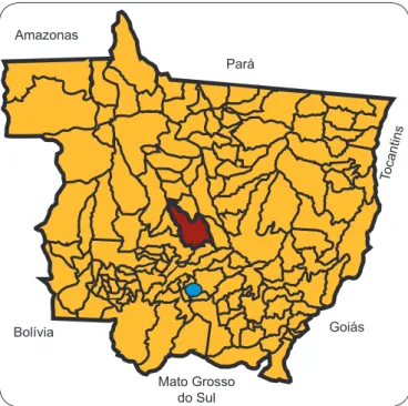
Nova Mutum (13o05’04’’S, 56o05’16’’W) is a 9,572.69km2
municipality that is located in the mid-Northern area of the Brazilian State of Mato Grosso (Figure 1). Our study was developed in a rural area of Cerrado (tropical savannah) that has an area of 16,000 hectares, and is located at 42km from the city center of Nova Mutum. The location has a native forest that consists of small to large trees (Figure 2) and has an ample amount of decaying vegetal organic matter. Various animal species can be found there, including monkeys, wild pigs, snakes, rats, scouts and armadillos. The area attracts people who engage in activities that put them
in close contact with nature, such as swimming and fi shing.
Phlebotomine sandfl y capture and identifi cation
Six entomological captures were performed bimonthly for three
consecutive nights between June 2011 and April 2012. Ten Center
for Disease Control (CDC) light traps were set fi ve feet from the ground and 100m from one another at a transect of approximately
1,000 meters between the edge and the interior of the forest. The capture sites were plotted by GPS. The captured phlebotomine
sandfl ies were packed in insulated containers and taken to the laboratory for adequate assembling and identifi cation, as described
by Young and Duncan15. The specimens were incorporated into
the collection of the Entomology laboratory from the Health
Department of Mato Grosso. The phlebotomine female sandfl ies were identifi ed by dissecting the last three segments to allow for
the visualization of the abdominal spermathecae and by head
dissection to allow for the examination of specifi c taxonomic characteristics. Damaged specimens were identifi ed at the genus level only. One to 10 specimens of phlebotomine female sandfl ies
were pooled according to species, date and site of capture, and were
then stored in 6% dimethyl sulfoxide (DMSO) at -20oC until use.
Pará
Tocantins
Goiás
Mato Grosso do Sul Bolívia
Amazonas
FIGURE 1 - Geographical localization of the municipality of Nova Mutum (in red) in the State of Mato Grosso, Brazil. The capital of Mato Grosso, Cuiabá, is indicated by a solid blue circle.
FIGURE 2 - Aerial view of the area under study in Nova Mutum, State of Mato Grosso, Brasil. The sites of entomological captures are indicated by red balloons.
Lutzomyia constitutive gene (cacophony)
The adequacy of DNA extraction from the phlebotomine sandfl ies was checked by polymerase chain reaction (PCR) with specifi c primers to the IVS6 region of the Lutzomyia genus (cacophony), as described by Lins16.
Detection of Leishmania DNA in phlebotomine sandfl ies
Pooled samples of phlebotomine female sandfl ies were submitted to total DNA extraction using a commercial kit (GE
HealthCare, Uppsala, Sweden). The presence of Leishmania
DNA was tested by nested PCR (LnPCR) with primers that were directed at the small subunit ribosomal ribonucleic acid (SSUrRNA) gene17,18. The fi rst amplifi cation step was
performed using R221 and R332 primers that are specifi c to the Kinetoplastida order but not exclusively to the Leishmania genus.
RESULTS
TABLE 1 - Phlebotomine sandfl y species captured with CDC light
traps in a rural area of Nova Mutum, State of Mato Grosso, Brazil. Period: June 2011 to April 2012.
Female Male Total
Species n % n % n %
B. brumpti 14 0.51 6 0.60 20 0.53
L. antunesi 1,265 46.25 436 43.26 1,701 45.45
L. aragaoi 4 0.15 - - 4 0.11
L. ayrozai 33 1.21 - - 33 0.88
L. begonae 28 1.02 - - 28 0.75
L. bourrouli 6 0.22 1 0.10 7 0.19
L. chagasi 15 0.55 - - 15 0.40
L. claustrei 14 0.51 20 1.98 34 0.91
L. complexa 1 0.04 4 0.40 5 0.13
L. dasypodogeton 12 0.44 6 0.60 18 0.48
L. davisi 3 0.11 2 0.20 5 0.13
L. fl aviscutellata 146 5.34 97 9.62 243 6.49
L. furcata 37 1.35 1 0.10 38 1.02
L. hermanlenti 9 0.33 9 0.89 18 0.48
L. lenti 1 0.04 - - 1 0.03
L. llanosmartinsi 46 1.68 2 0.20 48 1.28
L. longipennis 9 0.33 6 0.60 15 0.40
L. octavioi - - 5 0.50 5 0.13
L. punctigeniculata 2 0.07 - - 2 0.05
L. runoides 2 0.07 2 0.20 4 0.11
L. sallesi - - 3 0.30 3 0.08
L. saulensis 677 24.75 93 9.23 770 20.57
L. shannoni 34 1.24 - - 34 0.91
L. shawi 1 0.04 - - 1 0.03
L. sordellii 16 0.58 3 0.30 19 0.51
L. spp. 16 0.58 9 0.89 25 0.67
L. ubiquitalis 43 1.57 83 8.23 126 3.37
L. umbratilis 1 0.04 - - 1 0.03
L. walkeri 282 10.31 212 21.03 494 13.20
L. whitmani 1 0.04 6 0.60 7 0.19
L. yuilli yuilli 17 0.62 2 0.20 19 0.51 Total 2,735 100.0 1,008 100.0 3,743 100.0
B.: Brumptomyia; L.: Lutzomyia; CDC: Center for Disease Control.
The PCR products were then tested using a new amplifi cation
step with R233 and R333 primers18. All amplifi cations were
performed with the Illustra PuRe Taq Ready-To-Go PCR Beads kit (GE Healthcare, Uppsala, Sweden), and the products were analyzed using electrophoresis in agarose gels. DNA from
Le. braziliensis (M2903 strain) and sterile distilled water were used as the positive and negative controls, respectively.
Identifi cation of the Leishmania species in
phlebotomine sandfl ies
The amplifi ed bands from the PCR steps were extracted from the gels using the QIAquick extraction kit (QIAGEN,
Hilden, Germany) and submitted for DNA sequencing using an ABI3130 analyzer (Applied Biosystems Inc., Foster City, California, USA). The sequences were analyzed using the Blast Nucleotide Standard software (blast.ncbi.nlm.nih.gov).
Minimum rates of Leishmania infection in
phlebotomine sandfl ies
The minimum rates of Leishmania infection in the captured
phlebotomine sandfl ies were calculated by dividing the number
of positive pools of each sandfly species by the number of specimens of that species in that pool and then multiplying by 10019.
A total of 3,743 phlebotomine sandfl y specimens were
captured in Nova Mutum. Of the captured specimens, 1,008
(26.9%) were males (M) and 2,735 (73.1%) were females (F), which resulted in an overall M/F ratio of 0.4% (Table 1). The
phlebotomine sandfl y fauna consisted of 31 different species.
The predominant species was Lutzomyia antunesi (Coutinho,
1939), which accounted for approximately 45% of the total
captured specimens. L. fl aviscutellata (Mangabeira, 1942),
L. whitmani (Antunes & Coutinho, 1939) and L. umbratilis (Ward & Fraiha, 1977), all known ACL vectors, were also
captured at the following percentages: 6.49%, 0.19% and 0.03%,
respectively (Table 1).
A total of 2,419 sandflies females were dissected and pooled for molecular analysis. Then, 293 pooled samples were obtained, which were distributed by species as follows: 196 of
L. antunesi, 71 of L. fl aviscutellata, eight of L. yuilli yuilli, 16 of
L. ubiquitalis, one of L. umbratilis and one of L. whitmani. After LnPCR, the 353bp DNA fragment of the Leishmania genus was observed in 13 of the pooled samples (11 from L. antunesi and two from L. ubiquitalis [Figure 3]). No amplifi cation products
were detected in the remaining samples. The minimum rates of
Leishmania infection were 0.88% and 6.67% for L. ubiquitalis
and L. antunesi, respectively. The adequacy of the Lutzomyia
DNA extraction was confi rmed by the amplifi cation of the
cacophony gene in all 13 positive Leishmania samples (data not shown).
After the DNA sequencing and nucleotide alignment of the 353bp amplicons from the Leishmania-containing phlebotomine samples (Figure 3), the infecting Leishmania
species were identifi ed as follows: Le. braziliensis (GQ332355)
or Le. guyanensis (GQ332358) in six pooled L. antunesi
samples (numbered 190, 224, 233, 246, 256 and 275) and in two pooled L. ubiquitalis samples(numbered 264 and 265);
Le. infantum (GQ 332359) was identifi ed in one pooled sample
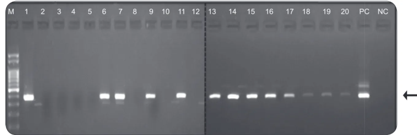
FIGURE 4 - Agarose gel electrophoresis of the PCR products obtained after amplifying the total phlebotomine sandfl y DNA with primers for the cacophony IV6 gene of Lutzomyia. The characteristic 220bp fragment is indicated by an arrow. Samples: L. umbratilis (11 and 12); L. antunesi (1 to 10, 13); PC: Positive control (L. longipalpis DNA); NC: negative control (no DNA); and M: 100 bp DNA ladder.
FIGURE 3 - Agarose gel electrophoresis of the LnPCR products after amplifying the total phlebotomine sandfl y DNA for the SSUrRNA gene of Leishmania sp. The characteristic 353bp fragment (indicated by an arrow) was present in the following samples: L. umbratilis (18 and 19); L. antunesi (1, 6, 7, 9, 11, 13, 14, 15, 16, 17 and 20). PC: positive control (Le. braziliensis M2903); NC: negative control (no DNA); and M: 100bp DNA ladder.
DISCUSSION
The phlebotomine sandfl y fauna of the State of Mato Grosso is quite diversifi ed, consisting of more than 100 species whose
spatial distribution varies according to the different biomes present21,24. In the rural Cerrado (tropical savannah), which was under study, roughly 50% of the captured specimens were
L. antunesi.L. longipalpis and L. cruzi, which areboth proven vectors of the visceral leishmaniasis (VL) in Brazil, were not captured at that location. This result differed from the capture reports in urban areas with Cerrado fragments23,25. Surprisingly,
even in the absence of L. longipalpis and L. cruzi and no reported cases of VL, Le. infantum was present in a pooled sample of
L. antunesi from Nova Mutum. A previous fi nding of promastigotes
in L. antunesi with a VL focus in the Marajó Island (State of
Pará) led the authors to suggest that the sandfl y species could be
involved as a secondary VL vector on that island. The authors also suggested the possibility of Le. infantum infection in
L. antunesi, although the inoculation of isolated promastigotes
from the sandfl ies produced no skin lesions in hamsters26.
Regarding ACL vectors, L. whitmani (vector of
Le. braziliensis, Le. guyanensis and Le. shawi), L. fl aviscutellata (vector of Le. amazonensis), L. ubiquitalis (vector of
Le. lainsoni), L. umbratilis (vector of Le. guyanensis) and
L. antunesi (epidemiologically shown to transmit Le. lindenbergi) were captured in Nova Mutum. L. ubiquitalis has been previously found in forested areas in Brazil, Peru and Bolivia and is the only known vector of Le. lainsoni so far27.
Studies on natural Leishmania infection in the State of Mato
Grosso are rare, with the exception for two that were published
by Missawa12,13 regarding proven VL vectors12. In Brazil,
estimated infection rates of ACL vectors by Leishmania ranged
from 0.16% to 7.1%: 0.4% in Bahia11 and Maranhão State28, from
0.8% to 7.1% in Minas Gerais29, 2% in Rio de Janeiro30, 0.3% in
Porto Alegre31, and 0.16% in Serra dos Carajás, Pará32. In Nova
Mutum (MT) we found minimum infection rates of 0.8% for
Le. (V.) braziliensis or Le. (V.) guyanensis and 0.08% for Le. (L.) infantum in L. antunesi, For L. ubiquitalis, the minimum rate
of infection was 6.67% for Le. braziliensis or Le. guyanensis.
There are no previous reports of Leishmania infection in any of
these phlebotomine sandfl ies species. Vásquez-Trujillo33 found L. antunesi naturally infected with Leishmania spp. and suggested its involvement in the transmission cycle of ACL in Colombia.
Genetically speaking, Le. braziliensis and Le. guyanensis are closely related species. We attempted to distinguish them in the
Leishmania infected sandfl ies using hsp7034 methodology. However,
only one pooled sample of L. antunesi showed the expected
amplifi ed product (1300bp) (data not shown). After gel extraction,
that DNA fragment provided insufficient sequencing results. In a study conducted at the Reference Center for Leishmaniasis in Cuiabá (MT) using isozyme typing and
molecular analysis, most (94.1%) ACL cases were because
of infection by Le. braziliensis, whereas Le. amazonensis
was the causative parasite in the others35. In another study
that investigated patients from MT that were diagnosed with
Le. amazonensis, Le. braziliensis and Le. shawi were determined to be the infecting species36. Although we were unable to
discriminate between Le. braziliensis and Le.guyanensis in our
M 1 2 3 4 5 6 7 8 9 10 11 12 13 14 15 16 17 18 19 20 PC NC
infected sandfl y samples, it seems probable that the parasite was
Le. braziliensis, as opposed of Le. guyanensis.
Based on the occurrence of autochthonous human cases of ACL and on the presence of L. antunesi and L. ubiquitalis that are infected by Leishmania (most likely Le. braziliensis), it is
possible to hypothesize that those phlebotomine sandfl y species
might be involved in the zoonotic cycle of ACL in the rural
Cerrado of Nova Mutum (MT).
REFERENCES
The authors declare that there is no confl ict of interest.
1. Medeiros ACR, Rodrigues SS, Roselino AMF. Comparison of the especifi city of PCR and histopathological detection of leishmania for the diagnosis of American cutaneous leishmaniasis. Braz J Med Biol Res 2002; 35:421-424.
2. World Health Organization. Control of leishmaniasis. Report of the secretariat. [Cited 2007 September 09] 2006; Available from: http://www. who.int/gb/ebwha/pdf_fi les/EB118/B118_4-en.pdf.
3. Lainson R, Shaw JJ. Evolution, classifi cation and geographical distribution,
In: W. Peters, Killick-Kendrick, editors. The Leishmaniasis in Biology and Medicine. Vol 1 - Biology and Epidemiology. London, UK: Academic Press; 1987. p. 1-20.
4. Ministério da Saúde. Secretaria de Vigilância em Saúde. Departamento de Vigilância Epidemiológica. Atlas de leishmaniose tegumentar americana: diagnósticos clínico e diferencial. Brasília: Editora do Ministério da Saúde; 2006.
5. Missawa NA, Maciel GBML, Rodrigues H. Distribuição de Lutzomyia (Nissomyia) withmani (Antunes e Coutinho, 1939) no estado de Mato Grosso. Rev Soc Bras Med Trop 2008; 41:369-373.
6. Ribeiro ALM, Missawa NA. Spatial distribuition of Phlebotomine species in the state of Mato Grosso, Brazil, in the period of 1996 to 2011. Entomol y Vectores 2002; 9:33-34.
7. Perez JE, Ogusuku E, Inga R, Lopez M, Monje J, Paz L, et al. Natural
Leishmania infection of Lutzomyia spp. in Peru. Trans R Soc Trop Med Hyg 1994; 88:1614.
8. Silva AC, Grunewald GMS. Contribuition to the sand fl y fauna (Diptera:Phlebotominae) of Rio Grande do Sul, Brazil, and Leishmania (Viannia) infections. Mem Inst Oswaldo Cruz 1999; 94:579-5825. 9. Rodriguez N, Aguilar CM, Barrios MA, Barker DC. Detection of
Leishmania braziliensis in naturally infected individual sandfl ies by the polymerase chain reaction. Trans R Soc Trop Med Hyg 1999; 93:47-49.
10. Aransay AM, Scoulica E, Tselentis Y. Detection and identifi cation of
Leishmania DNA within naturally infected sand fl ies by seminested
PCR on minicircle kinetoplastic DNA. Appl Environ Microbiol 2000; 66:1933-1938.
11. Miranda JC, Reis E, Schreifer A, Gonçalves M, Reis MG, Carvalho L, et al. Frequency of infection of Lutzomyia phlebotomines with Leishmania braziliensis in a Brazilian endemic area as assessed by pinpoint capture and polymerase chain reaction. Mem Inst Oswaldo Cruz 2002; 97: 185-188.
12. Missawa NA, Michalsky EM, Fortes-Dias CL, Dias ES. Lutzomyia
longipalpis naturally infected by Leishmania (L.) chagasi in Várzea Grande, Mato Grosso State, Brazil, an area of intense transmission of visceral leishmaniasis. Cad Saude Publica 2010; 26:2414-2419. 13. Missawa NA, Veloso MAE, Maciel GBML, Michalsky EM, Dias ES.
Evidência de transmissão de leishmaniose visceral por Lutzomyia cruzi no município de Jaciara, Estado de Mato Grosso, Brasil. Rev Soc Bras Med Trop 2011; 44:76-78.
14. Pereira TR apud Azevedo ACR, Souza NA, Meneses CRV, Costa WA,
Costa SM, Lima JB, et al. Ecology of sand fl ies (Diptera: Psychodidae:
Phlebotominae) in the North of the state of Mato Grosso, Brazil. Mem Inst Oswaldo Cruz 2002; 97:459.
15. Young PG, Duncan MA. Guide to the identifi cation and geographic distribution of Lutzomyia sandfl ies in México, the West India, Central
and South America (Diptera: Psychodidae). Florida: Publishers American Entomological Institute; 1994.
16. Lins RMMA, Oliveira SG, Souza NA, Queiroz RG, Justiniano SCB, Ward RD, et al. Molecular evolution of the cacophony IVS6 region in
sand fl ies. Insect Mol Biol 2002; 11:117-122.
17. Van Eys GJJM, Schoone GJ, Kroon NCM, Ebeling SB. Sequence analysis of small subunit ribosomal RNA genes and its use for detection
and identifi cation of Leishmania parasites. Mol Biochem Parasitol 1992; 51:133-142.
18. Cruz I, Canãvate C, Rubio JM, Morales MA, Chicharro C, Laguna F, et al. A nested polymerase chain reaction (Ln-PCR) for diagnosing and monitoring Leishmania infantum infection in co-infected patients with
human immunodefi ciency virus. Trans R Soc Trop Med Hyg 2002;
96:185-189.
19. Paiva BR, Secundino NFC, Pimenta PFP, Galati E, Andrade Jr HF, Malafronte RS. Padronização de condições para detecção de DNA de
Leishmania spp. em flebotomíneos (Diptera, Psychodidae) pela reação
em cadeia da polimerase. Rev Parasitol Vet 2007; 23:41-45.
20. Missawa NA, Maciel GB. List of species in the genus Lutzomyia, França, 1924 (Psychodidae, Phlebotominae) from the State of Mato Grosso. Rev Soc Bras Med Trop 2007; 40:11-14.
21. Ribeiro ALM, Missawa NA, Zeilhofer P. Distribution of phlebotomine
sandfl ies (Diptera: Psychodidae) of medical importance in Mato Grosso
State, Brazil. Rev Inst Med Trop São Paulo 2007; 49:5.
22. Maciel GBML, Missawa NA. Fauna fl ebotomínica (Diptera: Psychodidae) em aldeias indígenas do Estado de Mato Grosso. Rev Soc Bras Med Trop 2009; 42:597-602.
23. Amaral AFS, Varjão JR, Silva GB, Arrais-Silva WW. Phlebotomine fauna (Diptera: Psychodidae: Phlebotominae) in a residential area and in a fragment of savanna vegetation in the municipality of Pontal do Araguaia, Mato Grosso, Brazil. Rev Bras Parasitol Vet 2011; 20:165-167. 24. Alves GB, Oshiro ET, Leite MC, Melão AV, Ribeiro LM, Mateus NLF,
et al. Phlebotomine sandfl ies fauna (Diptera: Psychodidae) at rural
settlements in the municipality of Cáceres, state of Mato Grosso, Brazil. Rev Soc Bras Med Trop 2012; 45: 437-443.
25. Queiroz MFM, Varjão JR, Moraes SC, Salcedo GE. Analysis of sand
fl ies (Diptera: Psychodidae) in Barra do Garças, State of Mato Grosso, Brazil, and the infl uence of environmental variables on the vector density
of Lutzomyia longipalpis (Lutz & Neiva, 1912). Rev Soc Bras Med Trop 2012; 45:313-317.
26. Ryan L, Silveira FT, Lainson R, Shaw JJ. Leishmanial infections in
Lutzomyialongipalpis and Lu. antunesi (Diptera: Psychodidae) on the
island of Marajó, Pará State, Brazil. Transc Roy Soc Trop Med Hyg 1984; 78:547-548.
27. Silveira FT, Lainson R, Shaw JJ, Braga RR, Ishikawa EEA, Souza AAA. Cutaneous leishmaniasis in Amazonian: isolation of Leishmania (Viannia)
lainsoni from the rodent Agouti paca (Ro dentia: Dasyproctidae) in the state of Pará, Brazil. Rev Inst Med Trop São Paulo 1991; 33:18-22. 28. Oliveira-Pereira YN, Rebêlo JMM, Moraes JLP, Pereira SRF. Diagnóstico
molecular da taxa de infecção natural de fl ebotomíneos (Psychodidae,
Lutzomyia) por Leishmania sp. na Amazônia maranhense. Rev Soc Bras Med Trop 2006; 39:540-543.
FINANCIAL SUPPORT
Fundação de Amparo à Pesquisa de Mato Grosso (FAPEMAT; process 505189/2009) and of the Health Department of Mato Grosso.
29. Carvalho GML, Andrade-Filho JD, Falcão AL, Gontijo CMF. Naturally infected Lutzomyia sandfl ies and the transmission of leishmaniasis in an
endemic area of Brazil. Vector Borne Zoonotic Dis 2008; 8:407-414.
30. Pita-Pereira D, Alves CR, Souza MB, Brazil RP, Bertho AL, Barbosa AF, et al. Identifi cation of naturally infected Lutzomyia intermedia and Lutzomyia migonei with Leishmania (Viannia) braziliensis in Rio de Janeiro (Brazil) reveled by PCR multiples non-isotopic hibrydisation assay. Trans R Soc Trop Med Hyg 2005; 99:905-913.
31. Pita-Pereira D, Souza GD, Zwetsch A, Alves CR, Britto C, Rangel EF. First report of Lutzomyia (Nyssomyia) neivai (Diptera: Psychodidae:Phlebotominae) naturally infected by Leishmania (Viannia)
braziliensis in a periurban area of south Brazil using a multiplex polymerase chain reaction assay. Am J Trop Med Hyg 2009; 80:593-595.
32. Souza AAA, Silveira FT, Lainson R, Barata IR, Silva MGS, Lima JAN, et al. Fauna fl ebotomínica da Serra dos Carajás, Estado do Pará, Brasil, e sua possível implicação na transmissão da leishmaniose tegumentar americana. Rev Pan-Amaz Saúde 2010; 1:45-51.
33. Vásquez-Trujillo A, Santamaría-Herreño E, González-Reina AE, Bultrago-Álvarez LS, Góngora-Orjuela A, Cabrera-Quintero OL. Lutzomyia antunesi, probable vector de Leishmaniasis Cutánea em el área Rural de Villavicencio. Rev Salud Publica 2008; 10:625-632.
34. Garcia L, Kindt A, Bermudez H, Llanos-Cuentas A, Doncker S, Arevalo J, et al. Culture-independent species typing of neotropical Leishmania for clinical validation of a PCR-based assay targeting heat shock protein 70 genes. J Clin Microbiol 2004; 42:2294-2297.
35. Carvalho MLR, Andrade ASR, Fontes CJF, Hueb M, Silva SO, Melo MN.
Leishmania (Viannia) braziliensisis the prevalent species infecting patients with tegumentary leishmaniasis from Mato Grosso State, Brazil. Acta Tropica 2006; 98:277-285.