2016 | Lavras | Editora UFLA | www.editora.ufla.br | www.scielo.br/cagro
Temperature and light intensity interaction on
Cercospora
coffeicola
sporulation and conidia germination
Interação da temperatura e intensidade luminosa na esporulação e germinação de conídios de Cercopora coffeicola
Marília Goulart da Silva1*, Edson Ampélio Pozza1, Caio Vitor Rodrigues Vaz de Lima1, Tales Jesus Fernandes2
1Universidade Federal de Lavras/UFLA, Departamento de Fitopatologia/DFP, Lavras, MG, Brasil 2Universidade Federal de Lavras/UFLA, Departamento de Ciências Exatas/DEX, Lavras, MG, Brasil
*Corresponding author: [email protected] Received in july 13, 2015 and approved in september 14, 2015
ABSTRACT
Difficulty in obtaining abundant sporulation in culture of many species of Cercospora may be the limiting factor for studies of biology, systematics, and inoculation of the genus. Therefore, it is necessary to understand the nutritional and environmental requirements that influence mycelial growth, sporulation and germination. As it is difficult to obtain conidia of Cercospora coffeicola in vitro, different
temperatures (17, 22, 27, and 32 °C) and light intensities (80, 160, 240, and 320 μmol m-2 s-1) were evaluated to optimize pathogen sporulation and assess favorable conditions for spore germination, aiming for a strategy of disease control. The dark treatment (0 μmol m-2 s-1) was added for sporulation. A significant interaction was found between temperature and light intensity for both variables. The highest sporulation rate of C. coffeicola occurred at a light intensity of 240 μmol m-2 s-1 and air temperature of 22 °C, reaching 5.9x10⁶
con mL-1. Germination was higher at temperature 17 °C and light intensity of 320 μmol m-2 s-1, reaching 52%. Interaction between light intensity and temperature proved to influence the processes of sporulation and germination of C. coffeicola.
Index terms:Environmental variables; Cercospora leaf spot; Coffea arabica.
RESUMO
Obter abundante esporulação em meio de cultura, para muitas espécies de Cercospora, pode ser o fator limitante em estudos de biologia, sistemática e inoculação das espécies do gênero. Por isso, faz-se necessário o entendimento dos requerimentos nutricionais e ambientais, os quais influenciam o crescimento micelial, a esporulação e a germinação Em razão da dificuldade de obter conídios
de Cercospora coffeicola in vitro, objetivou-se avaliar diferentes condições de temperatura (17, 22, 27 e 32 °C) e intensidade luminosa (80,
160, 240 e 320 µmol m-2 s-1), com o intuito de otimizar a esporulação do patógeno e avaliar as condições favoráveis à germinação dos conídios, visando a táticas de controle da doença. Para a esporulação, adicionou-se o tratamento escuro (0 µmol m-2 s-1). Observou-se interação significativa entre temperatura e intensidade luminosa para ambas as variáveis testadas. A maior esporulação de C. coffeicola
foi observada na intensidade de 240 µmol m-2 s-1 e na temperatura de 22 °C, atingindo 5,9x10⁶ con mL-1. Com relação à germinação, esta foi maior na temperatura de 17 °C e na intensidade luminosa de 320 µmol m-2s-1, atingindo 52%. A interação entre intensidade luminosa e temperatura do ar, influenciou os processos de esporulação e germinação de C. coffeicola.
Termos para indexação: Variáveis ambientais; cercosporiose; Coffea arabica.
INTRODUCTION
Cercospora leaf spot of coffee caused by the fungus
Cercospora coffeicola Berk. & Cooke occurs from nursery
seedlings to adult plants in the field, resulting in serious losses to farmers (Pozza; Carvalho; Chalfoun, 2010). Intensity of symptoms as well as growth, development, pathogen infectivity, and plant physiological responses to colonization and penetration can be influenced by environmental variables.
Air temperature, relative humidity, heat stress, and factors influencing plant nutrition such as drought, soil
type, and nutritional imbalance have direct or indirect effect on disease progression and on growth, sporulation, and germination of fungi (Waggoner, 1960; Schumann; D’arcy, 2012). The infectious process of C. coffeicola
Most light-sensitive fungi sporulate when exposed to continuous light but diurnal esporulators also require a period of darkness, usually with variability between species and within each species itself. Other species require light to
initiate conidiophore formation and sporogenesis; however, sporulation is inhibited by light (Dhingra; Sinclair, 1995). Kilpatrick and Johnson (1956) studied various Cercospora
species in carrot-decoction agar. Abundant sporulation was found when cultures were exposed to light compared to those kept in the dark. In the literature, reports on the need of
C. coffeicola for light to sporulate are controversial. Initially,
Echandi (1959) found no influence of light regime (constant darkness, constant light, light/dark) in sporulation. However, Buitrago Jaramillo and Fernandéz Borrero (1982) reported higher sporulation under 9 hours of light alternating with 15 hours of darkness. Del Peloso et al. (1989) found higher sporulation under continuous light. The authors did not study interaction with temperature, thus there was no consensus on need or length of light exposure for pathogen sporulation. In addition, the incubation temperature required for increasing conidial production and facilitating host inoculation under these conditions remains to be determined.
With respect to germination of Cercospora
species, a study of the effect of temperatures 12, 18, 24, 30, and 36 °C on conidia germination of C. coffeicolain
vitro showed 100% germination at 30 °C, followed by
97% germination at 24 °C (Echandi, 1959). Conidia of
Cercospora arachidicola germinated on the leaf surface
(Abdou; Gregory; Cooper, 1974; Jenkins, 1938) and on glass slide (Oso, 1972) at temperatures 25- 30 °C and three-six-hour intervals of incubation under near saturated moisture conditions. The optimum temperature range for germination was 20-30 °C where as low germination occurred above 30 °C (Oso, 1972). However, the authors did not assess interaction with light.
Interaction with light has been already studied in other species of this pathogen, such as Cercospora sojina. The time required for this fungus to germinate varied according to temperature, light regimes and exposure to light. Camera et al. (2013) assessed temperatures 0, 5, 10, 15, 20, 25, 30, 35, and 40 °C with exposure times 3, 6, 9, and 12 hours, under constant light, and in the darkness. The highest percentages of conidial germination occurred under constant light and darkness in all exposure times at temperatures 22.4 °C and 23 °C, respectively. Studies like this for C. coffeicola were not found on the literature. Thus, this study evaluated the interaction between air temperature and light intensity on sporulation and germination of C. coffeicola.
MATERIAL AND METHODS
The experiments described below were conducted twice. Isolates of C. coffeicola were previously tested for pathogenicity. The study isolate (CML 2984), isolate from South Minas Gerais – Brazil. This isolate in conditions of light, is capable to produce a large amount of cercosporin and in consequence of this, is highly aggressive. The isolatewas inoculated in Petri dishes in malt medium.
Experimental design of sporulation and germination tests
Both sporulation and germination tests used completely randomized design with four replications, each replication consisting of a Petri dish.
Interaction of five light intensities, 0, 80, 160, 240, and 320 µmol m-2 s-1 and four temperatures, 17, 22, 27, and 32 °C were assessed for sporulation using factorial analysis of variance 5x4 in 20 treatments. The same interaction was evaluated for conidia germination, using four light intensities, 80, 160, 240, and 320 µmol m-2 s-1 and four temperatures, 17, 22, 27, and 32 °C, in factorial analysis of variance 4x4 in 16 treatments.
Both experiments used a metal stepped structure with light bulbs totaling 400 Watts placed at 100, 80, 60, and 40 cm from the plates to obtain light intensities 80, 160, 240, and 320 µmol m-2 s-1, respectively, in grow chambers with a 12-hour photoperiod.
Pathogen sporulation
Twelve mycelium discs 0.5 cm were taken from the edge of each colony in the chosen isolate of C. coffeicola,
which had been cultured in malt medium for 14 days. Discs were transferred to 30 mL of tomato juice agar (STT) in 125 mL Erlenmeyer flasks. The medium was composed of 200 mL of tomato juice Super Bom®, 3.2 g CaCO
3 and 800 mL distilled water. The flasks containing tomato juice agar and fungi were kept under constant stirring at 120 rpm, at around 25 ºC. After four days, the content of each flask was poured in a mixture of solid tomato juice and previous medium plus 16 g agar per liter in two Petri dishes, which remained open in the respective treatments in growth chambers. When the medium dehydrated after two days, 5 mL distilled water was added to each dish, which was then lightly scraped with a glass rod, and the suspension was filtered through a layer of sterile gauze. Conidia were counted under optical microscope with hemocytometer, and the concentration per ml in each treatment was estimated by the average of readings in both counting areas.
Conidial germination
1x10⁴ conidia mL-1. Dishes 60x15 mm containing 10 mL water-agar were prepared and 100 uL of suspension was poured and spread with Drigalski handle. Then, dishes were placed in the treatments in growth chamber. Germination was halted after 11 h incubation with lactoglycerol trypan blue for subsequent conidia counting. Conidia with germ tubes equal to or longer than half their length were considered to have germinated (Beckman; Payne, 1983). Germination rate was obtained through two counts of 100 conidia germination per dish at random.
Statistical analysis of the experiments
Joint analysis of both experiments and their repetitions over time was performed for sporulation and germination. Data were subjected to Levene’s test to assess homogeneity of residual variance. As the data followed the assumptions of analysis of variance (Anova), F-test was performed for all variables in SAS software without needing to transform data. Using the significant variables in the F-test, linear and nonlinear regression models were fitted. Goodness of fit was tested with both variables together (polynomial model) and separately in all analyses. Separate models were combined to generate response surface (Cardoso; Reis; Moreira, 2008). The best models were selected according to the highest R2 and the lowest Akaike’s Information Criterion (AIC), same used by Anco, Madden and Ellis (2013). The adjusted models were obtained in R software.Response surface cutting and data graphs were plotted in Sigma Plot.
For sporulation data, in function of the temperature (Temp) and light intensity (Lint) was fitted the quadratic regression model (Equation 1 and 2):
y= a + b*Temp (4)
y= a*Temp + b*Temp2 (1)
y= a*Lint + b*Lint2 (2)
y= ae(-e(-k*(Lint-b))) (5)
The linear polynomial model was fitted to data of conidia germination, according temperature (Temp) and light intensity (Lint) (Equation 6): The linear polynomial model was fitted to data of sporulation, according temperature (Temp) and light intensity (Lint) (Equation 3): Y= a*Temp + b*Lint + c*Temp2 + d*Lint2 (3)
Y= a*Temp + b*Lint + c*(Temp*Lint) (6)
RESULTS AND DISCUSSION
Joint analysis of the experiments was not significant (P>0.05), therefore results of both sporulation and germination refer to their respective averages.
Sporulation of C. coffeicola conidia in vitro
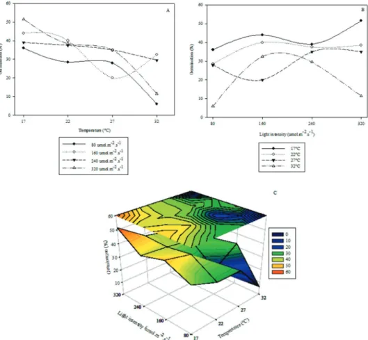
The isolate showed significant interaction between light intensity and temperature. The highest sporulation was achieved at 22 °C and light intensity of 240 µmol m-2 s-1, reaching 5.9 x 10⁶ conidia mL-1.The lowest sporulation values were obtained with the extremes of light intensity, 0 and 320 µmol m-2 s-1 in all temperatures. From these extremes, minimum and maximum values of light intensity, sporulation increased up to the value cited above. At temperature 32 °C sporulation was at its lowest (Figure 1). Based on quality evaluators R2 and AIC, the response surface generated by the polynomial model (Equation 3) provided the best fit (Tables 1 and 2).
Akaike’s Information Criterion (AIC): It is also a quality evaluator based on residual standard deviation of fit; the lower the better.
Other authors studied these variables individually in sporulation de C. coffeicola, but did not evaluate their
interaction. Souza (2011) obtained conidia for this species using a light intensity of 165 µmol m-2 s-1, which is under the ideal for higher conidia production in this study. Echandi (1959) reported higher sporulation of C. coffeicola in coffee leaves at 30 °C and
24 °C in vitro, which differed from was found in this study, 22
°C; however, light intensity was not cited. Furthermore, neither of the authors reported interaction with other environmental variables. Light intensity can exert both inductor and inhibitor effects on the formation of reproductive structures. Trione and Leach (1969) reported direct action of light on the activation of key enzymes in fungi with sporulation induced by light. The same authors state that the quantity and quality of light needed to induce formation of reproductive structures vary according to the fungi species. Beckman and Paine (1983) tested three light regimes using white fluorescent bulbs for optimizing conidia production of an isolate of C. zeae-maydis. Treatments
were 12-hour photoperiod, ten days of constant light followed by three days of darkness, and constant darkness. The highest conidia production occurred in 12-hours photoperiod. For conidia germination data, in function of the
Figure 1: Sporulation of C. coffeicola according to temperature (A), light intensity (B) and interaction between temperature and light intensity (C).
Table 1: Estimates of parameters of the quadratic regression modelfitted to data according to temperature (1), light intensity (2), and linear polynomial model (3) fitted to data of sporulation according to luminosity and temperature.
Models Parameters Estimates Standard Error
Quadratic Lint a 30,565.40 4,487.43
b -85.84 16.33
Quadratic Temp a 23,0753.00 55,859.00
b -6,303.00 2,038.00
a 120,487.68 35,497.02
Polynomial b 26,588.45 4,190.45
c -4,091.20 1,126.18
Table 2: Goodness of fit of response surface generated by combining quadratic-linear models and the polynomial model (multiple linear).
Evaluators* Ra2 AIC
Quadratic*Quadratic 0.5361 2,480.79
Polynomial 0.7865 1,839.16
*Coefficient of determination of model (R2): means the
amount of data variation which the model is able to describe; the closer to 1 the better.
extract agar (Calpouzos; Stallknecht 1967). Previously, these authors (Calpouzos; Stallknecht 1965) had already found significant interaction between temperature and light. Under constant light at 15 °C and 30 °C fungus sporulation was stimulated and inhibited, respectively. Thus, interaction of temperature with luminosity is essential for sporulation of Cercospora species.
Conidia Germination in C. coffeicola
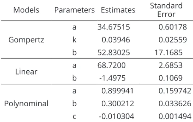
Significant interaction between light intensity and temperature was also found in the isolate germination. The highest percentage of germination occurred at temperature 17 °C and light intensity 320 µmol m-2 s-1, reaching 52%. Regardless of light intensity, germination decreased with increased temperature and at 32 °C the lowest germination occurred (Figure 2). Based on the quality evaluators, R2 and AIC, the response surface generated by combining Gompertz (Equation 5) models and Linear models (Equation 4) provided the best fit (Tables 3 e 4).
Figure 2: Germination of C. coffeicola according to A, temperature and B, light intensity. C, and interaction between temperature and light intensity.
Studies on interaction between temperature and luminosity were performed for other Cercospora species. In studies of C. arachidicola, the highest sporulation was
found in peanut leaf-oatmeal agar at 28 °C under constant light regime (Moraes and Salgado 1978; Smith, 1971). The effect of luminosity was also demonstrated for C. beticola
Table 3: Estimates for parameters of linear regression (4) fitted to data of temperature, the non-linear regression model by Gompertz (5) fitted in according to light intensity, and the polynomial model (6) fitted to data of germination according to light intensity and
air temperature.
Models Parameters Estimates Standard Error
Gompertz
a 34.67515 0.60178
k 0.03946 0.02559
b 52.83025 17.1685
Linear a 68.7200 2.6853
b -1.4975 0.1069
Polynominal
a 0.899941 0.159742
b 0.300212 0.033626 c -0.010304 0.001494
Table 4: Goodness-of-fit of response surface generated by combining Gompertz and linear models and the polynomial model (multiple linear).
Evaluators* Ra2 AIC
Gompertz Linear 0.9585 194.29
Polinomial 0.9261 238.18
* Coefficient of determination (R2): amount of data variation
which the model is able to describe; the closer to 1 the better. AIC:Akaike’s Information Criterion is also a quality evaluator based on residual standard deviation of fit; the lower the better.
The result in this study differed from that obtained by Echandi (1959), who found higher germination of
C. coffeicola at 30 °C and 24 °C, with 100% and 97%
germination respectively. However, the study did not assess interaction with light intensity. This variation in ideal temperature was also reported in other Cercospora
species in different phases of the pathogen circle. Gobina and Melouk (1981) evaluated temperatures 15, 20, 25, 30, and 35 °C for C. arachidicola. After 12 hours of
incubation, the authors found germination in less than 1% conidia at 15 °C and 84% at 35 °C. However, after 48 hours of incubation in aqueous suspension at temperatures 15-35 °C no significant difference was found in conidia germination. Alderman and Beute (1986) reported optimal conditions for conidia germination and germ tube elongation at 19-25 °C, which was over the optimum found in this experiment. Germination was low between 28 °C
and 32 °C, thus agreeing with the lowest germination at 32 °C in this study.
Regarding luminosity, constant light inhibited spore germination, germ tube elongation and sporulation
in C. zeae-maydis (Beckman and Payne 1983). This
variation in behavior at different temperatures found
in Cercospora species and between isolates could be
due to occurrence of genetic variability (Souza; Maffia; Mizubuti, 2012).
Thus, to the isolate under study, there was interaction between the variables temperature and light intensity in sporulation and germination of C. coffeicola.
CONCLUSIONS
There was interaction of temperature and light intensity variables on sporulation and germination of
C. coffeicola. The highest sporulation was obtained at a
temperature of 22 °C and light intensity of 240 μmol m-2 s-1, reaching 5.9x10⁶ con mL-1. The highest percentage of spore germination was obtained at 17 °C and light intensity of 320 μmol m-2 s-1, reaching 52%.
ACKNOWLEDGMENTS
The authors would like to acknowledge the financial support and scholarships provided by CNPq, Capes, Fapemig and INCT- Café for their financial support and scholarship.
REFERENCES
ABDOU, Y. A. M.; GREGORY, W. C.; COOPER, W. E. Sources and nature of resistance to Cercospora arachidicola Hori and Cercospora personatum (Beck and Curtis) Deighton in Arachis species. Peanut Science, 1(1):6-11, 1974.
ALDERMAN, S. C.; BEUTE M. K. Influence of temperature and moisture on germinaton and germ tube elongation of
Cercospora arachidicola. Phytopathology, 76(7):715-719, 1986.
ANCO, D. J.; MADDEN, L. V.; ELLIS, M. A. Effects of temperature and wetness duration on the sporulation rate of Phomopsis viticola on infected grape canes. Plant Disease, 97(5):579-589, 2013.
BECKMAN, P. M.; PAYNE, G. A. Cultural techniques and conditions influencing growth and sporulation of
Cercospora zeae-maydis and lesion development in corn. Phytopathology, 73(2):286-289, 1983.
BUITRAGO J. H. L.; FERNÁNDEZ B. O. Esporulacion in vitro de Cercospora coffeicola Berkeley & Cooke. Cenicafé,
CALPOUZOS, L.; STALLKNECHT, G. F. Sporulation of Cercospora beticola affected by an interaction between light and
temperature. Phytopathology, 55(12):1370-1371, 1965.
CALPOUZOS, L.; STALLKNECHT G. F. Symptoms of cercospora
leaf spot of sugar beets influenced by light intensity. Phytopathology, 57(7):799-780, 1967.
CAMERA, J. N. de et al. Limiares térmicos para a germinação de conídios de Cercospora sojina em dois regimes luminosos. Summa Phytopathologica, 39(1):58-61, 2013.
CARDOSO, C. A. A.; REIS, E. M.; MOREIRA, E. N. Development of a warning system for wheat blast caused by
Pyricularia grisea. S u m m a P h y t o p a t h o l o g i c a, 34(3):216-221, 2008.
CUSTODIO, A. A. P. de et al. Intensidade da ferrugem e da cercosporiose em cafeeiro quanto à face de exposição das
plantas. Coffee Science, 5(3):214-228, 2010.
DAUB, M. E.; HERRERO, S.; CHUNG, K. R. Photoactivated perylenequinone toxins in fungal pathogenesis of plants. FEMS Microbiology Letters, 252(2):197-206, 2005.
DEL PELOSO, M. C. de et al. Esporulação de Cercospora coffeicola
em diferentes meios de cultura. Fitopatologia Brasileira,
14(1):41-44, 1989.
DHINGRA, O. D.; SINCLAIR, J. B. Basic plant pathology methods. Boca Raton, FL,USA: Lewis Publisher, 1995. 448p. ECHANDI, E. La chasparria de los cafetos causada por el hongo
Cercospora coffeicola Berk. & Cooke. Turrialba, 9(2):54-67,
1959.
GOBINA, S. M.; MELOUK, H. A. Effect of temperature on the sporulation and conidial germination of Cercospora arachidicola. (Abstr.) Phytopathology, 71(8):876, 1981.
JENKINS, W. A. Two fungi causing leaf spot of peanut. Journal of Agricultural Research, 56(5):317-332, 1938.
KILPATRICK, R. A.; JOHNSON, H. W. Sporulation of Cercospora
species on carrot leaf decoction agar. Phytopathology,
46(3):180-181, 1956.
MORAES, S. A.; SALGADO, C. L. Influência da luz sobre a esporulação de Cercospora arachidicola Hori. Summa Phytopathologica, 4(2):128-135, 1978.
OSO, B. A. Conidial germination in Cercospora arachidicola Hori. Transactions of the British Mycologycal Society,
59(1):169-172, 1972.
POZZA, A. A. A. de et al. Influência da nutrição mineral na
intensidade da mancha-de-olho-pardo em mudas de
cafeeiro. Pesquisa Agropecuária Brasileira, 36(1):53-60, 2001.
POZZA, E. A; CARVALHO, L. C; CHALFOUN, S. M. Sintomas de injúrias causadas por doenças em cafeeiro. In: GUIMARÃES, R.J; MENDES A.N.G; BALIZA D.P. (eds). Semiologia do cafeeiro: Sintomas de desordens nutricionais, fitossanitárias e fisiológicas. Lavras: Editora UFLA, 2010. p. 68-106.
SALGADO, B. G. de et al. Progresso da ferrugem e da cercosporiose do cafeeiro consorciado com grevílea, com injazeiro e a pleno sol em Lavras, MG. Ciência e Agrotecnologia, 31(4):1067-1074, 2007.
SCHUMANN, G. L.; D’ARCY, C. J.; Review of hungry planet: Stories of Plant Diseases Essential Plant Pathology, St.
Paul, MN, USA: APS Press, 2012. 306p.
SMITH, D. H. A simple method for producing Cercospora arachidicola conidial inoculum. Phytopathology,
61(11):1414, 1971.
SOUZA, A. G. C.; MAFFIA, L. A.; MIZUBUTI, E. S. G. Cultural and aggressiveness variability of Cercospora coffeicola. Journal of Phytopathology, 160(10):540-546, 2012.
SOUZA, A. G. C. Infection process of Cercospora coffeicola on
coffee leaf. Journal of Phytopathology, 159(1):6-11, 2011.
TRIONE, E. J.; LEACH, C. M. Light-induced sporulation and sporogenic substances in fungi. Phytopathology,
59(8):1077-1083, 1969.
WAGGONER, P. E. Forecasting Epidemis. In: HORSFALL, J,G.; COWLING, E.B. Plant Pathology, San Francisco and