Vol.57, n.3: pp. 349-356, May-June 2014 http://dx.doi.org/10.1590/S1516-89132014005000006
ISSN 1516-8913 Printed in Brazil
BRAZILIAN ARCHIVES OF BIOLOGY AND TECHNOLOGY
A N I N T E R N A T I O N A L J O U R N A L
In vitro Antimicrobial Activity and Characterization of
Mangrove Isolates of Streptomycetes Effective against
Bacteria and Fungi of Nosocomial Origin
Arijit Das
*, Sourav Bhattacharya, Abuelgasim Yegoup Hassan Mohammed and
Subbaramiah Sundara Rajan
Department of Microbiology; Genohelix Biolabs; A Division of Centre for Advanced Studies in Biosciences; Jain University; Bangalore - Karnataka - India
ABSTRACT
The study aimed at determining the in vitro antimicrobial activity of alkaliphilic and halotolerant actinomycetes isolated from a mangrove ecosystem and identification of a potent strain. Twenty five isolates of actinomycetes were isolated from the sediment samples of Valapattanam mangrove swamp in Kerala, India. Antimicrobial activity of four selected actinomycete isolates was determined against bacterial and fungal pathogens of nosocomial origin by agar well diffusion method. Molecular characterization of the potent isolate was performed by 16S rDNA sequencing. Isolate no I-1 significantly inhibited Staphylococcus aureus ATCC 25923 (12 mm), S. aureus (15±0.05 mm), S. citreus (20±0.5 mm), Bacillus cereus (17±0.2 mm) and Serratia marcescens (12 mm). It also demonstrated effective antifungal action against Penicillium sp. (12±0.2 mm), Candida albicans (20±0.5 mm), C. parapsilosis (12 mm) and Cryptococcus neoformans (12 mm). Morphological study revealed that all the isolated actinomycetes belonged to the genus Streptomyces. Based on 16S rDNA sequence data, the selected isolate I-1 was shown to be closely related to Streptomyces xiamenensis. The results revealed that the mangrove ecosystem of Valapattanam harboured a rich consortium of many potent actinomycetes, which could synthesize novel bioactive compounds of pharmacological significance.
Key words:Streptomyces xiamenensis, Actinomycetes, Antimicrobial activity, Mangrove swamp
*Author for correspondence: [email protected]
INTRODUCTION
Due to emergence of multidrug-resistant strains of bacteria and fungi, especially those causing nosocomial infections, there is an ever-increasing demand for novel antibiotics with broad antimicrobial spectra. Actinomycetes constitute a diverse group of microorganisms that are widely distributed in terrestrial, freshwater and marine habitats (Radhika et al. 2011). Compared to terrestrial actinomycetes, very few studies have been conducted on marine actinomycetes. Marine ecosystem constitutes oceans, the deep sea and the
sea floor, estuaries and lagoons, salt marsh and intertidal zones, coral reefs and mangrove swamps.
opportunity for finding rare actinomycetes with unique properties, capable of producing many novel bioactive compounds such as antibiotics,
enzymes and antitumor agents. Rare
actinomycetes are considered as the strains whose isolation frequency by conventional methods is much lower than that of commonly occurring
actinomycete strains(Naikpatil and Rathod 2011).
Among the actinomycetes, species of
Streptomyces constitute second highest percentage
after Nocardia in terms of distribution in the
mangrove ecosystem (George et al. 2012). They are prolific producers of secondary metabolites and about 80% of total antibiotics are produced
from the species of Streptomyces (Sathiyaseelan
and Stella 2011).
The coastal states of peninsular India are lined with numerous mangrove forests. These habitats offer unique ecological niche supporting the occurrence and interactions between many rare microbial forms. Therefore, the present study
aimed at determining the in vitro antimicrobial
activity of alkaliphilic and halotolerant
actinomycetes isolated from a mangrove
ecosystem and identification of a potent strain of actinomycete effective against the bacterial and fungal pathogens of nosocomial origin.
MATERIALS AND METHODS
Collection and processing of sediment samples
Seven samples of fresh sediments were collected from the Valapattanam mangrove ecosystem (11° 54′ 0″ N, 75° 22′ 0″ E) in Kannur district of Kerala, India during September 2011. Sediment samples (rich in plant debris) were collected with an auger (down to 10 cm depth) after removing approximately 5.0 cm litter of fallen leaves near
the root system of Avicennia marina. Samples
were stored in sterile zip-lock polythene packets, transported to the laboratory and stored at 4°C. After the determination of pH values, the sediment samples were air dried at room temperature, mixed thoroughly and sieved through a 2-mm pore size sieve (Retsch, Haan, Germany) to remove the large debris. The sieved samples were used for the isolation of actinomycetes.
Isolation of actinomycetes
Sieved sediment sample (1.0 g) was suspended in 100 mL sterile distilled water and incubated in an orbital shaker (Orbitek) at 28°C and 200 rpm for 1 h (Boroujeni et al. 2012). Particles were allowed
to settle and then serial dilutions of the spore
suspension were prepared up to 10-4. From each
dilution, 0.1 mL was spread evenly over the surface of starch casein nitrate (SCN) agar plates
(supplemented with cycloheximide 50 µg mL-1)
with sterile L-shaped glass rod and incubated at 28°C for 10 days (Kuster and Williams 1964). Dilutions that yielded 30-300 colonies were chosen for further study. Actinomycete isolates were purified by streak-plate technique and the pure cultures were maintained on SCN agar slants at 4°C for further use.
Preliminary screening of actinomycetes for antagonistic activity
The test bacterial pathogens included S. aureus
ATCC 25923, S. aureus, S. citreus, B. cereus,
Escherichia coli, S. marcescens, Proteus mirabilis, Pseudomonas aeruginosa, Salmonella typhi and Klebsiella pneumoniae. The test fungal
pathogens included Aspergillus niger, A. flavus,
Penicillium sp., Fusarium sp., C. albicans, C. parapsilosis, Trichophyton rubrum, Trichophyton mentagrophytes and Cryptococcus neoformans. All these test bacterial and fungal pathogens were hospital isolates obtained from the Kempegowda Institute of Medical Sciences, Bangalore. For screening of antibacterial activity, pure isolates of actinomycetes were streaked at the centre of sterile SCN agar plates and incubated at 28°C for 5 days. Broth cultures of the test bacterial pathogens were inoculated into molten Mueller Hinton agar and overlaid on SCN agar plates. After solidification, the plates were incubated at
37°C for 24 h. Screening for the antifungal
activity was performed by agar double layer method using Sabouraud dextrose agar medium and incubated at 27°C for 48 h. Antimicrobial activity was noted by observing the area of inhibition surrounding the streaked actinomycete isolate.
Production and extraction of antimicrobial metabolites
The selected actinomycete isolates were
(2007). The purified extracts were stored at 4°C for further assay.
Antimicrobial activity of actinomycete isolates
The antimicrobial activity of purified extracts
derived from the actinomycetes was determined against all the test bacterial and fungal pathogens using agar well diffusion method. Test cultures of the bacterial pathogens were prepared in Mueller Hinton broth and incubated at 37°C. The 24 h old bacterial cultures were swabbed onto sterile Mueller Hinton agar plates. Wells were punched with a sterile cork borer (6 mm internal diameter)
and 35 µL of the extract was added to each well.
Ampicillin and streptomycin (50 mg/mL) were used as standard antibiotics for Gram positive and Gram negative bacteria, respectively. Similarly, spore suspensions of the fungal pathogens were prepared in Sabouraud dextrose broth. After swabbing the sterile Sabouraud dextrose agar
plates with the suspensions, 35 µL of the extract
was added to each well. Fluconazole (20 mg/mL) was used as the standard antifungal. The inoculated plates containing the extracts were incubated first under refrigeration for 6 h for diffusion of the extracts. The bacterial plates were then incubated at 37°C for 24 h. The fungal plates were incubated at 27°C for 48 h. Following incubation, diameters of the inhibitory zones were measured to the nearest millimetre and recorded.
Characterization of actinomycete isolates
The actinomycete isolates that exhibited effective
antimicrobial activities were characterized
morphologically by observing the spore chains of the Gram stained smears of 10 days old colonies. The organisms were identified by observing the structures and comparing with Bergey’s Manual of Determinative Bacteriology (Bergey and Holt 2000). Colour of aerial mycelia was determined from the mature, sporulating aerial mycelia of the actinomycete colonies on SCN agar (Hamedani et al. 2012). Colour of the substrate mycelia (reverse of the plate) was also observed along with diffusible pigments, if any (Padmadhas and Ragunathan 2010). Physiological characterization such as the effect of pH (3.3-9.3), temperature tolerance (4-42°C), salt tolerance (0.1-10% w/v NaCl) and melanin production were analyzed.
Extraction of genomic DNA from the
actinomyceteisolate
The most potent actinomycete isolate was inoculated in SCN broth and incubated at 28°C for 7 days in an orbital shaker at 130 rpm. Genomic DNA was extracted using the Fungal Genomic DNA Isolation Kit RKT 41/42 (Chromous Biotech Pvt. Ltd., Bangalore, India) according to the manufacturer instructions and visualized using 0.8% agarose gel (with ethidium bromide) electrophoresis (Hamedani et al. 2012).
PCR amplification
DNA amplification by polymerase chain reaction (PCR) was performed in a total volume of 25 µL. Each reaction mixture contained the following
solutions: 1.5 µL genomic DNA, 1.0 µL 10 pmol
forward universal 16S rDNA primer
(5’-AGAGTTTGATCCTGGCTCA-3’) (Sigma,
USA); 1.0 µL of 10 pmol reverse universal 16S
rDNA primer
(5’-ACGGCTACCTTGTTACGACT-3’) (Sigma,
USA); 1.0 µL of 30 mm deoxyribonucleoside
5’-triphosphate (N= A,T,G,C) (dNTP’s); 2.5 µL of
10X PCR buffer and 1 µL Taq polymerase (1 U)
(Chromous Biotech Pvt. Ltd., Bangalore, India)
and water was added up to 25 µL. The thermal
cycler (MJ Research PTC 200, USA) was programmed as follows: 2 min initial denaturation at 94°C, followed by 30 cycles that consisted of denaturation for 1 min at 94°C, annealing for 30 s at 57°C and extension at 74°C for 1 min and a final extension of 5 min at 74°C. The PCR-amplified product was detected by 1.2% agarose gel (with ethidium bromide) electrophoresis and the results were visualized under UV light using gel documentation system (Herolabs, Germany) (Hamedani et al. 2012).
Partial 16S rDNA sequencing and analysis of sequenced data
The partial 16S rDNA sequencing of the PCR-amplified product was performed at Chromous Biotech Pvt. Ltd., Bangalore, India. The 16S rDNA sequence data was aligned manually with the available nucleotide sequences retrieved from
NCBI database by using BLASTn (Prapagdee et
RESULTS AND DISCUSSION
The rapid emergence and widespread occurrence of multidrug-resistant strains of bacteria and fungi, especially those causing nosocomial infections, has threatened the treatment regimen with conventional antibiotics. Several such cases are being reported worldwide (Ruscher et al. 2010). A recent study reported that nosocomial infections at the surgical sites were responsible for 42.9% of the mortality cases in paediatric living donor liver transplantation (Nafady-Hego et al. 2011).
Actinomycetes are ubiquitously prevalent diverse groups of prokaryotic microorganisms, which are known to synthesize a wide range of bioactive
metabolites such as enzymes, antibiotics,
pigments, antitumor compounds and
immunosuppressive agents (Valli et al. 2012).
They are specially recognized for their ability to synthesize various secondary metabolites, which possess antibacterial, antifungal and antiprotozoal activities (Ravikumar et al. 2011). Actinomycetes are present in terrestrial, marine as well as fresh water habitats (Boroujeni et al. 2012). They are also found in brackish water and estuarine ecosystems.
Mangrove ecosystem consists of pneumatophores bearing woody plants, which grow in waterlogged saline soil of intertidal coasts in the tropical and subtropical zones. They mainly occur along the coastline at the confluence of rivers and sea. This ecosystem produces large amounts of detritus (organic matter) due to the autolysis and microbial decomposition of fallen leaves, twigs, flowers and fruits. Abundance of these organic matters, salinity and high degree of moisture content
favour the prevalence of actinobacterial
population and other life forms in the mangrove ecosystem (Nag et al. 2012). These alkaliphilic actinomycetes are naturally capable of producing different plant fibre hydrolyzing enzymes and
secondary metabolites(Tsujibo et al. 2003).
Isolation of actinomycetes from mangrove sediment
All the sediment samples were collected post monsoon in order to obtain a diverse population of actinomycetes. The sediment samples showed pH values ranging from 6.5 to 8.3. Twenty five isolates of actinomycetes were recovered from the sediment samples collected from the rhizosphere of A. marina. Some previous studies have
reported the isolation of novel species of actinomycetes from the mangrove habitats. In a study investigating the actinobacterial population from the Pitchavaram mangrove forests in Tamil Nadu, India, maximum number of actinomycetes
were recovered from the rhizosphere of A. marina.
About 50% of the isolates revealed activity
against S. aureus, Bacillus subtilis, E. coli, Vibrio
cholerae, K. pneumoniae, Proteus vulgaris and S. typhi (Balagurunathan et al. 2010). It could be
deciphered from these findings that a diverse
group of pharmacologically potent actinomycetes remained in close association with the roots of these plants, protecting them from the adverse
environmental conditions and successfully
competing with various soil borne pathogens
(Kumaresan and Suryanarayanan 2001).
Ravikumar et al. (2011) also studied the biodiversity of actinomycetes in the Manakkudi mangrove ecosystem located in Tamil Nadu, India and found maximum population in the rhizospere soil.
Antibacterial activity of actinomycete isolates
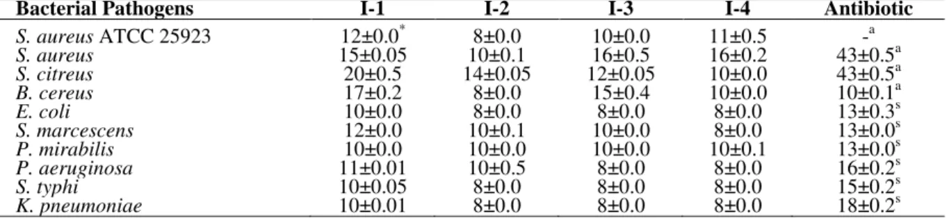
Preliminary screening of all the actinomycetes for antagonistic activity against the pathogenic bacteria and fungi revealed four isolates, which showed better antimicrobial spectra. These four isolates were designated as I-1, I-2, I-3 and I-4. The secondary metabolites produced by these four isolates were extracted and partially purified. The antimicrobial activity of these four isolates is
presented in Table 1. Among the test bacteria, S.
citreus was maximally inhibited by I-1 (20±0.5
mm) and I-2 (14±0.05 mm), whereas, S. aureus
was inhibited byI-3 (16±0.5 mm) and I-4 (16±0.2
mm), respectively. Interestingly, among the four actinomycete isolates, I-1 inhibited most of the Gram positive and Gram negative test bacteria, which indicated its broad antibacterial spectra. This was in agreement with that of another recent study wherein only five actinomycetes among a
total of 107 marine isolates inhibited B. subtilis, S.
aureus, Proteus vulgaris, E. coli, Klebsiella aerogenes and P. aeruginosa (Gulve and Deshmukh 2012). An earlier study revealed that
Enterococcus sp., multidrug-resistant P. aeruginosa and methicillin-resistant S. aureus
were the predominant bacterial pathogens causing nosocomial infections in liver transplantation patients (Nafady-Hego et al. 2011). In the present
study, the streptomycete isolate I-1 inhibited S.
though no inhibition was recorded with ampicillin. Overall, the antagonistic effect of I-1 against the Gram positive bacteria was significantly higher than that of Gram negative bacteria. On the other hand, extracts from the I-2, I-3 and I-4 showed higher inhibition against the Gram positive
bacteria than the Gram negative pathogens, with zone diameters ranging between 8-20 mm. The greater degree of resistance exhibited by the Gram negative bacteria might be due to the presence of
an outer membrane consisting of
lipopolysaccharide(Parunago et al. 2007).
Table 1 - Antibacterial activity of selected actinomycete isolates against bacterial pathogens showing diameters of inhibitory zones (in mm) by agar well diffusion method.
Bacterial Pathogens I-1 I-2 I-3 I-4 Antibiotic
S. aureus ATCC 25923 12±0.0* 8±0.0 10±0.0 11±0.5 -a
S. aureus 15±0.05 10±0.1 16±0.5 16±0.2 43±0.5a
S. citreus 20±0.5 14±0.05 12±0.05 10±0.0 43±0.5a
B. cereus 17±0.2 8±0.0 15±0.4 10±0.0 10±0.1a
E. coli 10±0.0 8±0.0 8±0.0 8±0.0 13±0.3s
S. marcescens 12±0.0 10±0.1 10±0.0 8±0.0 13±0.0s
P. mirabilis 10±0.0 10±0.0 10±0.0 10±0.1 13±0.0s
P. aeruginosa 11±0.01 10±0.5 8±0.0 8±0.0 16±0.2s
S. typhi 10±0.05 8±0.0 8±0.0 8±0.0 15±0.2s
K. pneumoniae 10±0.01 8±0.0 8±0.0 8±0.0 18±0.2s
*
: values are mean ± standard deviation (n=3); a: ampicillin; s: streptomycin; -: no zone.
A total of 55 actinomycetes comprising of
Actinomyces, Nocardia, Streptomyces and
Micromonospora were isolated from the soil sample of Karanjal region in Sundarbans
(Arifuzzaman et al. 2010). Among these, 20
actinomycete isolates showed antibacterial activity against the Gram negative bacteria such as
Shigella boydii, S. flexneri, S. sonnei,
Pseudomonas sp., S. dysenteriae type-1, Vibrio cholerae, S. typhi, Plesiomonas, Hafnia sp. and E.
coli. Their study revealed that three isolates
exhibited broad spectrum antibacterial activity. A previous study reported the isolation of 42 actinomycetes from the mangrove sediments of Andaman and Nicobar Islands, India, among which 22 species showed antagonistic activities
against S. aureus, B. subtilis, S. typhi and K.
pneumoniae (Baskaran et al. 2011). Another investigation revealed the isolation of an actinomycete from the mangrove forest soil of Guangxi Beihai, China, with designated strain no.
BH0954, which was 99% related to Streptomyces
sindenensis. This isolate inhibited S. aureus, S. epidermidis, E. coli and P. vulgaris, with no effect
against P. aeruginosa and C. albicans(Dalin et al.
2010). A recent study also reported the isolation of
Streptomyces sp. from the Coringa mangrove forest, Andhra Pradesh, India, whose metabolite
was inhibitory to S. aureus, P. fluorescens, P.
aeruginosa, Lactobacillus acidophilus, L. casei, C. albicans, Streptococcus mutans, B. subtilis, B. megaterium and Xanthomonassp. (Deepthi et al. 2012).
Antifungal activity of actinomycete isolates
Extract from the isolate I-1 also exhibited antifungal activity against all the test fungal pathogens as illustrated in Table 2. Molds such as
A. niger, A. flavus, Penicillium sp. and Fusarium
sp. were inhibited with zones ranging between 8-12 mm. Some important systemic fungal
pathogens affecting the humans are C. albicans, C.
neoformans and A. fumigatus, some of these causing nosocomial infections in the patients during the postoperative months (Oskay 2009; Nafady-Hego et al. 2011). Among the pathogenic
yeasts, C. albicans was inhibited by I-1 (20±0.5
mm). The extract of I-1 was equally effective
against C. parapsilosis and C. neoformans.
Isolates I-2 and I-4 also demonstrated maximum
activity against C. albicans but I-3 revealed
highest inhibition against C. neoformans. Extracts
of the isolates I-2, I-3 and I-4 either demonstrated moderate to weak, or no antagonistic activity against the other fungi.
The antifungal activity of a rare actinomycete has
been reported recently (Mangamuri et al. 2012).
This actinomycete strain, isolated from the mangrove sediments of Nizampatnam, India, was
found closely related to Pseudonocardia
endophytica and inhibited A. niger, F. oxysporum
Table 2 - Antifungal activity of selected actinomycete isolates against fungal pathogens showing diameters of inhibitory zones (in mm) by agar well diffusion method.
Fungal Pathogens I-1 I-2 I-3 I-4 Fluconazole
A. niger 10±0.0* 9±0.0 9±0.05 7±0.0 17±0.0
A. flavus 8±0.0 7±0.0 7±0.0 - 18±0.0
Penicillium sp. 12±0.2 10±0.0 7±0.0 8±0.0 21±0.5
Fusarium sp. 10±0.0 8±0.0 9±0.0 7±0.0 -
C. albicans 20±0.5 12±0.05 9±0.0 12±0.05 10±0.05
C. parapsilosis 12±0.0 11±0.02 8±0.0 9±0.0 37±1.0
T. rubrum 8±0.0 8±0.0 7±0.05 - 19±0.03
T. mentagrophytes 9±0.1 - - 7±0.0 18±0.05
C.neoformans 12±0.05 8±0.0 11±0.0 7±0.0 22±0.0
*
: values are mean ± standard deviation (n=3); -: no zone.
Characterization of actinomycete isolates
The morphological characterization of the
actinomycete isolates was performed based on the study of colony characters and observation of Gram stained smears. On SCN agar, all the actinomycetes produced dry, compact, chalk-like
colonies, similar to those of genus Streptomyces.
The colonies on SCN agar showed hues of white, cream, grey, pale brown, dark brown, yellow, tan, pink and purple shades. The four selected strains had white to grey aerial mycelia, whereas the colour of their substrate mycelia showed distinct
difference. The dark brown hue of the substrate mycelium as exhibited by the actinomycete isolate I-1 indicated its ability to produce unique pigment and/or any secondary metabolite. This observation could be correlated to the fact that among all the mangrove isolates, the isolate I-1 revealed the
highest antagonistic activity against the
nosocomial pathogens. Gram stained smears revealed the presence of filamentous structures bearing the spores in verticillate and spiral arrangements. The morphological characters of the actinomycete isolates are shown in Table 3.
Table 3 -Morphological characterization of selected actinomycete isolates.
Characteristics I-1 I-2 I-3 I-4
Colour of aerial mycelium Grey Pale yellow Light grey Dark grey Colour of substrate mycelium Dark brown Yellow Dark purple Dark Grey
Gram’s reaction Positive Positive Positive Positive
Spore chain Spirales Spirales Spirales Spirales
The selected actinomycete isolates exhibited optimum growth within the mesophilic range of 25-37°C. Interestingly, all the selected isolates grew well at pH ranging from 5.3 to 9.3 but optimum growth was observed between pH 7.3-9.3. Furthermore, they were also tolerant to 0.1-5% NaCl but failed to grow at 10% salt concentration. This could probably be due to the hypertonic medium wherein the isolates would have suffered from severe osmotic shock. On par
with the present findings, a marine strain of S.
rochei, isolated from Visakhapatnam coast in India, was reported to be tolerant to pH 10.5 and 6% NaCl (Reddy et al. 2011). From these observations, it was clearly evident that mangrove actinomycetes were generally alkaliphilic and halotolerant. Furthermore, the isolates I-1, I-3 and I-4 also demonstrated melanin production. The physiological characters of four selected isolates are presented in Table 4.
Table 4 - Physiological characterization of selected actinomycete isolates.
Characteristics I-1 I-2 I-3 I-4
Growth at 4°C - - - -
25°C + + + +
30°C + + + +
37°C + + + +
42°C - - - -
Growth at pH 3.3 - - - -
4.3 + - + +
5.3 + + + +
6.3 + + + +
7.3 + + + +
8.3 + + + +
9.3 + + + +
Growth at NaCl (%) 0.1 + + + +
0.5 + + + +
1 + + + +
3 + + + +
5 + + + +
10 - - - -
Melanin production + - + +
Recently, molecular methods have been extensively used for the identification of bacterial species. They are preferred over the conventional methods because they yield faster and accurate results. 16S rDNA sequencing is one of the molecular methods that has revolutionized the bacterial systemics and classification. Based on the results of antimicrobial assay, the most potent alkaliphilic and halotolerant actinomycete isolate I-1 was subjected to molecular characterization. PCR amplification of its 16S rDNA gene sequence showed an amplicon of around 1500 bps. The automated sequencing of this PCR-amplified product was carried out. The sequence data when manually aligned in NCBI database using BLASTn revealed that the isolate I-1 was closely
related to S. xiamenensis MCCC 1A01550 with
96% homology. Previously, many antimicrobial metabolite-producing actinomycetes isolated from
marine sediments were identified as S.
roseoverticillatus, S. roseorubens and S. septatus
(Valan Arasu et al. 2012). The nucleotide
sequence of the isolate I-1, designated as S.
xiamenensis GHBA11, was provided a GenBank accession number JX827497. Most of the
morphological, cultural and physiological
characters of the isolate I-1 were in agreement
with that of a rare actinomycete, S. xiamenensis
sp. nov., isolated from the sediment of national mangrove reserve in Fujian Province, China (Xu et al. 2009). They reported its uniqueness in having a very high DNA G+C content of 71.6 mol%. To the best of our knowledge, this is the first report illustrating the antibacterial and
antifungal activity of S. xiamenensis.
CONCLUSIONS
Mangrove ecosystem harbours many rare
actinomycetes with interesting physiological properties. It is suggested that these unique strains of mangrove actinomycetes be further studied in search for some broad-spectrum, novel antibiotics, which could be effective in the treatment of nosocomial infections.
ACKNOWLEDGMENTS
We wish to extend our sincere gratitude to Dr. R. Chenraj Jain, Chairman, Jain Group of Institutions,
Bangalore; Dr. N. Sundararajan, Vice-Chancellor of Jain University, Bangalore; Prof. K. S. Shantamani, Chief Mentor, Jain Group of
Institutions and Dr. S. Sundara Rajan, Director of
Genohelix Biolabs, A Division of Centre for Advanced Studies in Biosciences, Jain University, for their encouraging support. We express our heartfelt thanks to Mrs. K. Prashanthi for her assistance in molecular characterization.
REFERENCES
Arifuzzaman M, Khatun MR, Rahman H. Isolation and screening of actinomycetes from Sundarbans soil for antibacterial activity. Afr J Biotechnol. 2010; 9(29): 4615-4619.
Balagurunathan R, Masilamani Selvam M, Kathiresan K. Bioprospecting of mangrove rhizosphere actinomycetes from Pitchavaram with special reference to antibacterial activity. J Pharm Res. 2010; 3(5): 909-911.
Baskaran R, Vijayakumar R, Mohan PM. Enrichment method for the isolation of bioactive actinomycetes from mangrove sediments of Andaman Islands, India.
Mal J Microbiol. 2011;7(1): 26-32.
Bergey DH, Holt JG. Bergey’s Manual of Determinative Bacteriology. 9th ed. Philadelphia: Lippincott Williams and Wilkins; 2000.
Boroujeni ME, Das A, Prashanthi K, Suryan S, Bhattacharya S. Enzymatic screening and random amplified polymorphic DNA fingerprinting of soil streptomycetes isolated from Wayanad district in Kerala, India. J Biol Sci. 2012; 12(1): 43-50.
Dalin H, Guifeng Y, Yajuan X, Naixiang L, Jianhong C, Jiang lianxiu L, Senzhou C. Isolation and identification of Actinomycetes sp. BH0954 from the mangrove forest soil of Guangxi Beihai. Chin Agric Sci Bull. 2010; 26(18): 406-409.
Deepthi MK, Sudhakar MS, Devamma MN. Isolation and screening of Streptomyces sp. from Coringa mangrove soils for enzyme production and antimicrobial activity. Int J Pharmaceut Chem Biol Sci. 2012; 2(1): 110-116.
George M, Anjumol A, George G, Hatha AAM. Distribution and bioactive potential of soil actinomycetes from different ecological habitats. Afr J Microbiol Res. 2012; 6(10): 2265-2271.
Giri C, Ochieng E, Tieszen LL, Zhu Z, Singh A, Loveland T, Masek J, Duke N. Status and distribution of mangrove forests of the world using earth observation satellite data. Global Ecol Biogeogr.
Gulve RM, Deshmukh AM. Antimicrobial activity of the marine actinomycetes. IMRJ. 2012; 2(3):16-22. Hamedani K, Soudbakhsh MN, Das A, Prashanthi K,
Bhattacharya S, Suryan S. Enzymatic screening, antibacterial potential and molecular characterization of streptomycetes isolated from Wayanad district in Kerala, India. Int J Pharm Bio Sci. 2012; 2(1): 201-210.
Kumaresan V, Suryanarayanan TS. Occurrence and distribution of endophytic fungi in a mangrove community. Mycol Res. 2001; 105:1388-1391. Kuster E, Williams ST. Selection of media for isolation
of streptomycetes. Nature. 1964;202: 928-929. Mangamuri UK, Muvva V, Poda S, Kamma S.
Isolation, identification and molecular characterization of rare actinomycetes from mangrove ecosystem of Nizampatnam. Mal J Microbiol. 2012; 8(2): 83-91.
Muiru WM, Mutitu EW, Mukunya DM. Characterization of antibiotic metabolites from actinomycete isolates. African Crop Science Conference Proceedings. 2007; 8: 2103-2107. Nafady-Hego H, Elgendy H, El Moghazy W, Fukuda
K, Uemoto S. Pattern of bacterial and fungal infections in the first 3 months after pediatric living donor liver transplantation: an 11-year single-center experience. Liver Transplant. 2011; 17(8): 976-984. Nag C, Bhattacharya S, Das A. Evaluation of
antagonistic activities of microbes from Vallapattanam and Pappinishery mangrove ecosystems of Kannur district in Kerala, India. Int J Pharm Life Sci. 2012; 3(5):1650-1659.
Naikpatil SV, Rathod JL. Selective isolation and antimicrobial activity of rare actinomycetes from mangrove sediment of Karwar. J Ecobiotechnol. 2011; 3(10): 48-53.
Oskay M. Antifungal and antibacterial compounds from
Streptomyces strains. Afr J Biotechnol. 2009; 8(13): 3007-3017.
Padmadhas R, Ragunathan R. Isolation, characterisation and identification of novel actinomycetes collected from Western Ghats region of India. J Pharm Biomed Sci. 2010;1(7): 1-7.
Parunago MM, Maceda EBG, Villano MAF. Screening of antibiotic-producing actinomycetes from marine, brackish and terrestrial sediments of Samal Island, Philippines. J Res Sci Comp Engg. 2007; 4(3): 29-38. Prapagdee B, Kuekulvong C, Mongkolsuk S.
Antifungal potential of extracellular metabolites produced by Streptomyces hygroscopicus against phytopathogenic fungi. Int J Biol Sci. 2008; 4(5): 330-337.
Radhika S, Bharathi S, Radhakrishnan M, Balagurunathan R. Bioprospecting of fresh water actinobacteria: isolation, antagonistic potential and characterization of selected isolates. J Pharm Res. 2011; 4(8): 2584-2586.
Ravikumar S, Fredimoses M, Gokulakrishnan R. Biodiversity of actinomycetes in Manakkudi mangrove ecosystem, southwest coast of India.
Annals Biol Res. 2011; 2(1): 76-82.
Ravikumar S, Inbaneson SJ, Uthiraselvam M, Priya SR, Ramu A, Banerjee MB. Diversity of endophytic actinomycetes from Karangkadu mangrove ecosystem and its antibacterial potential against bacterial pathogens. J Pharm Res. 2011; 4(1): 294-296.
Reddy NG, Ramakrishna DPN, Raja Gopal SV. A morphological, physiological and biochemical studies of marine Streptomyces rochei (MTCC 10109) showing antagonistic activity against selective human pathogenic microorganisms. Asian J Biol Sci. 2011; 4(1): 1-14.
Ruscher C, Lübke-Becker A, Semmler T, Wleklinski CG, Paasch A, Soba A, et al. Widespread rapid emergence of a distinct methicillin- and multidrug-resistant Staphylococcus pseudintermedius (MRSP) genetic lineage in Europe. Vet Microbiol. 2010; 144(3-4): 340-346.
Sathiyaseelan K, Stella D. Isolation, identification and antimicrobial activity of marine actinomycetes isolated from Parangipettai. Rec Res Sci Tech. 2011; 3(9): 74-77.
Tsujibo H, Kubota T, Yamamoto M, Miyamoto K, Inamori Y. Characteristics of chitinase genes from an alkaliphilic actinomycete, Nocardiopsis prasina
OPC-131. Appl Environ Microbiol. 2003; 69(2): 894-900.
Valan Arasu M, Asha KRT, Duraipandiyan V, Ignacimuthu S, Agastian P. Characterization and phylogenetic analysis of novel polyene type antimicrobial metabolite producing actinomycetes from marine sediments: Bay of Bengal India. Asian Pac J Trop Biomed. 2012; 2012: 803-810.
Valli S, Suvathi Sugasini S, Aysha OS, Nirmala P, Vinoth Kumar P, Reena A. Antimicrobial potential of actinomycetes species isolated from marine environment. Asian Pac J Trop Biomed. 2012; 2012: 469-473.
Xu J, Wang Y, Xie S, Xu J, Xiao J, Ruan J.
Streptomyces xiamenensis sp. nov., isolated from mangrove sediment. Int J Syst Evol Micr. 2009; 59(Pt 3): 472-476.