Printed version ISSN 0001-3765 / Online version ISSN 1678-2690 www.scielo.br/aabc
Protists and bacteria interactions in the presence of oil
JOSÉ A.P. BITENCOURT1, FREDERICO SOBRINHO SILVA2, INÁCIO D. DA SILVA-NETO3 and MIRIAN A.C. CRAPEZ1
1Laboratório de Microbiologia Marinha, Departamento de Biologia Marinha, Instituto de Biologia, Universidade Federal Fluminense, Outeiro São João Batista, s/nº, Caixa Postal 100644, Campus do Valonguinho, 24001-970 Niterói, RJ, Brasil
2
Laboratório de Palinofácies and Fácies Orgânica, Departamento de Geologia, Instituto de Geociências, CCMN, Universidade Federal do Rio de Janeiro, Av. Athos da Silveira Ramos, 274, Campus Ilha do Fundão,
Cidade Universitária, 21941-916 Rio de Janeiro, RJ, Brasil
3Laboratório de Protistologia, Departamento de Zoologia, Instituto de Biologia, CCS, Universidade Federal do Rio de Janeiro, Av. Carlos Chagas Filho, 373, Campus Ilha do Fundão, Cidade Universitária, 21941-590 Rio de Janeiro, RJ, Brasil
Manuscript received on June 20, 2012; accepted for publication on September 2, 2013
ABSTRACT
Little is known about the role of protists and bacteria interactions during hydrocarbon biodegradation. This work focused on the effect of oil on protists from three different locations in Guanabara Bay and bacteria from Caulerpa racemosa (BCr), Dictyota menstrualis (BDm) and Laurencia obtusa (BLo) during a 96
h bioassay. Cryptomonadida (site 1, 2 and 3), Scuticociliatida (site 2) and Euplotes sp.1 and Euplotes sp.2 (site 3) appeared after incubation. The highest biomass observed in the controls was as follows: protist site 3 (6.0 µgC.cm-3
, 96 h) compared to site 3 with oil (0.7 µgC.cm-3
, 96 h); for bacteria, 8.6 µgC.cm-3
(BDm, 72 h)
and 17.0 µgC.cm-3 (B
Cr with oil, 24 h). After treatment, the highest biomasses were as follows: protists at
site 1 and BLo, 6.0 µgC.cm -3
(96 h), compared to site 1 and BLo with oil, 3.31 µgC.cm -3
(96 h); the bacterial biomass was 43.1 µgC.cm-3
at site 2 and BDm (96 h). At site 3 and BLo with oil, the biomass was 18.21 µgC.
cm-3 (48 h). The highest biofilm proportions were observed from B
Cr 1.7 µm (96 h) and BLo with oil 1.8 µm
(24 h). BCr, BLo and BDm enhanced biofilm size and reduced the capacity of protists to prey.
Key words: bacterial consortia, biomass, free living protist, Guanabara Bay, microbial loop, petroleum.
Correspondence to: José Augusto Pires Bitencourt E-mail: [email protected]
INTRODUCTION
Protists are unicellular eukaryotes that are wide-spread in all types of habitats (Buck et al. 2000, Laybourn-Parry et al. 2000, Pedros-Alio et al. 2000, Scott et al. 2001). They are an essential component of microbial food webs, and their phagotrophic activities release waste products into the environment, both as dissolved and particulate organic matter from the undigested components of prey bacteria (Nagata and Kirchman 1992a, b) and as dissolved inorganic
nutrients, particularly ammonium and phosphate (Caron and Goldman 1990, Dolan 1997). Thus,
protist grazing provides substrates for the further
growth of prey, which include both heterotrophic bacteria (Jumars et al. 1989, Christaki et al. 1999) and autotrophic cells (Dolan 1997).
Protists are similar in size to their microbial
prey, including bacteria, algae, and other heterotrophic protists. Their growth potential is the same as their microbial prey, and the high rate of protist metabolism
facilitates carbon and energy flux through ecosystems
746 JOSÉ A.P. BITENCOURT, FREDERICO SOBRINHO SILVA, INÁCIO D. DA SILVA-NETO and MIRIAN A.C. CRAPEZ
Bacteria are present in sediment in large numbers (approximately 1010 cells.g-1). Their biomass is greater than the biomass of all other benthic orga-nisms due to the structure and function of microbial
biofilms. They possess a high surface to volume
ratio, which is indicative of high metabolic rates. Dissolved inorganic and organic substrates can
be metabolized with high substrate affinity and specificity. Particulate organic matter can be
decomposed in close contact with the substrate by
using hydrolytic enzymes (Silva et al. 2010, Guerra
et al. 2011). Other than oxygen, microorganisms use alternative electron acceptors (nitrate, manganese, iron, sulfate, and carbon dioxide) for the assimilation of organic material (Edwards et al. 2005).
Oil is a complex mixture of recalcitrant and toxic substances (Wilkinson et al. 2002) and is able to disrupt cellular homeostasis (Sikkema et al. 1995,
Crapez 2001). Some microorganisms utilize oil as a carbon and energy source (Crapez et al. 2001, Ron and
Rosenberg 2002), and oil degradation is a multi-step process in which each step is performed via distinct processes performed by different functional groups from various microbial organisms (Dalby et al. 2008).
Despite the ecological importance of protists in aquatic and terrestrial ecosystems, relatively little is known about their role in hydrocarbon degra-dation compared to the reported role of bacteria. A more detailed knowledge of the role of protists in microbial interactions with hydrocarbons is essential to trace and model the fate of hydrocarbon contaminants in terrestrial and aquatic ecosystems.
This work aims to study the influence of oil
(Light Arabian oil) on bacteria isolated from three seaweed samples and protists isolated from the sediment of Guanabara Bay during a 96 hour bioassay.
MATERIALS AND METHODS
STUDY AREA
Guanabara Bay is located in the state of Rio de
Janeiro in Southeast Brazil, between 22°40`-23°00`S latitude and 043°00`–043°18`W longitude.
It is one of the largest bays along the Brazilian
coastline and has an area of approximately 384 km2 including its islands. The bay has a complex
bathymetry with a relatively flat central channel
that is 400 m wide that stretches more than 5 km
into the bay and is defined by the 30 misobath. The
deepest point of the bay (58 m) is located within this channel (Kjerfve et al. 1997). According to the same authors, north of the Rio de Janeiro -Niterói Bridge, the channel loses its characteristic features as the bay rapidly becomes shallower due to high rates of sedimentation, with an average depth of 5.7 m, which has accelerated in the past century due to anthropogenic activities in the catchment area.
The drainage basin of Guanabara Bay has an area of 4,080 km2 and consists of 32 separate sub-watersheds (Kjerfve et al. 1997). However, only six of the rivers are responsible for 85% (JICA 1994) of the 100 m3.s-1 of the total mean annual freshwater input. Currently, 11 million inhabitants live in the greater Rio de Janeiro metropolitan area, which discharges tons of untreated sewage directly into the bay (Carreira et al. 2002). There are more than 12,000 industries located in the drainage basin accounting for 25% of the organic pollution released
into the bay. The bay also hosts two oil refineries
along its shore that process 7% of the national oil. At least 2,000 commercial ships dock in the port of Rio de Janeiro every year, making it the second largest
harbor in Brazil. The bay is also the homeport of two
naval bases, a shipyard, and a large number of ferries,
fishing boats, and yachts (FEEMA 1990). Recently,
the chronically stressed environment has selected microorganisms able to cleave toxic compounds and
form inter- or intra-specific microbial consortia to improve their survival (Crapez 2001, Crapez 2002,
Fontana et al. 2006, Krepsky et al. 2007).
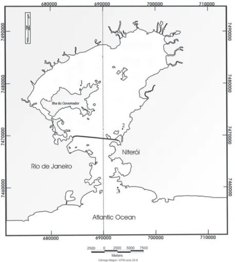
In this study, sediments at three different loca-tions were sampled (Fig. 1). Site 1 is located in the northwest of the bay at Ilha do Governador
(22°46.589' S, 43°11.424' W). This site has fine sand,
the sediment dry weight, and an input of 458 g.C.m-2. year-1 of organic matter (Carreira et al., 2002).
Site 2 is located in the east of the bay at
São Gonçalo’s eutrophic river (22°51.064' S, 43°06.697' W). This site has medium silt and
large space heterogeneity in sediment properties. The organic carbon measured was approximately 1.6% in this sediment (Carreira et al. 2002).
Site 3 is located at the entrance of the bay
(22°59'01.1"S, 43° 04'59.8” W). This site has
moderately well-sorted medium sand and a very low percentage of total organic carbon, ranging from 0.82-3.1% (Carreira et al. 2002).
MEDIUM AND SELECTION OF PROTISTS
The sediment samples were prepared according to Dragesco and Dragesco-Kernéis (1986) 3-4 hours
after sampling. When free-living protists excysted,
they were stored at 30°C for the maintenance of
living bacteria and the short microbial loop (Fenchel 1987, Krepsky et al. 2007). All protist specimens were selected using glass micropipettes after six months of incubation. The protists were maintained in the laboratory in liquid medium containing coarse powdered rice at room temperature (Dragesco and
Dragesco-Kernéis 1986). The identification of the
protists was performed with a light microscope (Dragesco and Dragesco-Kernéis 1986), a BX 41 from Olympus, using the Protargol technique (Silva-Neto 2000).
MEDIUM AND SELECTION OF BACTERIAL CONSORTIA
748 JOSÉ A.P. BITENCOURT, FREDERICO SOBRINHO SILVA, INÁCIO D. DA SILVA-NETO and MIRIAN A.C. CRAPEZ
(Forsskål) (Agardh 1873), Laurencia obtusa (Huds) (Lamouroux 1813), and Dictyota menstrualis (Hoyt) (Shnetter et al. 1987), according to Silva et al. (2005). They were maintained according to Krepsky et al. (2007), and oil was the source of both carbon and energy (Silva et al. 2005). Throughout the manuscript, the bacterial consortia are termed BCr (C. racemosa), BLo (L. obtusa) and BDm (D. menstrualis).
BIOASSAY
The organic carbon or biomass (µgC.cm-3) of the protists and the bacteria BCr, BLo and BDm were
quan-tified according to Gomes et al. (2007), Mauclaire et
al. (2003), Carlucci et al. (1986) and Kepner and Pratt (1994), respectively. The bioassay was performed at 0, 24, 48, 72 and 96 h at room temperature (Krepsky et al. 2007) in absence of oil. In the bioassay including oil (treatment group), 1 mL of oil was added (Light Arabian oil, from PETROBRAS S.A) and the sample was incubated with shaking for 1 minute (Krepsky et
al. 2007). The control groups were analyzed protists
with or without consortia. The bacterial proportion,
including the capsule that forms the biofilm, and the bacterial cell size were measured with an Olympus
microscope, model BX 41, using the phase contrast,
PH3 filter (1000 X) (Madigan et al. 2004).
STATISTICAL ANALYSES
The data were analyzed with Statisoft Statistica 7, using ANOVA and MANOVA tests to analyze the carbon and biofilm results, respectively. Organic
carbon data were transformed with arc-sen. Data
were considered significant at p≤0.05.
RESULTS
The sediment samples from Guanabara Bay did not contain vegetative protists. They appeared after 3 weeks of laboratory incubation. In the northwest of the bay, at Ilha do Governador (site 1), Cryptomonadida (Senn 1900) was isolated (Kugrens et al. 2000).
In the east of the bay, at São Gonçalo`s eutrophic
river, (site 2), Cryptomonadida and Scuticociliatida (Small 1967) were isolated (Lynn and Small 2000). At the entrance of the bay, Cryptomonadida and two different ciliate species, Euplotes sp.1 and Euplotes sp.2, were isolated (Lynn and Small 2000).
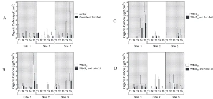
At site 1, Cryptomonadida produced 1.5 µgC. cm-3(48 h) of biomass in the bioassay control, but in the presence of oil, it only produced 0.4 µgC.cm-3(24 h). At site 2, Cryptomonadida and Scuticociliatida produced 0.7 µgC.cm-3 (72 h) of biomass in the bioassay control, but in the presence of oil, they produced 0.4 µgC.cm-3 (96 h). At site 3, Cryptomonadida, Euplotes sp.1 and Euplotes sp.2 produced 6.0 µgC.cm-3 of biomass in the bioassay control, but this value was reduced to 0.7 µgC.cm-3 in the presence of oil. The highest biomass was generally observed at the end of the bioassay (72-96 h) (Fig. 2a).
On the surface of C. racemosa (BCr), L. obtusa (BLo) and D. menstrualis (BDm) bacterial consortia, hydrocarbon-degrading bacteria was observed. The bacterial organic carbon values for the bioassay control were 8.4, 8.4 and 8.6 µgC.cm-3 produced by BCr in 96 h, and by BLo and BDm in 72 h, respectively. In the presence of oil, the organic carbon produced by BCr, BLo and BDm was 17.0 µgC.cm-3in 24 h, 7.1 µgC.cm-3in 72 h and 10.9 µgC.cm-3 in 24 h, respectively (Fig. 3a).
At site 1, Cryptomonadida in contact with the bacterial consortia showed enhanced biomass at the end of the bioassay. In the presence of BCr, BLo and BDm, Cryptomonadida produced 5.0, 6.0 and 0.2 µgC.cm-3 of biomass, respectively, but in the presence of oil, Cryptomonadida produced 2.0, 3.3 and 0.4 µgC.cm-3, respectively, at 96 h (Fig. 2b, c, d).
At site 2, Cryptomonadida and Scuticociliatida in the presence of both BCr and BLo produced 1.2 µgC.cm-3 at 72 and 48 h. In the presence of B
Dm, Cryptomonadida and Scuticociliatida produced 2.2 µgC.cm-3 of biomass at 72 h. In the presence of oil, their biomass was reduced to 0.0 µgC.cm-3 in the presence of BCr, and 0.37 µgC.cm-3 in the presence of BLo after 48 h and BDm at 72 h (Fig. 2b, c, d).
At site 2, the bacterial organic carbon produced by BCr was 53.53 µgC.cm-3 of biomass at 72 h, and the carbon produced by BLo and BDm was 29.5 and 43.0 µgC.cm-3 of biomass after 48 h, respectively. However, in the presence of oil, BCr, BLo and BDm produced 10.57, 11.47 and 3.05 µgC.cm-3 at 72 h, respectively (Fig. 3b, c, d).
Cryptomonadida, Euplotes sp.1 and Euplotes sp.2 at site 3 in the presence of BCr or BLo produced a protist organic biomass of 1.9 and 1.1 µgC.cm-3 of biomass at 72 h, respectively. In the presence of BDm, Cryptomonadida, Euplotes sp.1 and Euplotes sp.2 produced 1.5 µgC.cm-3 of biomass at 24 h. In the presence of oil, they produced 1.9 and 0.4 µgC.cm-3 of biomass in the presence of
BCr or BLo at 72 h, respectively, and 0.4 µgC.cm-3 after 48 h with BDm (Fig. 2b, c, d).
At site 3, the organic carbon produced by BCr, BLo and BDm was 11.35, 21.34 and 13.90 µgC.cm-3 of biomass at 96, 72 and 48 h, but in the presence of oil, BCr, BLo and BDm produced 10.78, 18.21 and 10.64 µgC.cm-3 at 96, 48 and 72 h, respectively (Fig. 3b, c, d).
At the end of the bioassay, the size of the BCr, BLo and BDm biofilm was 1.5 µm (48 h), 1.5 µm (72 h) and 1.5 µm (72 h), respectively; however, in the
presence of oil, the capsule size was 1.7 µm (96 h),
1.6 µm (24 h) and 1.8 µm (24 h), respectively. The BCr, BLo and BDm cell size was 2.5 µm (96 h), 2.0 µm (72 h) and 2.0 µm (48 h), but in the presence
of oil, the cell size was 2.3 µm (72 h), 2.2 µm (72 h)
and 2.3 µm (96 h), respectively (Fig. 4a, b, c).
Statistical analyses showed significant
diffe-rences between the biomass of protists and bacteria
(ANOVA, p≤0.05), but their biomasses were
signifi-cantly linked at 48 and 72 h of the bioassay (Tukey test, p<0.05). The organic carbon produced by protists
was not significantly affected in the bioassays by the
presence of oil (Tukey test, p>0.05). Figure 2 - Biomass from protists sampled from Guanabara Bay under influence of Bacterial consortia
750 JOSÉ A.P. BITENCOURT, FREDERICO SOBRINHO SILVA, INÁCIO D. DA SILVA-NETO and MIRIAN A.C. CRAPEZ
Figure 3 - Biomass from Bacterial consortia sampled from seaweed under influence of protists sampled
from Guanabara Bay and oil. A-Control; B-With BCr; C-With BDm; D-With BLo.
Figure 4 - Bacterial proportions measured during oil degradation. A-Total size; B-Bacterial
capsula size; C-Bacterial cell size.
The quantity of organic carbon produced by the bacteria BCr, BLo and BDm was significantly different. The bacterial carbon from BCr and BDm
showed significant differences in the bioassay
in the presence of protists and oil (Tukey test,
p<0.05). Oil also significantly affected the
DISCUSSION
Free-living protists are very common in bodies of water (Fenchel 1987, Sigee 2005). Scuticociliatida, Cryptomonadida and Hipotrichea are very
common in eutrophized bodies of water, such as the Guanabara Bay (Slàdeček 1973, Foissner et al.
1995, Paiva and Silva-Neto 2004).
In the bioassay controls, protists showed a reduced ability to live in the presence of oil, and
their associated bacteria did not utilize oil as a
source of carbon and energy (Zarda et al. 1998, Mauclaire et al. 2003).
The hydrocarbon-degrading bacteria C. racemosa (BCr), L. obtusa (BLo) and D. menstrualis (BDm) emul-sify hydrocarbon-water mixtures, which enable them
to grow on the oil droplets (Crapez 2002, Krepsky et al. 2007, Rahman et al. 2003). These emulsification
properties have also been demonstrated to enhance hydrocarbon degradation in the environment, making them potential tools for oil spill pollution control (Banat 1995, Krepsky et al. 2007).
The hydrocarbon-degrading bacteria are cosmopolitan, and seaweed offers a suitable place for bacterial establishment (Atlas 1995, Armstrong et al. 2000). Seaweeds offer a large surface for the settlement of bacteria, oxygen and dissolved organic matter (Armstrong et al. 2000, Sigee 2005). On the other hand, the bacteria consume the organic matter, releasing carbon, nitrogen, and phosphorus and provide defense against pollutants, such as oil and aromatic compounds, and epiphytic organisms (Bell et al. 1974, Atlas 1995, Sigee 2005).
Protists release some nitrogen and/or phos-phorus-containing compounds when preying on
inactive bacterial cells (Hahn and Höfle 2001, Kujawinski et al. 2002, Matz and Jürgens 2001). Protist
feeding rates can be affected by characteristics of the prey particles, such as electrostatic charge
(Hammer et al. 1999), cell shape (Kolaczyk and
Wiackowski 1997) and exopolymer secretions (Liu and Buskey 1999).
During the bioassays, the hydrocarbon-degrading bacteria BCr, BLo and BDm showed
significant size differences in the bioassays
in the presence and absence of oil. These
differences manifested in the increasing size
of the biofilm. The biofilms contain bacteria depositing exopolysaccharides (EPS), which exhibit amphiphilic properties allowing these macromolecules to interface with hydrophobic substrates, such as hydrocarbons. These macro-molecules effectively increase the solubility of aromatic hydrocarbons and enhance their biodegradation by the microbial community
(Pacwa-Płociniczak et al. 2011, Gutierrez et al. 2013).
In the presence of oil, the production of a
biofilm with surfactant activity (Krespsky et al. 2007) and an increase in cell size resulted in an
increase in the biomass of the bacteria BCr, BLo,BDm and a decrease in the biomass of Scuticociliatida, Cryptomonadida (site 2), Euplotes sp. and Cryptomonadida, (site 3), suggesting a reduction
in grazing and a decrease in the transfer of
carbon to higher trophic levels. Site 2 had high concentrations of coprostanol due to the input of sewage from the city of São Gonçalo (Carreira et al. 2001), which selects the microorganisms capable of living in environments with high concentrations of organic matter.
In environments polluted by oil, such as site 1, hydrocarbon-degrading bacteria and their predators, such as Cryptomonadida, are selected (Crapez et al. 2001). The growth of the hydrocarbon-degrading bacteria BCr, BLo and BDm was followed by the
growth of grazing Cryptomonadida. Site 1 is the
nearest to the REDUC oil refinery, and sediment samples may be classified as moderately to highly contaminated (250 to 500 μg.kg-1 total PAH) (Silva et al. 2007).
CONCLUSIONS
752 JOSÉ A.P. BITENCOURT, FREDERICO SOBRINHO SILVA, INÁCIO D. DA SILVA-NETO and MIRIAN A.C. CRAPEZ
Oil pollution selects the hydrocarbon-degrading bacteria, and this resulted in an increase in the biomass of Cryptomonadida at site 1, sugges-ting a transfer of carbon to higher trophic levels.
Site 2 was polluted by sewage, which prevented Cryptomonadida survival in the presence of oil, suggesting a bottom-up effect.
Although hydrocarbon-degrading bacteria
showed an increase in cell size and produced a biofilm with surfactant activity, the biomass of
Scuticociliatida, Cryptomonadida (site 2), Euplotes sp. and Cryptomonadida (site 3) decreased. These
sites showed no significant contribution of oil to
select hydrocarbon-degrading microorganisms or oil-resistant protists.
ACKNOWLEDGMENTS
The authors wish to thank Antônio de Paula Filho from the IQ institute (UFRJ) and Camila M. da Silva, who mediated the transfer of Light Arabian oil from PETROBRAS to UFF. This study was supported
by grants from the Brazilian Petroleum Agency
(ANP) and its scholarship program (PRH-11), grants from Fundação Carlos Chagas Filho de Amparo à Pesquisa do Estado do Rio de Janeiro (FAPERJ) and
Conselho Nacional de Desenvolvimento Científico e
Tecnológico (CNPq).
RESUMO
Pouco se sabe sobre o papel dos protistas e as interações bacterianas durante a biodegradação de hidrocarbonetos. Este trabalho se concentrou no efeito do óleo sobre protistas de três localidades diferentes na Baía de Guanabara e bactérias de Caulerpa racemosa (BCr),
Dictyota menstrualis (BDm) e Laurencia obtusa (BLo)
durante 96 h de bioensaio. Cryptomonadida (locais 1 , 2 e 3), Scuticociliatida (local 2) e Euplotes sp.1 e Euplotes sp.2 (local 3) apareceram após incubação. As biomassas mais elevadas observadas nos controles foram como se
segue: protista local 3 (6,0 μgC.cm-3
, 96 h ) comparado
com o local 3 com óleo (0,7 μgC.cm-3
, 96 h); para as
bactérias, 8,6 μgC.cm-3
(BDm, 72 h ) e 17,0 μgC.cm -3
(BCr
com óleo, 24 h). Após o tratamento, as maiores biomassas foram como se seguem: protistas no local 1 e BLo, 6,0
μgC.cm-3
(96 h), em comparação com o local 1 e BLo
com óleo, 3,31 μgC.cm-3
(96 h), a biomassa bacteriana
foi de 43,1 μgC.cm-3
no local 2 e BDm (96 h). No local
3 e BLo com óleo, a biomassa foi 18,21 μgC.cm -3
(48 h).
As maiores proporções de biofilme foram observadas de
1,7 µm BCr (96 h) a 1,8 µm BLo com óleo (24 h). BCr, BLo
e BDm aumentaram o tamanho do biofilme e reduziram a
capacidade dos protistas predarem.
Palavras-chave: consórcio bacteriano, biomassa, protista de vida livre, Baía de Guanabara, alça microbiana, petróleo.
REFERENCES
ARMSTRONG E, ROGERSON A AND LEFTLEY JW. 2000. The abundance of heterotrophic protists associated with intertidal seaweeds. Estuar Coast Shelf Sci 50: 415-424. ATLAS RM. 1995. Petroleum biodegradation and oil spill
bioremediation. Mar Pollut Bull 31: 178-182.
BANAT IM. 1995. Biosurfactants production and possible uses in microbial enhanced oil recovery and oil pollution remediation: A Review Biores Tech 51: 1-12.
BELL W, LANG JM AND MICHELL R. 1974. Selective stimulation of marine bacteria by algal extracellular products. Limnol Oceanogr 19: 833-839.
BUCK KR, BARRY JP AND SIMPSON AGB. 2000. Monterey
Bay cold seep biota: Euglenozoa with chemoautotrophic
bacterial epibionts. Eur J Protistol 36: 117-126.
CARLUCCI AF, CRAVEN DB, ROBERTSON KJ AND WILLIAMS PM. 1986. Surface-film microbial populations: diel amino acid metabolism, carbon utilization, and growth rates.
Mar Biol 92: 289-297.
CARON DA AND GOLDMAN JC. 1990. Protozoan nutrient regeneration. In: Capriulo GM (Ed), Ecology of Marine
Protozoa, New York: Oxford University Press, p. 283-306.
CARREIRA R, WAGENER ALR, FILEMAN T AND READMAN JW. 2001. Distribuição de coprostanol (5β(H)-colestan-3β-OL) em sedimentos superficiais da Baía de Guanabara: indicador da poluição recente por esgotos domésticos. Quim Nova 24: 37-42.
CARREIRA RS, WAGENER ALR, READMAN JW, FILEMAN TW, MACKO SA AND VEIGA A. 2002. Change in the
sedimentary organic carbon pool of a fertilized tropical estuary, Guanabara Bay, Brazil: an elemental, isotopic and
molecular marker approach. Mar Chem 79: 207-227. CHRISTAKI U, VAN WAMBEKE F AND DOLAN JR. 1999.
Nanoflagellates (mixotrophs, heterotrophs, and
CRAPEZ MAC. 2001. Efeitos de hidrocarbonetos de petróleo na biota marinha. In: MORAES R, CRAPEZ MAC, PFEIFFER W, FARINA M, BAINY A AND TEIXEIRA V (Eds), Efeitos dos Poluentes em Organismos Marinhos, São Paulo: Arte e Ciência-Vilipress, p. 255-270.
CRAPEZ MAC. 2002. Bactérias marinhas. In: Pereira RC and Soares-Gomes A (Eds), Biologia Marinha, Rio de Janeiro: Interciência, p. 81-101.
DALBY AP, KORMAS KAR, CHRISTAKI U AND KARAYANNI H. 2008. Cosmopolitan heterotrophic microeukaryotes
are active bacterial grazers in experimental oil-polluted
systems. Environ Microbiol 10: 47-56.
DOLAN JR. 1997. Phosphorus and ammonia excretion by planktonic protists. Mar Geol 139: 109-122.
DRAGESCO J AND DRAGESCO-KERNÉIS A. 1986. Ciliés libres de
l`Afrique intertropicale. Introduction à connaissance et à l`etude des ciliés. Faune Tropicale 26: 1-559.
EDWARDS KJ, BACH W AND MCCOLLOM TM. 2005. Geomicrobiology in oceanography: microbe-mineral
interactions at and below the seafloor. Trends Microbiol
13: 449-455.
FEEMA 1990. Projeto de recuperação gradual da Baía de Guanabara, v. 1. Rio de Janeiro: Fundação Estadual de Engenharia do Meio Ambiente, 203 p.
FENCHEL T. 1987. Ecology of Protozoa: The Biology of Free-living Phagotrophic Protists. Berlin: Springer, 197 p. FOISSNER W, BERGER H, BLATTERER H AND KOHMANN F.
1995. In: Taxonomische und ökologische revision der ciliaten des saprobiensystems - Band IV: Gymnostomates, Loxodes. Munich: Informationsberichte des Bayer
Landesamtes für Wasserwirtschaft, 540 p.
FONTANA LF, SILVA FS, KREPSKY N, BARCELOS MA AND CRAPEZ MAC. 2006. Natural attenuation of aromatic hydrocarbon from sandier sediement in Boa Viagem,
Guanabra Bay, RJ, Brazil. Geochimica Brasiliensis 20:
78-86, 2006.
GOMES EAT, SANTOS VSD, TENENBAUM DR AND VILLAC MC. 2007. Protozooplankton characterization of two contrasting sites in a tropical coastal ecosystem (Guanabara Bay, RJ).
Braz J Oceanogr 55: 29-38.
GUERRA LV, SAVERGNINI F, SILVA FS, BERNARDES MC AND CRAPEZ MAC. 2011. Biochemical and microbiological tools for the evaluation of environmental quality of a
coastal lagoon system in Southern Brazil. Braz J Biol 71:
461-468.
GUTIERREZ T, BERRY D, YANG T, MISHAMANDANI S, MCKAY L, TESKE A AND AITKEN MD. 2013. Role of Bacterial Exopolysaccharides (EPS) in the Fate of the Oil Released
during the Deepwater Horizon Oil Spill. PLoS One 8(6):
e67717. doi:10.1371/journal.pone.0067717
HAHN MW AND HÖFLE MG. 2001.Grazing of protozoa and it’s eject on populations of aquatic bacteria. FEMS Microbiol Ecol 35: 113-121.
HAMMER A, GRUTTNER C AND SCHUMANN R. 1999. The effect of electrostatic charge of food particles on capture
efficiency by Oxyrrhis marina Dujardin (dinoflagellate).
Protist 150: 375-382.
JICA 1994. The study on recuperation of the Guanabara Bay ecosystem, vol 8. Japan International Cooperation Agency, Kokusai Kogyo Co., Ltd., Tokyo.
JUMARS PA, PENRY DL, BAROSS JA, PERRY MJ AND FROST BW. 1989. Closing the microbial loop: dissolved carbon pathway to heterotrophic bacteria from incomplete ingestion, digestion and absorption in animals. Deep-Sea Res 36: 483-495.
KJERFVE B, RIBEIRO CA, DIAS GTM, FILIPPO A AND QUARESMA VS. 1997. Oceanographic characteristics of an impacted
coastal bay: Baia de Guanabara, Rio de Janeiro, Brazil.
Cont Shelf Res 17: 1609-1643.
KEPNER RL AND PRATT JR. 1994. Use of fluorochromes for direct enumeration of total bacteria in environmental samples: past and present. Microbiol Rev 58: 603-615. KOLACZYK A AND WIACKOWSKI K. 1997. Induced defense
in the ciliate Euplotes octocarinatus is reduced when alternative prey are available to the predator. Acta
Protozool 36: 57-61.
KREPSKY N, FONTANA LF, SILVA FS AND CRAPEZ MAC. 2007. Alternative methodology for biossurfactant production.
Braz J Biol 67: 117-124.
KUGRENS P, LEE R AND HILL DR. 2000. Flagellated. In: LEE JJ, LEEDALE GF AND BRADBURY P (Eds), The
illustrated guide to the protozoa, 2nd ed., Lawrence:
Society of Protozoologists, p. 1111-1250.
KUJAWINSKI EB, FARRINGTON JW AND MOFFETT JW. 2002. Evidence for grazing-mediated production of dissolved
surface-active material by marine protists. Mar Chem 77: 133-144.
LAYBOURN-PARRY J, MELL EM AND ROBERTS EC. 2000. Protozoan growth rates in Antarctic lakes. Polar Biol 23:
445-451.
LIU H AND BUSKEY EJ. 1999. The exopolymer secretions (EPS) layer surrounding Aureoumbra lagunensis cells
affects growth, grazing and behavior of protozoa. Limnol
Oceanogr 45: 1187-1191.
LYNN DH AND SMALL E. 2000. Ciliophora. In: LEE JJ, LEEDALE GF AND BRADBURY P (Eds), The illustrated
guide to the protozoa, 2nd ed., Lawrence: Society of
Protozoologists, p 371-676.
MADIGAN TM, MARTINKO JM AND PARKER J. 2004. Hábitat microbianos, ciclos de nutrientes e interacciones con plantas y animales. In: Microbiología de los micro-organimos, 10th ed., Madri: Prentice Hall, p. 624-687. MATZ C AND JÜRGENS K. 2001. Effects of hydrophobic and
electrostatic cell surface properties of bacteria on feeding
rates of heterotrophic nanoflagellates. Appl Environ
Microbiol 67: 814-820.
MAUCLAIRE L, PELZA O, THULLNERA M, ABRAHAMB W AND ZEYER J. 2003. Assimilation of toluene carbon along a bacteria-protist food chain determined by 13C-enrichment
of biomarker fatty acids. J Microbiol Methods 55: 635-549. NAGATA T AND KIRCHMAN DL. 1992a. Release of dissolved
organic matter by heterotrophic protozoa: implications for
754 JOSÉ A.P. BITENCOURT, FREDERICO SOBRINHO SILVA, INÁCIO D. DA SILVA-NETO and MIRIAN A.C. CRAPEZ
NAGATA T AND KIRCHMAN DL. 1992b. Release of macro-molecular organic complexes by heterotrophic marine
flagellates. Mar Ecol Prog Ser 83: 233-240.
PACWA-PŁOCINICZAK M, PLAZA GA, PIOTROWSKA-SEGET Z AND CAMEOTRA SS. 2011. Environmental applications of biosurfactants: Recent Advances Int J Mol Sci 12: 633-654. PAIVA TS AND SILVA-NETO ID. 2004. Ciliate protists from
Cabiúnas lagoon (Restinga de Jurubatiba, Macaé, Rio de Janeiro) with emphasis on water quality indicator species and description of Oxytrichamarcili sp. Braz J Biol 64: 465-478.
PEDROS-ALIO C, CALDERON-PAZ I, MACLEAN MH, MEDINA G, MARRASE C, GASOL JM AND GUIXA-BOIXEREU N. 2000. The microbial food web along salinity gradients. FEMS Microbiol Ecol 32: 143-155.
RAHMAN KSM, RAHMAN TJ, KOURKOUTAS Y, PETSAS I, MARCHANT R AND BANAT IM. 2003. Enhanced bioremediation of n-alkane in petroleum sludge using bacterial consortium amended with rhamnolipid and micronutrients. Bioresour Technol 90: 159-168.
RON EZ AND ROSENBERG E. 2002. Biosurfactants and oil bioremediation. Curr Opin Biotechnol13: 249-252. SCOTT FJ, DAVIDSON AT AND MARCHANT HJ. 2001. Grazing
by the Antarctic sea ice ciliate Pseudocohnilembus. Polar Biol 24:127-131.
SHERR EB AND SHERR BF. 1994. Bacterivory and herbivory: key roles of phagotrophic protists in pelagic food webs. Microb Ecol 28: 223-235.
SIGEE DC. 2005. Freshwater microbiology: biodiversity and dynamic interactions of microorganisms in the aquatic environment. West Sussex: J Wiley & Sons, 537 p. SIKKEMA J, BONT JA AND POOLMAN B. 1995. Mechanisms of
membrane toxicity of hydrocarbons. Microbiol Rev 59: 201-222.
SILVA FS, KREPSKY N, TEIXEIRA VL AND CRAPEZ MAC. 2005. Estímulo da produção de biossurfactante por extrato da alga vermelha Digenea simplex (WULFEN) C. AGARDH em comunidades bacterianas da praia de Boa Viagem (RJ). In: Série Livros 10 do Museu Nacional do Rio de Janeiro, Rio de Janeiro: Museu Nacional do Rio de Janeiro, p. 469-483.
SILVA FS, SANTOS ES, LAUT LLM, SANCHEZ-NUÑES ML, FONSECA EM, BAPTISTA-NETO JA, MENDONÇA-FILHO JG AND CRAPEZ MAC. 2010. Geomicrobiology and Biochemical Composition of Two Sediment Cores from
Jurujuba Sound - Guanabara Bay – SE Brazil. Anu Inst
Geocienc 33:73-84.
SILVA-NETO ID. 2000. Improvement of silver impregnation technique (Protargol) to obtain morphological features of
protists ciliates, flagellates and opalinates.Rev Bras Biol 60: 451-459.
SILVA TF, AZEVEDO DA AND AQUINO NETO FR. 2007. Distribution of polycyclic aromatic hydrocarbons in surface sediments and waters from Guanabara Bay, Rio
de Janeiro, Brazil. J Braz Chem Soc 18: 628-637.
SLÀDEČEK V. 1973. System of water quality from the biological point of view. Arch Hydrobiol Beih Ergebn Limno 7: 1-218.
WILKINSON S, NICKLIN S AND FAULL JL. 2002. Biodegradation of fuel oils and lubricants: Soil and water bioremediation options. In: Singh VP and Stapleton RD (Eds), Progress in Industrial Microbiology 36: 69-100.