UNIVERSIDADE DA BEIRA INTERIOR
Ciências
Novel therapeutic approaches for
skin regeneration
Sofia Mendes Saraiva
Dissertação para obtenção do Grau de Mestre em
Bioquímica
(2º ciclo de estudos)
Orientador: Professor Doutor Ilídio Joaquim Sobreira Correia
Coorientadores: Professora Doutora Paula Isabel Teixeira Gonçalves Borges
Mestre Maximiano José Prata Ribeiro
UNIVERSIDADE DA BEIRA INTERIOR
Ciências
Novas abordagens terapêuticas para a
regeneração da pele
Sofia Mendes Saraiva
Dissertação para obtenção do Grau de Mestre em
Bioquímica
(2º ciclo de estudos)
Orientador: Professor Doutor Ilídio Joaquim Sobreira Correia
Coorientadores: Professora Doutora Paula Isabel Teixeira Gonçalves Borges
Mestre Maximiano José Prata Ribeiro
iv
vi
Acknowledgments
First, I would like to thank to Professor Ilídio Correia for the possibility of developing my master thesis in his group, the unconditional support, guidance and for the patience. During this master thesis Professor Ilídio helped me to grow as an investigator but also as a person. As he says, we have to work hard every single day to achieve our goals. Thank you for all the understanding during all the difficult times.
I thank to Maximiano Ribeiro, my co-supervisor, for his support, guidance and for being a friend. All the advices were very important to overcome the difficulties.
I would also like to thank Sónia Miguel not only for her help in acquiring scanning electron microscopy images of the hydrogels, but also for all the guidance since the beginning of this work, endless support and for being such a comprehensive partner and friend.
Moreover, I would like to thank to my group colleagues for their support and knowledge sharing, at any time. To Ricardo Fradique, Mariana Vallejo and specially Elisabete Costa, thank you for the support, the funny moments and the company during the endless days and nights of work. To Vitor Gaspar, thank you for all the help and patience.
To my closest friends that accompanied me during the academic life. Tânia Pascoal, João Salvado and Sara Maruje, thank you for all the help, support and encouragement during all these years and specially this one. A huge and special thank to Raquel Tavares, whose friendship remained during all these years and no matter what she always there.
Finally, and foremost, a very special thanks to my parents José and Rosa Saraiva and my brother Francisco Saraiva for their unlimited and unconditional support, patience, love and affection. Thank you for never stop believing in me, for making me a strong and better person every day. Nothing would matter without them on my side. To them I dedicate this thesis.
viii
Abstract
The integrity of skin, as the largest organ of the human body, must be preserved in order to play its role as a protective barrier, in the maintenance of fluid homeostasis and temperature regulation. Skin lesions can occur due to different causes, being burns those responsible for extensive skin loss, infection, electrolyte imbalances and respiratory failure. In order to decrease the effects of skin damages, new skin substitutes have been developed to accelerate the healing process and thus restore the native structure of skin. Among the existent materials, hydrogels own the most desirable characteristics of an “ideal dressing”, such as biocompatibility and biodegradability. Currently, new photocrosslinkable hydrogels have been developed for tissue engineering purposes. Taking into account the intrinsic properties of hydrogels, the work plan developed during this master thesis allowed the production of photocrosslinkable hydrogels, composed of chitosan and gelatine that present some of the properties required for wound regeneration. Initially, methacrylate groups were added to the chitosan and gelatine primary amine groups, leading to the synthesis of methacrylamide chitosan (MAC) and methacrylamide gelatine (MAG). The chemical modification of the polymers was confirmed by proton nuclear magnetic resonance (1H NMR).
Then, MAC and MAG hydrogels were produced using ultraviolet (UV) light in the presence of a photoinitiator (Irgacue 2959). The hydrogels were subsequently characterized by scanning electron microscopy and Fourier transform infrared spectroscopy. Porosity and swelling properties were also analyzed and revealed that the hydrogels with a higher content of chitosan had higher porosity and swelling capacity. The cytotoxic profile of the hydrogels was evaluated through an MTS assay, using human fibroblast cells. Cell adhesion on the surface of the hydrogels was visualized through scanning electron microscopy. The results obtained demonstrated that the hydrogels developed herein possess suitable properties for being used as wound dressings. Therefore, in the future in vivo studies will be performed to evaluate the histocompatibility of the hydrogels and their capacity to improve the wound healing process. In addition, growth factors and antimicrobial agents can be incorporated in the hydrogels, in order to improve wound repair and prevent bacterial infections, respectively.
ix
Keywords
xi
Resumo
A pele é o maior órgão do corpo humano e devido ao seu papel protetor, regulador da temperatura do corpo e manutenção da homeostase de líquidos, a sua integridade estrutural deve ser preservada de forma a desempenhar corretamente as suas funções. As lesões na pele podem ser causadas por diferentes fatores, sendo as queimaduras a causa mais comum. A perda da integridade estrutural do sistema tegumentar pode provocar a desidratação do doente, infeções, distúrbios eletrolíticos e falhas respiratórias. Com o intuito de ultrapassar os problemas associados às lesões cutâneas, têm sido desenvolvidos novos substitutos de pele que permitam acelerar o processo de cicatrização e assim restaurar a estrutura nativa da pele. De entre os materiais existentes, os hidrogéis são os que possuem as características mais adequadas para serem usados como substitutos de pele. Na atualidade, novos hidrogéis fotopolimerizáveis têm sido desenvolvidos para serem usados na área da engenharia de tecidos. Tendo em conta as propriedades intrínsecas deste tipo de hidrogéis, no presente plano de trabalhos foram desenvolvidos hidrogéis fotopolimerizáveis constituídos por quitosano e gelatina, com o intuito de serem usados na regeneração de feridas. Inicialmente, grupos metacrilato foram incorporados nas aminas primárias do quitosano e da gelatina, permitindo a síntese do quitosano metacrilamida (MAC) e da gelatina metacrilamida (MAG). A modificação dos polímeros foi confirmada através de ressonância magnética nuclear (1H NMR).
Os hidrogéis MAC e MAG foram produzidos na presença de um fotoiniciador (Irgacure 2959), usando luz ultravioleta (UV). Os hidrogéis foram caracterizados por microscopia eletrónica de varrimento e espetroscopia de infravermelho com transformada de Fourier. A porosidade e capacidade dos hidrogéis absorverem água também foram analisadas, demonstrando que o hidrogel com maior teor de quitosano apresentava maior porosidade e capacidade de absorção de água. O perfil citotóxico dos hidrogéis foi avaliado através de ensaios MTS, usando fibroblastos humanos como células modelo. A adesão celular na superfície dos hidrogéis foi visualizada através de microscopia eletrónica de varrimento. Os resultados obtidos demonstraram que os hidrogéis desenvolvidos possuem propriedades adequadas para serem usados na regeneração de feridas. No futuro, serão efetuados testes in vivo para avaliar a histocompatibilidade dos hidrogéis e a sua capacidade para promover o processo de regeneração de feridas. Fatores de crescimento e agentes antimicrobianos podem ser incorporados nos hidrogéis, por forma a melhorar o processo de regeneração e prevenir infeções bacterianas, respetivamente.
xii
Palavras-chave
xiv
Resumo alargado
A pele é o maior órgão do corpo humano, apresentando uma área aproximada de 2 m2 nos
adultos. Devido ao seu papel protetor, regulador da temperatura do corpo e manutenção da homeostase de líquidos, a sua integridade estrutural deve ser preservada de forma a desempenhar corretamente as suas funções. As lesões da pele são o resultado da disrupção da sua estrutura anatómica e podem ser causadas por diferentes fatores, sendo as queimaduras a causa mais comum. A perda da integridade estrutural do sistema tegumentar pode provocar a desidratação do doente, infeções, distúrbios eletrolíticos e falhas respiratórias.
Consoante o tipo e duração do processo de cicatrização das feridas, as lesões da pele podem ser classificadas como agudas ou crónicas. A cicatrização de feridas é realizada através de uma cascata de eventos complexos e dinâmicos, que incluem a coagulação, inflamação, síntese e deposição de matriz extracelular, angiogénese, fibroplasia, epitelização, contração e remodelação. Após a ocorrência de uma lesão a pele deve ser revestida, a fim de diminuir a dor e o risco de infeção e promover o restabelecimento da sua integridade, minimizando assim a perda das suas funções. A terapêutica “standard” para o tratamento da perda de grandes quantidades de pele, por exemplo devido a queimaduras, continua a ser o uso de auto-, alo- e xeno-enxertos. Contudo esta abordagem terapêutica apresenta limitações como rejeição e transmissão de doenças. Para colmatar estas limitações, nas últimas décadas, vários substitutos de pele têm sido desenvolvidos de forma a acelerar o processo de cicatrização e restabelecer as funções da pele.
De acordo com a camada da pele que pretendem substituir, os substitutos de pele podem ser classificados como epidérmicos, dérmicos e dermo-epidérmicos. Os substitutos de pele podem ser constituídos à base de polímeros naturais e/ou sintéticos, sendo que alguns possuem ainda células. Contudo, nenhum dos substitutos atualmente disponíveis no mercado é capaz de reestabelecer totalmente as propriedades funcionais e anatómicas da pele.
De entre os materiais existentes, os hidrogéis são os que possuem as características que mais se enquadram nas propriedades de um “substituto ideal”, uma vez que são biocompatíveis e biodegradáveis, apresentam uma estrutura semelhante à da matriz extracelular e têm a capacidade de prevenir a desidratação da ferida e de remover exsudados. Além disso, alguns hidrogéis são particularmente interessantes devido à possibilidade de serem formados in situ e aplicados através de processos minimamente invasivos, como é o caso dos hidrogéis fotopolimerizáveis.
A aplicação de materiais fotopolimerizáveis tem sido extensivamente usada em medicina dentária devido à rápida reticulação dos materiais sob uma breve exposição à luz ultravioleta.
xv
Recentemente, os hidrogéis fotopolimerizáveis têm sido alvo de grande atenção na engenharia de tecidos devido à sua biocompatibilidade, rápida reticulação e aplicação in situ. A fotopolimerização permite ainda controlar a estrutura dos materiais, produzindo hidrogéis com as propriedades mecânicas desejadas.
O presente plano de trabalhos teve como objetivo o desenvolvimento de hidrogéis fotopolimerizáveis, constituídos por quitosano e gelatina, com o intuito de serem aplicados na regeneração de feridas. Estes polímeros naturais foram usados devido ao seu baixo custo, biocompatibilidade e biodegradabilidade, e pelo fato de não induzirem uma resposta imunológica. Inicialmente, grupos metacrilato foram adicionados às aminas primárias do quitosano e da gelatina, permitindo a síntese do quitosano metacrilamida (MAC) e da gelatina metacrilamida (MAG). A modificação dos polímeros foi confirmada e determinada através de ressonância magnética nuclear (1H NMR). Os hidrogéis MAC e MAG foram produzidos na
presença de um fotoiniciador (Irgacure 2959), usando luz ultravioleta (UV). O Irgacure 2959 foi usado como iniciador uma vez que é conhecido pela baixa toxicidade para as células. Os hidrogéis foram caracterizados por microscopia eletrónica de varrimento e espetroscopia de infravermelho com transformada de Fourier. A porosidade e capacidade dos hidrogéis absorverem água também foram analisadas, demonstrando que o hidrogel com maior teor de quitosano apresentava maior porosidade e capacidade de absorção de água. O perfil citotóxico dos hidrogéis foi avaliado in vitro através de ensaios MTS, usando fibroblastos humanos como células modelo. A adesão celular na superfície dos hidrogéis foi visualizada através de microscopia eletrónica de varrimento. Os resultados obtidos demonstraram que o hidrogel com mais quitosano apresenta uma estrutura mais porosa e com maior capacidade de absorção de água. Os hidrogéis revelaram ser biocompatíveis e capazes de promover a adesão e proliferação dos fibroblastos. Tais resultados sugerem que os hidrogéis fotopolimerizáveis desenvolvidos possuem propriedades adequadas para serem usados na regeneração de feridas. No futuro, serão realizados testes in vivo para avaliar a histocompatibilidade dos hidrogéis e a sua capacidade para promover o processo de regeneração de feridas. Fatores de crescimento e agentes antimicrobianos podem ser incorporados nos hidrogéis, por forma a melhorar o processo de regeneração e prevenir infeções bacterianas, respetivamente.
xvii
Index
Abstract ... viii Keywords ... ix Resumo ... xi Palavras-chave ... xiiResumo alargado ...xiv
Index ... xvii
Figure Index ... xxi
Table Index ... xxiii
List of abbreviations ... xxv
Chapter I - Introduction 1. Introduction ... 1
1.1. Skin ... 1
1.1.1. Functions and structure ... 1
1.1.2. Epidermis ... 2
1.1.3. Dermis ... 3
1.1.4. Hypodermis ... 4
1.2. Wounds ... 4
1.2.1 Types of wound healing ... 5
1.2.2. Wound healing mechanism ... 6
1.2.2.1. Haemostasis ... 6
xviii
1.2.2.3. Cell migration and proliferation ... 8
1.2.2.4. Remodelling ... 8
1.3. Tissue engineering and skin substitutes ... 9
1.4. Hydrogels ... 17
1.4.1. Photocrosslinkable Hydrogels ... 18
1.5. Chitosan... 20
1.6. Gelatine ... 21
1.7. Aims ... 23
Chapter II - Materials and Methods 2. Materials and Methods ... 25
2.1. Materials ... 25
2.2. Methods ... 25
2.2.1. Synthesis and characterization of deacetylated Chitosan ... 25
2.2.2. Synthesis of methacrylamide chitosan ... 26
2.2.3. Synthesis of methacrylamide gelatine ... 26
2.2.4. Nuclear Magnetic Resonance ... 26
2.2.5. Hydrogel production ... 27
2.2.6. Fourier transform infrared spectroscopy analysis... 27
2.2.7. Water uptake capacity of the hydrogels (Swelling) ... 28
2.2.8. Porosity evaluation ... 28
2.2.9. Characterization of the cytotoxic profile of the hydrogels ... 29
2.2.10. Proliferation of fibroblast cells in the presence of the hydrogels ... 29
2.2.11. Scanning electron microscopy analysis ... 30
xix Chapter III - Results and Discussion
3. Results and Discussion ... 32
3.1. Characterization of photocrosslinkable chitosan ... 32
3.2. Characterization of photocrosslinkable gelatine ... 35
3.3. Hydrogel production and morphology characterization ... 37
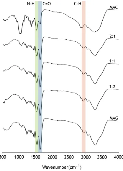
3.4. FTIR analysis of hydrogels ... 40
3.5. Swelling profile of the hydrogels ... 41
3.6. Porosity evaluation ... 42
3.7. Characterization of the cytotoxic profile of the hydrogels ... 43
3.8. Evaluation of cell proliferation in the presence of hydrogels ... 45
Chapter IV - Conclusion and Future Perspectives 4. Conclusion and Future Perspectives ... 50
Chapter V - References 5. References ... 52
xxi
Figure Index
Chapter I - Introduction
Figure 1: Representation of the structure of the skin ... 1
Figure 2: Representation of epidermis layers ... 3
Figure 3: Schematic (A) and illustrative (B) representations of the main phases of the wound healing process ... 7
Figure 4: Representation of a tissue engineering model that involves seeding cells within a porous scaffold. ... 10
Figure 5: Representation of chitosan structure ... 20
Figure 6: Representation of gelatine structure ... 21
Chapter III - Results and Discussion Figure 7: 1H NMR spectra of deacetilated chitosan (A) and methacrylamide chitosan (B) ... 34
Figure 8: 1H NMR spectra of unmodified gelatine (A) and methacrylamide gelatine (B) ... 36
Figure 9: Macroscopic image of the 2:1 (MAC:MAG) photocrosslinkable hydrogel produced in this study ... 38
Figure 10: SEM images of MAC:MAG (2:1, 1:1, 1:2) hydrogels ... 39
Figure 11: FTIR spectra of MAC and MAG polymers, and MAC:MAG (2:1, 1:1, 1:2) lyophilized hydrogels ... 40
Figure 12: Swelling profile of the produced MAC:MAG (2:1, 1:1, 1:2) hydrogels ... 42
Figure 13: Porosity of the MAC:MAG (2:1, 1:1, 1:2) hydrogels ... 43
Figure 14: Evaluation of cellular viability through an MTS assay, after 24 and 72 h. ... 45
Figure 15: Microscopic images of NHDF cells after 24 and 72 h in contact with MAC, MAG and MAC:MAG (2:1, 1:1, 1:2) hydrogels ... 47
xxiii
Table Index
Chapter I - Introduction
Table 1: Examples of epidermal skin substitutes currently available in the market ... 14 Table 2: Examples of dermal skin substitutes currently available in the market ... 15 Table 3: Examples of dermo-epidermal skin substitutes currently available in the market ... 16
Chapter II - Materials and Methods
Table 4: Volumes of MAC and MAG stock precursor solutions used for the preparation of
hydrogels precursor solutions ... 27
Chapter III- Results and Discussion
Table 5: Degree of deacetylation of the commercial MMW chitosan and the produced
xxv
List of abbreviations
1DUVS First derivative UV-spectroscopy 3D Three-dimensional
APS Ammonium persulfate
Az-CH-LA Azidobenzoic acid-modified lactose chitosan BSA Bovine serum albumin
DD Degree of deacetylation DM Degree of methacrylation
DMEM-F12 Dulbecco’s modified Eagle’s medium ECM Extracelular matrix
EDTA Ethylenediaminetetraacetic acid FBS Fetal bovine serum
FGF Fibroblast growth factor
FTIR Fourier transform infrared spectroscopy GAGs Glycosaminoglycans
GFs Growth factors HA Hyaluronic acid
HAM Hyaluronic acid membrane
Irgacure 2959 2-hydroxy-1-(4-(hydroxyethoxy)phenyl)-2-methyl-1-propanone K- Negative Control
K+ Positive Control
MA Methacrylic anhydride MAC Methacrylamide chitosan MAG Methacrylamide gelatine MMPs Metalloproteinases MMW Medium molecular weight
xxvi
MTS 3-(4,5-dimethylthiazol-2-yl)-5-(3-carboxymethoxyphenyl)-2-(4-sulfophenyl) -2H-tetrazolium
NHDF Normal Human Dermal Fibroblasts NIH-3T3 Mouse embryonic fibroblast NMR Nuclear Magnetic Ressonance PAM Polyacrylamide
PBS Phosphate-buffered saline solution PDGF Platelet-derived growth factor PEG Poly(ethylene glycol)
PEO Poly(ethylene oxide) PGA Poly(glycolic acid)
PHEMA Poly(2-hydroxyethyl methacrylate) PI Photoinitiator
PLA Poly(lactic acid) PVA Poly(vinyl alcohol) RT Room temperature SC Stratum corneum
SEM Scanning electron microscopy SMBS Sodium metabisulfite
TIMP Tissue inhibitors of metalloproteinases WHO World Health Organization
UV Ultraviolet
Chapter I
Introduction
Chapter I -Introduction
1
1. Introduction
1.1. Skin
1.1.1. Functions and structure
Skin is the largest organ of the human body, occupying almost 2 m2 of surface area and
accounting for 7% of body’s mass [1]. Its primary function is to serve as a protective barrier against external insults from the environment, such as ultraviolet (UV) light radiation, chemicals and pathogens. Skin is also involved in fluid homeostasis maintenance, thermoregulation, immune surveillance, sensory detection and self-healing [2-5]. Such demands require that the structure of this tissue is not compromised [6]. Moreover, since skin is the outward appearance of an individual to the world, it is also an indicator of well being and health state of the individual [7]. The injuries or diseases that lead to the loss of the integrity of large portions of skin may result in significant morbidity and mortality [4].
The skin has a complex structure (figure 1) and is organized in three distinct layers, known as epidermis, dermis and hypodermis [8]. Associated with the epidermis and dermis are various appendages such as nails, hair follicles, sweat and sebaceous glands, nerves, lymphatic and blood vessels, that play different functions [9, 10].
Chapter I -Introduction
2
1.1.2. Epidermis
The epidermis is the outermost layer of skin (figure 1) and contains no blood vessels. This stratified layer has evolved to provide a physical barrier that is fundamental for organisms’ survival, since it prevents the loss of fluids and infections [9, 12].
keratinocytes are the most abundant cell type in epidermis (90 – 95%). Their ability of undergoing self-renewal through a continuous process of proliferation, differentiation and cell death, enables the compartmentalization of epidermis into different strata (figure 2) that represent different stages of keratinocyte maturation [13]. Epidermal differentiation begins with the migration of keratinocytes from the stratum basale, which is the first layer of epidermis counting from the inside to the outside. As the basal keratinocytes proliferate, the “daughter” cells migrate towards and differentiate, forming the other four sub-layers of epidermis, known as [2, 9, 10, 12]:
stratum spinosum, a layer of spinous cells where the keratinization process begins;
stratum granulosum, a granular layer where differentiated keratinocytes become granular
cells;
stratum lucidum, a thin and translucent layer of dead cells (mostly seen in palms and
soles);
stratum corneum (SC), also known as cornified layer, represents the final result of
keratinocyte differentiation. SC is formed by completely differentiated dead keratinocytes (corneocytes) lacking nuclei and organelles, and interspersed with intercellular lipids (mainly ceramides and sphingolipids). This layer provides the most significant contribution to the permeation barrier property of human skin.
Besides keratinocytes, there are other cells that have significant roles in the epidermis. Melanocytes are present in the stratum basale and produce melanin, which contributes for skin pigmentation and protects the skin from harmful ultraviolet radiation. Specialized epidermal cells, known as Merkel cells, are also present in the stratum basale and are associated with nerve endings that are responsible for detecting superficial pressure. The Langerhan’s cells are dendritic cells present in the stratum spinosum that have an essential role in the skin immune defence system [9, 10, 14].
Chapter I -Introduction
3
Figure 2: Representation of epidermis layers (adapted from [11]).
The epidermis layer is connected to the underlying dermis via basement membrane, at the dermal-epidermal junction. The basement membrane, can be divided into lamina lucida, the layer closer to the epidermis, which is made up of laminin, integrins, entactins, and dystroglycans and lamina densa, a sheet-like structure composed mainly of collagen type IV [10, 14].
1.1.3. Dermis
The dermis lies between the epidermis and the hypodermis (figure 1). This skin layer is enriched with nerve fibres, blood and lymphatic vessels [1, 2, 8-10]. The dermis’ cellular components include fibroblasts, adipocytes, macrophages and mast cells. Fibroblasts are the major cell type present in the dermis and they are capable of producing different collagens, elastic fibres, glycosaminoglycans, glycoproteins and remodelling enzymes (proteases and collagenases). The collagens and elastic fibres present in the dermis layer confer support and elasticity to the skin [15].
Dermis is divided in papillary and reticular layers. The superficial papillary dermis is composed by thin fibres that are loosely arranged and contains blood vessels that supply the epidermis with nutrients, remove waste products and help in body temperature regulation.
Chapter I -Introduction
4
The deeper reticular dermis accounts for 80% of dermis and is composed by dense collagen and elastic fibre matrix, conferring strength and flexibility to the skin [1, 14, 15].
1.1.4. Hypodermis
Below the dermis is the hypodermis, also known as subcutaneous tissue, is mainly made up of fat and connective tissues (figure 1). This layer is highly vascularised, providing blood vessels and nerves to the skin and also allowing its connection to the underlying bones and muscles, contributing to the thermoregulatory and mechanical properties of the skin [8, 14].
1.2. Wounds
According to the Wound Healing Society, a wound is the outcome of the “disruption of normal anatomic structure and function” of the skin [16]. Skin lesions are traumatic events that can occur due to several reasons, including genetic disorders, surgical interventions, acute trauma or chronic wounds [17, 18].
Depending on the nature of the repair process, wounds can be classified as acute or chronic wounds. Acute wounds include mechanical lesions like abrasions, surgically created wounds and traumatic injuries like burns, among others. Usually this kind of wounds heals completely within 8-12 weeks. Acute wound healing is a timely ordered process that involves a complex series of events, regulated by several mediators such as platelets, inflammatory cells, cytokines and growth factors (GFs) [16, 19, 20]. On the other hand, some wounds fail to heal due to the interruption of the orderly sequence of events of the healing process, caused by the occurrence of an infection or an underlying disease process. These non-healing-wounds, known as chronic wounds, require continued management. Decubitis ulcers and leg ulcers are some examples of chronic wounds [16, 20, 21].
Wounds can also be classified according to the extent of surface area and depth involved [17]:
Epidermal – typical of sunburns, light scalds and abrasions. In this type of wounds only
the epidermis is affected, and they are characterized by erythema and minor pain. Such injuries will regenerate rapidly without scaring;
Chapter I -Introduction
5
Superficial partial-thickness – the epidermis and superficial parts of the dermis are
affected, causing severe pain and epidermal blistering like in the case of thermal trauma. Healing will occur through the epithelialization from the margins and the edge of the wound, by keratinocytes;
Deep partial-thickness – a large area of skin is affected. Due to the depth of the injury,
it will require long periods to heal and the scaring will be more pronounced than in the case of superficial partial-thickness wounds;
Full-thickness – there is a complete destruction of the skin appendages. Such injuries
heal by contraction, and the epithelialization process occurs only from the edge of the wound. Since these injuries cannot epithelialize on their own, they require skin grafting and may lead to cosmetic (extensive scaring) and functional defects.
1.2.1 Types of wound healing
Skin lesions lead to the increase of fluid loss, hypothermia, locally immunocompromised regions, infections and scarring that causes the change of body image [22]. Due to the crucial role of skin in maintaining the homeostasis, its integrity must be restored [20].
The mode of repair is primarily resultant from the depth of the injury [20, 21]:
Primary healing occurs when a wound, created by laceration or surgical incision, causes
only focal disruption of the continuity of the epithelial basement membrane. The wound is closed within 12-24 h of its occurrence;
Delayed primary healing involves wounds that remain open for some days as a
consequence of bacterial contamination. The closure is performed after the debridment by host defences. After 3-4 days, phagocytic and inflammatory cells are recruited to the wound site to fight and eliminate the cause of the infection. Collagen metabolism is usually unaffected and the wound retains its tensile strength;
Secondary healing occurs when the wound edges cannot be approximated due to the
extensive loss of soft tissue, caused by a major trauma like severe burns and some surgical procedures. This type of wound healing is common in patients with underlying co-morbidities such as vascular, diabetic and pressure ulcers. The wound is left open and thus prone to infections. The epithelial cells aren’t capable of restoring the original architecture by themselves and the defect will slowly be filled with granulation tissue.
Chapter I -Introduction
6
Myofibroblasts that have structural properties, similar to that of fibroblast and smooth muscle cells, are thought to play a crucial role in the healing of this type of injuries. The secondary healing is slower and may lead to skin functional defects.
1.2.2. Wound healing mechanism
Wound healing is an extremely dynamic, interactive and complex biological process that includes a wide range of mechanisms, such as coagulation, inflammation, matrix synthesis and deposition, angiogenesis, fibroplasia, epithelialization, contraction and remodelling. These mechanisms involve complex interactions of components from extracellular matrix (ECM), GFs, resident cells and infiltrating leukocytes for the re-establishment of the integrity of the damaged tissue [18, 22]. The wound healing process comprises four phases: haemostasis, inflammation, cell migration-proliferation and remodelling (figure 3) [20, 21, 23, 24].
1.2.2.1. Haemostasis
After the occurrence of an injury, haemostasis begins spontaneously. The loss of structural integrity triggers platelet aggregation, fibrin clot formation and activation of the coagulation cascade. Within seconds, vasoconstriction occurs to reduce blood loss (figure 3). The platelets trapped in the clot degranulate and release their alpha granules that secrete several GFs, which in turn will attract and activate fibroblasts, endothelial cells and macrophages. Moreover, the platelet plug confers support to the infiltrating cells [20, 21, 23, 25].
1.2.2.2. Inflammation
The inflammatory phase occurs almost simultaneously with haemostasis (figure 3) and its main purpose is to clean the wound by removing the necrotic tissues and to prevent infection. The activation of coagulation and complement system leads to the release of chemoattractants that recruit neutrophils into the wound site. Once in the wound, they start the phagocytosis of bacteria and kill them by releasing enzymes and free radicals. After 2-3 days, blood monocytes enter to the wound and differentiate into macrophages. These cells play a crucial role in the later stages of inflammation, since they act as phagocytic cells and secrete GFs that are responsible for the proliferation of endothelial and smooth muscle cells and for the production of ECM by fibroblasts. They also release enzymes that help to debride the wound [16, 20, 24, 25].
Chapter I -Introduction
7
Figure 3: Schematic (A) and illustrative (B) representations of the main phases of the wound healing
process. ECM: Extracellular matrix; MMP: Metalloproteinases; TIMP: Tissue inhibitors of metalloproteinases (adapted from [20, 26].
Chapter I -Introduction
8
1.2.2.3. Cell migration and proliferation
The migration stage occurs 2-10 days after injury and begins the reestablishment of the wound. Epithelial cells and fibroblasts, from the edge of the wound, migrate into the injured local to restore the damaged and lost tissue. Almost simultaneously the proliferation phase starts (figure 3). In this phase, the wound is filled with granulation tissue. The endothelial cells of the adjacent venules initiate the angiogenesis process. These cells also synthesize remodelling enzymes that perform the breakdown of the ECM, creating defects into which new capillary vessels will grow and form a network, thus restoring the vasculature.
The migration of fibroblasts and ECM deposition are also characteristics of this stage. The fibroblast growth factor (FGF) and platelet-derived growth factor (PDGF) produced by macrophages attract fibroblasts into the wound site and some of them differentiate into myofibroblasts [16, 20, 21, 23, 24]. Once in the wound, fibroblasts and myofibroblasts interact, proliferate and produce matrix proteins like fibronectin, elastin, hyaluronan, collagen and proteoglycans. ECM is essential for cell adhesion and also for the regulation of cell growth, migration and differentiation within it. Moreover, fibroblasts also secrete FGF that along with vascular endothelial growth factor (VEGF), secreted by platelets and neutrophils, act as an angiogenic factor stimulating the endothelial cell proliferation and migration. Myofibroblasts are responsible for wound contraction, bringing the edges together. The final stage of the proliferative phase is the epithelialization, where keratinocytes from the margin will proliferate and sustain wound epithelization [16, 20, 21, 23, 24].
1.2.2.4. Remodelling
Remodelling, also known as maturation, is the last step of wound healing (figure 3). It starts 2-3 weeks after injury and lasts for months to years. At this stage, all of the processes that were activated after injury cease, through the apoptosis of the majority of endothelial cells, macrophages and myofibroblasts leaving a mass that consists mostly of collagen and other ECM proteins. Metalloproteinases (MMPs) secreted by fibroblasts, macrophages and endothelial cells are continuously replacing collagen type III by mature collagen type I, allowing the constant remodelling of ECM. The initial granulation tissue is weak, but due to the collagen replacement, it becomes stronger. Sometimes, during the remodelling process an imbalance may occur and lead to abnormal scar formation [20, 23, 24].
Chapter I -Introduction
9
1.3. Tissue engineering and skin substitutes
As previously described, skin damages can have different causes. One of the most common reasons for major skin loss are burns, specially thermal traumas [17]. The World Health Organization (WHO) estimates that each year over 300 000 people die from fire-related burn injuries and millions more suffer from physical and emotional consequences thereof [6]. The gold standard therapy used for burn wounds treatment and repair has been, and remains, the patients’ own skin, known as Autograft [27]. In the case of severe burned patients, they lack sufficient adequate skin donor sites and so there is a clinical need for human cadaver skin, such as Allograft. Yet, the cadaver skin provides only temporary coverage, due to host immune rejection. Besides limited availability in skin banks, Allograft has other drawbacks, like variable quality, inconvenience of harvesting skin in the mortuary and the risk of disease transmission. Another approach used so far is Xenogeneic skin grafting or Xenograft, which involves the transfer of tissues between different species, but it has also rejection problems associated [10, 17, 28].
The demand for tissues and organs that can be used for regenerative purposes led to the appearance of tissue engineering. This interdisciplinary field applies the principles of engineering and life sciences toward the development of artificial tissues and its main purpose is to restore the function of damaged tissues or organs [29-31].
Chapter I -Introduction
10
Figure 4: Representation of a tissue engineering model that involves seeding cells within a porous
scaffold. a) Cells are isolated from the patient. b) In vitro cell culture on two-dimensional surfaces. c) Cells are seeded in a porous scaffold together with GFs, small molecules, and micro- and/or nanoparticles. The scaffold serves as a mechanical support and a shape-determining material, and it porous nature provides high mass transfer and waste removal. d) The cell construct is then cultivated in bioreactors to provide optimal conditions for it become a functioning tissue. e) Once the tissue has been successfully engineered, the construct is transplanted into the defect to restore tissue function (adapted from [30]).
The creation of tissue-engineered skin substitutes revolutionized the therapy of wounds that are not able to heal on their own (large - deep burns and chronic wounds) [3, 32]. The skin substitutes aim is to restore the structure and functions of native skin, in patients where these have been compromised [33].
Currently, different wound dressings and devices are available in the market, with new ones appearing each year. In the past, dressings like bandages, cotton wool, lint and gauze were used, being their primary function to keep the wound dry, allowing evaporation of exudates, and prevent the entry of harmful bacteria [16]. Nowadays it is well known that a warm moist wound environment is better than a dry one, since it achieves more rapid and successful the healing of the wound. Among the enormous list of wound dressings available, there isn’t a single one that is able for the management of all wounds, since there are different types of
Chapter I -Introduction
11
wounds (acute, chronic, epidermal, full-thickness, exuding and dry) and the healing process comprises various phases [16]. Therefore dressings can be classified in several ways, depending on their function in the wound (debridement, antibacterial, occlusive, absorbent and adhesive) [16], the type of the applied material (hydrocolloid, alginate, collagen) [34] and the physical form of the dressing (ointment, film, foam, gel) [35].
In the development of tissue-engineered materials it is necessary to take into account three major requirements: the safety of the patient, the clinical efficacy and the convenience of handling and application. In general, such biomaterials must be non toxic, do not induce immune response or cause excessive inflammation, and should also have a low risk of disease transmission [17, 33].
An ideal wound dressing should be able to play the role of ECM until host cells can repopulate a new natural matrix [36]. It should also maintain a moist environment, provide pain relief, protect the wound from infection, remove exudates, and be nonadherent to the wound in order to be removed without causing trauma [17, 37]. The biomaterial should be biodegradable and have similar mechanical and physical properties to the skin that it replaces. It is also of great interest the production of cost-effective skin substitutes, readily available, user-friendly and stored with a long-term storage [17].
According to Shakespeare [27] the functions that tissue-engineered skin products can offer are:
I. Protection — establishment of the mechanical barrier at the wound surface that restricts the access of microorganisms and also reduces fluid loss;
II. Procrastination — following early wound debridement, the cover of the wound is needed until its closure. For this purpose skin grafts and cultured autologous cells have been used in extensive burns;
III. Promotion— the delivery of dermal matrix components, cytokines and GFs to the wound bed can promote the wound healing process;
IV. Provision — supply new structures (dermal collagen or cultured cells) to be incorporated into the wound during healing process and persist thereafter.
Chapter I -Introduction
12
The currently available skin substitute products are classified according to [17]:
Anatomical structure that they are able to replace (epidermal, dermal, dermo-epidermal);
Duration of the cover (temporary, permanent, semi-permanent);
Type of biomaterial (biological or synthetic);
Cellular component (cellular or acellular);
Primary cellular loading of the biomaterial (in vitro or in vivo).
A key step in the design and production of epidermal substitutes is the isolation of keratinocytes from a donor. Cultured human keratinocytes (cultured epidermal autographs) were first used in 1981 for the treatment of extensive burns [17, 23]. The use of autologous keratinocytes is advantageous since the risk of rejection is very low. However, this kind of substitute presents some disadvantages, such as long culture time (about 3 weeks), difficulty to handle/apply and high production costs. To overcome the need to obtain a biopsy from each patient and then wait 2 to 3 weeks, epidermal cells from cadavers and adult donors have been used, despite the risk of rejection [3, 17, 28]. There are some approaches that make the keratinocytes handling and application easier, such as an aerosol of cell suspension (Cell Spray) and delivery membranes (MySkin) that can be made of synthetic materials (silicone) with a biological surface coating (collagen, fibrin glue, hyaluronic acid). Moreover, these delivery systems allow the reduction of culture and preparation times and thus an earlier clinical cell application. Other examples of some of the available epidermal substitutes are listed in table 1. These are being used to treat partial-thickness burns and chronic ulcers [17, 23].
Dermal substitutes (table 2) are usually acellular, based on allogeneic, xenogeneic or
synthetic materials [38]. Dermal skin replacements present advantages, such as reduced costs, easier manufacture and rigorous quality control. They also add mechanical stability and prevent the wound from contracting [3, 17]. However, they can be rejected by the host and be involved in diseases transmission [8]. There are some dermal substitutes available, such as AlloDerm and Integra that are used to treat full- and partial-thickness wounds. Dermagraft, is applied for the treatment of full-thickness diabetic foot ulcers, Biobrane and Transcyte serve as temporary covering of partial-thickness burns and wounds [23].
Dermo-epidermal skin substitutes or composite skin substitutes (table 3) aim to mimic the
native structure of skin (epidermal and dermal layers). These substitutes are more advanced and sophisticated than the epidermal and dermal ones, although they are the most expensive. Usually they are based on allogeneic skin cells incorporated into a dermal scaffold [17]. In
Chapter I -Introduction
13
addition, Allograft, Apligraf and OrCel are examples of composite substitutes and can be used to treat burn wounds and venous and diabetic foot ulcers, respectively [23].
The selection of the scaffolding materials, with natural or synthetic origin, also plays an important role in the production of skin substitutes. Natural materials like chitosan, alginate, collagen, fibronectin, glycosaminoglycans (GAGs), hyaluronic acid (HA) and hydroxyapatite have a low toxicity level. The synthetic materials include polyglycolide, polylactide and polylactide coglycolide, among others [8].
Despite the different skin substitutes currently available, none of them can fully replace the functional and anatomical (layers, appendages, vascular and nerve network) properties of the native skin [17]. Still, significant progress has been made in the development of these products and in their clinical uses [3, 33].
Chapter I -Introduction
14
Table 1: Examples of epidermal skin substitutes currently available in the market. Hyaluronic acid membrane (HAM); autologous (auto); recombinant (recomb);
Chapter I -Introduction
15
Table 2: Examples of dermal skin substitutes currently available in the market. Polyglycolic acid (PGA); polylactic acid (PLA); extracellular matrix (ECM) derived
from fibroblasts; glycosaminoglycans (GAGs); growth factors (GFs); allogeneic (allo); xenogeneic (xeno); synthetic (synth); permanent (perm); semi-permanent (semi-perm); temporary (temp) [8, 17].
Chapter I -Introduction
16
Table 3: Examples of dermo-epidermal skin substitutes currently available in the market. Growth factors (GFs); allogeneic (allo); xenogeneic (xeno); temporary
Chapter I -Introduction
17
1.4. Hydrogels
Hydrogels are insoluble three-dimensional (3D) networks of crosslinked hydrophilic polymers that are promising to be used in tissue engineering applications [39]. Hydrogels own the most desirable characteristics of an “ideal dressing”, like biocompatibility and biodegradability, structural similarity to ECM, permeability and water uptake capacity. Hydrogels are able to maintain a moist environment that is crucial for improving the wound-healing process, by preventing tissue dehydration, as well as the capacity to absorb tissue exudates, avoiding fluid accumulation that could lead to skin maceration and microbial proliferation. The moist environment will also encourage rapid granulation tissue formation and re-epithelialization [16, 36]. In addition, hydrogels are highly permeable to gases, like oxygen that will help in angiogenesis, nutrients and waste products [40]. The biocompatibility and biomimetic microstructure of hydrogel, resembling natural tissues, provides a 3D structure for cell adhesion and proliferation [36]. However, hydrogels present some drawbacks related to sterilization issues and low mechanical strength, being too soft and weak to be applied in some tissue engineering applications [41].
A significant progress has been made in the design and synthesis of hydrogels, allowing their use in several biomedical applications such as wound dressings, drug delivery vehicles (antibiotics and GFs), cell based therapies and anti-adhesion materials [39, 42-44]. Furthermore, the ability of hydrogels to be formed in situ and administered through a minimally invasive procedure (injectable system) extends its range of applications. Depending on the pretended application, the materials used for the production of hydrogels must be different and subsequently their properties will also be varied [44-46].
Hydrogels can be classified into different categories depending on their macromolecular precursors and the synthesis process used for their manufacture. They can be produced with natural and synthetic sources [42, 47]. Natural polymers include alginate, chitosan, chondroitin sulphate, collagen, elastin, fibrin, gelatine, heparin and HA. These polymers have attracted a lot of attention for biological applications due to their resemblance to the ECM components, high chemical versatility and biodegradability. However, they present some disadvantages such as low mechanical properties and inability to control their degradation and structure. On the other hand, synthetic polymers like poly(ethylene glycol) (PEG), poly(ethylene oxide) (PEO), poly(vinyl alcohol) (PVA), poly(2-hydroxyethyl methacrylate) (PHEMA), and polyacrylamide (PAM), generally show high mechanical properties, but lack biological moieties. Due to the natural and synthetic polymers’ characteristics, they can be blended in order to create hydrogels with unique properties for biomedical and biotechnological applications [39, 47].
Chapter I -Introduction
18
These polymers can be physically or chemically crosslinked to produce polymeric networks and preserve their 3D structures [42, 47]. Physical crosslinking is achieved through the formation of physical bonds between the polymer chains. Physically crosslinked hydrogels are formed through changes in environmental conditions (pH, temperature and ionic interactions), hydrogen bonds and protein interactions, without the need of chemical crosslinkers. In situ forming physical hydrogels, after injection, instantaneously change from sol to gel state under physiological temperature and pH conditions. However, they present low mechanical properties, which limit their application in the area of tissue engineering. On the other hand, chemical crosslinking creates stable covalent bonds between the polymers chains. Chemically crosslinked gels can be obtained by radical polymerization, chemical reactions, energy irradiation and enzymatic crosslinking. In situ forming chemical hydrogels can have their mechanical properties adjusted by the degree of crosslinking. Despite the improved mechanical properties, these hydrogels can have some cytotoxicity due to the presence of some residues of the crosslinkers [39, 41, 45, 48].
The physical, chemical and biological properties of hydrogels can be modulated by varying the combination of materials and the degree of crosslinking, resulting in a series of biomaterials with specific mechanical and functional requirements that satisfy the demands of a target tissue [49].
1.4.1. Photocrosslinkable Hydrogels
Recently photocrosslinkable hydrogels have attracted a lot of attention for many biomedical applications, like tissue engineering scaffolds, drug delivery and cell therapy. This increased interest is due to in situ delivery of polymeric solutions containing cells and/or bioactive molecules with a minimally invasive method and fast crosslinking under brief exposure to UV or visible light [43, 50, 51]. The use of light to cure materials in vivo has been used extensively in dentistry to form sealant and dental restorations. Photopolymerization has also been used for other applications, such as membranes, blood vessel adhesives, coatings, and for encapsulation of pancreatic islet cells [40, 52].
Photocrosslinkable hydrogels are covalently crosslinked by exposing a precursor photocurable polymer solution to light, in the presence of a non-toxic photoinitiator (PI) [51]. The UV light is the most commonly used to induce the photoinitiator to generate free radicals. These free radicals subsequently attack the alkene groups of the precursor polymer, creating covalent bonds that crosslink the hydrogel network [47]. Photocrosslinkable hydrogels offer some
Chapter I -Introduction
19
advantages over other types of crosslinking methods like biocompatibility, in situ forming at physiological temperature and pH, fast curing rates, temporal/spatial control and low cost. However, they present some drawbacks such as the production of free radicals during the crosslinking reaction that may lead to DNA damage and the limited light penetration through tissues [40, 41, 47, 53].
Synthetic (PEG and PHEMA) and natural (alginate, chitosan, collagen, dextran, gelatine, HA) polymers have been modified with photocrosslinkable functional groups, like methacrylate groups, to yield photocurable gels [39, 47]. Since unreacted monomers from synthetic polymers can be toxic, natural polymers have receiving an increasing attention [54].
Furthermore, the choice of the more appropriated PI, light intensity and exposure time are critical for limiting possible deleterious effects on cells and bioactive molecules, and also control hydrogels mechanical properties [43, 55, 56]. Bryant and colleagues investigated the viability of NIH-3T3 cells (mouse embryonic fibroblasts) where different PIs were used and concluded that one in particular, 2-hydroxy-1-[4-(hydroxyethoxy)phenyl]-2-methyl-1-propanone (Irgacure 2959), displayed a minimal toxicity for cell encapsulation in hydrogels [57]. Williams et al. evaluated the toxicity profiles of some PIs on six different cells lines (bovine chondrocytes, goat bone marrow-derived mesenchymal stem cells, human bone marrow-derived mesenchymal stem cells, rabbit corneal epithelial cells, human fetal osteoblasts and human embryonic germ cells). Their study also revealed that the photoinitiator Irgacure 2959, was cytocompatible for the tested cells [52].
In vivo, the formation of photopolymerized hydrogels can be performed using bulk or
interfacial photopolymerization. The most commonly used is bulk photopolymerization, where the PI is dissolved in the precursor polymer solution and under exposure to light, the precursor solution is converted to a hydrogel. In interfacial photopolymerization, the PI is adsorbed onto the surface of tissues/cells and then the hydrogel precursor solution is added. The subsequently exposition to light allows the creation of thin hydrogel linings on the surface of tissues/cells [40]. Moreover, photopolymerization through transdermal illumination is a promising way to induce polymerization of hydrogels that are injected subcutaneously without large incisions (using laparoscopic devices, catheters or subcutaneous injection). Furthermore this also improves the intimate contact of the polymer with the surface of the tissue at the implantation site [39-41, 58, 59].
Ishihara et al. evaluated a photopolymerizable chitosan hydrogel as a dressing for wound occlusion to improve the healing process, using a mouse model with full thickness-skin incisions. The study revealed that the photocrosslinkable chitosan hydrogel is a promising dressing for wound occlusion and for being used as a tissue adhesive, especially suitable for
Chapter I -Introduction
20
situations where urgent haemostasis is required [60]. Fujita and colleagues developed a photocrosslinkable chitosan hydrogel containing FGF-2, as a myocardial angiogenic therapy using a rabbit model of myocardial defect. The results indicated that the FGF-2/chitosan hydrogel efficiently induces angiogenesis and collateral circulation and protects the myocardium, thus suggesting a potential therapeutic value, especially to diffuse myocardial ischemia or residual ischemic lesion in surgical bypass [61].
1.5. Chitosan
Chitosan is a natural polysaccharide composed of glucosamine and N-acetyl-glucosamine (figure 5). It was produced for the first time in 1859 by Rouget and colleagues through the deacetylation of chitin [62]. Chitin is the second most abundant polysaccharide in nature, being found in the shells of crustaceans, exoskeleton of insects and cell walls of fungi [63-65].
Figure 5: Representation of chitosan structure (adapted from [66]).
The deacetylation degree (DD) of commercial chitosan is usually between 70% and 95%. The different degrees of deacetylation are defined in terms of the percentage of primary amine groups in the polymer backbone [64]. Chitosan with higher DD presents a greater number of free amino groups. This allows the interaction between chitosan and cells, stimulating their adhesion and proliferation and also improves its antimicrobial and haemostatic activities, thus enhancing tissue regeneration [67].
Chapter I -Introduction
21
Chitosan is gradually degraded in vivo, by certain human enzymes, especially lysozyme, releasing N-acetyl-glucosamine, which initiates fibroblasts proliferation, stimulates HA and type IV collagen synthesis at the wound site. Thus, improving wound healing and also scar prevention [36, 67, 68].
A disadvantage of this polymer is its pH-sensitive behaviour, due to the large quantities of amine groups on its chain, chitosan dissolves easily at low pH and it is insoluble in basic solutions. However, as chitosan consists of both reactive amine and hydroxyl groups, it can be chemically and physically modified, increasing its versatility [64, 69].
Furthermore, due to its low cost and availability, biocompatibility, biodegradability and bioadhesive properties, chitosan has attracted great attention for the tissue engineering field. Chitosan has been used to produce hydrogels, scaffolds, sponges, membranes, nanofibers, beads, microparticles and nanoparticles, to be applied for different biomedical applications in skin, bone, cartilage and vascular grafts [64, 68, 70].
1.6. Gelatine
Gelatine is a natural polymer (figure 6) derived from collagen, which is the most abundant protein in skin, bone, cartilage and tendons.
Figure 6: Representation of gelatine structure (adapted from [71]).
This polymer has several advantages, such as low cost and low immunogenicity. It is soluble at 40°C and at physiological pH. Moreover, gelatine is biocompatible and biodegradable (by collagenases). The large number of amine, carboxyl and hydroxyl groups, allows gelatine
Chapter I -Introduction
22
chemical modification and increases its versatility [72]. Moreover, gelatine contains Arg–Gly– Asp (RGD)-like sequences that promote cell adhesion, proliferation and migration [63, 72, 73].
Gelatine has widespread applications in food (emulsifiers, foaming agents, colloid stabilizers, fining agents, biodegradable packaging materials and micro-encapsulating agents), cosmetic and pharmaceutical industry, specially due to its gel-forming properties [74]. Since 1970, gelatine has also being used in the tissue engineering field to produce scaffolds and wound dressings for both soft and hard tissue engineering [36, 75].
Two relevant gelatine products, commercially available are Gelfilm, an absorbable film for use in thoracic, ophthalmic and neuro-surgery, and Gelfoam, a compressed gelatine sponge that is used as a haemostatic device. Recently, gelatine scaffolds, specially the photocrosslinkable ones, have been used in several tissue engineering applications such as cartilage repair, blood vessels generation and cardiac tissue development [76].
Chapter I -Introduction
23
1.7. Aims
The overall aim of the present thesis was to design and develop new photopolymerizable hydrogels based on natural polymers, to be used as skin substitutes. The specific aims of this study were:
Modification and characterization of chitosan and gelatine;
Development of chitosan-gelatine photopolymerizable hydrogels;
Evaluation and characterization of the physicochemical properties of the produced hydrogels;
Characterization of roughness and porosity of the developed hydrogels through scan electron microscopy analysis;
Evaluation and characterization of the biological properties of the produced hydrogels;
Chapter II
Materials and Methods
Chapter II – Materials and Methods
25
2. Materials and Methods
2.1. Materials
Antibiotic-antimycotic (penicillin and streptomycin), Bovine serum albumin (BSA), Chitosan (medium molecular weight (MMW) 190.000-310.000 g.mol-1), Cellulose dialysis membrane,
Dulbecco’s modified Eagle’s medium (DMEM-F12), Ethylenediaminetetraacetic acid (EDTA), Gelatine, Glutaraldehyde, Irgacure 2959, Methacrylic anhydride (MA), Phosphate-buffered saline solution (PBS), Trypan blue and Trypsin were purchased from Sigma-Aldrich (Sintra, Portugal). Acetic acid and Sodium hydroxide were acquired from Pronalab (Barcelona, Spain). Normal Human Dermal Fibroblasts (NHDF) were bought from PromoCell (Labclinics, S.A.; Barcelona, Spain). Fetal bovine serum (FBS) was obtained from Biochrom AG (Berlin, Germany). 3-(4,5-dimethylthiazol-2-yl)-5-(3-carboxymethoxyphenyl)-2-(4-sulfophenyl)-2H-tetrazolium (MTS) was purchased from Promega (Canada, USA). Tris Base was purchased from Fisher Scientific (Portugal).
2.2. Methods
2.2.1. Synthesis and characterization of deacetylated Chitosan
The deacetylation of chitosan was performed through a method previously described in the literature [77]. Briefly, 500 mg of MMW chitosan were mixed with 10 ml of 1 M NaOH. The mixture was heated at 50°C, under magnetic stirring, for 3 h and then filtered with a 0.44 μm filter and a Buchner funnel. The remaining material was washed extensively with ultrapure water until a neutral pH was obtained. The samples were then dried at 40°C overnight. In order to determine the DD, the recovered chitosan was dissolved in 1 M acetic acid, filtered with a 0.22 μm filter to remove any solid particles. Subsequently, the pH was adjusted to 7 with 1 M NaOH, resulting in chitosan precipitation. The product was then centrifuged three times at 4500 rpm (Sigma 3K18C centrifuge) and finally the recovered pellet was freeze-dried (Scanvac CoolSafe™, ScanLaf A/S, Denmark) for 24 h. The DD was measured by a first derivative UV-spectroscopy (1DUVS) method [78].
Chapter II – Materials and Methods
26
2.2.2. Synthesis of methacrylamide chitosan
Methacrylamide chitosan (MAC) was synthesized using a method previously described in the literature with slight modifications [79]. Briefly, the deacetilated chitosan was dissolved 3% (wt/v) in acetic acid overnight. After complete dissolution, the MA was added dropwise and allowed to react for 4 h. The mixture was then dialyzed against ultrapure water using dialysis tube (molecular weight cutoff: 12-14 KDa), for 5 days at room temperature (RT), to remove unreacted MA and additional water soluble contaminants. The water was changed three times per day. Finally, the solution was freeze-dried to obtain pure MAC and stored at -80°C, until further use.
2.2.3. Synthesis of methacrylamide gelatine
Similarly, methacrylamide gelatine (MAG) was synthesized as previously described in the literature with slight modifications [41]. Briefly, gelatine was dissolved 10% (wt/v) in ultrapure water, at 40°C. After gelatine was completely dissolved, MA was added dropwise, with vigorous stirring. The methacrylation reaction took 1 h and the resultant solution was afterwards dialyzed against ultrapure water in 12–14 kDa cut-off dialysis tube, for 5 days at 40°C, to remove unreacted MA and additional water soluble contaminants. The water was changed three times per day. Finally, the solution was freeze-dried to obtain pure MAG and stored at -80°C, until further use.
2.2.4. Nuclear Magnetic Resonance
The degree of methacrylation (DM) of MAC and MAG polymers were quantified by proton Nuclear Magnetic Ressonance (1H NMR) spectroscopy, by using a Brüker Advance III 400 MHz
spectrometer (Brüker Scientific Inc., USA). Prior to spectra acquisition, chitosan and MAC were dissolved in 1 mL of deuterium chloride (DCl) and deuterium oxide (D2O) at RT, and
gelatine and MAG were dissolved in 1mL of D2O, at 40°C. Then samples were carefully
transferred to 5 mm NMR glass tubes. All the 1H spectra were acquired at a constant
temperature of 25°C, using pre-saturation for water suppression. The data was recorded with a spectral width of 0 to 7.5 kHz. Then, the recorded spectra were processed and integrated with the TOPSPIN 3.1 software (Brüker Scientific Inc). The DM was calculated from the integration of the appropriate peaks [79, 80].
Chapter II – Materials and Methods
27
2.2.5. Hydrogel production
Initially a MAC stock precursor solution of 2% (wt/v) was prepared by dissolving the freeze-dried MAC polymer in ultrapure water containing 0.25% (wt/v) of PI Irgacure 2959. Likewise, a MAG stock precursor solution of 20% (wt/v) was prepared by dissolving the freeze-dried MAG polymer in ultrapure water containing 0.25% (wt/v) of PI Irgacure 2959. MAC:MAG precursor solutions were also prepared using different volume ratios, 2:1, 1:1 and 1:2, of MAC and MAG stock solutions.
Table 4: Volumes of MAC and MAG stock precursor solutions (containing Irgacure 2959 0.25% (wt/v))
used for the preparation of hydrogels precursor solutions.
Hydrogels precursor solutions
MAC stock precursor solution 2% (wt/v)
MAG stock precursor solution 20% (wt/v) MAC 12 mL - 2:1 8 mL 4 mL 1:1 6 mL 6 mL 1:2 4 mL 8 mL MAG - 12 mL
To fabricated MAC, MAG and MAC:MAG hydrogels, 100 µL of the previously prepared “hydrogels precursor solutions” (see table 5) were pipetted into a silicon mould and subsequently exposed to UV light, 254 nm, ~7 mW/cm2 (UVC 500 Ultraviolet Crosslinker) for
20 and 10 min, respectively.
2.2.6. Fourier transform infrared spectroscopy analysis
Fourier transform infrared spectroscopy is extensively applied for identifying the chemical structure of hydrogels by comparing the materials used in their production. In this technique, the chemical bonds can be excited and absorb the infrared light at characteristic frequencies. The resulting spectra represent the frequencies that are characteristics of each type of
Chapter II – Materials and Methods
28
chemical bond [81]. FTIR analysis of MAC and MAG polymers, and MAC:MAG freeze-dried hydrogels was performed. The FTIR spectra were acquired in a Nicolet iS10 spectrometer (Thermo Scientific Inc., USA) equipped with a Smart iTR auxiliary, by recording 256 scans with a spectral width from 500 - 4000 cm-1, at a spectral resolution of 4 cm-1.
2.2.7. Water uptake capacity of the hydrogels (Swelling)
The swelling properties of MAC:MAG hydrogels were assessed in Tris buffer (pH 5) solution. After being produced, the hydrogels were lyophilized and weighted. The dry hydrogels (n=3) were placed in eppendorfs with 1 mL of the swelling solution at 37°C, under constant shaking (60 rpm). At predetermined time intervals the hydrogels were bottled out, gently wiped with filter paper, to remove the excess of liquid in the samples surface, weighted and re-immersed in the solution. The process was repeated until the hydrogels reached equilibrium [55]. The swelling ratios were evaluated through equation (1):
Where Wd is the initial weight of dry hydrogel and Ws is the weight of swollen hydrogel.
2.2.8. Porosity evaluation
The total porosity of the MAC:MAG hydrogels was determined by adapting a displacement method previously reported [82]. The porosity of the lyophilized hydrogels was determined, after 24 h of immersion in ethanol, using the following equation (2):
Where Wd is the initial weight of dry hydrogel and Ws is the weight of swollen hydrogel, dethanol
the density of the ethanol at RTand Vhydrogel is the volume of the swollen hydrogel. For each
hydrogel, three replicates were analyzed and the data represent the average of each replicate.
![Figure 1: Representation of the structure of the skin (adapted from [11]).](https://thumb-eu.123doks.com/thumbv2/123dok_br/18848869.929348/29.892.238.705.667.1033/figure-representation-structure-skin-adapted.webp)
![Figure 5: Representation of chitosan structure (adapted from [66]).](https://thumb-eu.123doks.com/thumbv2/123dok_br/18848869.929348/48.892.188.753.543.819/figure-representation-chitosan-structure-adapted.webp)
![Figure 6: Representation of gelatine structure (adapted from [71]).](https://thumb-eu.123doks.com/thumbv2/123dok_br/18848869.929348/49.892.152.783.701.967/figure-representation-gelatine-structure-adapted.webp)