João Pedro Alves Rodrigues dos Reis
Master’s Degree in Cell and Molecular BiologyDepartment of Biology 2019
Supervisor
Pedro Nuno Leão, Principal Investigator, FCUP, CIIMAR Cosupervisor
Ralph Urbatzka, CIIMAR
A novel
biocatalytic
esterification
involved in the
biosynthesis of
branched
bartolosides
Todas as correções determinadas pelo júri, e só essas, foram efetuadas. O Presidente do Júri,
Acknowledgments
First and foremost, I would like to thank my supervisor Pedro Leão for allowing me to develop my master thesis in his lab. During these two years, he always pushed me to go forward and taught me a lot of things that I will carry with me throughout my scientific and personal life. Your leadership and guidance allowed me to develop a work which makes me proud of.
It has indeed been a pleasure to be part of such a wonderful team that shares so much scientific knowledge and cultivates such a friendly environment. Sara Freitas, Teresa Martins and Adriana Rego I am very glad to have had the opportunity to help assemble our lab, thank you for all your support and motivation. Sandra Figueiredo for helping me achieve all our isolations and teaching me how to work independently with expensive equipment. Ana Vieira for always helping me get my material in time, allowing me faster results. Raquel Castelo Branco for all the support before and after joining CNP. Kathleen Abt and Jorge Antunes for all the kind gestures and friendship. Nádia Eusébio for helping me deal with both my thesis and Halversity project. Thank you all for the shared scientific and social experiences!
I am also thankful for having had the opportunity to work with my co-supervisor Ralph Urbatzka and for his patience when my master thesis’ objectives changed. Additionally, I would like to acknowledge all the people from BBE for helping to create a cooperative environment. Individually I want to thank Lígia Sousa for teaching me how to work with mammalian cells and for staying with me until I was able to work independently.
I would also to thank my friends from university/college for all the extracurricular fun and experiences. Filipa Timóteo specially for being such a good and kindhearted friend and Gonçalo Outeiro for all the companionship and gestures throughout our academic and personal journeys. I would like give a special thanks to Eugénia Rocha for always being with me in the good and bad times and for trying to listen to me and help even though you don’t understand a word of what I am saying when I talk about science. You are the best in all areas, love you so much. Last but not least, I would like to thank my parents for all the investments and for believing in me and helping me to grow throughout my personal life and for always wanting the best for me. You are the best parents in the universe.
This work was supported by Fundação para a Ciência e a Tecnologia (FCT), through project HALVERSITY (PTDC/BIA-BQM/29710/2017) and by the European Research Council (ERC) through a Starting Grant (759840, FattyCyanos).
Abstract
Cyanobacteria have been extensively studied due to their prolific capacity to biosynthesize natural products with potent biological activities and fascinating chemistry behind their biosynthesis. Bartolosides are a group of halogenated dialkylresorcinols found in certain cyanobacteria that rely on the recruitment of fatty acid derivatives from the primary metabolism. Armed with this knowledge, we envisioned that this could be harnessed to incorporate terminal alkyne moieties into the alkyl chains of bartolosides, generating click chemistry-accessible versions that could be used for probing their biological role. To achieve this, we supplemented cultures of a bartoloside producing cyanobacterium – Synechocystis salina LEGE 06099 – with terminal alkyne-containing fatty acids. Instead, our experiments led us to detect and later isolate bartoloside esters of the supplemented 6-heptynoic acid. We then demonstrated that naturally occurring esterified versions of bartolosides could be found in S. salina LEGE 06099 in the absence of fatty acid supplementation. Such findings encouraged us to focus our attention on the bartolosides biosynthetic gene cluster, brt, particularly the enzyme BrtB which had no ascribed function. Through several in vitro assays we established that BrtB is responsible for the esterification of bartolosides. BrtB is a novel enzyme that alkylates free fatty acids using secondary alkyl chlorides (the bartolosides). It is the second enzyme of its family to be characterized following the Friedel-Crafts C-alkylating enzyme, CylK. Phylogenetic analysis of these enzymes revealed a series of cyanobacterial biosynthetic gene clusters that may represent opportunities for the discovery of novel natural products and enzymes.
Resumo
As cianobactérias têm sido extensivamente estudadas devido à sua capacidade prolífica de produzir compostos naturais derivados de processos bioquímicos únicos associados à sua biossíntese. As bartolosidas são um grupo de dialquilresorcinóis halogenados encontrados em certas cianobactérias que dependem do recrutamento de ácidos gordos derivados do metabolismo primário. Com isto em mente, previmos que isso poderia ser aproveitado para incorporar alcinos terminais nas cadeias alifáticas das bartolosidas, gerando versões acessíveis a “click chemistry” que poderiam ser utilizadas para investigar a sua função biológica. Para isso, suplementamos as culturas de uma cianobactéria produtora de bartolosidas - Synechocystis
salina LEGE 06099 - com ácidos gordos contento alcinos terminais. Contudo, as nossas
experiências levaram à deteção e, posterior, isolamento de ésteres de bartolosides com o ácido 6-heptinóico suplementado. Em seguida, demonstramos a ocorrência natural de versões esterificadas das bartolosidas, encontradas na cultura de S. salina LEGE 06099 sem suplementação de ácidos gordos. Estes resultados encorajaram-nos a dedicar nossa atenção no “cluster” biossintético das bartolosidas, brt, particularmente na enzima BrtB, inicialmente sem função conhecida. Através de vários ensaios in vitro, estabelecemos que a BrtB é responsável pela esterificação de bartolosidas. BrtB é uma nova enzima que alquila ácidos gordos livres usando as cadeias alifáticas cloradas presentes nas bartolosidas. Esta é a segunda enzima da sua família a ser caracterizada após a enzima CylK que catalisa a alquilação de Friedel-Crafts. A análise filogenética dessas enzimas revelou uma série de “clusters” biossintéticos cianobacterianos que podem representar oportunidades para a descoberta de novos compostos e enzimas naturais.
Key words
Natural products, secondary metabolites, polyketide, non-ribosomal peptide, terminal alkyne, dialkylresorcinol, esterification, alkylation, structural elucidation, heterologous expression.
Abbreviations list
ATP adenosine triphosphate
BGC biosynthetic gene cluster
CAI codon adaptation index
COSY COrrelated SpectroscopY
DAR dialkylresorcinol
DNA deoxyribonucleic acid
EIC extracted ion chromatogram
FDA Food and Drug Administration
HMBC Heteronuclear Multiple Bond Correlation
HRESIMS high resolution electron spray ionization mass spectrometry
HSQC heteronuclear single quantum correlation
IR infrared
LC liquid chromatography
MS mass spectrometry
NMR nuclear magnetic resonance
NP Natural product
NRPS non-ribosomal peptide synthetases
OD optical density
PCR polymerase chain reaction
PKS polyketide synthases
RiPPS Ribosomally synthesized and post-translationally modified peptides
SAM S-adenosyl methionine
SDS-PAGE sodium dodecyl sulfate–polyacrylamide gel electrophoresis
TLC thin layer chromatography
Table of contents
Acknowledgments ... v
Abstract ... vi
Resumo ... vii
Key words ... viii
Abbreviations list ... viii
Table and Figure List ... xi
I. Introduction ... 1
Impact of natural products in the biological sciences and industry ... 1
Cyanobacteria are prolific natural product producers ... 4
Approaches for natural product and drug discovery ... 7
Bartolosides, a group of cyanobacterial dialkylresorcinols ... 8
II. Results and Discussion ... 12
Incorporation of terminal alkyne-containing fatty acids into bartolosides ... 12
Isolation of branched bartolosides formed under supplementation ... 16
Structural elucidation of esterified bartolosides 3 and 4 ... 18
Incorporation of additional substrates into the bartolosides ... 22
Detection of naturally occurring bartoloside esters ... 24
Isolation of bartoloside A monoester 5a ... 27
Structural elucidation of bartoloside A monopalmitate 5a ... 30
Bioactivity of compounds 3, 4 and 5a ... 32
Cloning of NStrep-BrtB ... 35
Expression and purification of NStrep-BrtB ... 37
Enzymatic assays using recombinant NStrep-BrtB ... 39
Phylogenetic Analysis of BrtB homologs ... 43
IV. Materials and Methods ... 46
Cyanobacterial culture conditions ... 46
Feeding experiments ... 46
Large-scale S. salina LEGE 06099 cultivation ... 47
Organic extraction ... 47
LC-HRESIMS analysis ... 47
HRESIMS/MS analysis ... 48
NMR, spectrometry and spectroscopy analysis... 49
Fractionation and isolation of branched bartolosides ... 49
Cloning of brtB ... 50
Small-scale NStrep-BrtB expression tests ... 51
Expression and purification of NStrep-BrtB in E. coli Rosetta(DE3) ... 52
Enzymatic assays ... 53
Phylogenetic analysis ... 53
Bioactivity assays ... 53
V. References ... 55
Table and Figure List
Table 1 - Primers used for the amplification of NStrep-brtB.
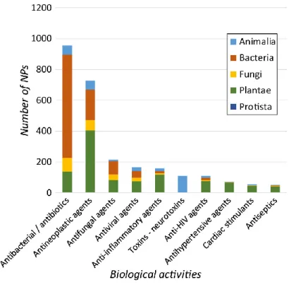
Figure 1 - Major bioactivities of natural products and their distribution throughout the different kingdoms of life.
Figure 2 – Examples of enzymatic reactions used in natural product biosynthesis.
Figure 3 – Examples of structural and chemical diversity of cyanobacterial secondary metabolites. Figure 4 – Click chemistry using azides and terminal alkynes
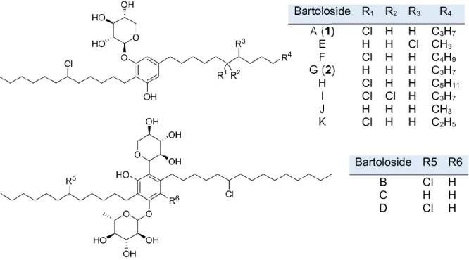
Figure 5 – Bartolosides chemical structures.
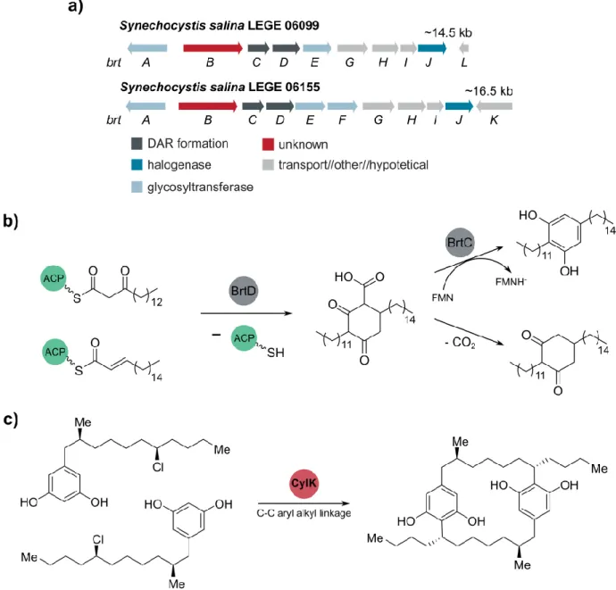
Figure 6 – Biosynthesis of bartolosides and cylindrocyclophanes
Figure 7 - S. salina LEGE 06099 growth over 30 days after exposing the culture to both 5-hexynoic and 6-heptynoic acid.
Figure 8 - Supplementation of terminal alkyne-containing fatty acids was expected to lead to the incorporation of terminal alkyne moieties into the alkyl chains of bartolosides produced by S. salina LEGE 06099.
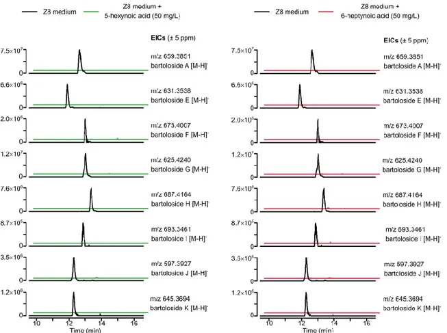
Figure 9 - Depletion of bartolosides levels in S. salina LEGE 06099 supplemented with fatty acids. Figure 10 - Total Ion Chromatograms obtained from LC-HRESIMS analysis of feeding experiments.
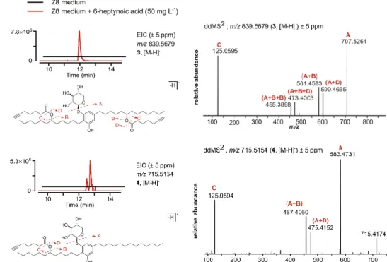
Figure 11 - Formation of bartoloside A and G esters upon supplementation of S. salina LEGE 06099 with 6-heptynoic acid.
Figure 12 - Formation of bartoloside A and G esters upon supplementation of S. salina LEGE 06099 with 5-hexynoic acid.
Figure 13 - Extracted Ion Chromatograms (EICs) of m/z 839.5678 [M-H]- (compound 3) and m/z
715.5154 [M-H]- (compound 4).
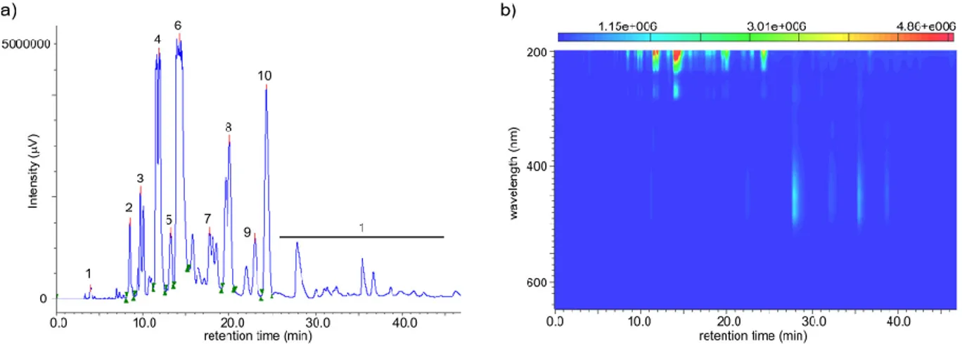
Figure 14 - Reverse phase HPLC chromatogram and PDA heat map spectra of derived from the semipreparative chromatography of fraction 5 performed for the isolation of compounds 3 and 4. Figure 15 - Key HMBC and COSY correlations of bartoloside A-17,29-diyl bis(hept-6-ynoate) (3). Figure 16 - HRESIMS/MS spectra for the in-source (collision energy of 90 eV) generated fragment of 3 providing the required confirmation and validation of the chemical structure of compound 3. Figure 17 - Key HMBC and COSY correlations of bartoloside G-17-yl hept-6-ynoate (4).
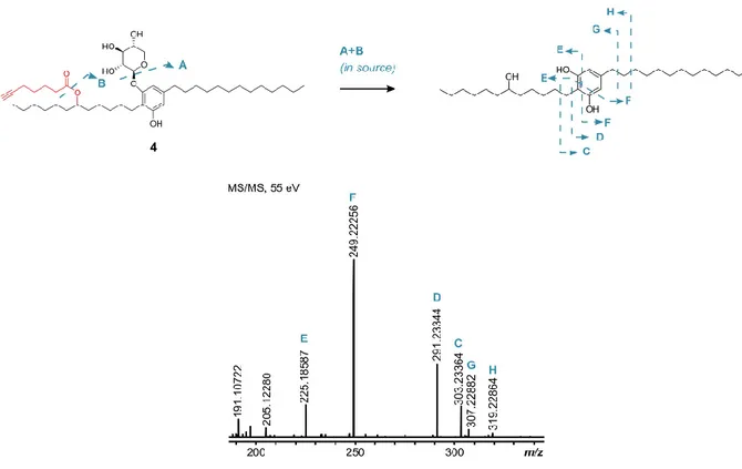
Figure 18 - HRESIMS/MS spectra for the in-source (collision energy of 65 eV) generated fragment of 4 providing the required confirmation and validation of the chemical structure of compound 4.
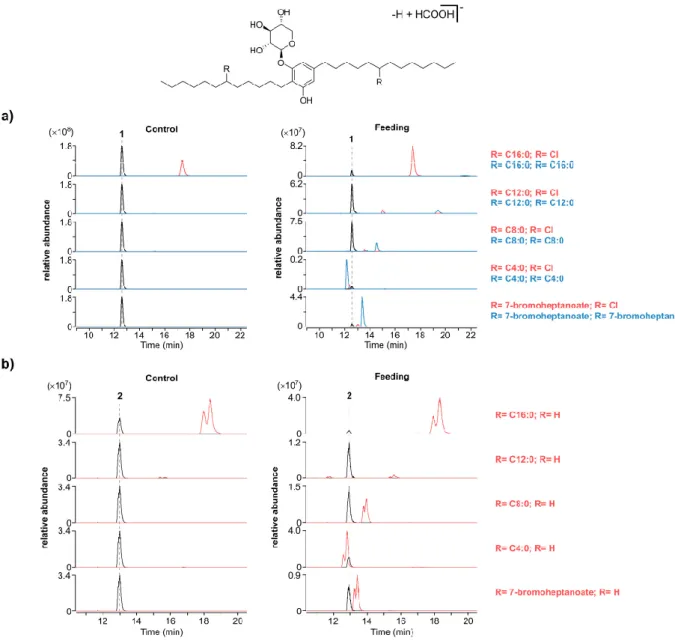
Figure 19 - Bartoloside esters formed in S. salina LEGE 06099 cells upon supplementation with different fatty acids commonly present in bacteria and with the halogenated fatty acid 7-bromoheptanoate.
Figure 20 - Detection and HRESIMS/MS based structural assignment of naturally occurring bartoloside A and G esters of palmitate in S. salina LEGE 06099.
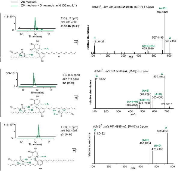
Figure 21 - Detection and HRESIMS/MS based structural assignment of naturally occurring bartoloside esters produced by S. salina LEGE06099.
Figure 22 - Relative abundance of naturally occurring bartoloside esters found in S. salina LEGE06099 cells.
Figure 23 - Extracted Ion Chromatograms (EICs) of putative esterified bartoloside A monopalmitate.
Figure 24 - Chromatograms obtained after RP-HPLC of fraction 15 using 99% acetonitrile in water and 98% methanol in water.
Figure 25 - Chromatogram of purified subfraction 15.7.2 after a reverse phase semipreparative HPLC of fraction 15.7 using 98% acetonitrile in water.
Figure 26 - HRESIMS/MS spectra for the in-source (collision energy of 90 eV) generated fragment of 5a providing the required confirmation and validation of the chemical structure of compound 5a. Figure 27 - Key HMBC and COSY correlations of bartoloside A-17-yl palmitate (5a).
Figure 28 - 1H NMR-based assignment of which alkyl chain in 5a is esterified with palmitic acid.
Figure 29 - Bioactivity assay results of exposed HT-29 cells to compounds 3, 4 and 5a after 24h and 48h.
Figure 30 - Bioactivity assay results of exposed hCMEC/D3 cells to compounds 3 and 4 after 24h and 48h.
Figure 31 - Bioactivity assay results of exposed HTC116 cells to compounds 3 and 4 after 24h and 48h.
Figure 32 - Tested bacterial and fungi strains.
Figure 33 - Results from1% agarose gel electrophoresis of cloning procedures of NStrep-brtB. Figure 34 - SDS-PAGE results of NStrep-BrtB expression after 4h and 16h induction with IPTG and CaCl2 of both E. coli BL21(DE3)pLysS and Rosetta(DE3).
Figure 35 - SDS-PAGE of the affinity chromatography performed and the NStrep-BrtB after desalting and concentration.
Figure 36 - BrtB converts bartoloside A (1) and 6-heptynoic acid into bartoloside esters in vitro. Figure 37 - BrtB converts bartoloside A (1) and palmitic acid into bartoloside esters in vitro. Figure 38 - Selected O-alkylating enzymes.
cluster containing BrtB/CylK homologs.
Annex Table 1 – NMR Spectroscopic Data (1H 400 MHz, 13C 100 MHz, CDCl
3) for bartoloside
A-17,29-diyl bis(hept-6yonate) (3).
Annex Table 2 - NMR Spectroscopic Data (1H 400 MHz, 13C 100 MHz, CDCl
3) for bartoloside
G-17-yl hept-6-ynoate (4).
Annex Table 3 - NMR Spectroscopic Data (1H 400 MHz, 13C 100 MHz, CDCl
3) for bartoloside
A-17-yl palmitate (5a).
Annex Figure 1 - Detection and HRESIMS/MS based structural assignment of naturally occurring bartoloside A esters produced by S. salina LEGE06099.
Annex Figure 2 - Detection and HRESIMS/MS based structural assignment of naturally occurring bartoloside G esters produced by S. salina LEGE06099.
Annex Figure 3 - 1H NMR spectra for subfraction 15.7 obtained during the isolation of bartoloside
A monopalmitate (5a).
Annex Figure 4 - HRESIMS/MS of fraction 15.7.2 obtained during the isolation of compound 5a with respective structural assignment.
Annex Figure 5 - LC-HRESIMS/MS analysis of the bartoloside esters 3 and 8a/8b (a) and 5a (b) formed in the full assay.
Annex Figure 6 - 1H NMR (CDCl
3, 400 MHz) spectrum of compound 3.
Annex Figure 7 - 13C NMR (CDCl
3, 100 MHz) spectrum of compound 3.
Annex Figure 8 - HSQC (CDCl3, 400 MHz) spectrum of compound 3.
Annex Figure 9 - HMBC (CDCl3, 400 MHz) spectrum of compound 3.
Annex Figure 10 - COSY (CDCl3, 400 MHz) spectrum of compound 3.
Annex Figure 11 - 1H NMR (CDCl
3, 400 MHz) spectrum of compound 4.
Annex Figure 12 - 13C NMR (APT, CDCl
3, 100 MHz) spectrum of compound 4.
Annex Figure 13 - HSQC (CDCl3, 400 MHz) spectrum of compound 4.
Annex Figure 14 - HMBC (CDCl3, 400 MHz) spectrum of compound 4.
Annex Figure 15 - COSY (CDCl3, 400 MHz) spectrum of compound 4.
Annex Figure 16 - 1H NMR (CDCl
3, 400 MHz) spectrum of compound 5a.
Annex Figure 17 - 13C NMR (APT, CDCl
3, 100 MHz) spectrum of compound 5a.
Annex Figure 18 - HSQC (CDCl3, 400 MHz) spectrum of compound 5a.
Annex Figure 19 - HMBC (CDCl3, 400 MHz) spectrum of compound 5a
I. Introduction
Impact of natural products in the biological sciences and industry
Natural products have impactful roles in our society. With the serendipitous discovery of penicillin by Alexander Fleming, 19291, modern natural products research flourished with potential
applications in several fields, namely drug discovery, nutrition, chemical biology and also in the industry.2-3 However, the understanding of what a natural product is may not be clear and
consistent throughout different scientific areas. From the chemical point of view, a natural product can be described as a small molecule derived from a biological source and that is not strictly required for the development and viability of the producing organism.2, 4
From a historical point of view, mankind has always entrusted Nature to supply not only food but also ways to improve life quality. Natural products have been used in traditional medicine from ancient times, with records of plant-based substances dating 2600 B.C.5 However, secondary
metabolite chemical diversity and biological importance started to gain more importance after 1817 with the isolation of morphine from Papaver somniferum by a German pharmacist Friedrich Sertuerner.6 Such discovery motivated scientists to pursue plant based compounds capable of
helping the treatment of a wide spectrum of diseases, for example, the discovery of quinine, a promising antimalarial drug from the bark of Cinchona species.7
Plant derived natural products were undoubtedly fundamental to the improvement of life quality throughout the times, but advances in microbiology research stimulated the screening for bioactive secondary metabolites from microbial sources. 8 As mentioned above, the discovery (Alexander
Fleming) and isolation (Howard Florey) of penicillin from the fungus Penicillium notatum triggered a massive interest in studying microbial organisms’ secondary metabolism, with a special focus on bacteria.9 The huge diversity of bacteria was, by itself, a motivation to seek for bioactive
compounds that could increase and expand the scientific horizons in terms of chemical and structural diversity together with the promising biological and industrial role of bacterial metabolites. In fact, even though natural products found in the Plantae kingdom are approximately eight times the ones found in the Bacteria kingdom, the relative amount of bacterial bioactive secondary metabolites is heavily superior.9-10 Many bioactive compounds obtained from bacterial
sources present antibacterial properties9-10 (Figure 1), representing 51% of all FDA-approved
Actinomycetes, a group of filamentous gram positive bacteria, are responsible for the production of almost all antibiotics used in the pharmaceutical industry, with almost 75% of reported bioactive compounds being produced by a single genus, Streptomyces.12-13 One of the most significant
antibiotics with bacterial origin is streptomycin, initially isolated by Albert Schatz and Selman Waksman in 1944, which was later introduced in the market as a tuberculosis treatment as result of its strong activity against Mycobacterium tuberculosis.14-15
As discussed above, secondary metabolites were initially studied with the aim to find novel compounds, with special consideration to antibacterial and anticancer compounds. However, the applications of such diverse and complex natural products goes beyond their direct application to the pharmaceutical industry. The fast increase in global population demanded more intense farming practices and with that the need for products capable of both protecting the cultivations and conserving the produced food.10, 16 Further concern was also related with the handling of
environmentally friendly compounds due to their low toxicity when compared to other industrial chemicals.17 With that in mind, natural products started to gain relevance in these fields and
secondary metabolite producers started to be screened for potential applications in agricultural
Figure 1. Major bioactivities of natural products and their distribution throughout
activities and food industry.16 For instance, natural products have been actively applied as
pesticides and herbicides as well as food additives and preservatives with the financing dedicated to these fields usually rounding 50 billion dollars per year10, 17-18, once again with a special focus
on bacterial secondary metabolites.19 From the reported and approved natural products used as
pesticides, avermectin was one of most recognized compounds due to its potent anthelmintic activity.20 This secondary metabolite, produced by Streptomyces avermitilis, causes the
hyperexcitation of invertebrates’ nervous system and effectively paralyzes them, protecting plantations from being damaged.16, 21 Secondary metabolites have also been extensively studied
and employed as food ingredients, as in the application of pigments as natural colorants, which can also be derived from microbial sources.22 In some cases, secondary metabolites may be used
both as food additives to confer specific colors but also as bioactive compounds – the cyanobacteria and microalgae-derived scytonemin and astaxantin, respectively, serve as good examples due to their anti-inflammatory properties.23-24
Although natural products unarguably have a clear role in our daily lives, their potential applications go beyond their direct use. Advances in interdisciplinary domains, such as chemical sciences, have been driven by attempts to mimic the intricate and complex chemistry and biosynthesis of natural products.25 The use of enzymes in chemical synthesis (biocatalysis) is
currently a very promising field of academic and industrial research. Biocatalysis offers several advantages for the pharmaceutical industry, for example, due to the economic and environmental benefits over traditional “non-green processes”.26 Enzymes allow the synthesis of different
products due to their regio- and stereo- selectivity without the need for protection steps as in organic synthesis.26-27
The use of biocatalysis over traditional chemical catalysts also allows the expansion of organic synthesis’ toolbox with the use of several reactions such as oxidations, reductions, halogenations among other, with a special focus on alkylation reactions. For instance, chemical synthesis reactions that require either O-alkylation, C-alkylation and N-alkylation are time-consuming and are not economical and environmentally viable due to the use of toxic alkylating agents.27 A great
example of enzymatic catalysts are methyltransferases, a widespread group of enzymes involved in the transfer of methyl groups, which can be green alternatives for Friedel-Crafts alkylations (figure 2).28-29 The catalytic mechanism of enzymatic oxidations are also of great interest to the
pharmaceutical industry since they avoid undesired side-products due to their high stereoselectivity.30 However, enzymatic catalysis is not adapted to industrial processes leading to
and specificity of the enzymatic reactions. Additionally, innovative methodologies that improve the biocatalysts performance have also been explored, for example in the scale up of Bayer-Villiger oxidations through the use of bioreactors and resin based methodologies (figure 2).30, 32
This reaction is performed by flavin-dependent monooxygenases, which are usually related with natural product biosynthesis and can be adapted to organic synthesis and in prodrug activation. 33-34 Besides, studying proteins and enzymes allows to better understand how reactions are carried
in terms of substrate affinity to active centers as well as substrate interactions and enzymatic folding.
Cyanobacteria are prolific natural product producers
Terrestrial bacteria are the most well-known source of bioactive metabolites, but extensive exploration programmes led to a decrease in the number of new natural products discovered from these sources each year.35 On the other hand, the development of efficient strategies and
technologies for marine sampling allowed to expand the secondary metabolite discovery to the oceans due to the high biological and chemical diversity associated.36-37
Cyanobacteria are widespread photosynthetic gram-negative prokaryotes that can be found in different environments, such as freshwater and marine ecosystems as well as in extreme
Figure 2. Examples of enzymatic reactions used in natural product biosynthesis. a) Methyltransferase NovO performs an
enzymatic Friedel-Crafts alkylation, using S-Adenosyl methionine (SAM) as a cofactor, to introduce a methyl moiety in aromatic substrates. 29 b) Cyclohexanone monooxygenase performs Bayer-Villiger oxidations.33 Adapted from Tengg et al. 2016 and
habitats.38-39 They have had an important role in the production of oxygen dating at least 2.3 billion
years ago, which make these organisms one of the most ancient and diverse groups of organisms on our planet. 40-41 Cyanobacteria are also unique among bacteria since some species are capable
of cell differentiation into structures called heterocysts and akinetes.41 The ability of
compartmentalization allows these microorganisms to fix nitrogen, in the heterocysts, and survive under extreme conditions (e.g. light deprivation) by entering a vegetative-like status through the specialized cells akynetes.41
Although they are commonly recognized as oxygen producing microorganisms, cyanobacteria are also a prolific source of secondary metabolites. 38-39, 42 Richard Moore was a pioneer in this field
with his group’s work in cyanobacterial derived compounds and was responsible for the discovery of dozens of cyanobacterial secondary metabolites, such as majusculamides A and B43,
oscillatoxins or debromoaplysiatoxin.44-46 There are many applications for cyanobacterial
compounds due to their varied biological properties such as UV protection conferred by the scytonemins47 or anticancer activity, e.g. dolastatin 10, the pharmacophore of the anticancer drug
brentuximab vedotin.48-50 However, the question of what distinguishes cyanobacteria from other
secondary metabolite producers arises. The high chemical diversity of cyanobacteria is in fact one of the reasons that make such organisms great targets for natural product discovery.38-39, 51 The
vast and unique mechanisms involved in the biosynthesis of cyanobacterial secondary metabolites allow these microorganisms to generate varied compounds that are of interest to chemists and biologist alike.
The secondary metabolite production relies on a biosynthetic machinery consisting of a contiguous arrangement of a set of genes defined as biosynthetic gene clusters (BGCs).52-53
Bacterial genes codifying for secondary metabolites are physically clustered while in plants the biosynthesis of natural products does not follow this assembly line, which contributed to the focus on bacterial genomes.54 The rationale behind BGCs is that the biosynthetical pathway involves
the interaction of each enzyme of the cluster with substrates to promote the “brick by brick” construction of the resulting secondary metabolite (figure 3). Additionally, biosynthetic gene clusters can include enzymes capable of performing halogenation reactions (e.g. chlorine and bromine) as well as other mechanistically interesting reactions that enable the production of a broad range of metabolites with different chemical moieties.55
Apart from the compounds generally present in other organisms such as terpenes and polysaccharides, these photosynthetic microorganisms produce metabolites that may enclosure
chemically diverse structures that range from lipid- to amino acid-based skeletons., among others.38, 51, 56 In fact, cyanobacterial metabolites can be classified according to their chemical
structural and divided into 10 classes including alkaloids, polyketides and peptides, as well as hybrids thereof.38 Most of the metabolites produced by cyanobacteria are peptides38 that are
mainly derived from non-ribosomal peptides synthetases (NRPS) or ribossomally-produced and post-translationally modified peptide (RiPP) systems. NRPs are synthesized by the action of several enzymes encoded in their respective BGCs, without the involvement of ribosomal RNA as in the usually seen peptides.56-57 On the other hand, RiPPs biosynthesis relies on a ribosomally
generated peptide containing recognition sequences that will later promote post-translationally modifications, such as cyclization, yielding the final natural product (e.g. sphaerocyclamide).58-59
Additional secondary metabolites are the fatty acid-based compounds called polyketides (PKs), e.g. oscillatoxins45. Their biosynthesis relies on the action of modular systems consisting on the
elongation of activated fatty acids and posterior modifications resulting in a broad structural diversity.60-61
Figure 3. Examples of structural and chemical diversity of cyanobacterial secondary metabolites. a) depicted is scheme of a partial
biosynthetic gene cluster. b) Dolastatin 10 is a lipopeptide derived from marine cyanobacterium Symploca sp. that has potent anticancer activity.58Oscillatoxin A is a toxic metabolite derived from PKS systems46. Sphaerocyclamide is a RiPP found in the
In addition, cyanobacteria are capable of producing remarkable compounds such as lipopeptides, which contain lipid chains bound to peptide moieties, e.g. the previously mentioned dolastatin 10.62
The cyanobacterial chemical diversity encourages the discovery of natural products not only for the novelty of the enzymatic chemistry behind their biosynthesis but also for the opportunity to discover new potent activities.
Approaches for natural product and drug discovery
The discovery of penicillin caused the emergence of the Golden Age of natural products where the main focus was drug discovery. This generated a need for developing efficient approaches capable of providing a high rate of natural product discovery that could be used in the industry. During these years, the application of bio-guided drug discovery was the main and only source of potential candidates due to the simple assays and methodologies that could be applied.63 Put
simply, target cells were exposed to microbial extracts and the phenotype of the assay would dictate the compounds activity and determine the need for further isolation efforts.63 However,
such methodologies soon started to be ineffective due to the time consuming and expensive methodologies applied, which led to the need for high throughput screening approaches.63-65
When the scientists started to focus on the chemical diversity of natural products techniques such as mass spectrometry (MS) and nuclear magnetic resonance (NMR) were extensively used and allowed faster dereplication processes and secondary metabolite discovery.66 In fact, the
applicability of combinatorial bio-guided and mass spectrometry approaches is still used for the discovery of natural products.e.g.67
Nevertheless, with the fast development in DNA sequencing technologies68 the natural products
area witnessed a resurgence of interest relying on genomic and bioinformatic approaches. For instance, metagenomics allowed the discovery of an unprecedented number of compounds from uncultured microorganisms.69 Additionally, genome mining approaches allowed the fast expansion
in knowledge of both biosynthetic gene cluster architechture and natural product chemistry in the beginning of the 21st century.70 However, predicting the end-product of a gene cluster can be
sometimes challenging. Such problem could be addressed, for example, through the use of heterologous systems: entire BGC can be cloned into suitable heterologous hosts (e.g.
Streptomyces sp.) to access the secondary metabolite derived from the gene cluster inserted. e.g.71-72 In the case of cyanobacterial secondary metabolites such bioinformatic approaches have been
extensively used for the characterization of PKS and NRPS derived compounds. These strategies have allowed the discovery of a huge variety of cyanobacterial natural products but such methodologies are still non-universal, can be time consuming or even discouraging.
One other subject of natural products research is the study of their mode of action, in particular, regarding their ecological or pharmacological relevance. Achieving this, however, is not trivial. Click chemistry has emerged as a powerful set of biorthogonal reactions (i.e. chemical reactions occurring in living systems without affecting their biological processes75) that are acknowledged
for their fast and simple procedures with a wide-range of applications in chemical biology, e.g. deciphering the mode of action of a natural product.76-77 One of the most common approaches
relies on heterocycle formation between azides and terminal alkyne moieties through a copper-catalyzed Huisgen ,3-dipolar cycloaddition (figure 4).78 Click chemistry advancements have
allowed the creation of studies based in bio-conjugation of functionalities of interest to the azide, later generating a tagged version of the desired compound which is suitable for both in vivo and
in vitro studies.77, 79-80
Bartolosides, a group of cyanobacterial dialkylresorcinols
Bartolosides are a recently discovered and unprecedented group of cyanobacterial secondary metabolites present in some cyanobacteria with uncommon features such as a chlorinated dialkylresorcinol (DAR) core or C-glysosyl moieties.81-82 Non-halogenated DAR scaffolds are
somewhat common in other bacteria but only a few DAR compounds have been discovered from cyanobacteria, e.g. paracyclophanes, microcarbonin A and nostocyclyne A. 83-86
Bartolosides can be divided into two groups according to their structure: i) monoglycosylated versions, containing an linked β-D-xylosyl moiety and ii) diglycosylated versions featuring an O-linked α-L-rhamnose and a C-O-linked xylose while additional differences consist in the alkyl chain length that can range from 11 to 15 carbons.81-82 Bartolosides A-K have been discovered and
Figure 4. Click chemistry using azides and terminal alkynes. For example, terminal alkyne labelled natural products (NP) react with
azides to form a heterocycle through a copper-catalyzed Huisgen 1,3-dipolar cycloaddition. Azides can be tagged with several moieties such as biotin and fluorophores.
isolated from the coccoid cyanobacterium Synechocystis salina LEGE 06155 and Synechocystis
salina LEGE 06099 as well as from the filamentous Nodosilinea sp. LEGE 06102.
The bartolosides biosynthetic gene cluster (brt) (Figure 6) encodes two enzymes essential for the dialkylresorcinol formation (DAR). The ketosynthase BrtD performs the condensation between two fatty acyl thioester derivatives leading to the ring formation while the flavin-dependent aromatase BrtC finalizes the resorcinol formation.82 Additional genes brtGHI encoded in the brt cluster are
involved in the glycolipid export while brtAEF encode glycosyltransferases. Halogenation of the alkyl chains in the bartolosides is predicted to be performed by BrtJ since some of its homologs (CylC, ColD/ColE) have been found in cyanobacterial biosynthetic gene cluster encoding for halogenated secondary metabolites, cylindrocyclophanes and columbamides.87-88 Previous
bioinformatic analysis also supports that this enzyme belongs to a class of dimetal halogenases acting on unactivated carbons.81-82
Figure 5. Bartolosides chemical structures enclose several chlorinations, different alkyl chain lengths and can be mono- or
diglycosylated. Bartoloside R1 R2 R3 R4 A (1) Cl H H C3H7 E H H Cl CH3 F Cl H H C4H9 G (2) H H H C3H7 H Cl H H C5H11 I Cl Cl H C3H7 J H H H CH3 K Cl H H C2H5 Bartoloside R5 R6 B Cl H C H H D Cl H
An additional enzyme – BrtB – is encoded in the brt cluster. Phylogenetic analysis indicates that BrtB and CylK - an enzyme that participates in paracyclophane biosynthesis – are homologs.89
The paracyclophanes are cyanobacterial natural products that contain aryl-alkyl linkages in their core structure, however, the biocatalytic mechanisms behind paracyclophane assembly were only recently clarified, in particular the function of CylK.87, 89 Through several in vitro assays this enzyme
(annotated as hemolysin-type calcium-binding protein) was shown to be responsible for the
Figure 6. Biosynthesis of bartolosides and cylindrocyclophanes. a) Bartolosides biosynthetic gene cluster, brt, from S. salina
LEGE 06099 and S. salina LEGE 061155 with annotated gene functions. b) proposed mechanism of dialkylresorcinol formation in bartolosides B-D. Leão et al. 2015.c) CylK catalyzes the paracyclophane formation in the cylindrocyclophanes.
paracyclophane formation through a Friedel-Crafts alkylation between the chlorinated chains of the intermediates and the resorcinol ring through a SN2 nucleophilic substitution (Figure 6c).89-90
Despite its homology with CylK, the function of BrtB has remained unclear. The reasons behind this are that a CylK-like mechanism is not necessary for the biosynthesis of the known bartolosides and that CylK requires a free C-2 position in the alkylresorcinol, which is unavailable in the bartolosides.
Objective: The biosynthesis of the dialkylresorcinol skeleton in the bartolosides involved
recruitment of fatty acids derivatives from primary metabolism. We envisioned that this could be exploited to incorporate terminal alkyne moieties into bartolosides and generate click-chemistry accessible versions of these secondary metabolites for probing their biological role. However, while attempting this we encountered unexpected results that led us to diverge from this initial aim and focus on the:
Isolation of novel bartolosides from S. salina LEGE 06099; Biosynthesis of the novel bartolosides in S. salina LEGE 06099.
II. Results and Discussion
Incorporation of terminal alkyne-containing fatty acids into bartolosides
As previously mentioned, the biosynthesis of bartolosides involves the recruitment of fatty acid derivatives from primary metabolism to assemble the dialkylresorcinol skeleton. We hypothesized that this could be exploited to incorporate terminal alkyne moieties into these natural products, making them accessible for click chemistry experiments to gain insights in their biological role. In order to understand if cyanobacteria could incorporate the desired terminal alkynes we initially assessed whether they could be toxic to the cyanobacterial cells. To this end, we supplemented the bartoloside producer Synechocystis salina LEGE 06099, with both 5-hexynoic and 6-heptynoic acids at three different concentrations (5, 10 and 50 mg L-1). The OD
750nm was measured in a
96-well plate in a microplate reader (Synergy HT, BioTek) at day 0 (day of supplementation) and every 2 days until reaching the 30-day period.
0 0,2 0,4 0,6 0,8 1 0 5 10 15 20 25 30 OD 75 0n m Time (days) 5 mg/L 10 mg/L 50 mg/L 0 0,2 0,4 0,6 0,8 1 0 5 10 15 20 25 30 O D750 nm Time (days) 5 mg/L 10 mg/L 50 mg/L
Figure 7. S. salina LEGE 06099 growth over 30 days after exposing the culture to both 5-hexynoic
We observed that supplementation with both acids did not affect the normal growth of S. salina LEGE 06099 since they presented the normal growth curve tending to the stationary phase after 20 days, approximately. Additionally, the highest concentration applied (50 mg/L) was still soluble in water and did not precipitate. These allowed us to proceed with the experiments and analyze the incorporation of the terminal alkyne-containing fatty acids into the cells.
After harvesting the cells and performing the extraction of organic compounds with a mixture of MeOH/CH2Cl2 the samples were analyzed by LC-HRESIMS. Since the target compounds
(bartoloside derivatives) have long aliphatic chains we adapted the LC method to be able to observe highly non-polar species (see “Materials and Methods” section). The output analysis revealed a massive depletion of the major metabolite bartoloside A (1) levels as well as those of several other analogues (figure 9). However, when searching for the m/z values corresponding to terminal-alkyne versions of bartoloside A and other major metabolites we did not detect any ions compatible with these structures. Bartoloside A (1) is one of the most abundant secondary metabolites in this strain, representing around 0.6% dry weight81, which means that the feeding
experiment performed could either have affected its biosynthesis or have been incorporated in an unexpected region.
Figure 8. Supplementation of terminal alkyne-containing fatty acids was expected to lead to the incorporation of terminal
In order to understand the reason behind bartolosides depletion, we sought to find new masses that were only present in the samples corresponding to supplemented cultures. Further analysis of the obtained chromatograms revealed a very different profile between samples and led to the detection of several peaks that were highly abundant in the cultures exposed to the fatty acids but were absent in the controls (figure 10). We verified that these ions were consistent with the incorporation of one or two units of the supplemented fatty acids with the concomitant loss of one or both Cl atoms in bartolosides A (1) and G (2) (most abundant metabolites), respectively. In order to support our hypothesis, we performed fragmentation of some of the new abundant ions through LC-HRESIMS/MS analysis of the crude extracts (figures 11 and 12). The data showed fragments corresponding to the intact alkyne precursors or their neutral loss in the bartolosides core structure. This suggested that the detected peaks could correlate with esterified versions of bartolosides 1 and 2 with 5-hexynoate and 6-heptynoate.
Figure 9. Depletion of bartolosides levels in S. salina LEGE 06099 supplemented with fatty acids. Analysis was carried by
LC-HRESIMS of the crude extracts obtained and presented are the extracted ion chromatograms (EICs) of the m/z correspondent to the [M-H]- ions of bartolosides.
Figure 10. Total Ion Chromatograms obtained from LC-HRESIMS analysis. Supplementation
with 5-hexynoic and 6-heptynoic acids led to the formation of new abundant masses absent in the control. Additionally, bartoloside A level was depleted in the exposed cultures.
Figure 11. Formation of bartoloside A and G esters upon supplementation of S. salina LEGE 06099 with
6-heptynoic acid. Here we present the proposed chemical structures and respective annotated LC-HRESIMS/MS data for compounds 3 and 4.
Isolation of branched bartolosides formed under supplementation
The previously performed experiments showed that supplementation with 5-hexynoic and 6-heptynoic acids did not generate terminal alkyne moieties in the DAR moiety of the bartolosides but instead led to the formation of new compounds. To unambiguously establish the chemical structure of the newly observed metabolites we sought to isolate and purify the major metabolites found under supplementation with fatty acids.
To fulfill our objective, we performed a 20 L cultivation of S. salina LEGE 06099 under supplementation with 6-heptynoic acid at a concentration of 50 mg L-1 in order to try to obtain a
Figure 12. Formation of bartoloside A and G esters upon supplementation of S. salina LEGE 06099 with 5-hexynoic acid. Here we
sufficient amount of compound for structure elucidation. The harvested freeze-dried biomass (5.6 g, d. w.) was extracted by repeated percolation using a mixture of CH2Cl2/MeOH (see “Materials
and Methods” section) resulting in a crude extract of 754.6 mg. Subsequent fractionation was achieved by normal phase flash chromatography using a gradient of increased polarity from hexane to ethyl acetate (EtOAc) to methanol (MeOH). The fractionation originated eight fractions (1-8) with similar TLC (thin layer chromatography) profiles. Fractions 4 (13.5 mg), 5 (102.2 mg) and 6 (14.5 mg) eluting with a 2:3 mixture of EtOAc/hexane were analyzed by LC-HRESIMS and indicated the presence of the desired m/z corresponding to the hypothetical branched bartolosides A and G (m/z 839.5678 [M-H]- and 715.5154 [M-H]-, respectively). At this stage, depending on the
relative abundance of each compound, we could have pooled the fractions together but considering that both compounds were mostly present in fraction 5 (figure 13) we decided to only go forward with this sample for further fractionation.
The isolation of the desired metabolites involved a new chromatographic step using a reverse-phase semipreparative HPLC which allows a more efficient peak collection since we can track the separation progress in real time with the help of a PDA (photodiode array) detector. The HPLC program was adapted to allow the proper resolution of all peaks. Initially we tested an isocratic gradient of 80% acetonitrile in water during 25 minutes but peak separation was insufficient. After increasing the acetonitrile concentration to 90% we verified a more efficient separation with the
Figure 13. Extracted Ion Chromatograms (EICs) of m/z 839.5678 [M-H]- (compound 3) and m/z 715.5154 [M-H]- (compound 4)
advantage of a faster fractionation since the compounds were eluting faster due to the decreased polarity of the mixture. Finally, we decided to use an isocratic gradient of 92% acetonitrile in water during 25 minutes followed by a washing step with 100% acetonitrile (see “Materials and Methods” section), affording 10 subfractions. All subfractions were submitted to 1H NMR to evaluate their
composition and purity. Fraction 5.6 (48.0 mg, figure 14), eluting at 13.5-15.0 minutes, was spectroscopically pure (> 95%, 1H NMR) and showed characteristic signals of bartolosides
regarding resorcinol and xylosyl protons.81-82 Additionally, fraction 5.10 (4.3 mg, figure 14), with a
retention time of 24.0-25.0 minutes, was also pure and presented a similar 1H NMR profile to the
subfraction 5.6 and consequently to the bartolosides.
Structural elucidation of esterified bartolosides 3 and 4
The 1H NMR analysis of both 5.6 and 5.10 subfractions indicated that they were suitable for further
structural elucidation. Therefore, both samples were submitted to 1D 13C NMR and 2D NMR
(HSQC, HMBC and COSY) as well as HRESIMS/MS analysis in order to unequivocally establish their chemical identity.
bartoloside A-17,29-diyl bis(hept-6-ynoate) (3)
HRESIMS analysis of compound 3 (isolated as an orange oil) showed a [M-H]- peak at m/z
839.5685, compatible with a molecular formula of C50H80O10 (calculated for C50H79O10-, m/z
839.56867) and 11 degrees of unsaturation. The 1H and 13C NMR spectra showed typical
bartoloside resonances, namely for the aromatic ring (δC 155.8, 154.6, 141.9, 116.3, 110.4 and
Figure 14. Reverse phase HPLC chromatogram (a) and PDA heat map spectra (b) of derived from the semipreparative
107.8) and xylosyl (δC 101.4, 72.6, 74.9, 69.7 and 64.5) (annex table 1). Characteristic resonances
for two ester/acid carbonyls (δC 174.1 and 173.9) and two alkyne moities (δC 68.8, δH 1.95) were
also observed. The HRESIMS derived molecular formula and isotope pattern of 3 dictated the absence of halogens in the molecule. The molecular formula had an additional C14H18O4 when
compared to the major bartoloside 1 produced by S. salina LEGE 06099 (bartoloside A) consistent with the incorporation of two 6-heptynoic acid moieties. This initial analysis strongly suggested that compound 3 could correspond to a bartoloside derivative where the chlorinated positions in 1 were substituted by the supplemented 6-heptynoic acid. To obtain further insights regarding the structure of 3, we performed 2D NMR experiments (HSQC, HMBC, COSY). Correlational data for the glycosylated dialkylresorcinol moiety were highly similar to those of the bartolosides and it was also possible to deduce the structures of two intact 6-heptynoate spin systems from COSY and HMBC (figure 15). Notably, two conspicuous protons resonating at δH 4.87 (H-17 and H-29) in 3
are not found in any of the previously reported bartolosides. Each of these protons was positioned in the middle of an alkyl chain (figure 15, annex table 1) and showed HMBC correlations to the carbonyls C-1’ (δC 174.1) and C-1’’ (δC 173.9). Strong deshielding indicated that these were
oxymethine protons and established the directionality of the ester linkages. HRESIMS/MS data, which show fragments corresponding to 6-heptynoate (m/z 125.05 [M-H]-, figure 11) are in
accordance with this observation. Additionally, the resonances for the chlorinated carbons in bartoloside A (δC 64.9, δH 4.01)81-82 are not present in 3, further implying ester linkages in positions
17 and 29. While it was not possible to fully connect the oxymethines to the dialkylresorcinol rings, the HRESIMS/MS data of in-source fragments of 3 (figure 16) matches the data for bartoloside A (1) and other bartolosides in our previous structural elucidation efforts.81-82 As such, we propose
that the esterified positions in 3 correspond to the chlorinated positions in 1. Optical rotation, infrared (IR) and ultraviolet (UV) fingerprint:
[α] 25
D -5.0 (c 0.60, MeOH); IR (thin film) ʋmax 3398, 3308, 2923, 2857, 1726, 1703, 1590, 1428,
1052, 1033, 1017, 627 cm-1. UV (MeOH) λ
Figure 15. Key HMBC and COSY correlations of bartoloside A-17,29-diyl bis(hept-6-ynoate) (3)
Figure 16. HRESIMS/MS spectra for the in-source (collision energy of 90 eV) generated fragment of 3 providing the required
bartoloside G-17-yl hept-6-ynoate (4)
HRESIMS data of compound 4 (isolated as a light orange oil) showed a prominent peak at m/z 715.5161 [M-H]- which was compatible with a molecular formula of C
43H72O8 (calculated for
C43H71O8-, m/z 715.5162) and 8 degrees of unsaturation. The 1H and 13C NMR data (annex table
2) were highly similar to those from compound 3, notably featuring a carbonyl resonance (δC
174.1) and an alkyne moiety signal (δC 68.8, δH 1.95). Additionally, the typical bartoloside
resonances for the aromatic ring (δC 155.7, 154.3, 142.5, 116.1, 110.4, 108.1) and xylosyl group
(δC 101.4, 72.6, 74.9, 69.7 and 64.5) were present. The HRESIMS-derived molecular formula and
isotope pattern of 4 indicated the absence of halogens in the molecule and an additional C7H9O2
when compared to another bartoloside produced by S. salina LEGE 06099 (bartoloside G, 2). This would be consistent with 4 resulting from the incorporation of one 6-heptynoic acid moiety into 2. Two-dimensional NMR experiments (HSQC, HMBC and COSY) showed the presence of an intact 6-heptynoate-derived spin system from COSY and HMBC correlations (figure 17). The HMBC correlations for H-17 (δH 4.88) with C-1’ (δC 174.1) indicated an identical ester linkage to that of 3,
at the C-17 (δC 74.7) position (figure 17). In line with these observations, the HRESIMS/MS data
for 4 showed a 6-heptynoate-derived fragment (m/z 125.05 [M-H]-, figure 11). By isolating and
carrying out HRESIMS/MS analysis on an in-source fragment of 4, we obtained diagnostic fragments indicating a fully aliphatic alkyl chain with 13 carbons (m/z 249.22 and m/z 291.23, figure 18), as well as fragments consistent with an alcohol group in the C12 alkyl chain (e.g. m/z 225.18 and m/z 319.23, figure 18). We thus propose that metabolite 4 is a derivative of the naturally occurring 2, in which the chlorinated position is substituted by a 6-heptynoate moiety. Optical rotation, infrared (IR) and ultraviolet (UV) fingerprint:
[α] 25
D -4.6 (c 0.65, MeOH); IR (thin film) ʋmax 3410, 3313, 2923, 2855, 1731, 1704, 1590, 1428,
1054, 1033, 1014, 627 cm-1; UV (MeOH) λ
max (log ε) 216 (3.1), 221 (3.1), 273 (2.8).
Incorporation of additional substrates into the bartolosides
We hypothesized whether additional substrates could be incorporated in the same fashion as the terminal alkyne-containing fatty acids supplemented. With this in mind, we set out to investigate which type of substrates could be incorporated in the bartolosides alkyl halide chains.
The previously used S. salina LEGE 06099 strain was exposed to several substrates containing not only carboxylate moieties (7-bromoheptanoic, trans-3-hexenedioic, butyric, caprylic, lauric and palmitic acids) but also other related nucleophiles commonly used in organic synthesis (hexylalcohol and hexanamide). After supplementing the cultures, the cells were harvested, extracted as carefully described in the “Materials and Methods” section and analyzed by LC-HRESIMS. As expected, the results showed the formation of corresponding mono- and diesters of both bartoloside A (1) and G (2) with the fatty acids supplemented (figure 19). Since trans-3-hexenedioic acid contains two carboxylate moieties we hypothesized that the branching could occur between two bartolosides or even inside the same molecule through the esterification of both alkyl halide chains to the same substrate. However, we could not detect any ion that could
Figure 18. HRESIMS/MS spectra for the in-source (collision energy of 65 eV) generated fragment of 4 providing the required
correlate with branched versions of bartoloside A and G with hexylalcohol, hexanamide nor trans-3-hexenedioic indicating selectivity towards fatty acids. Overall, our results show that exogenously provided fatty acids are converted in vivo into esterified versions of bartolosides by S. salina LEGE 06099.
Figure 19. Bartoloside esters formed in S. salina LEGE 06099 cells upon supplementation with different fatty acids commonly present
in bacteria and with the halogenated fatty acid 7-bromoheptanoate. LC-HRESIMS of extracted ion chromatograms (EICs) of [M-H+HCOOH]- adducts of the proposed bartoloside A esters (a) and bartoloside G esters (b). Controls without supplementation are
Interestingly, while analyzing the LC-HRESIMS data of our non-supplemented controls we verified the presence of two abundant compounds with m/z values and retention times consistent with both bartoloside A monopalmitate (5a/5b) and bartoloside G palmitate (6). Such findings led us to hypothesize that esterified bartolosides occur naturally in the cyanobacteria and not only while under supplementation with exogenous fatty acids. Therefore, we set out to investigate whether the detected species was in fact the branched version of bartoloside A monopalmitate (5a/5b).
Detection of naturally occurring bartoloside esters
The previously obtained LC-HRESIMS results of the control samples presented in figure 18 directed us to search for the naturally occurring bartoloside A monopalmitate (5a/5b) ester (m/z 879.6486 [M-H]-). Further analysis of the CH
2Cl2/MeOH (2:1) cellular extract of S. salina LEGE
06099 without supplementation used in the chapter revealed the presence of a m/z consistent with the diesterified version of bartoloside A with palmitic acid (7, m/z 1099.9177 [M-H]-). The
higher retention time corroborated with the hypothetical structure due to the putative compound’s lower polarity owing to the four alkyl chains.
With these data in our hands we decided to perform the mass fragmentation of the detected ions (m/z 879.6486 [M-H]- and 1099.9177 [M-H]-). The LC-HRESIMS/MS data revealed the presence
of intact fragments of palmitic acid (m/z 255.2330 [M-H]-) as well as fragments corresponding to
the previously presented data of the bartoloside A core (figure 20). Fragmentation of the hypothetical bartoloside G palmitate (6, m/z 845.6876 [M-H]-) also revealed the presence of
palmitic acid and bartoloside G fragments, without a chlorine atom, in line with what had been observed for the previously isolated compound 4.
Knowing this, we extensively examined the LC-HRESIMS data for additional esterified versions of bartolosides A and G with fatty acids known to be present in cyanobacterial cells.91 This revealed
20 additional peaks matching with m/z of branched versions of 1 and 2 with fatty acids with varying chain length and saturation, which were then analyzed by LC-HRESIMS/MS to confirm the presence of corresponding fragments (figure 21, annex figures 1 and 2 ). Putative bartoloside A and G esters 5a/5b and 6, respectively, seem to be the most abundant ones as verified by the integration of the corresponding peak area (figure 22).
Figure 20. Detection and HRESIMS/MS based structural assignment of naturally occurring bartoloside A and G esters of palmitate in S. salina LEGE 06099. a) Annotated MS/MS fragments for the structures of bartoloside A mono- (5a/5b) and diester (7) and bartoloside
G palmitate (6). Depicted are the extracted Ion Chromatograms (EICs) for the [M-H+HCOOH]- ions and MS/MS spectra for the [M-H]
Figure 21. Detection and HRESIMS/MS based structural assignment of naturally occurring bartoloside esters produced by S. salina
LEGE06099. a) Annotated MS/MS fragments for the structures of bartoloside A and G esters. b-d) Extracted Ion Chromatograms (EICs) for the [M-H+HCOOH]- ion and MS/MS spectra for the [M-H]- specie of some of the detected naturally occurring bartoloside A
esters. e-g) Extracted Ion Chromatograms (EICs) for the [M-H+HCOOH]- ion and MS/MS spectra for the [M-H]- specie of some
Even though S. salina LEGE 06099 produces other bartolosides we could not detect any peaks corresponding to hypothetical esterified versions of such metabolites due to their low abundance in the used strain. 81 On the other hand, proving that the detected peaks were indeed bartoloside
esters required purification and structure elucidation.
Isolation of bartoloside A monoester 5a
To undoubtedly establish that the LC-HRESIMS detected compounds were indeed branched versions of bartolosides we cultivated 20 L of S. salina LEGE 06099 without the supplementation of exogenous fatty acids. After extracting the organic compounds from the freeze-dried biomass (574.31 mg d.w. crude extract) using a mixture of CH2/Cl2:MeOH (2:1) we performed another flash
chromatography to fractionate our sample (see “Materials and Methods” section).
At this point, we had to adapt the solvent concentration since our target compounds are more apolar due to their long aliphatic chains. Since we used a normal phase chromatography and started with apolar solvents (hexane) our compounds were predicted to have eluted earlier than compounds 3 and 4. Therefore, we tried to extend the separation using higher volumes of the more apolar mixtures (until 40% EtOAc, 60% hexane). The derived samples were pooled according to their TLC profiles originating 23 fractions (1-23) and analyzed by LC-HRESIMS. The derived data showed a very weak separation of compounds when compared to the flash chromatography performed in the isolation of 3 and 4 since the target compound (m/z 879.6486 [M-H]-) had eluted throughout several fractions. Fraction 15 (18.81 mg) contained the highest
relative abundance of the desired peak (putative bartoloside A monopalmitate 5a/5b, m/z
879.6486 [M-H])and was selected for further fractionation through semipreparative reverse phase HPLC (figure 23).
Since we had previously applied a concentration of 92% acetonitrile in water for the isolation of compounds 3 and 4 we decided to try and use a higher concentration of acetonitrile (99%) due to the non-polar nature of the target modified bartoloside 5a/5b. However, this percentage was not sufficient to cause the elution of all compounds present in fraction 15 under 120 minutes (figure 24a). Therefore, we decided to try methanol for the mobile phase, which, although more polar than acetonitrile, is known to be more efficient in eluting compounds when applied in high proportion in C18 columns.92 In fact, after adjusting the methanol concentration to an isocratic
gradient of 98% MeOH in water we obtained a good separation in under 60 minutes (figure 24b). From the collected 11 peaks, subfraction 15.7 (retention time 32.1-34.5 minutes) contained one of the major peaks and likely the target species with m/z 879.6486 [M-H]-. To validate this, we
analyzed the eluting peak by HRESIMS/MS using direct injection into the mass spectrometer device. Again, the target m/z value was observed and fragments corresponding to palmitic acid as well as its neutral loss were detected along with bartoloside A dialkylresorcinol fragments (annex figure 4).
Figure 23. Extracted Ion Chromatograms (EICs) of putative esterified bartoloside A monopalmitate m/z 879.6486 [M-H]-.
Depicted are fractions 14-20 derived from the flash chromatography performed with the crude extract obtained from 20 L of non-supplemented S. salina LEGE 06099 containing the putative esterified bartoloside A monopalmitate m/z 879.6486 [M-H]-.
The obtained subfraction 15.7 (6.52 mg d.w.) was submitted to 1H NMR so that we could verify its
purity and forward it to 2D NMR analyses for the structural elucidation. However, the data showed that a minor bartoloside co-eluted (likely the structural isomer of bartoloside A monopalmitate 5b) since the proton resonances were extremely close and overlapping (annex figure 3), thus further separation was necessary.
Although methanol was efficient for a faster separation of fraction 15 we decided to test acetonitrile since it promoted a much slower peak elution. In fact, after some program adjustments we verified that 98% acetonitrile in water was the ideal to cause the proper separation of subfraction 15.7, even though the elution was occurring only after 120 minutes. Fraction 15.7.2 (3.1 mg, retention time ~122 minutes, figure 25) was then submitted to 1H NMR and since it was spectroscopically
pure we proceeded with the 1D and 2D NMR analysis for the structural elucidation.
Figure 24. Chromatograms obtained after RP-HPLC of fraction 15 using 99% acetonitrile in water (a) and 98% methanol in water (b).
the collected subfractions (15.1-15.11) are evidenced by the numbers on top of the peaks or peak region in b).
a)
Structural elucidation of bartoloside A monopalmitate 5a
The HRESIMS performed for compound 5a (isolated as a white glassy solid) presented a peak with m/z 879.6504 [M-H]- (calculated for C52H92O8Cl- m/z 879.6499), compatible with a molecular
formula of C52H93O8Cl and 6 degrees of unsaturation. The 1H and 13CNMR data indicated the
presence of the characteristic aromatic resorcinol ring and xylosyl group observed in bartolosides from S. salina LEGE 06099 (annex table 3). A carbonyl resonance was also present (δC 174.7) as
observed for bartoloside esters 3 and 4. The additional C16H31O2 (compared to 1) was consistent
with the esterification of one palmitic acid moiety with bartoloside A (1) and the concomitant loss of one chlorine atom (as seen for 3 and 4). Support to this hypothesis was gained from HRESIMS/MS analysis of the in-source-formed species with m/z 473.40032, which showed diagnostic fragments that are consistent with a backbone similar to that of 1 (figure 26), as well as from 2D NMR experiments: an HSQC correlation between δH 3.88 and δC 64.5, likely
corresponding to a chlorinated methine, was part of a spin system that expanded, on both directions into degenerate CH2 resonances, indicated that this corresponded to a mid-chain
chlorination, characteristic of the bartolosides (figure 27). HRESIMS/MS data was consistent with the presence of a palmitate-derived fragment (m/z 255.23 [M-H]-, figure 20). The 1D and 2D NMR
data showed an additional methyl group (compared to 1) at the terminal position of an alkyl chain, as well as a bigger CH2 envelope, both in line with the presence of a palmitate moiety in compound
5a. The oxymethine proton resonating at δH 4.88 presented HMBC correlations to C-1’ (δC 174.7)
in the palmitate moiety and was itself part of another spin system that degenerated into CH2
envelopes; hence it was likely positioned in the middle of the non-chlorinated alkyl chain. All these
Figure 25. Chromatogram of purified subfraction 15.7.2 after a reverse phase semipreparative HPLC of fraction 15.7 using
observations supported our proposal of 5a being a palmitate ester of 1. Because 1 has two halogenated methine moieties, to clarify which chain contained the esterification in 5a, we compared the 1H NMR spectra for 5a with the previously reported81-82 data for 1. Interestingly, we
found that the multiplicity of H2-12 (δH 2.58) changed from a complex multiplet in bartoloside A to
a triplet in 5a, while the multiplicity of H2-24 (δH 2.50) remained unaffected (figure 28). As such,
we propose that position 17, in the same alkyl chain as the H2-12 benzylic protons, is esterified in
bartoloside A-17-yl palmitate (5a). Further HRESIMS/MS analysis is consistent with this assignment (figure 26).
Optical rotation, infrared (IR) and ultraviolet (UV) fingerprint: [α] 25
D -9.2 (c 0.42, MeOH); IR (thin film) ʋmax 3417, 2923, 2854, 2360, 2340, 1730, 1704, 1592,
1463, 1455, 1430, 1159, 1048, 1033 cm-1; UV (MeOH) λ
max (log ε) 217 (3.3), 222 (3.3), 273 (2.9).
Figure 26. HRESIMS/MS spectra for the in-source (collision energy of 90 eV) generated fragment of 5a
![Figure 13. Extracted Ion Chromatograms (EICs) of m/z 839.5678 [M-H] - (compound 3) and m/z 715.5154 [M-H] - (compound 4) from the LC-HRESIMS of fractions obtained from the flash chromatography performed for the isolation of 3 and 4](https://thumb-eu.123doks.com/thumbv2/123dok_br/18731651.919782/31.892.109.791.522.869/extracted-chromatograms-compound-compound-fractions-chromatography-performed-isolation.webp)