ABSTRACT
http://dx.doi.org/10.1590/1678-775720140351
New methodology for evaluating osteoclastic
activity induced by orthodontic load
Adriele Silveira ARAÚJO1, Alline Birra Nolasco FERNANDES1, José Vinicius Bolognesi MACIEL1, Juliana de Noronha
Santos NETTO2, Ana Maria BOLOGNESE1
1- Department of Orthodontics and Pediatric Dentistry, School of Dentistry, Federal University of Rio de Janeiro, Rio de Janeiro, RJ, Brazil. 2- Department of Oral Pathology and Oral Diagnosis, School of Dentistry, Federal University of Rio de Janeiro, Rio de Janeiro, RJ, Brazil.
Corresponding address: Adriele Silveira Araújo - Departamento de Ortodontia e Odontopediatria – Faculdade de Odontologia da Universidade Federal do Rio de Janeiro - UFRJ - Av. Professor Rodolpho Rocco, 325 - Ilha do Fundão - 21941-617 - Rio de Janeiro - RJ - Brazil - Phone: +55 21 2590-9771 / +55 21 98070-4358 - e-mail: [email protected]
Submitted: September 9, 2014 - Modiication: October 29, 2014 - Accepted: October 30, 2014
O
rthodontic tooth movement (OTM) is a dynamic process of bone modeling involving osteoclast-driven resorption on the compression side. Consequently, to estimate the inluence of various situations on tooth movement, experimental studies need to analyze this cell. Objectives: The aim of this study was to test and validate a new method for evaluating osteoclastic activity stimulated by mechanical loading based on the fractal analysis of the periodontal ligament (PDL)-bone interface. Material and Methods: The mandibular right irst molars of 14 rabbits were tipped mesially by a coil spring exerting a constant force of 85 cN. To evaluate the actual inluence of osteoclasts on fractal dimension of bone surface, alendronate (3 mg/Kg) was injected weekly in seven of those rabbits. After 21 days, the animals were killed and their jaws were processed for histological evaluation. Osteoclast counts and fractal analysis (by the box counting method) of the PDL-bone interface were performed in histological sections of the right and left sides of the mandible. Results: An increase in the number of osteoclasts and in fractal dimension after OTM only happened when alendronate was not administered. Strong correlation was found between the number of osteoclasts and fractal dimension. Conclusions: Our results suggest that osteoclastic activity leads to an increase in bone surface irregularity, which can be quantiied by its fractal dimension. This makes fractal analysis by the box counting method a potential tool for the assessment of osteoclastic activity on bone surfaces in microscopic examination.Keywords: Tooth movement. Osteoclasts. Alendronate. Fractals.
INTRODUCTION
The arrival of osteoclasts is the required irst step in orthodontic tooth movement (OTM)15, and
any interference with the function of these cells results in decreased eficiency and effectiveness of orthodontic treatment8. Experimental studies
that aim to understand the inluence of distinct mechanisms involved in the regulation of OTM require, therefore, a meticulous evaluation of the osteoclastic activity. Most of these studies make this assessment by counting osteoclasts in histological sections5,6 or by histomorphometry, calculating the
osteoclast surface per bone surface12,17.
It is well known that the biomechanical and cellular cascades initiated by orthodontic forces
reshape the bony contour of the alveolus18. After
osteoclasts gain access to the mineralized bone, matrix dissolution occurs and the resorption lacuna is formed. The frontal resorption of bone may thus cause structural changes at the bone surface, resulting in its increased irregularity. The irregular morphology of this region makes it dificult to measure. Thus, few investigations have studied the periodontal ligament (PDL)-bone interface because its complexity does not allow for numerical assessment by Euclidean geometrical principles21.
Studies have evaluated the irregularity of the junctions between the tissues in order to discriminate normal tissue or benign from malignant tumors11,13,23. With the use of fractal analysis, a
thus capable of quantifying morphologies that are generally considered irregular21, larger fractal
dimensions have been found with increasing border irregularity. The fractal dimension of an object characterizes its self-similarity and describes its
space-illing properties19; the more space the object
occupies, the higher the fractal dimension.
Due to the bone surface recontouring promoted by
osteoclasts, we hypothesized that the quantiication
of surface irregularity can be measured by fractal
analysis and that this value may relect the level
of osteoclastic activity. Thus, the purpose of this study was (1) to estimate the degree of bone resorption by osteoclast counts, (2) to analyze the fractal dimension of the PDL-bone interface, and (3) to verify whether there is a relationship between osteoclastic activity and fractal dimension.
To consider the possible inluence of other factors
related to orthodontic tooth movement that could interfere with the fractal dimension, we proposed the administration of alendronate, a compound that promotes suppression of osteoclasts20, allowing the
assessment of the actual inluence of osteoclastic activity in the fractal dimension of the PDL-bone interface.
MATERIAL AND METHODS
Animals and alendronate administration
This in vivo experimental study sample consisted of 14 male New Zealand white rabbits, 16-week-old, with a mean weight of 2.6±0.3 kg. The animals were randomly divided into two groups, the control group and the alendronate-treated experimental group. They were acclimatized for 1 week, individually housed in cages inside a room with a 12-hour light-dark cycle and fed with water and powdered commercial pellets (to avoid damage to the orthodontic appliance) ad libitum. The study protocol was reviewed and approved by the Ethics Committee on Animal Use in Scientiic Research at the Heath Center of the Federal University of Rio de Janeiro (protocol number LABCE01), and was carried out in accordance with the Directive 2010/63/EU of The European Parliament and of The Council of The European Union on the protection of animals used for scientiic purposes.
Alendronate sodium trihydrate (Alendronate sodium trihydrate A4978, Sigma-Aldrich, Saint Louis, MO, USA) in crystalline form was dissolved in distilled water and vortexed for 60 seconds to obtain a solution containing 3 mg per milliliter of alendronate. The solution was prepared immediately before each administration in order to prevent interference in its stability caused by environmental and storage conditions. The experimental group received weekly subcutaneous injections of alendronate at a dosage of 3 mg/kg
for 6 weeks (3 doses before and 3 after appliance activation), whereas saline solution was injected in the control group under the same protocol.
Experimental tooth movement
The mandibular right irst molars of all animals were mesially tipped with a constant force of 85 cN for 3 weeks. The orthodontic appliance was composed of a nickel titanium closed coil spring (Mola Ortodôntica Fechada NiTi para Miniparafuso – 12 mm, Morelli, Sorocaba, SP, Brazil) stretched from the cervical area of the molar to a staple adapted to the mandibular incisors, until the desired strength was achieved, as measured by a dynamometer accurately calibrated (Dynamometer 040-711, Dentaurum, Ispringen, Baden-Württemberg, Germany). The coil spring was tied to both sites with ligature wire. The mandibular left irst molars without orthodontic force application served as the negative controls. All procedures were carried out under general anesthesia using intramuscular injection of a mixed solution of ketamine (35 mg/ kg), acepromazine (1 mg/kg) and xylazine (5 mg/ kg), an adequate dose to produce anesthesia during the entire procedure. Every week, a veterinarian examined the animals to evaluate their physical conditions, and the researcher performed oral hygiene, evaluated the integrity of tissues and appliances, and the amount of tooth movement.
Microscopic examination
After 21 days of load application, the rabbits were euthanatized by intracardiac injection of a lethal dosage of thiopental with prior sedation. Tissue sections of the left and right quadrants of the mandible were fixed in 10% neutral buffered formalin for 48 hours, decalcified in 14% ethylenediaminetetraacetic acid (EDTA) for 60 days and embedded in parafin. Longitudinal 6-μm thick sections in a mesiodistal direction were obtained from the buccal surface of the mandibles. The irst section that included the entire height of the alveolar bone, from the alveolar crest to the mandibular basal bone on the mesial side of the irst molar, was selected and stained with hematoxylin and eosin.
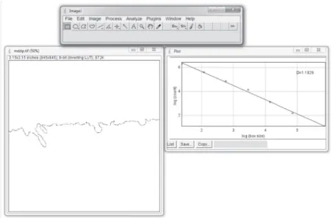
Fractal analysis
Light micrographs of the interface between bone and periodontal ligament of the same area of osteoclast counting were captured with a 10x objective lens. The lateral edge of the images coincided with the top of the mesial bone crest of
the irst molar, and the top edge was positioned
parallel to the cementum of this tooth (Figure 1A). The PDL-bone interface within this image was
drawn using the Photoshop software (Photoshop CS6, Adobe Systems, San Jose, CA, USA) as 1 pixel thick line (Figure 1B). The corresponding PDL-bone interface outlines were saved in TIFF format, 300 dpi and 945x945 pixel dimension (Figure 1C).
The Image J software (version 1.46r, National Institutes of Health, Bethesda, MD, USA) was used to measure the fractal dimension (FD) of the PDL-bone interface by the automated box counting method, using square grids with side lengths of 4, 8, 16, 32, 64, 128 and 356 pixels. To obtain the fractal dimension, we irst selected the
File>Open option to locate and open the image ile.
Next, the image was converted to binary image (Process>Binary>Make Binary), and then the fractal dimension was calculated (Analyze>Tools>Fractal Box Counter). The values of Box Sizes described above were determined and the D value, which represents the fractal dimension, was acquired (Figure 2). The entire process was performed by a single and blinded examiner (ASA).
Statistical analysis
All measurements were repeated with a two weeks interval, and the intraclass correlation coeficients for “single measures” were found to be 0.976 for osteoclast counts and 0.902 for FD of the PDL-bone interface. The mean between the 2 measurements was used for further statistical analysis, performed at a 5% level of signiicance.
Intergroup and intragroup comparisons were analyzed with unpaired and paired Student’s t-test, respectively. Correlation analysis, using the Pearson correlation coeficient, was performed to evaluate relationships between FD of the PDL-bone interface and number of osteoclasts in order to estimate the inluence of osteoclastic activity on this variable.
RESULTS
After induction of tooth movement, the number of osteoclasts increased only in the control group (p=0.008) (Table 1). There was no signiicant difference between the groups on the side without OTM. However, on the side with OTM there was a signiicant reduction in osteoclasts in the experimental group (p=0.041), showing the inhibitory effect of alendronate on these cells.
Similarly, as with the number of osteoclasts, tooth movement increased the FD of the PDL-bone interface in the control group, but did not cause changes in the experimental group (Figure 3). The osteoclast activity was responsible for the increase in FD, due to an increment in boundary irregularity caused by the presence of more active resorption sites. In the Pearson correlation, the number of osteoclasts was strongly related to FD (r=0.808, p<0.01), and accounted for 65.3% (r2=0.653) of
Figure 1- Obtention of the periodontal ligament (PDL)-bone interface contour. A: micrographs captured with a 10x objective lens of the PDL-bone interface of the mesial
of the lower irst molar; B: PDL-bone interface outlined; C:
image used to calculate the fractal dimension
AB=alveolar bone; PDL=periodontal ligament;
Table 1- Number of osteoclasts on the mesial of the lower irst molar
Orthodontic tooth movement
Control animals Experimental animals Group
differences
Mean SD Mean SD p
No 11.25 4.04 13.36 8.35 0.559
Yes 27.43* 13.55 14.14 2.75 0.041
*Signiicant (p=0.008) change in the number of osteoclasts after orthodontic tooth movement. SD= standard deviation.
There was no difference within experimental group (p=0.803).
Figure 2- ImageJ software used to obtain the fractal dimension, represented by the value D
Figure 3- periodontal ligament (PDL)-bone interface outlines and fractal dimension. A: left and B: right side of a control animal; C: left and D: right side of an experimental animal. Note the increase in bone surface irregularity in the control group and almost no change in the experimental group after 21 days of induced tooth movement; E: values represent the mean ±SD (standard deviation) of FD.
#Signiicant (p=0.003) change in the FD after orthodontic tooth movement was found only in the control group
(OTM-=1.0726±0.021; OTM+=1.1491±0.048). Fractal dimension was not altered in the experimental group (OTM-=1.0924±0.028; OTM+=1.0976±0.028). *p=0.03. OTM+, with OTM; OTM-, without OTM
Figure 4- Scatterplot showing the correlation between fractal dimension and number of osteoclasts. Strong correlation was found for both control (r=0.86, p<0.01) and alendronate groups (r=0.813, p<0.01), as well as the entire sample (r=0.808, p<0.01). Note that with the increase in the number of osteoclasts, there was an increase in fractal dimension
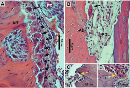
Figure 5- Micrograph of the compression side of a right irst molar from A: control and B: experimental group. The number of osteoclasts (black arrows) was signiicantly lower in the alendronate-treated rabbits. C, D: osteoclasts with nuclear
condensation, a sign of apoptosis, were found (yellow arrows); (hematoxylin and eosin staining, 400x)
the variability in the FD value (Figure 4).
DISCUSSION
In the present study, the complexity of the PDL-bone interface was measured and proposed as a new method for evaluating osteoclastic activity induced by mechanical loading. After OTM, there was an increase in the bone surface irregularity due to the frontal resorption promoted by osteoclasts, and this irregularity could be measured by fractal analysis.
Few studies10,21 have used fractal analysis to
assess the periodontal region after orthodontic tooth movement, and only one21 evaluated fractal
dimension change of the PDL-bone interface after mechanical stresses produced by an orthodontic appliance. That paper showed an increase in the fractal dimension caused by the mechanical loading and directly proportional to the magnitude of force applied. However, unlike the present study, the authors stated that the increase occurred apart from mechanisms of bone cell directed remodeling. Actually, cellular interference could not be analyzed since the force was applied for only 6 hours, insuficient time for the manifestation of signiicant osteoclastic activity. At 6 hours, many osteoclasts and pre-osteoclastic cells are still observed in vascular canals22. The number of osteoclasts in the
periodontal ligament and adjacent alveolar bones increases only on day 122, with a peak level about
50 hours after orthodontic force application. In this study, the mechanical loading was applied until day 21, when frontal bone resorption had already signiicantly occurred and osteoclastic activity could be evaluated.
To eliminate the possibility of misinterpretation and ensure that the change in fractal dimension was due to the activity of osteoclasts, apart from other factors involved in the orthodontic tooth movement such as the mechanical loading, the administration of alendronate was performed in the experimental group. Alendronate is a nitrogen-containing bisphosphonate, orally administered, and commonly prescribed for the prophylaxis and treatment of osteoporosis4. After administration, it
is redistributed to the bone, particularly to areas of increased bone turnover, and subsequently incorporated into osteoclasts involved in bone resorption8. Within these cells, the compound
interferes with specific intracellular pathways, resulting in disruption of cytoskeletal function and intracellular signaling16,20. The consequences of
these events include the suppression of osteoclastic activity, loss of osteoclast cytoskeletal integrity and rufled border, and ultimately cell apoptosis3. As
expected, the number of osteoclasts on the mesial bone surface of the right irst molar was signiicantly
higher in the control group (Figure 5A) than in the experimental group (Figure 5B). This result corroborates the inding of other studies7,9 that also
revealed a decline in the number of osteoclasts on the pressure side along the bone surface. Moreover, osteoclasts with chromatin condensation were occasionally observed in the alendronate-treated group, suggesting the presence of cells in apoptosis (Figures 5C, 5D).
The inhibitory effect of bisphosphonates on osteoclasts results is the initiation of fewer active remodeling sites on bone surfaces2,14, the most
prominent effect of these compounds seen in histological analysis1. Moreover, in addition to
reducing the number of active resorption units, bisphosphonates decrease the size of these sites1.
Due to the initiation of resorption cavities in smaller quantity and size, administration of alendronate resulted in lower bone surface complexity than that in the control group during tooth movement, because the osteoclastic activity was inhibited by the bisphosphonate. Therefore, the current research does not corroborate the inding of Wagle, et al.21 (2005), who attributed the change of the
fractal dimension of the PDL-bone interface merely to mechanical loading. The action of osteoclasts was necessary to increase the fractal dimension, otherwise an increase in this variable would also have occurred in the experimental group in which orthodontic force was applied, but the mechanical loading was not suficient to increase the fractal dimension. Increase in the fractal dimension was observed only in the control group.
Fractal dimension was strongly related to the number of osteoclasts. Thus, it is suggested that the larger number of resorption sites produced as a consequence of the increased frequency of osteoclasts activation led to a signiicant increment in fractal dimension. This makes fractal analysis by the box counting method a potential tool for the assessment of osteoclastic activity on bone surfaces in microscopic examination.
The merit of this study was to present a new methodology for evaluating osteoclastic activity induced by orthodontic force. The method is practical, simple, reproducible, inexpensive, and easier than osteoclast counting, a method that requires more time for its execution, greater attention to the correct recognition of the cells or the use of speciic staining techniques (tartrate resistant acid phosphatase) to facilitate identiication. These points make fractal analysis a complementary tool to the classic histological methodologies.
surface in other situations. Further studies might prove this hypothesis.
CONCLUSIONS
Through this study on the fractal analysis as a method for evaluating osteoclastic activity, we can reach the following conclusions:
The action of osteoclasts enhanced the fractal dimension of the PDL-bone interface as a consequence of increasing irregularity of the bone surface.
This study provides bases for the use of fractal analysis by the box counting method as a tool for the evaluation of osteoclastic activity.
ACKNOWLEDGMENTS
We would like to express our grateful appreciation to: the Coordination of Higher Education and Graduate Training (CAPES); Dental Morelli Ltda., for the donation of the coil springs, and Professor Paulo Cesar Silva for animal support.
REFERENCES
1- Allen MR, Erickson AM, Wang X, Burr DB, Martin RB, Hazelwood SJ. Morphological assessment of basic multicellular unit resorption parameters in dogs shows additional mechanisms of bisphosphonate effects on bone. Calcif Tissue Int. 2010;86(1):67-71.
2- Allen MR, Iwata K, Phipps R, Burr DB. Alterations in canine vertebral bone turnover, microdamage accumulation, and biomechanical properties following 1-year treatment with clinical treatment doses of risedronate or alendronate. Bone. 2006;39(4):872-9.
3- Boivin G, Meunier PJ. Effects of bisphosphonates on matrix mineralization. J Musculoskelet Neuronal Interact. 2002;2(6):538-43.
4- Epstein S. Update of current therapeutic options for the treatment of postmenopausal osteoporosis. Clin Ther. 2006;28(2):151-73.
5- Hakami Z, Kitaura H, Kimura K, Ishida M, Sugisawa H, Ida H, et al. Effect of interleukin-4 on orthodontic tooth movement and associated root resorption. Eur J Orthod. 2014 Jul 29. Epub ahead of print.
6- Han KH, Park JH, Bayome M, Jeon IS, Lee W, Kook YA. Effect of frequent application of low-level laser therapy on corticotomized tooth movement in dogs: a pilot study. J Oral Maxillofac Surg. 2014;72(6):1182.e1-12.
7- Igarashi K, Mitani H, Adachi H, Shinoda H. Anchorage and retentive effects of a bisphosphonate (AHBuBP) on tooth movements in rats. Am J Orthod Dentofacial Orthop. 1994;106(3):279-89.
8- Karras JC, Miller JR, Hodges JS, Beyer JP, Larson BE. Effect of alendronate on orthodontic tooth movement in rats. Am J Orthod Dentofacial Orthop. 2009;136(6):843-7.
9- Liu L, Igarashi K, Haruyama N, Saeki S, Shinoda H, Mitani H. Effects of local administration of clodronate on orthodontic tooth movement and root resorption in rats. Eur J Orthod. 2004;26(5):469-73.
10- Madan MS, Liu ZJ, Gu GM, King GJ. Effects of human relaxin on orthodontic tooth movement and periodontal ligaments in rats. Am J Orthod Dentofacial Orthop. 2007;131(1):8.e1-10. 11- Mart́n-Landrove M, Pereira D, Caldeira ME, Itriago S, Juliac M. Fractal analysis of tumoral lesions in brain. Conf Proc IEEE Eng Med Biol Soc. 2007;2007:1306-9.
12- Murphy CA, Chandhoke T, Kalajzic Z, Flynn R, Utreja A, Wadhwa S, et al. Effect of corticision and different force magnitudes on orthodontic tooth movement in a rat model. Am J Orthod Dentofacial Orthop. 2014;146(1):55-66.
13- Piantanelli A, Maponi P, Scalise L, Serresi S, Cialabrini A, Basso A. Fractal characterisation of boundary irregularity in skin pigmented lesions. Med Biol Eng Comput. 2005;43(4):436-42. 14- Rodan GA, Fleisch HA. Bisphosphonates: mechanisms of action. J Clin Invest. 1996;97(12):2692-6.
15- Rody WJ, Jr., King GJ, Gu G. Osteoclast recruitment to sites of compression in orthodontic tooth movement. Am J Orthod Dentofacial Orthop. 2001;120(5):477-89.
16- Rogers MJ. New insights into the molecular mechanisms of action of bisphosphonates. Curr Pharm Des. 2003;9(32):2643-58. 17- Sato T, Miyazawa K, Suzuki Y, Mizutani Y, Uchibori S, Asaoka R, et al. Selective beta2-adrenergic Antagonist Butoxamine Reduces Orthodontic Tooth Movement. J Dent Res. 2014;93(8):807-12. 18- Toms SR, Lemons JE, Bartolucci AA, Eberhardt AW. Nonlinear stress-strain behavior of periodontal ligament under orthodontic loading. Am J Orthod Dentofacial Orthop. 2002;122(2):174-9. 19- Updike SX, Nowzari H. Fractal analysis of dental radiographs to detect periodontitis-induced trabecular changes. J Periodontal Res. 2008;43(6):658-64.
20- Van Beek ER, Cohen LH, Leroy IM, Ebetino FH, Löwik CW, Papapoulos SE. Differentiating the mechanisms of antiresorptive action of nitrogen containing bisphosphonates. Bone. 2003;33(5):805-11.
21- Wagle N, Do NN, Yu J, Borke JL. Fractal analysis of the PDL-bone interface and implications for orthodontic tooth movement. Am J Orthod Dentofacial Orthop. 2005;127(6):655-61; quiz 754. 22- Yokoya K, Sasaki T, Shibasaki Y. Distributional changes of osteoclasts and pre-osteoclastic cells in periodontal tissues during experimental tooth movement as revealed by quantitative immunohistochemistry of H(+)-ATPase. J Dent Res. 1997;76(1):580-7.