Full Terms & Conditions of access and use can be found at
https://www.tandfonline.com/action/journalInformation?journalCode=sgra20
Grana
ISSN: 0017-3134 (Print) 1651-2049 (Online) Journal homepage: https://www.tandfonline.com/loi/sgra20
Paisia
, an Early Cretaceous eudicot angiosperm
flower with pantoporate pollen from Portugal
Else Marie Friis, Mário Miguel Mendes & Kaj Raunsgaard Pedersen
To cite this article: Else Marie Friis, Mário Miguel Mendes & Kaj Raunsgaard Pedersen (2018) Paisia, an Early Cretaceous eudicot angiosperm flower with pantoporate pollen from Portugal, Grana, 57:1-2, 1-15, DOI: 10.1080/00173134.2017.1310292
To link to this article: https://doi.org/10.1080/00173134.2017.1310292
© 2017 The Author(s). Published by Informa UK Limited, trading as Taylor & Francis Group
Published online: 01 Jun 2017.
Submit your article to this journal
Article views: 731
Paisia, an Early Cretaceous eudicot angiosperm flower with
pantoporate pollen from Portugal
ELSE MARIE FRIIS
1, MÁRIO MIGUEL MENDES
2,3& KAJ RAUNSGAARD PEDERSEN
41
Department of Palaeobiology, Swedish Museum of Natural History, Stockholm, Sweden, 2Centre for Interdisciplinary Development and Research on Environment, Applied Management and Space, Lusófona University of Humanities and Technologies, Lisboa, Portugal, 3Centre for Marine and Environmental Research, University of Algarve, Campus de Gambelas, Faro, Portugal,4Department of Geosciences, University of Aarhus, Aarhus, Denmark
Abstract
A new fossil angiosperm, Paisia pantoporata, is described from the Early Cretaceous Catefica mesofossil flora, Portugal, based on coalified floral buds, flowers and isolated floral structures. The flowers are actinomorphic and structurally bisexual with a single whorl offive fleshy tepals, a single whorl of five stamens and a single whorl of five carpels. Tepals, stamens and carpels are opposite, arranged on the same radii and tepals are involute at the base clasping the stamens. Stamens have a massivefilament that grades without a joint into the anther. The anthers are dithecate and tetraspor-angiate with extensive connective tissue between the tiny pollen sacs. Pollen grains are pantoporate and spiny. The carpels are free, apparently plicate, with many ovules borne in two rows along the ventral margins. Paisia pantoporata is the oldest known flower with pantoporate pollen. Similar pantoporate pollen was also recognised in the associated dispersed palynoflora. Paisia is interpreted as a possibly insect pollinated, herbaceous plant with low pollen production and low dispersal potential of the pollen. The systematic position of Paisia is uncertain and Paisia pantoporata most likely belongs to an extinct lineage. Pantoporate pollen occurs scattered among all major groups of angiosperms and a close match to the fossils has not been identified. The pentamerous floral organisation together with structure of stamen, pollen and carpel suggests a phylogenetic position close to the early diverging eudicot lineages, probably in the Ranunculales.
Keywords: Almargem Formation, apocarpous, basal eudicots, Ranunculales, SRXTM, synchrotron radiation x-ray microtomography
Pantoaperturate (pantoporate and pantocolpate) pol-len grains occur scattered among all major groups of angiosperms and are reported both among early diverging lineages and in more derived groups. Their occurrence in basal lineages is particularly interesting in the light of the diversity of this kind of pollen in the Early Cretaceous, but the impor-tance of pantoaperturate pollen in early angiosperm evolution has not yet been explored. The dispersed pollen record documents pantoporate/polyporate pollen already in the Aptian and by the Albian this
pollen type is almost globally distributed and repre-sented by a diversity of forms (Chlonova1986; Ibra-him et al.2017). Among early diverging angiosperms pantoporate pollen is reported for Trimeniaceae (Austrobaileyales) and Chloranthaceae (Sampson & Endress1984; Endress1986), and in early diverging monocots pantoporate pollen characterises all extant
Alismataceae and Limnocharitaceae (Argue 1973,
1974, 1976; Chanda et al. 1988; Furness & Banks
2010). Pantocolpate and pantoporate pollen grains are also common in early diverging lineages of
Correspondence: Else Marie Friis, Department of Palaeobiology, Swedish Museum of Natural History, Box 50007, 10405 Stockholm, Sweden. E-mail:else.marie.friis@nrm.se
(Received 16 August 2016; accepted 8 March 2017)
Vol. 57, Nos. 1–2, 1–15, https://doi.org/10.1080/00173134.2017.1310292
© 2017 The Author(s). Published by Informa UK Limited, trading as Taylor & Francis Group
This is an Open Access article distributed under the terms of the Creative Commons Attribution License (http://creativecommons.org/licenses/by/4.0/), which permits unrestricted use, distribution, and reproduction in any medium, provided the original work is properly cited.
eudicots including the Ranunculales and Buxales. In the Ranunculales, pantoaperturate pollen is recorded for the Eupteleaceae, Papaveraceae (Fumarioideae and Papaveroideae), Berberidaceae and Ranuncula-ceae (e.g. Wodehouse1936; Praglowski1974; Now-icke & Skvarla 1981, 1982; Blackmore et al. 1995; Emadzade et al.2010). Euptelea, usually resolved as sister to all other members of the Ranunculales, has mainly apolar and pantocolpate or sometimes partly pantoporate pollen, while tricolpate pollen is rare (Praglowski 1974). Nevertheless, Eupteleaceae and all other families of Ranunculales were scored as having tricolpate pollen in the phylogenetic discus-sion of pollen characters by Doyle (2005) and pan-toporate aperture configuration was not considered in the character matrix of later phylogenetic discus-sions by Doyle (e.g. Doyle & Endress2014).
We here describe a new fossilflower, Paisia panto-porata sp. nov., from the Early Cretaceous Catefica mesofossil flora, Portugal, with pantoporate in situ pollen. It shares the actinomorphic flower
arrange-ment and free floral parts with many other Early
Cretaceous floral structures such as Kajanthus and Kenilanthus (Mendes et al. 2014; Friis et al. 2017) and Paisia adds to the diversity of Early Cretaceous floral structures with an apocarpous gynoecium. It is, however, distinguished from all other Early Cretac-eousflowers recorded so far by its pantoporate pollen and provides thefirst information of a flower produ-cing pantoporate pollen in the Early Cretaceous. Pai-sia pantoporata is restricted to the Catefica mesofossil flora and is another unique taxon for this flora.
The systematic position of Paisia pantoporata is uncertain. It may represent an extinct lineage close to the base of the eudicot angiosperms, most likely in the Ranunculales or among other early diverging eudicots. This position is inferred from the
penta-merous and isopenta-merous organisation of the flower,
stamens with a massivefilament and an apocarpous
gynoecium with plicate carpels, as well as the com-mon occurrence of pantoaperturate pollen at this grade.
Material and methods
The fossil floral structures described here are from the Lusitanian Basin, western Portugal, and were collected at the Catefica locality (39° 3ʹ 16″ N; 09° 14ʹ 24″ W) situated near Torres Vedras on the wes-tern margin of the Runa Basin. The plant bearing
strata at the Catefica locality were previously
assigned to the‘Grés de Torres Vedras’ (Carta Geo-lógica de Portugal, Folha 30-D Alenquer; Zbys-zewski & Torre de Assunção 1965) that is now
included in the Almargem Formation (Rey 1992,
1993). The Almargem Formation is of Early
Cretac-eous (late Barremian–Albian) age, but the exact stra-tigraphic position of the Catefica deposits within the Almargem Formation is not yet established. Accord-ing to Jacques Rey (personal communication, June
2012) the Almargem Formation at the Catefica
locality may be equivalent to the basal part of the Figueira da Foz Formation and of late Aptian–early
Albian age, but although the Catefica mesofossil
flora does share some elements with the mesofossil floras of the Figueira da Foz Formation, there are
many taxa that are unique to the Catefica plant
assemblages, both among the mesofossils (EMF and KRP, own observation) and the microfossils (MMM, own observation). There are also elements in the Catefica mesofossil flora that are shared with
the probably older Torres Vedras mesofossil flora
(EMF; KRP, P. R. Crane, own observation) and that are not known for the slightly younger mesofos-silfloras of the Figueira da Foz Formation.
Plant mesofossils were first reported from the
Catefica locality by Friis et al. (1994). They are typically small and three-dimensionally preserved either as charcoalifications or lignitised. The fossils were extracted from the sediments and prepared for examination following standard methods for Cretac-eous mesofossils (Friis et al. 2011). The mesofossil
assemblages are rich in angiosperm flowers and
inflorescences as well as isolated fruits, seeds and stamens of angiosperms, in addition to a diversity of seeds related to the Bennettitales –Erdtmanithe-cales–Gnetales complex, conifer seeds and twigs of Cheirolepidiaceae as well as many fern fragments and megaspores (e.g. Friis et al.1994, 2009, 2010,
2013,2014b,2015a,2015b; Friis & Pedersen2014). About 60 specimens were studied. For scanning electron microscopy (SEM), fossil specimens were mounted on aluminium stubs with nail polish, sput-ter coated with gold and examined using a Hitachi Field S-4300 FE-SEM at 2 kV. Organisation and internal details of 15 fossils (accession numbers: S101214, S118680, S171514, S171515, S171519, S171523-S171526, S171529, S171530, S174439, S174743, P0298, P0338) were studied using syn-chrotron radiation X-ray tomographic microscopy (SRXTM) at the TOMCAT Beamline, Swiss Light Source, at the Paul Scherrer Institute, Villigen, Swit-zerland (Stampanoni et al. 2006). The fossils were mounted with nail polish on brass stubs and mea-sured at 10 keV using a 10× objective with isotropic pixel size of 0.65μm, a sCMOS detector and a 20μm thick LAG:Ce scintillator screen (for details on SRXTM work on Cretaceous plant mesofossils at TOMCAT see Friis et al.2014a). Virtual slices and reconstructions based on the SRXTM data were made using Avizo software (versions 5–9.1.1). The specimens are housed in the palaeobotanical
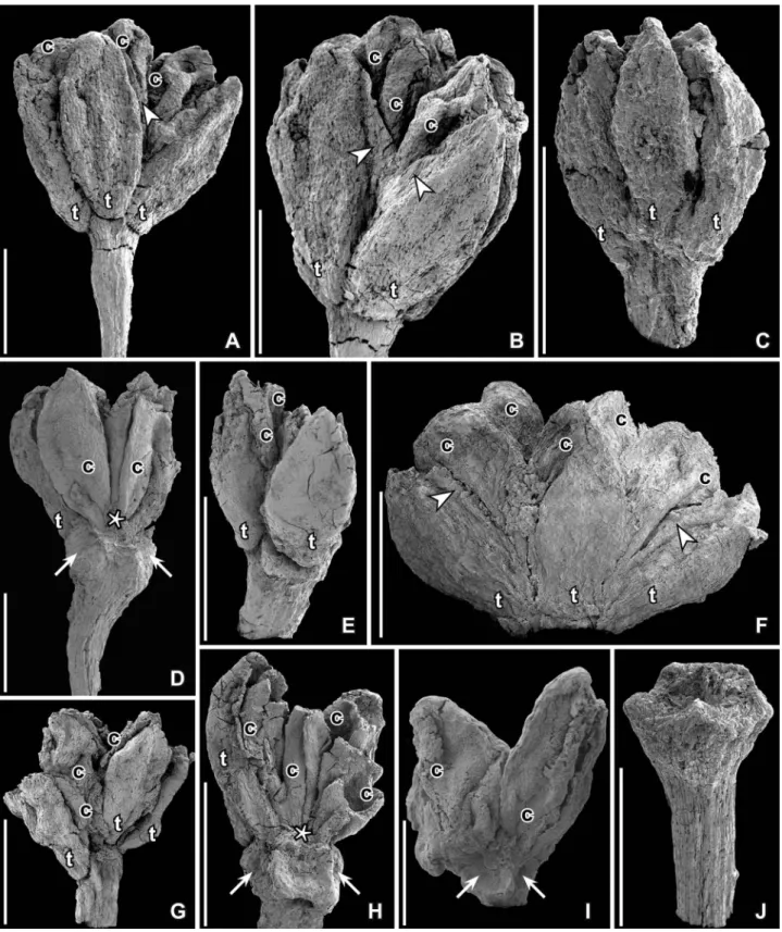
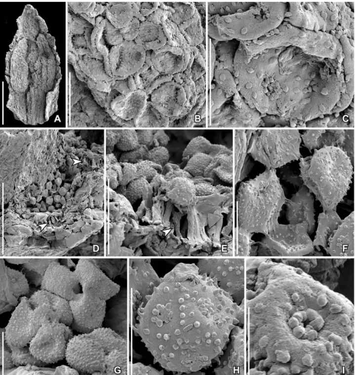
collec-Figure 1. SEM images of Paisia pantoporata gen. et sp. nov.,flowers from the Early Cretaceous Catefica locality, Portugal. A, B. Holotype; antheticflower with pedicel, tepals (t) and carpels (c) preserved; margins of tepals are incurved at base and very thin (arrow heads) further up (S101214; sample Catefica 49). C. Floral bud with all floral parts and part of pedicel preserved; tepals (t) cover stamens and carpels (S118680; sample Catefica 49). D. Fragmentary anthetic flower with pedicel preserved; one tepal (t) and two carpels (c) are exposed and the ventral suture of one carpel is visible; asterisk marks base of slit; facets on receptacle (arrows) show scars from tepals and stamens (S174739; sample Catefica 50). E. Anthetic flower with pedicel preserved showing tepals (t) with incurved margins and carpels (c) (S170429; sample Catefica 342). F. Anthetic flower showing tepals (t) and carpels (c); note the thin margins of tepals (arrowheads) (S174302; sample Catefica 242). G. Antheticflower with tepals (t) and carpels (c) preserved; base of tepals incurved; note downwards expanded base of tepals (S171519; sample Catefica 342). H. Same specimen as in (E) from opposite side showing receptacle with scars from tepals and stamens (arrows); tepals (t) and carpels (c) only partly preserved; asterisk shows base of ventral slit (S170429; sample Catefica 342). I. Anthetic flower with two carpels (c) preserved and scars from tepals and stamens (arrow) (S118679; sample Catefica 49). J. Isolated pedicel with swollen, angular receptacle (S171530; sample Catefica 151). Scale bars – 500 µm.
tions of the Palaeobiology Department of the Swed-ish Museum of Natural History, Stockholm (S) and the Geological Museum of Lisbon, Portugal (P).
Systematic palaeontology Angiospermae
Eudicots Paisia gen. nov.
Derivation of generic name. — In honour of Professor João Pais (1949–2016) for his contribution to the palaeobotany and geology of Portugal.
Generic and specific diagnosis. — Flower small, pedi-cellate, actinomorphic, pentamerous and isomerous and functional bisexual with one whorl offive tepals, one whorl offive stamens and one whorl of five free carpels, all opposite on the same radii. Receptacle distinctly five-angled. Perianth of a single whorl of five tepals; aestivation involute-valvate. Tepal base extended downwards and margins incurved, clasping the stamens. Stamens with a single massive bundle extending from base to apex. Stamens differentiated into a short, stout filament and an elongated, basi-fixed anther. Anthers dithecate and tetrasporangiate; sporangia minute, separated by a massive connec-tive. Anther dehiscence latrorse by longitudinal slits. Pollen pantoporate, tectate-punctate, spiny. Gynoecium superior; carpels free, sessile, elongate, and plicate. Ovules many, borne in two longitudinal rows on either side of the ventral suture. Stigma sessile, decurrent, indistinct.
Type species designated here. — Paisia pantoporata sp. nov.
Paisia pantoporata sp. nov. (Figures 1–6,8)
Derivation of specific name. — From the pantoporate pollen observed in situ in the stamens.
Specific diagnosis. — See combined generic and
specific diagnosis.
Dimensions. — Mature flowers without pedicel
about 1 mm long and up to 1.2 mm wide.
Holotype. — S101214 (Catefica sample 49;
illu-strated here onFigures 1A,B,2A–G).
Paratypes. — S118679, S118680, S118682,
S171526, S174746, S174747 (Catefica 49);
S174739, S174740, S174742 (Catefica 50);
S171530 (Catefica 151); S174302 (Catefica 242);
S170429, S171519 (Catefica 342); S171514,
S171515 (Catefica 343); S174743, S174344
(Cate-fica 361). Totally, about 60 specimens; several speci-mens may be stored in box slides under the same number.
Type locality. — Catefica (39° 03ʹ 30ʺ N; 09°14ʹ 30ʺ W), between the villages of Catefica and Mugideira, about 4 km south of Torres Vedras, Portugal.
Type horizon and age. — Almargem Formation,
Early Cretaceous (late Barremian–early Albian).
Description and remarks. — The material includes
several complete flowers (Figures 1A–C, F, G, 2A– G) or partly preservedflowers (Figures 1D,E,H,I,
3A–D, H-J, 4A–C) as well as isolated floral parts, such as tepals either empty or enclosing a single stamen (Figure 5A), pedicels where all floral parts are shed (Figures 1J, 6A–D), young carpels and sta-mens (Figure 3E–G) and mature follicles. Most flowers are preserved in the anthetic or post-anthetic stage, while a few are preserved in the pre-anthetic stage.
Flowers are pedicellate, small, about 1 mm long without pedicel and up to 1.2 mm in diameter, actinomorphic, pentamerous and isomerous with an
apocarpous and superior gynoecium (see floral
reconstruction and floral diagramFigure 8). Tepals and the enclosed stamens and carpels are inserted on
a five-angled receptacle with facetted sides with
tepals and stamens borne on the receptacle facets. Thefloral organs are apparently arranged in whorls. The perianth consists of a single whorl of parts, described here as tepals. Tepals are elliptical to ovate in dorsal view (Figure 1A–C, E–H) with pointed apex and rounded base that is distinctly separated from the pedicel and curving slightly downwards forming a dorsal extension that is some-times prominent (Figures 1A,B,E,G,2C). In cross-section, the tepals are more or less M-shaped with a broad base and incurved, thinner margins (Figures 2F–G, 3H–J). In young flowers, the tepal margins are incurved along the full length of the tepals (Figures 3B,H–J, C), while in more matureflowers the margins are incurved only at the base (Figures 1B, 2A, G) and in all specimens the tepal margins are thinner than the middle part of the tepals (Figures 1A, B, F, 2A, E, F). The tepals are fleshy composed of equiaxial, rounded parenchyma cells (Figures 2C,3H–J). There are three bundles
extend-ing almost for the full length of the tepals
(Figure 3I). The inner surface of the tepals has densely spaced bulging papillae (Figure 3C,H).
The androecium consists offive stamens opposite the tepals and the carpels. Stamens are elongate,
about 0.5–0.8 mm long, embraced by the incurved
margins of the tepals. In youngflowers, they consist of a short, stoutfilament, about 0.25 mm long that
grades without a joint into the basally attached, elon-gate anther (Figures 3B,E,4A,B). Anthers in young flowers are about 0.35 mm long, dithecate and tetra-sporangiate with small pollen sacs (Figures 3G, I,
4A–C). The two thecae are separated by a broad
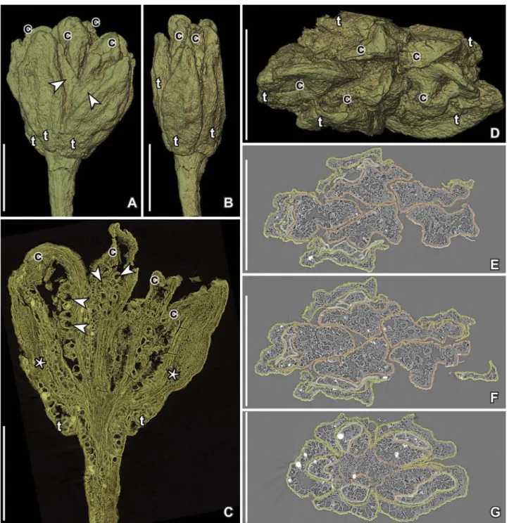
Figure 2. SRXTM volume renderings (A–D) and reconstructed transverse orthoslices (E–G) of Paisia pantoporata gen. et sp. nov., anthetic flower from the Early Cretaceous Catefica locality, Portugal (holotype, S101214; sample Catefica 49). A, B. Lateral views of flower at different angles showing well-preserved tepals (t) with thin margins (arrowheads) surrounding the carpels (c). C. Cut longitudinal volume rendering offlower (orthoslices yz500–520) showing sections of carpels with numerous small ovules/seeds (arrowheads) that do not fill out the ovary cavity; stamens (asterisk) are preserved between the tepals (t) and carpels. D. Apical view offlower showing the five tepals (t) and five carpels (c). E–G. Sections through flower at different levels from close to apex (E) to the base of the flower (G) showing the five tepals (light green) clasping thefive stamens (light yellow) and surrounding the five carpels (orange); tepal margins at base incurved, further up extended outwards. Scale bars– 500 µm.
Figure 3. SEM images (A–F) and SRXTM reconstructed transverse orthoslices (G–J) of Paisia pantoporata gen. et sp. nov., floral organs from the Early Cretaceous Catefica locality, Portugal. A–D, H–J. Pedicel and floral organs isolated from the same floral bud (S171526; sample Catefica 49). A. Pedicel in apical view showing swollen, slightlyfive-angled receptacle with scars from the floral organs. B. Single tepal clasping a stamen (asterisk); inner surface of tepal above stamen is papillate (arrowhead). C. Papillate inner surface of tepal enlarged. D. Young carpel with almost straight and parallel dorsal and ventral faces. H–J. Transverse orthoslices at different levels through two tepals; each tepal clasping a stamen; tepals fleshy with three vascular bundles (arrows in I); large, isodiametric parenchyma cells and incurved margins from apical part (H) to the base (J); stamens (asterisks) show massive connective and tiny pollen sacs (I) and a massivefilament with a single central strand (J). E. Isolated stamen (asterisk) and adhering carpel (c) in lateral view showing longitudinal dehiscence line of anther (S171514; sample Catefica 343). F. Cut longitudinal volume rendering of same specimen as shown in E (cut through median part of carpel and stamen) showing numerous, tiny ovules notfilling the ovary cavity. G. Transverse orthoslice (orthoslice xy1300) of the same specimen as shown in (E) showing stamen (asterisk) with massive connective with a single bundle and the four tiny, lateral pollen sacs (arrowheads) and section through middle part of carpel (c) with tiny ovules on both side of the ventral suture. Scale bars– 500 µm (B), 250 µm (H–J), 200 µm (A–F), 100 µm (G), 50 µm (C).
connective (Figures 3G, I, 4C). There is a single bundle extending from base almost to apex. The pollen sacs protrude and are laterally oriented with lateral dehiscence by longitudinal slits.
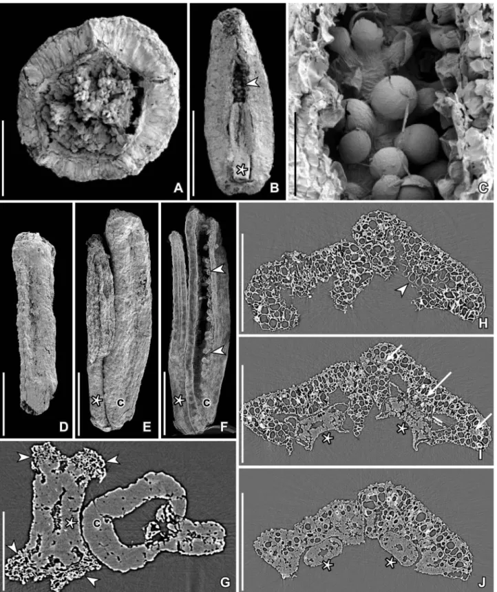
Pollen grains are found in situ in the anthers of severalflowers and in isolated fragments (Figure 5A– I). They are pantoporate, spheroidal and minute, about 11–14 µm in diameter, with tectate-punctate pollen wall and a supratectal ornamentation of spiny, conical elements, 0.7–1.0 µm long, constricted at the base, with a pointed or blunt tip, and with longitu-dinal ridges that give the elements an appearance of being compound of elongated elements (Figure 5I). The elements form a dense ring around each aper-ture, but are otherwise more irregularly scattered over the tectum surface (Figure 5C, F–I). In some specimens the pollen grains appear immature with supratectal elements of adjacent grains coalesced (Figure 5F, G). There are about six to eight pores, about 3 µm in diameter, and globally distributed over the grain. The aperture membrane is covered by irregular sculptural elements (Figure 5C, H) that are sometimes completely concealed by the surrounding spiny elements (Figure 5I). Orbicules were not observed.
There are five free carpels (Figures 1F, 2D–G). Carpels are sessile and elongated, interpreted as pli-cate. In youngflowers they are narrow and of almost the same width from base to apex and with almost straight ventral and dorsal margins (Figures 3D–F). In more maturefloral structures they are elliptical to obovate with slightly convex ventral margin and rounded dorsal margin (Figures 1D,F, I, 2C). The ventral slit extends for the full length of the carpel. This is obvious on broken specimens (Figure 1D,H) as well as in some of the specimens studied using SRXTM. There is no indication of an ascidiate zone in any of the specimen studied. The stigma is sessile and indistinct. The carpel wall is smooth and con-sists of almost equiaxial, rounded parenchyma cells. There are one dorsal and two ventral bundles. The outer epidermis is composed of tiny, equiaxial cells, apparently without stomata.
Ovules are numerous per carpel (about 20–30)
and tiny, apparently anatropous and both in young
stages (Figure 3E, F) and more mature flowers
(Figure 2C) ovules do not fill the ovary cavity. They are borne in two longitudinal rows, one on each side of the ventral suture extending from the carpel base to the apex.
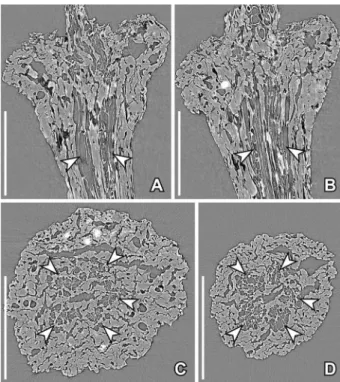
The pedicel is about 0.2 mm in diameter below the receptacle. The preservation does not allow a detailed description of the stem anatomy and com-parison with extant plants, but in all specimens stu-died by SRXTM it is clear that there arefive or more vascular bundles close to the centre of the pedicel
surrounding a small pith and surrounded by a broader zone of thick-walled parenchyma cells and a thick epidermis (Figure 6A–D).
?Paisia sp. (Figure 7)
One specimen (P0292) from the Catefica locality
(Figure 7A–E) consists of a small axis, about 3.2 mm long, with a terminal apocarpous gynoecium and a lateral bract further down the axis supporting a bud. The bud is in an early developmental stage and it is unknown whether it is vegetative or afloral bud. The axis is more or less circular in transection, about 0.2 mm in diameter. Vascular bundles are arranged in a ring around the small central pit and are sur-rounded by a tissue of thick-walled parenchyma cells.
Figure 4. SRXTM reconstructions of two tepals and two stamens of Paisia pantoporata gen. et sp. nov., from the Early Cretaceous Catefica locality, Portugal; floral organs from the same floral bud as inFigure 3A–D,H–J(S171526; sample Catefica 49). A. Long-itudinal voltex of tepals (t) and stamens (asterisk) and surface rendering (yellow) of one stamen showingfilament, massive con-nective and tiny pollen sacs. B. Transparent voltex showing thick parenchyma tissue of tepals and stamens enclosed in the tepals. C. Cut transverse voltex reconstruction at the level of the pollen sacs showing the two tepals with incurved margins clasping the stamens (asterisks); note massive connective with one central bundle and the two lateral thecae with tiny pollen sacs. Scale bars– 500 µm (A, B); 250 µm (C).
The terminal gynoecium appears to be in a post-anthetic stage. There are no perianth parts or sta-mens preserved. The receptacle below the carpels is
facetted with a number of scars (Figure 7A,B) indi-cating that otherfloral organs were originally present in theflower, but shed after anthesis. The gynoecium
Figure 5. SEM images of stamens and pollen in situ of Paisia pantoporata gen. et sp. nov., from the Early Cretaceous Catefica locality, Portugal. A. Isolated tepal and enclosed stamen with pollen in situ (S170388; sample Catefica 50). B, C. Pantoporate pollen from stamen shown in (A); note the scattered supratectal elements on the pollen body and the concentration of elements around the apertures (S170388; sample Catefica 50). D–H. Fragment of tepal enclosing stamen with pantoporate pollen in situ from floral structure shown in Figure 1I; cell wall thickenings of anther wall are seen in (D) and (E) (arrowheads); the pollen grains appear to be immature, sometimes grouped in tetrads and sometimes with supratectal elements of adjacent grains coalesced (F, G); supratectal elements with blunt or spiny apex (H) (S118679; sample Catefica 49). I. Detail of pollen grain from floral structure inFigure 1Gshowing apparent compound supratectal elements; note concentration of elements around the aperture (S171519; sample Catefica 342). Scale bars: 250 µm (A), 100 µm (D), 25 µm (E), 20 µm (B), 10 µm (C, F–H), 5 µm (I).
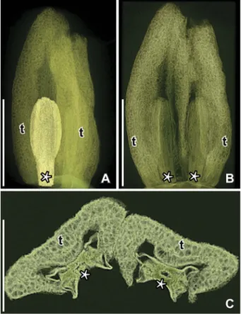
consists of three free carpels (Figure 7A–C, E), about 0.52 mm long and 0.34 mm wide, ovoid to elliptic in shape, each with a distinct ventral suture and an indistinct, sessile stigma (Figure 7C). No pollen grains were observed in the stigmatic area. There are about ten ovules/seeds borne in two
long-itudinal rows along the entire ventral suture
(Figure 7D, E). The ovules/seeds do not fill in the ovary cavity. The epidermal cells of the carpels are polygonal and more or less isodiametric in surface view.
The fossil specimen is similar to Paisia pantopor-ata in the facetted receptacle, large parenchymatic cells in pedicel, receptacle and carpels, and a ring of vascular bundles near the centre of the pedicel. The fossil is, however, distinguished from Paisia panto-porata by its trimerous gynoecium. It is possible that the specimen represents an aberrant tricarpellate form of the otherwise pentacarpellate Paisia panto-porata, but it is more likely that the specimen repre-sents a new species, either of Paisia or a new fossil genus. With the material currently available, a de fi-nite assignment of the specimen to Paisia is not possible and we therefore refer to the specimen as ?Paisia sp.
Discussion
Comparison of Paisia pantoporata with Early Cretaceousflowers and pollen
A number of isolated plicate carpels apparently from apocarpous gynoecia co-occur with Paisia pantopor-ata and ?Paisia sp. in the Catefica mesofossil flora and may represent related forms. A diversity of fossils with apocarpous gynoecia is also known from other Early Cretaceous mesofossil floras in Portugal and eastern North America (for Late Cretaceous follicular fruitlets see later). Some of them are known from flowers and have been assigned to eudicots based on their organisation and in situ tricolpate pollen. Kajanthus lusitanicus E.M.Friis, M.M.Mendes et K.R.Pedersen (Mendes et al.2014) from the Chical-hão locality (late Aptian–early Albian) of Portugal, and Kenilanthus marylandensis E.M.Friis, K.R.Peder-sen et P.R.Crane from early–middle Albian strata of the Kenilworth locality, Maryland, USA (Friis et al.
2017), both have structurally bisexualflowers and an apocarpous gynoecium of three tofive plicate carpels. Kajanthus is closely related to the extant Sinofranche-tia of the ranunculalean family Lardizabalaceae (Mendes et al.2014) and is distinguished from Paisia pantoporata by its trimerous flowers and tricolpate, reticulate pollen. Kenilanthus is similar to Paisia in having a pentamerous and isomerous organisation with five free carpels and numerous ovules that do
notfill out the ovary space. Kenilanthus may also have a single whorl of tepals as in Paisia pantoporata, but the nature of the perianth is not fully documented for
Kenilanthus. Kenilanthus differs in many other
respects from Paisia and they are probably not closely related. It has two whorls of apparently extrorse sta-mens and pollen grains are tricolpate-reticulate. Also, the epidermis of the Kenilanthus carpels have scat-tered stomata not observed for the carpels of Paisia.
Teixeiraea lusitanica K.R.Pedersen et E.M.Friis is another early eudicotflower with in situ pollen from the Early Cretaceous Vale de Água locality (late Aptian–early Albian) of Portugal. It is distinguished from Paisia in the multiparted nature of the androe-cium and the reticulate, tricolpate pollen. Further, Teixeiraea lusitanica is apparently unisexual and
cur-rently only the staminate flower is known (von
Balthazar et al. 2005). Specimens with an apocar-pous gynoecium from the Early Cretaceous (late Barremian–early Aptian?) Torres Vedras locality are multicarpellate, apparently with carpels in a helical arrangement (e.g. Friis et al.2011).
Two pentacarpellate and apocarpous structures with free plicate carpels were reported from the Early Cre-taceous (early–middle Albian) Puddledock locality,
Figure 6. SRXTM reconstructions of pedicel and receptacle anat-omy in Paisia pantoporata gen. et sp. nov., from the Early Cretac-eous Catefica locality, Portugal, same specimen as in Figure 1J
(S171530; sample Catefica 151). A–D. Longitudinal (A, B) and transverse (C, D) orthoslices showing thick-walled cells towards the periphery and ring of bundles (arrowheads) around the narrow central pith (A, orthoslice xz470; B, orthoslice xz640; C, ortho-slice xy700; D, orthoortho-slice 1015). Scale bars– 200 µm.
Virginia, USA (unnamed pistillate flower of Crane et al. [1994,figure 6a, 6b]). They may both be related to Paisia and particularly the specimen shown in Crane et al. (1994, figure 6a); has a comparable expanded receptacle. The other specimen is distinguished by its scattered stomata. In both specimens only the pistillate organs are preserved and it is uncertain whether the flowers were unisexual or bisexual.
Flowers with an apocarpous gynoecium are also known from several Early Cretaceous macrofossils floras including Sinocarpus decussatus Leng et E.M. Friis from the Aptian Yixian Formation of Liaoning, China (Leng & Friis 2003, 2006), Hyrcantha kar-atscheensis (Vakhram.) Krassilov, Shilin et Vakhram.
from the Albian of Kazakhstan (Vakhrameev 1952;
Krassilov et al.1983), Ranunculaecarpus
quinquecarpel-latus Samylina from the Albian of eastern Siberia, Rus-sia (Samylina 1960), and Ternariocarpus floribundus Krassilov et Volynets from the Albian of the Primorye Region, Russia (Krassilov & Volynets 2008). These fossils are all preserved in the post-anthetic stage and none of them show details of floral organisation (see Friis et al.2011).
In situ pantoporate pollen grains have also been discovered in stamens and coprolites from two Early Cretaceous mesofossilfloras. The spiny pantoporate grains from the Torres Vedras locality (Friis et al.
2010, plate 5, figures 1–2) are similar to those of Paisia pantoporata in the spiny supratectal ornamen-tation and aperture configuration, but details of the pollen wall is distinctly different from that of Paisia and the plants producing these two pollen types may
Figure 7. SEM illustrations (A–C) and SRXTM reconstructions (D–E) of ?Paisia sp., axis with tricarpellate gynoecium, from the Early Cretaceous Catefica locality, Portugal (P0292; sample Catefica Mendes 125). A. Axis with terminal floral structures and two lateral buds. B.Close up of terminalfloral structure consisting of a tricarpellate, apocarpous gynoecium and the angular, swollen receptacle with scars from otherfloral organs. C. Apical view of gynoecium showing the three free carpels and the ventral sutures; stigmatic area indistinct. D, E. Longitudinal (D) and transverse (E) orthoslices of gynoecium showing two rows of ovules along the ventral margins of the carpels (arrowheads); note ovules do notfill ovary cavity; fruit wall and receptacle with large isodiametric parenchyma cells (D, orthoslice; E, orthoslice xy). Scale bars– 1 mm (A), 500 µm (B, D), 250 µm (C, E).
not be closely related. Pantoporate pollen from the Famalicão flora (Friis et al. 1999, figures 75–77) is
reticulate with small, indistinct apertures and
without supratectal ornamentation. They are very similar to pollen of extant Sarcandra (Chlorantha-ceae) and clearly distinct from the pollen of Paisia.
Dispersed pantoporate grains similar to those observed in Paisia pantoporata have also been found in the Catefica palynoflora, but are so far unknown from other dispersed palynological assemblages asso-ciated with the Early Cretaceous mesofossilfloras of Portugal. The Paisiaflowers and pollen are not com-mon in the Catefica assemblages suggesting that the plant was not abundant in the vegetation. Pollen sacs are tiny comprising only a small part of the total stamen suggesting low pollen production for each flower. This together with an herbaceous habit sug-gested by the stem anatomy and possible insect pol-lination suggested by the spiny pollen would result in low pollen dispersal with no or only little pollen transported to the depositional basin.
Dispersed pantoporate or polyporate grains from the Cretaceous are typically assigned to the pollen genera Australopollis, Bochemiperiporis, Cretacaeiporites, Erdtmanipollis, Penetetrapites, Periporopollenites and Polyporites (e.g. Chlonova1986). None of them have spiny supratectal ornamentation similar to that of Paisia pantoporata, but detailed comparison with the dispersed grains is typically impeded by lack of SEM images that shows details of pollen wall. The several species of Cretacaeiporites reported from the dispersed palynological assemblages of the São Julião shallow marine section near Ericeira, western Portugal (Hor-ikx et al.2016), are all different from the in situ pollen of Paisia in the lack of spines. Other reports of
panto-porate pollen from the Iberian Peninsula include rare occurrences of Penetetrapites from the late Albian of north-eastern Spain (Sender et al. 2012). Early Cre-taceous reports from other regions shows that although pantoporate pollen are not common they were already geographically widespread in the Aptian with occurrences in Egypt and Columbia (Ibrahim et al. 2017) and by the Albian pantoporate pollen grains are diverse and almost global in distribution with occurrences in areas such as the former SSSR, Portugal, Egypt, Qatar, Sudan, Morocco, Tanzania, western Equatorial Africa, eastern North America, Brazil, Peru, and western China (e.g. Herngreen
1973; Chlonova 1986; Ibrahim et al. 2017; Zhang et al.2015; Ferreira et al.2016; Horikx et al.2016).
Systematic assessment of Paisia pantoporata
The arrangement of tepals, stamens and carpels in the same radii is unusual among extant angiosperms. Further, tepals, stamens and carpels of each radius appear almost as a separate synorganised unit with the tepal margin enveloping not only the stamens, but also part of the carpel base. An intriguing possi-bility is that the floral structure represents an inflor-escence offive simple flowers consisting of a bract, a stamen and a carpel instead of a single pentamerous flower (J. Schönenberger, personal communication, January 2017). Unfortunately, the fossil material cur-rently available does not allow for developmental studies and we therefore maintain the interpretation of the floral structure as a single flower. Uncertain-ties regarding floral organisation make comparison withflowers of extant plants difficult. For instance, it is unknown whether Paisia pantoporata is primitively monochlamydous or whether the single whorl of perianth parts is secondarily derived from a typical
heterochlamydous eudicot flower with two sets of
perianth parts by reduction of either the inner or outer whorl. This together with the relatively few morphological features impedes a precise phyloge-netic analysis of Paisia pantoporata.
Testing the position of Paisia using the morpholo-gical dataset and backbone constraint tree of Doyle and Endress (2010) by adding the pantoporate/pan-tocolpate aperture state resulted in a most parsimo-nious position between the Chloranthaceae and the remaining angiosperms, but most other positions were only a few steps less parsimonious. Using the Wang et al. (2009) dataset and backbone tree (Wang et al. 2009: figure 3: Strict consensus tree of eight most parsimonious trees, MPTs) suggested a posi-tion in the Ranunculales close to Berberidaceae and Ranunculaceae. The following characters of Wang et al. (2009) were scored for Paisia: 1–20: ?; 21,
inflorescence: ?; 22, sex: 0 = monoclinous; 23,
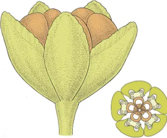
Figure 8. Reconstruction of open flower and floral diagram of Paisia pantoporata; light green, tepals and pedicel; light yellow, stamens; orange, carpels.
calyx: ?; 24, perianth phyllotaxis: 1 = whorled; 25, perianth whorls: 2 = one; 26, perianth arrangement: 3 = 5; 27, nectary petals: ?; 28, androecium phyllo-taxis: 1 = whorled: 29, stamen arrangement: 2 = in fives; 30, stamen fusion: 0 = free; 31, connective apex: 1 = truncate or smoothly rounded; 32, orienta-tion of dehiscence: 1 = latrorse; 33, pollen aperture type: 2 = porate; 34, pollen aperture number: 2 = more than three; 35, exine sculpturing: 3 = spi-nose; 36, pollen ektexine: ?; 37, pollen endexine: ?; 38, gynoecium phyllotaxis: 1 = whorled; 39, carpel number: 0 = more than 3; 40, carpel form: 0 = pli-cate; 41, carpel fusion: 0 = apocarpous; 42, pollen transmitting tissue: ?; 43, tanniferous tissue in car-pel: 0 = absent; 44, stylar scar: ?; 45, placentation type: 0 = marginal; 46, ovule number: 0 = more than 2; 47–65: ? (for method, see Friis et al. [2017]). None of the signals were very strong and the analysis was not conclusive.
Pantoporate pollen occurs scattered among all major groups of angiosperms and is reported both among early diverging lineages as well as in more derived groups. Among monocots, members of Alis-mataceae and Limnocharitaceae are characterised by having pantoporate pollen (Argue1973,1974,1976; Chanda et al. 1988; Furness & Banks 2010) and pollen in some genera such as Sagittaria and Hydro-cleys has spiny supratectal elements scattered over the tectum and surrounding the apertures in a similar way as seen in the pollen of Paisia. However, in Paisia, the elements appear compound and con-stricted at the base, while the microechinate-echinate pollen of Alismataceae and Limnocharitaceae has more conical and non-constricted supratectal ele-ments. A position of Paisia among monocots is highly unlikely due to the pentamerous arrangement of thefloral parts. Instead the pentamerous arrange-ment of perianth, androecium and gynoecium sug-gests that Paisia is related to eudicot angiosperms.
Pantoporate pollen together with most floral fea-tures of Paisia including the massive stamen
fila-ments, basifixed anthers with broad connective
between the thecae, longitudinal anther dehiscence, apocarpous gynoecium with plicate carpels and many small ovules, occur commonly among mem-bers of the earliest diverging eudicot order Ranun-culales (see discussion in Friis et al. [2017]). In Ranunculales,flowers with a single series of perianth parts are not common, but do occur in several taxa, sometime the outer set and sometimes the inner set are lost. Whether the single set of perianth parts in Paisia is a result of a secondary loss is unknown. The apparent presence of three vascular bundles might suggest that the tepals represents an outer set of perianth parts, but there is also some resemblance in the downwards extended base of the tepals to
young petals in certain Ranunculaceae such as Semi-aquilegia (Tucker & Hodges2005) and the petals of Sabia (Sabiaceae, see later), although the hollow spurs of Semiaquilegia are clearly distinct from the
solid tepal bases of Paisia. The isomerous floral
organisation also occur among Ranunculales, but are more common among rosid angiosperms (see discussion in Friis et al. [2017]). However, none of these isomerous rosids have pantoporate pollen and core eudicots with pantoporate pollen and spiny supratectal ornamentation such as members of the Caryophyllales, Malvales and Curcubitales are all clearly distinct from Paisia.
In the Ranunculales, pantoaperturate (pantopo-rate and pantocolpate) pollen occurs in the
Eupte-leaceae, Papaveraceae, Berberidaceae and
Ranunculaceae. The position of Eupteleaceae as sis-ter to all other Ranunculales and the position of Ranunculales as sister to all other eudicots (Stevens 2001 onwards) make the occurrence of pantoapertu-rate pollen in Eupteleaceae particularly interesting. In most species of Euptelea the grains are pantocol-pate, but partially pantoporate grains occur in Eupte-lea polyandra Siebold et Zucc., while tricolpate pollen is less common in Euptelea (Praglowski 1974) and the scoring of Euptelea pollen as basically tricolpate as in the analysis of pollen evolution by Doyle (2005) could be misleading. In Papaveraceae, pantoporate
pollen is known in Fumarioideae (Rupicapnos,
Fumaria) and Papaveroideae (Bocconia, Eomecon, Macleaya, Meconopsis, Papaver, Roemeria,
Sangui-naria) (Kadereit 1993; Blackmore et al. 1995;
Wang et al.2009) and in Berberidaceae, pantoporate pollen is reported for Ranzania (Nowicke & Skvarla
1981). In the Ranunculaceae, pantoporate pollen is known for species of Caltha (Smit & Punt 1969),
Thalictrum (Wodehouse 1936; Blackmore et al.
1995; Tatlidil et al. 2005), Clematis (Wang & Xie
2007; Xie & Li 2012), Coptis (Wodehouse 1936), Ranunculus (Nowicke & Skvarla 1979; Blackmore et al. 1995; Emadzade et al.2010), Krafia and Lac-copetalum (Emadzade et al. 2010) and pantocolpate pollen is known for Hepatica (Nowicke & Skvarla
1981). Many of the pantoporate grains in Ranuncu-laceae have a punctate-perforate tectum with micro-echinate-echinate supratectal ornamentation that in some species is superficially similar to the ornamen-tation in Paisia, but as for the alismatalean pollen, the supratectal elements are not constricted at their bases, do not appear compound, and are typically more densely distributed.
Flowers of modern Sabia (Sabiaceae), another early diverging eudicot, have somefloral structures that are similar to those of Paisia pantoporata. Particularly the shape and structure of the inner set of perianth parts (petals) in the flower buds of Sabia limoniacea Wall.
are M-shaped at the base embracing the massive sta-men filament as in Paisia. Also the inner surfaces of the petals of Sabia are papillate comparable to the tepal surface of Paisia (Ronse de Craene et al.
2015). However, in the preliminary phylogenetic ana-lysis using the Wang et al. (2009) dataset Paisia is not resolved as close to Sabia or Sabiaceae. Critical differ-ences are the two sets of perianth whorls, dithecal and disporangiate anthers, bicarpellate, syncarpous or sometimes monocarpellate gynoecium and the pre-sence of a nectary disk (Ronse de Craene et al.
2015) as well as tricolporate pollen in Sabia and other Sabiaceae (Furness et al.2007).
Apocarpous gynoecia occur in other early diver-ging eudicot lineages such as Proteales, but members of Proteaceae and Platanaceae all have triaperturate, or sometimes biaperturate, pollen. Five free carpels are common in fossil platanoid flowers, but flowers of Platanaceae are unisexual, the carpels uniovulate and the associated pollen tricolpate. In Proteaceae,
the flowers are typically tetramerous, sometimes
borne in pairs, with a monocarpellate or sometimes
bicarpellate gynoecium, and porate or more
rarely colporoidate pollen (Weston2007). The
folli-cular fruitlets of Agapitocarpus, Chontrocarpus,
Maiandrocarpus, Malliocarpus, Mitocarpus, Xylocarpus and Zeugarocarpus from the Late Cretaceous Åsen mesofossil flora (Leng et al. 2005) show superficial resemblance to the fruitlets of Paisia and were com-pared to extant Proteaceae, although they could not be included in the family. Some of these Late Cre-taceous fossils are among the most abundant plant remains in the Åsen mesofossil assemblages. They are commonly found isolated, but those that are found attached to the inflorescence/infructescence axis are borne either single or in pairs and probably not closely related to Paisia.
Pantoporate pollen also occurs among basal grade angiosperms in two species of Trimenia (Trimenia-ceae, Austrobaileyales) and in Sarcandra (Chlorantha-ceae; Sampson & Endress 1984; Endress 1986). Floral structures in these taxa are distinctly different from theflowers of Paisia and Trimenia and Sarcandra also differ from Paisia in their tectum ornamentation. The pantoporate pollen of Trimenia is weakly rugu-lose, while the pollen of Sarcandra is reticulate (Samp-son & Endress1984; Endress1986).
Supratectal sculptural elements similar to those of Paisia have not been observed in any of the panto-porate pollen of extant angiosperms that we have observed, but this kind of compound spines are known for inaperturate pollen of the two extant gen-era Peumus and Palmeria of the Monimiaceae in the Laurales (Sampson & Foreman 1990), but floral morphology of Peumus and Palmeria are distinct from that of Paisia in most features including their
unisexual organisation, cup-shaped receptacle, and irregular numbers of floral parts.
Conclusion
Paisia pantoporata described here from the Catefica locality, Portugal, is thefirst Early Cretaceous flower to be described with pantoporate in situ pollen. The combined floral and palynological features indicate that Paisia most probably belongs to an extinct line-age close to the base of the eudicot angiosperms, most likely close to the Ranunculales, but the exact position has not been established. Two other kinds of pantoporate pollen were reported from stamens and coprolites of the Early Cretaceous mesofossil floras of Torres Vedras and Famalicão, Portugal, and pantoporate pollen are also recorded from other parts of the Lusitanian Basin (Horikx et al.
2016). The occurrence of pantoporate pollen in
Early Cretaceous (Aptian–Albian) palynological
assemblages from other regions documents that plants producing this kind of pollen occur early in the history of angiosperms with dispersed grains geo-graphically widespread already in the Aptian (Ibra-him et al. 2017) and diverse and almost globally distributed by the Albian (Chlonova 1986; Ibrahim et al. 2017). The Early Cretaceous record of
panto-porate pollen in the Portuguese mesofossil floras
suggests that plants producing this kind of pollen may have been much more diverse and widespread in the Early Cretaceous than is apparent from the palynological record.
Acknowledgements
The authors thank Marco Stampanoni and Federica Marone, Swiss Light Source, Paul Scherrer Institute, Villigen, Switzerland, and Anna Lindström, Swedish Museum of Natural History, Stockholm, Sweden, for help with the SRXTM analyses performed at the Swiss Light Source, Paul Scherrer Institute, Villigen, Switzer-land. Thanks are also due to Jacques Rey, Université Paul-Sabatier, Toulouse, France, for helpful geological information, to Eduarda Ferreira, New University of Lisboa, Portugal, for technical assistance, to Pollyanna von Knorring, Swedish Museum of Natural History, Stockholm, Sweden, for preparing the linedrawings, and to Jürg Schönenberger, University of Vienna, Aus-tria, and Hervé Sauquet, Université Paris-Sud, Orsay, France, for useful suggestions to the manuscript.
Disclosure statement
No potential conflict of interest was reported by the authors.
Funding
This research was supported by the Swiss Light Source at the Paul Scherrer Institute (European Union FP6 projects 20130185 and 20141047), by the Swedish Research Council (2014-5228), the Por-tuguese Science Foundation (FCT) grant UID/ MAR/00350/2013 to CIMA of the University of Algarve and the CretaCarbo (PTDC/CTE-GIX/ 113983/2009) project.
ORCID
Else Marie Friis http://orcid.org/0000-0003-2936-2761
Mário Miguel Mendes http://orcid.org/0000-0002-5470-5634
Kaj Raunsgaard Pedersen http://orcid.org/0000-0003-3038-0967
References
Argue CL.1973. The pollen of Limnocharisflava Buch., Hydrocleis nymphaoides (Willd.) Buch., and Tenagocharis latifolia (Don) Buch. (Limnocharitaceae). Grana 13: 108–112. doi:10.1080/ 00173137309429885.
Argue CL.1974. Pollen studies in the Alismataceae (Alismaceae). Botanical Gazette 135: 338–344. doi:10.1086/336770. Argue CL.1976. Pollen studies in the Alismataceae with special
reference to taxonomy. Pollen et Spores 18: 161–201. Blackmore S, Stafford P, Persson V.1995. Palynology and
sys-tematics of Ranunculiflorae. Plant Systematics and Evolution [Supplement] 9: 71–82.
Chanda S, Nilsson S, Blackmore S.1988. Phylogenetic trends in the Alismatales with reference to pollen grains. Grana 27: 257– 272. doi:10.1080/00173138809429948.
Chlonova AF.1986. Distribution of several morphologic pollen types of the Cretaceous angiosperms. Review of Palaeobotany and Paly-nology 48: 365–372. doi:10.1016/0034-6667(86)90074-6. Crane PR, Friis EM, Pedersen KR.1994. Paleobotanical evidence
on the early radiation of magnoliid angiosperms. Plant Sys-tematics and Evolution Supplement 8: 51–72.
Doyle JA.2005. Early evolution of angiosperm pollen as inferred from molecular and morphological phylogenetic analyses. Grana 44: 227–251. doi:10.1080/00173130500424557. Doyle JA, Endress PK.2010. Integrating Early Cretaceous fossils
into the phylogeny of living angiosperms: Magnoliidae and eudicots. Journal of Systematics and Evolution 48: 1–35. doi:10.1111/jse.2010.48.issue-1.
Doyle JA, Endress PK.2014. Integrating Early Cretaceous fossils into the phylogeny of living angiosperms: ANITA lines and relatives of Chloranthaceae. International Journal of Plant Sciences 175: 555–600. doi:10.1086/675935.
Emadzade K, Lehnebach C, Lockhart P, Hörandl E. 2010. A molecular phylogeny, morphology and classification of genera of Ranunculeae (Ranunculaceae). Taxon 59: 809–828. Endress PK.1986. Reproductive structures and phylogentic
sig-nificance of extant primitive angiosperms. Plant Systematics and Evolution 152: 1–28. doi:10.1007/BF00985348. Ferreira NN, Ferreira EP, Ramos RRC, Carvalho IS.2016.
Paly-nological and sedimentary analysis of the Igarape Ipiranga and Querru 1 outcrops of the Itapecuru Formation (Lower Cretac-eous, Parnaíba Basin), Brazil. Journal of South American Earth Sciences 66: 15–31. doi:10.1016/j.jsames.2015.12.005.
Furness CA, Magallón S, Rudall PJ.2007. Evolution of endoa-pertures in early-divergent eudicots, with particular reference to pollen morphology in Sabiaceae. Plant Systematics and Evolution 263: 77–92. doi:10.1007/s00606-006-0477-y. Friis EM, Crane PR, Pedersen KR. 2011. Early flowers and
angiosperm evolution. Cambridge: Cambridge University Press.
Friis EM, Crane PR, Pedersen KR. 2017. Kenilanthus, a new eudicotflower with tricolpate pollen from the Early Cretaceous (early-middle Albian) of eastern North America. Grana 56(3): 161–173.
Friis EM, Crane PR, Pedersen KR, Stampanoni M, Marone F.
2015a. Exceptional preservation of tiny embryos documents seed dormancy in early angiosperms. Nature 528: 551–554. doi:10.1038/nature16441.
Friis EM, Grimm GW, Mendes MM, Pedersen KR.2015b. Can-rightiopsis, a new Early Cretaceous fossil with Clavatipollenites-type pollen bridge the gap between extinct Canrightia and extant Chloranthaceae. Grana 54: 184–212. doi:10.1080/ 00173134.2015.1060750.
Friis EM, Marone F, Pedersen KR, Crane PR, Stampanoni M.
2014a. Three-dimensional visualization of fossilflowers, fruits, seeds and other plant remains using synchrotron radiation X-ray tomographic microscopy (SRXTM): New insights into Cretaceous plant diversity. Journal of Paleontology 88: 684– 701. doi:10.1666/13-099.
Friis EM, Pedersen KR.2014. Blomster fra Kridttiden. Geologisk Tidsskrift 2013: 43–64.
Friis EM, Pedersen KR, Crane PR. 1994. Angiosperm floral structures from the Early Cretaceous of Portugal. Plant Sys-tematics and Evolution [Supplement] 8: 31–49.
Friis EM, Pedersen KR, Crane PR.1999. Early angiosperm diver-sification: The diversity of pollen associated with angiosperm reproductive structures in Early Cretaceousfloras from Portu-gal. Annals of the Missouri Botanical Garden 86: 259–296. doi:10.2307/2666179.
Friis EM, Pedersen KR, Crane PR.2009. Early Cretaceous meso-fossils from Portugal and eastern North America related to the Bennettitales-Erdtmanithecales-Gnetales group. American Journal of Botany 96: 252–283. doi:10.3732/ajb.0800113. Friis EM, Pedersen KR, Crane PR.2010. Cretaceous diversi
fica-tion of angiosperms in the western part of the Iberian Penin-sula. Review of Palaeobotany and Palynology 162: 341–361. doi:10.1016/j.revpalbo.2009.11.009.
Friis EM, Pedersen KR, Crane PR.2013. New diversity among chlamydospermous seeds from the Early Cretaceous of Portu-gal and North America. International Journal of Plant Sciences 174: 530–558. doi:10.1086/668250.
Friis EM, Pedersen KR, Crane PR.2014b. Welwitschioid diver-sity in the Early Cretaceous: Evidence from fossil seeds with pollen from Portugal and eastern North America. Grana 53: 175–196. doi:10.1080/00173134.2014.915980.
Furness CA, Banks H.2010. Pollen evolution in the early-diver-gent monocot order Alismatales. International Journal of Plant Sciences 171: 713–739. doi:10.1086/654848.
Herngreen GFW.1973. Palynology of Albian–Cenomanian strata of borehole 1-QS-1-MA, State of Maranhao, Brazil. Pollen et Spores 15: 515–555.
Horikx M, Hochuli PA, Feist-Burkhardt S, Heimhofer U.
2016. Albian angiosperm pollen from shallow marine strata in the Lusitanian Basin, Portugal. Review of Palaeo-botany and Palynology 228: 67–92. doi:10.1016/j. revpalbo.2015.12.008.
Ibrahim MIA, Zobaa MK, El–Noamani ZM, Tahoun SS.2017. A review of the angiosperm pollen genus Cretacaeiporites Hern-green, with one new species from the Upper Cretaceous of Egypt. Palynology 41: 101–116.
Kadereit JW.1993. Papaveraceae. In: Kubitzki K, Rohwer JG, Bittrich V, eds. The families and genera of vascular plants. II. Flowering plants– Dicotyledons. Magnoliid, hamamelid and caryophyllid families, 494–495. Berlin, Heidelberg, New York: Springer. Krassilov VA, Shilin PV, Vachrameev VA.1983. Cretaceous
flow-ers from Kazakhstan. Review of Palaeobotany and Palynology 40: 91–113. doi:10.1016/0034-6667(83)90005-2.
Krassilov VA, Volynets Y.2008. Weedy Albian angiosperms. Acta Palaeobotanica 48: 151–169.
Leng Q, Friis EM.2003. Sinocarpus decussatus gen. et sp. nov, a new angiosperm with syncarpous fruits from the Yixian For-mation of Northeast China. Plant Systematics and Evolution 241: 77–88. doi:10.1007/s00606-003-0028-8.
Leng Q, Friis EM.2006. Angiosperm leaves associated with Sino-carpus Leng et Friis infructescences from the Yixian Formation (mid-Early Cretaceous) of NE China. Plant Systematics and Evolution 262: 173–187. doi:10.1007/s00606-006-0461-6. Leng Q, Schönenberger J, Friis EM. 2005. Late Cretaceous
follicular fruits from southern Sweden with systematic af fi-nities to early diverging eudicots. Botanical Journal of the Linnean Society 148: 377–407. doi: 10.1111/j.1095-8339.2005.00428.x.
Mendes MM, Grimm GW, Pais J, Friis EM.2014. Fossil Kajanthus lusitanicus gen. et sp. nov. from Portugal: Floral evidence for Early Cretaceous Lardizabalaceae (Ranunculales, basal eudicot). Grana 53: 283–301. doi:10.1080/00173134.2014.932431. Nowicke JW, Skvarla JJ.1979. Pollen morphology: The potential
influence in higher order systematics. Annals of the Missouri Botanical Garden 66: 633–700. doi:10.2307/2398914. Nowicke JW, Skvarla JJ.1981. Pollen morphology and
phyloge-netic relationships of the Berberidaceae. Smithsonian Contri-butions to Botany 50: 1–83. doi:10.5479/si.0081024X.50. Nowicke JW, Skvarla JJ.1982. Pollen morphology and the
rela-tionships of Circaeaster, of Kingdonia, and of Sargentodoxa to the Ranunculaceae. American Journal of Botany 69: 990–998. doi:10.2307/2442896.
Praglowski J.1974. The pollen morphology of the Trochodendra-ceae, TetracentraTrochodendra-ceae, Cercidiphyllaceae with reference to tax-onomy. Grana 16: 449–467.
Rey J.1992. Les unités lithostratigraphiques du Crétacé inférieur de la région de Lisbonne. Comunicações dos Serviços Geoló-gicos de Portugal 78: 103–124.
Rey J.1993. Les unités lithostratigraphiques du groupe de Torres Vedras (Estremadura, Portugal). Comunicações Instituto Geo-lógico e Mineiro 79: 75–85.
Ronse de Craene LP, Quandt D, Wanntorp L. 2015. Flower morphology and anatomy of Sabia (Sabiaceae): Structural basis of an advanced pollination system among basal eudicots. Plant Systematics and Evolution 301: 1543–1553. doi:10.1007/s00606-014-1176-8.
Sampson FB, Foreman DB.1990. Pollen morphology of Peumus boldus (Monimiaceae)– A comparison with Palmeria scandens. Grana 29: 197–206. doi:10.1080/00173139009427752. Sampson FB, Endress PK. 1984. Pollen morphology in the
Trimeniaceae. Grana 23: 129–137. doi:10.1080/001731384 09427708.
Samylina VA.1960. Angiosperms from the lower Cretaceous of the Kolyma Basin. Botanicheskij Zhurnal 45: 335–352.
Sender LM, Villanueva-Amadoz U, Diez JB, Sanchez-Pellicer R, Bercovici A, Pons D, Ferrer J.2012. A new uppermost Albian flora from Teruel Province, northeastern Spain. Geodiversitas 34: 373–397. doi:10.5252/g2012n2a7.
Smit PG, Punt W.1969. Taxonomy and pollen morphology of the Caltha leptosepala complex. Mededelingen van het Botanisch Museum en Herbarium van de Rijksuniversiteit te Utrecht 312: 16–27.
Stampanoni M, Groso A, Isenegger A, Mikuljan G, Chen Q, Bertrand A, Henein S, Betemps R, Frommherz U, Bohler P, Meister D, Lange M, Abela R.2006. Trends in synchrotron-based tomographic imaging: the SLS experience. Proceedings of SPIE-The International Society for Optical Engineering 6318: 14 pp.
Tatlidil S, Bicakci A, Malyer H, Baser KHC.2005. Pollen mor-phology of Thalictrum L. species (Ranunculaceae) in Turkey. Pakistan Journal of Botany 37: 203–212.
Tucker SC, Hodges SA. 2005. Floral Ontogeny of Aquilegia, Semiaquilegia, Enemion (Ranunculaceae). International Journal of Plant Sciences 166: 557–574. doi:10.1086/429848. Vakhrameev VA.1952. Stratigraphy and fossilflora of the
Cretac-eous deposits in western Kazakhstan. Moscow, Leningrad: Akademia Nauk SSSR.
von Balthazar M, Pedersen KR, Friis EM.2005. Teixeiraea lusita-nica gen. et sp. nov., a ranunculalean flower from the Early Cretaceous of Portugal. Plant Systematics and Evolution 255: 55–75. doi:10.1007/s00606-005-0347-z.
Wang W, Lu A, Ren Y, Endress ME, Che Z.2009. Phylogeny and classification of Ranunculales: Evidence from four molecular loci and morphological data. Perspectives in Plant Ecology, Evolution and Systematics 11: 81–110. doi:10.1016/j. ppees.2009.01.001.
Wang W, Xie L. 2007. A revision of Clematis sect. Tubulosae (Ranunculaceae). Acta Phytotaxonomica Sinica 45: 425–457. doi:10.1360/aps06114.
Weston PH. 2007. Proteaceae. In: Kubitzki K, ed. Flowering plants, eudicots: Berberidopsidales, Buxales, Crossosomatales, Fabales p.p., Geraniales, Gunnerales, Myrtales p.p., Proteales, Saxifragales, Vitales, Zygophyllales, Clusiaceae Alliance, Passi-floraceae Alliance, Dilleniaceae, Huaceae, Picramniaceae, Sabiaceae, 364–404. Berlin, Heidelberg: Springer.
Wodehouse RP. 1936. Pollen grains in the identification and classification of plants. VII. The Ranunculaceae. Bulletin of the Torrey Botanical Club 63: 495–514. doi:10.2307/ 2480930.
Xie L, Li L.2012. Variation of pollen morphology, and its impli-cations in the phylogeny of Clematis (Ranunculaceae). Plant Systematics and Evolution 298: 1437–1453. doi:10.1007/ s00606-012-0648-y.
Zbyszewski G, Torre de Assunção C.1965. Carta geológica de Portugal na escala de 1/50 000. Notícia explicativa da folha 30-D Alenquer. Lisbon: Serviços Geológicos de Portugal. Zhang M, Ji L, Dub B, Dai S, Hou X.2015. Palynology of the
Early Cretaceous Hanxia Section in the Jiuquan Basin, North-west China: The discovery of diverse early angiosperm pollen and paleoclimatic significance. Palaeogeography, Palaeoclima-tology, Palaeoecology 440: 297–306. doi:10.1016/j. palaeo.2015.09.010.