341
CLINICS 2010;65(3):341-4
LETTER TO THE EDITOR
I Diagnostic Imaging Division, Hospital das Clínicas da Faculdade de
Medici-na de Ribeirão Preto, Universidade de São Paulo - Ribeirao Preto/SP, Brazil.
II Biomechanics, Medicine and Rehabilitation of the Locomotor System,
Faculdade de Medicina de Ribeirão Preto, Universidade de São Paulo - Ri-beirao Preto/SP, Brazil.
Email: marcello@fmrp.usp.br Tel.: 55 16 3602.2640
GIANT THROMBOSED VENOUS ANEURYSM IN THE
CALF: MRI CHARACTERISTICS AND THE TARGET
SIGN
doi: 10.1590/S1807-59322010000300017
Marcello Henrique Nogueira-Barbosa,I Edgard Eduard Engel,II Marcelo Novelino Simao,I Antonio Carlos dos Santos,I Jorge Elias JuniorI
INTRODUCTION
Primary venous aneurysms are not as common as arterial aneurysms. Venous aneurysms have been described in the popliteal, jugular and saphenous veins but are rarely observed in other veins such as the visceral veins.1-3
A popliteal venous aneurysm (PVA) is occasionally characterized by local signs and symptoms and notably may cause fatal complications, such as pulmonary embolism and other thromboembolic episodes, if it remains undiagnosed or untreated.
Although many cases of popliteal venous aneurysms have been reported, to date, there have been no previous descriptions of the MRI features of PVAs. The present case study describes a giant thrombosed venous aneurysm in the calf that simulated a soft tissue mass in its clinical presentation. The thrombosed venous aneurysm presentation, as observed via MRI, showed characteristics that could represent potential pitfalls resulting in a misinterpretation of the lesion as a peripheral nerve tumor.
CASE REPORT
A 24-year-old woman presented with calf pain over a period of ten days. Upon physical examination, a calf mass was observed, and the patient was subsequently referred to an emergency unit. There were no respiratory or
cardiovascular complaints.
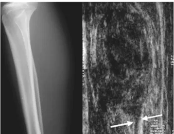
The X-ray images revealed a nonspeciic soft tissue prominence in the middle third of the calf, and ultrasound revealed a heterogeneous, predominantly hypoechoic, soft tissue mass (Figure 1).
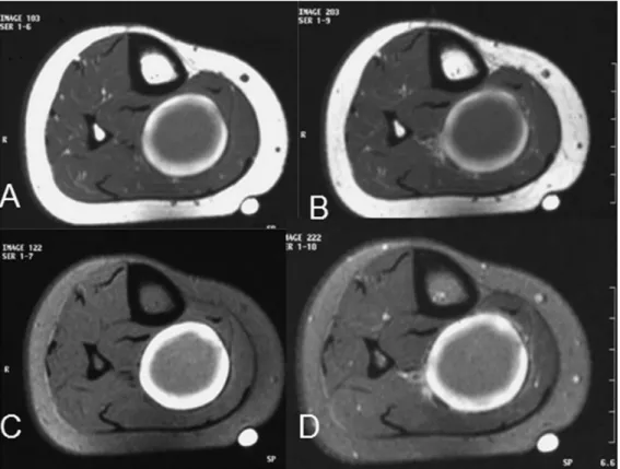
Furthermore, analysis of the MRI images revealed a soft tissue tumor with a tubular structure entering and exiting the mass (Figure 2). Retrospective evaluation of the ultrasound images also demonstrated that the mass was contiguous with a hypoechoic tubular structure. The transition between the lesion and this tubular structure of the neurovascular bundle had a contour aspect suggestive of a lesion that originated from the respective neurovascular element (Figures 1, 2A and 2B).
A discrete target sign was identiied in the tumoral lesion on the T2-weighted images that was characterized by a very
Figure 1 - A giant thrombosed venous aneurysm in the calf.Lateral
342
CLINICS 2010;65(3):341-4 Giant thrombosed venous aneurysm in the calf: MRI characteristics and the target sign
Nogueira-Barbosa MH et al.
bright periphery and relatively low signal in the central region. The pre- and post-contrast T1-weighted images showed a marked target pattern both with and without fat saturation (igures 2 and 3).
Surgery was then performed to biopsy the soft tissue
mass, and the resulting intra-operative evaluation suggested a giant thrombosed venous aneurysm, which was conirmed pathologically.
The low-signal central region observed in the MRI
Figure 2 - A giant thrombosed venous aneurysm in the calf as viewed by sagittal MRI sections. A. weighted image before contrast injection. B.
T1-weighted and fat-saturated images post-contrast injection. C. T2-T1-weighted and fat-saturated images. D. T2* echo gradient image. The thinner white arrows indicate the neurovascular bundle, and the thicker white arrows indicate a low-signal rim present only on the T2-weighted and fat-saturated and T2* echo gradient images.
Figure 3 - A giant thrombosed venous aneurysm in the calf as viewed by axial MRI sections. A. T1-weighted before contrast injection. B. T1-weighted
343
CLINICS 2010;65(3):341-4 Giant thrombosed venous aneurysm in the calf: MRI characteristics and the target sign Nogueira-Barbosa MH et al.
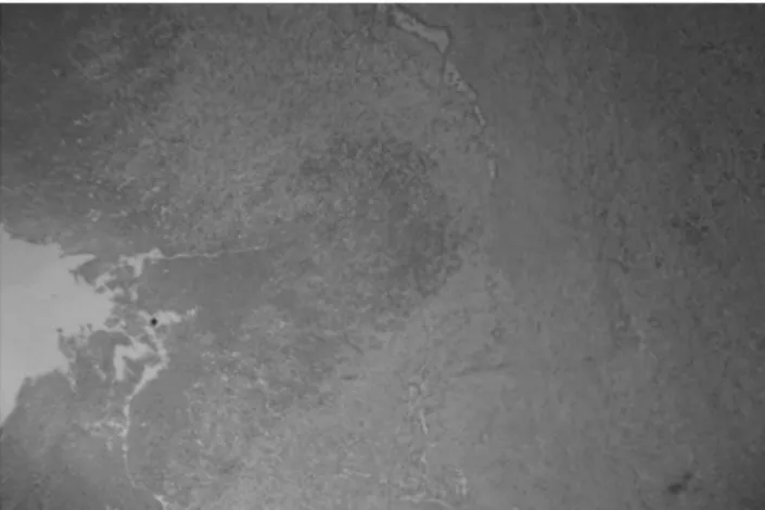
images correlated well with the area of the central thrombi that was verified by histology (figure 4). Both the T2-weighted fat-saturated fast spin-echo and the T2* echo gradient images of the soft tissue lesion had a thin low-signal rim related to the magnetic susceptibility of hemosiderin. It is also important to note that, because of the hemosiderin deposition, the T2* echo gradient images revealed the target sign signiicantly better than the T2-weighted fat-saturated fast spin echo.
DISCUSSION
Popliteal venous aneurysm is considered a rare condition, but it is a potentially fatal vascular disorder. The true incidence of this type of aneurysm may be underestimated in the literature because there is a group of patients who are known to be asymptomatic. Physician awareness, in addition to access to an ultrasound examination, may allow for an early diagnosis prior to the occurrence of any thromboembolic or other major complication. These aneurysms are more common in females and occur more frequently in people over 40 years of age.4-7
A retrospective analysis of 25 patients identiied that 24% of cases involved pulmonary embolism, and 76% of PVAs were discovered during an investigation for chronic venous disease.7 With the widespread use of venous duplex scanning, PVAs are increasingly found in patients with deep or supericial vein insuficiency.
Previously, popliteal venous aneurysms have been reported to mimic soft tissue popliteal masses.8,9 In this case study, the thrombosed venous aneurysm also presented clinically as a mass.
The thrombosed venous aneurysm in the calf showed a target sign on both the T2-weighted and T1-weighted images. In this case, the low-signal central region was correlated to the area of the central thrombi veriied during
Figure 4 - A giant thrombosed venous aneurysm in the calf. Histological
analysis showed vascular dilatation and luminal thrombus.
the histological analysis. It is interesting to note that the pre-contrast T1-weighted images already showed a marked target pattern. Moreover, due to hemosiderin deposition, the T2* echo gradient images better demonstrated the target sign than the T2-weighted fat-saturated fast spin echo.
Neurogenic neoplasms represent approximately 10% to 12% ofall benign soft-tissue neoplasms, while the most commonperipheral nerve tumors are neuroibromas and schwannomas.10,11 The MRI diagnosis of a peripheral nerve tumor may be suggested by the lesion location along a typical nerve distribution,12,13 and the identiication of a fusiform mass with an entering and exiting nerve is considered one of the most important imaging features suggestive of a peripheral nerve neoplasm.11,14,15 This sign has been reported in 88.2% to 94% of cases of benign peripheral nerve tumors.12,13
Analysis of MRI images has revealed that the signal intensity of peripheral nerve sheath tumors (PNSTs) may be nonspeciic. Heterogeneity may be present and in such cases would relect hypocellularity, hypercellularity, ibrous tissue, xanthomatous changes, cystic degeneration, necrosis and hemorrhage.11,12-14,16
In some cases of neuroibroma and schwannoma, it is possible to detect a characteristic target pattern on the T2-weighted images using a bright signal (myxomatous tissue) at the lesion periphery and a low to intermediate signal at the central region (ibrocollagenous tissue). Although it has not been precisely identiied, this target sign has been described as typical of peripheral nerve neoplasms.11,17 A target sign may also be present in benign peripheral nerve tumors imaged using high-resolution ultrasound.18
The imaging aspect of normal veins and of thrombosic veins on MRI is extremely variable, with many factors contributing to the signal intensity of the vessels.19 Precise anatomical landmarks eventually may be necessary to unequivocally differentiate the vessels from the nerve in the same neurovascular bundle.
344
CLINICS 2010;65(3):341-4 Giant thrombosed venous aneurysm in the calf: MRI characteristics and the target sign
Nogueira-Barbosa MH et al.
REFERENCES
1. Coffman SW, Leon SM, Gupta SK. Popliteal venous aneurysms: report of an unusual presentation and literature review. Ann Vasc Surg. 2000;14:286-90.
2. Bilotta W, Walker H, McDonald DJ, Sundaram M. Case report 651: Thrombosed, leaking popliteal aneurysm. Skeletal Radiol. 1991;20:71-2. 3. Sfyroeras GS, Antoniou GA, Drakou AA, Karathanos C, Giannoukas AD. Visceral Venous Aneurysms: Clinical Presentation, Natural History and Their Management: A Systematic Review. Eur J Vasc Endovasc Surg. 2009. article in press, epub ahead of print.
4. Chahlaoui J, Julien M, Nadeau P, Bruneau L, Roy P, Sylvestre J. Popliteal venous aneurysm: a source of pulmonary embolism. Am J Roentgenol. 1981;136:415-6.
5. Jack C, Sharma R, Vermuri RB. Popliteal venous aneurysms as a source of pulmonary embolism in a male: a case report. Angiology. 1984;35:54-7.
6. Ross GJ, Violi L, Barber LW, Vujic I. Popliteal venous aneurysm. Radiology. 1988;168:721-2.
7. Sessa C, Nicolini P, Perrin M, Farah I, Magne JL, Guidicelli H. Management of symptomatic and asymptomatic popliteal venous aneurysms: a retrospective analysis of 25 patients and review of the literature. J Vasc Surg. 2000;32:902-12.
8. Tsolakis JA, Kakkos SK, Panagiotopoulus E. Popliteal venous aneurysm mimicking a soft tissue tumour. A case report. Int Angiol. 1999;18:74-6. 9. Herrera L J, Davis JW, Livesay JJ. Popliteal Vein Aneurysm Presenting
as a Popliteal Mass. Tex Heart Inst J. 2006;33:246-8.
10. Kransdorf MJ. Benign soft-tissue tumors in a large referral population: distribution of speciic diagnoses by age, sex, and location. AJR. 1995;164:395-402.
11. Murphey MD, Smith WS, Smith SE, Kransdorf MJ, and Temple HT. From the Archives of the AFIP: Imaging of Musculoskeletal Neurogenic Tumors: Radiologic-Pathologic Correlation. RadioGraphics. 1999;19:1253-80.
12. Cerofolini E, Landi A, DeSantis G, Maiorana A, Canossi G, Romagnoli R. MR of benign peripheral nerve sheath tumors. J Comput Assist Tomogr. 1991;15:593-7.
13. Li CS, Huang GS, Wu HD, Chen WT, Shih LS, Lii JM, et al. Differentiation of soft tissue benign and malignant peripheral nerve sheath tumors with magnetic resonance imaging. Clinical Imaging. 2008;32:121-7.
14. Pilavaki M, Chourmouzi D, Kiziridou A, Skordalaki A, Zarampoukas T, Drevelengas A. Imaging of peripheral nerve sheath tumors with pathologic correlation Pictorial review. European Journal of Radiology. 2004;52:229-39.
15. Reynolds DL, Jacobson JA, Inampudi P, Jamadar DA, Ebeahim FS, Hayes CW. Sonographic Characteristics of Peripheral Nerve Sheath Tumors. AJR. 2004;182:741-4.
16. Jee WH, Oh SN, McCauley T, Ryu KN, Suh JS, Lee JH, et al. Extraaxial Neuroibromas Versus Neurilemmomas: Discrimination with MRI. AJR. 2004;183:629-33.
17. Suh JS, Abenoza P, Galloway HR, Everson LI and Griffiths HJ. Peripheral (extracranial) nerve tumors: correlation of MR imaging and histologic indings. Radiology. 1992;183:341-6.
18. Gruber H, Glodny B, Bendix N, Tzankov A, Peer S. High-resolution ultrasound of peripheral neurogenic tumors. European Radiology. 2007;17:2880-8.