DOI 10.1007/s00360-016-0991-3
ORIGINAL PAPER
Enzymatic capacities of metabolic fuel use in cuttlefish (Sepia
officinalis) and responses to food deprivation: insight into the
metabolic organization and starvation survival strategy
of cephalopods
Ben Speers‑Roesch1 · Neal I. Callaghan2 · Tyson J. MacCormack2 ·
Simon G. Lamarre3 · Antonio V. Sykes4 · William R. Driedzic1
Received: 14 December 2015 / Revised: 27 March 2016 / Accepted: 13 April 2016 / Published online: 30 April 2016 © Springer-Verlag Berlin Heidelberg 2016
de novo glucose synthesis (gluconeogenesis), important for glucose homeostasis, likely is restricted to the digestive gland, contrary to previous reports of widespread glucone-ogenesis among cephalopod tissues. Short-term starvation (3–5 days) had few effects on enzymatic capacities. Similar to vertebrates, lipid-based fuels, putatively mobilized from fat stores in the digestive gland, appear to be important energy sources for cephalopods, especially during starva-tion when glycolytic capacity is decreased perhaps to con-serve available glucose.
Keywords Cephalopod · Energy metabolism · Metabolic fuel preference · Enzyme activity · Starvation · Fasting · Lipid · Ketone body · Amino acid · Glucose · Glycolysis · Gluconeogenesis · Digestive gland
Introduction
Cephalopods are diverse and abundant molluscs that show functional convergence with fishes yet also differ in impor-tant ways extending from whole-animal locomotion to metabolic fuel preferences, possibly as a consequence of phylogenetic and life history constraints (O’Dor and Web-ber 1986). For example, whereas lipids are a key metabolic fuel in bony fishes (and most other vertebrates), the general consensus has been that cephalopods, like other molluscs, have a poor capacity to metabolize these high-energy nutri-ents (Storey and Storey 1983; O’Dor and Webber 1986; Hochachka 1995; Lee 1995). Cephalopods show relatively slow in vivo catabolism of lipids (O’Dor et al. 1984), have comparatively low tissue activities of the fatty acid cata-bolic enzyme 3-hydroxyacyl CoA dehydrogenase (HOAD) (Mommsen et al. 1981; Ballantyne et al. 1981; Mommsen and Hochachka 1981; Driedzic et al. 1990), and have Abstract Food limitation is a common challenge for
ani-mals. Cephalopods are sensitive to starvation because of high metabolic rates and growth rates related to their “live fast, die young” life history. We investigated how enzymatic capacities of key metabolic pathways are modulated dur-ing starvation in the common cuttlefish (Sepia officinalis) to gain insight into the metabolic organization of cephalo-pods and their strategies for coping with food limitation. In particular, lipids have traditionally been considered unimportant fuels in cephalopods, yet, puzzlingly, many species (including cuttlefish) mobilize the lipid stores in their digestive gland during starvation. Using a comprehen-sive multi-tissue assay of enzymatic capacities for energy metabolism, we show that, during long-term starvation (12 days), glycolytic capacity for glucose use is decreased in cuttlefish tissues, while capacities for use of lipid-based fuels (fatty acids and ketone bodies) and amino acid fuels are retained or increased. Specifically, the capacity to use the ketone body acetoacetate as fuel is widespread across tissues and gill has a previously unrecognized capacity for fatty acid catabolism, albeit at low rates. The capacity for
Communicated by H.V. Carey. * Ben Speers-Roesch bspeersr@gmail.com
1 Department of Ocean Sciences, Memorial University of Newfoundland, St. John’s, NL A1C 5S7, Canada
2 Department of Biology, Mount Allison University, Sackville, NB, Canada
3 Département de Biologie, Université de Moncton, Moncton, NB, Canada
4 CCMAR, Centro de Ciências do Mar do Algarve, Campus de Gambelas, Universidade do Algarve, Faro, Portugal
systemic hearts with negligible mitochondrial oxidation of fatty acids or activity of carnitine palmitoyltransferase (CPT), a key enzyme of mitochondrial fatty acid oxidation (Ballantyne et al. 1981; Mommsen and Hochachka 1981; Driedzic et al. 1990; Agnisola et al. 1991). Instead, cepha-lopod energy metabolism is thought to be fueled heavily by protein, as well as carbohydrate in particular to sustain exercise (Storey and Storey 1983; Lee 1995; Hochachka 1995). Yet, many cephalopods accumulate a large lipid store in their digestive gland (Lee 1995; Rosa et al. 2005), a voluminous organ analogous to the vertebrate liver, and the possibility of tissue-specific variation in activity of CPT, arguably the best enzymatic indicator of capacity for fatty acid oxidation, is largely unexplored.
The responses of cephalopods to starvation, in particu-lar, suggest that their use of lipid fuels perhaps has been underestimated. Food limitation is a common challenge for animals and responses to starvation provide valuable insight into metabolic organization and strategies for cop-ing with energy stress (McCue 2010; Secor and Carey 2016). In general, cephalopods are sensitive to starvation because their “live-fast, die-young” life history, which involves a high metabolic rate and fast growth, necessitates frequent food consumption (O’Dor and Webber 1986). Yet, cephalopods can encounter starvation periods resulting from environmental stress or life history events (Wells and Clarke 1996; Seibel et al. 2000). When deprived of food, cephalopods initially mobilize lipids (e.g. triacylglycerols), which are typically stored in the digestive gland (Boucher-Rodoni and Mangold 1985; Castro et al. 1992; Lee 1995; Lamarre et al. 2012; Morillo-Velarde et al. 2013) and appear to be used as metabolic fuel based on increased CO2 production from radiolabelled fatty acid in starved octopus (O’Dor et al. 1984). As starvation extends to 5–10+ days, triacylglycerol stores become depleted, body protein is increasingly called upon as a metabolic fuel, and metabolic rate is lowered (Boucher-Rodoni and Mangold 1985; Cas-tro et al. 1992; Grigoriou and Richardson 2009; Lamarre et al. 2012). The shift to lipid mobilization and eventually to protein mobilization appears broadly similar to the meta-bolic starvation response in vertebrates (Cahill 2006; Bar 2014; Secor and Carey 2016), but the apparent reliance upon lipid during starvation in cephalopods is paradoxi-cal given the current view of their poor capacity for fatty acid oxidation. Indeed, there are conflicting opinions about whether digestive gland lipids represent an energy store or an excretory product (Castro et al. 1992; Wells and Clarke 1996; Semmens 1998; Seibel et al. 2000; Moltschaniwskyj and Johnston 2006). Two potential, yet unexplored, expla-nations for the apparent reliance upon lipids during food deprivation are that starvation induces a greater capacity for fatty acid oxidation or that stored lipid could be
mobi-in mammals mobi-involves the hepatic conversion of fatty acids to ketone bodies, which are released into the blood to fuel metabolism in certain extrahepatic tissues (e.g. heart, brain) (Gibson and Harris 2002). The capacity of cephalopods to exploit ketone bodies as fuel during starvation is unknown, but tissues of one squid species showed activity of a key enzyme (succinyl coenzyme-A ketotransferase, SKT) involved in ketone body oxidation (Ballantyne et al. 1981).
Little is known about the importance of carbohydrates as metabolic fuels in starved cephalopods. Based on radi-olabel studies on starved octopus, O’Dor et al. (1984) sug-gested that available glucose is quickly depleted (within 1 day), while the large glycogen stores in the mantle are untouched and reserved for burst exercise even after 5 days of starvation. In starving mammals and fishes, glucose use typically is downregulated in non-essential tissues such as muscle while glycogen stores are mobilized to sustain glu-cose supply to specific tissues such as brain; as these stores are depleted, gluconeogenesis is increasingly relied upon to produce glucose in the liver from amino acids sourced from degradation of body protein (Gibson and Harris 2002; Bar and Volkoff 2012). Gluconeogenesis, a key metabolic path-way involved in regulation of circulating glucose levels, is of unknown importance in starved cephalopods. Interest-ingly, a limited number of available studies suggest that, unlike vertebrates, gluconeogenesis in cephalopods is not limited to the digestive gland (i.e. “liver”), based on activi-ties of gluconeogenic enzymes and incorporation of radi-olabelled amino acids into the glycogen pools in several tissues (although the terminal enzyme required for de novo glucose synthesis, glucose-6 phosphatase, has not been measured in cephalopod tissues) (Ballantyne et al. 1981; Fields and Hochachka 1982; Hochachka and Fields 1982). Gluconeogenesis therefore could be an important process during starvation in cephalopods.
We undertook a comprehensive survey of enzymatic capacities of tissue metabolism in fed and starved com-mon cuttlefish (Sepia officinalis) to elucidate the metabolic organization of cephalopods and the metabolic response that underlies survival of starvation. Profiles of maximal activities of metabolic enzymes provide a robust indicator of metabolic capacities and fuel preferences among animals (Newsholme and Crabtree 1986; Suarez 2012). Cuttlefish and certain other cephalopods are important food species whose nutrition and starvation responses are particularly relevant given the increasing interest in their culture (Nav-arro et al. 2014; Sykes et al. 2014). To clarify the paradox of lipid mobilization during starvation in cephalopods, we measured the enzymatic capacities of cuttlefish tissues to use fatty acids and ketone bodies as metabolic fuels and examined for the first time whether these capacities are modulated by starvation. We hypothesized that the
capaci-than previously recognized and enhanced during starvation. We also examined how starvation modulated enzymatic capacities of the glycolytic pathway, hypothesizing a down-regulation in line with a shift towards lipid-based fuels. Finally, we evaluated the notion that gluconeogenic capac-ity extends to tissues beyond the digestive gland in cepha-lopods and examined how starvation affects this capacity as well as key enzymes of biosynthetic pathways linked to glucose metabolism (pentose phosphate shunt and lipid biosynthesis).
Materials and methods Animals
Common cuttlefish (S. officinalis) were obtained from a captive reared population at the Ramalhete Aquaculture Station (Ria Formosa, southern Portugal—37°00′22.39″N; 7º58′02.69″W), according to the methods described by Sykes et al. (2014). The cuttlefish derived from eggs laid by a F3 captive stock. Cuttlefish were kept in large outdoor 500 l plastic tanks supplied with aerated, flow-through sea-water (21.0 ± 2.1 °C; salinity 36.3 ± 0.9 g l−1; dissolved
oxygen level was 94.8 ± 4.6 % air saturation) and were fed ad libitum daily with live grass shrimp (Palaemonetes varians). All experiments were conducted following Portu-guese animal care guidelines.
Starvation protocol
Experiments were carried out at the Ramalhete Aqua-culture Station in May 2014. Cuttlefish were housed individually in floating baskets (5.5 l water volume; 31 cm × 22 cm × 8 cm) with 1 mm mesh size, placed within four larger plastic holding tanks (500 l) that were outdoors and covered with netting, following stand-ards for good welfare (Fiorito et al. 2015). The cuttlefish were fed daily and allowed to acclimate to these condi-tions for a week before experimentation. Individual cut-tlefish were randomly selected to be subjected to one of three treatments: fed daily (Fed; n = 8; mass at sampling 43.9 ± 5.3 g), starved for 3–5 days (Short-starved; n = 7; 37.7 ± 1.3 g), or starved for 12 days (Long-starved; n = 8; 30 ± 0.8 g). The masses of fed and short-starved individu-als were statistically similar, but long-starved individuindividu-als weighed significantly less (one-way ANOVA with New-man–Keuls post-test, p < 0.05), consistent with previous results for starved cuttlefish (Castro et al. 1992). Following the treatments, cuttlefish were killed by anaesthesia in sea-water with 5 % ethanol, followed by bisection of the brain and severing of the ventral nerve and optical lobes. Hemo-lymph was withdrawn from the cephalic vein using a 25G
needle. Systemic heart, branchial hearts, gill, ventral man-tle muscle, and digestive gland were excised and frozen on dry ice and stored at −80 °C until analysis.
Triacylglycerol and ketone body assays
Triacylglycerol content of the digestive gland was measured as described by Lamarre et al. (2012). We attempted to measure two ketone bodies, acetoacetate and β-hydroxybutyrate, in acidified (6 % perchloric acid) and then neutralized (3 M K2CO3) extracts of hemolymph using well-established end-point or kinetic methods (McMur-ray et al. 1984). We discovered that these methods were unsuitable for cuttlefish because of substantial background rates of redox reactions in hemolymph, possibly due to an abundance of metal ions, which result in erroneously high measurements. Addition of up to 5 mM EDTA to the assay buffer did not improve assay resolution, whereas this has been shown to be effective in other pyridine nucleotide-linked metabolite assays (Storey et al. 1979).
Enzyme assays
Frozen tissues were weighed and homogenized in ten vol-umes of ice-cold homogenization buffer using a Polytron homogenizer. The homogenization buffer contained 50 mM imidazole (pH 7.4 at 22 °C), 1 mM disodium EDTA, and 40 μl/ml (digestive gland) or 20 μl/ml (all other tissues) of protease inhibitor (Sigma P2714; used 10× concentrate, i.e. bottle contents reconstituted in 10 ml). Homogenates were centrifuged at 4 °C and 1000×g for 10 min (man-tle) or 600×g for 5 min (all other tissues) and dilutions of the supernatant were used for the enzyme assays. Enzyme measurements were made for all sampled tissues from fed, short-starved, and long-starved individuals, with the fol-lowing exceptions. First, the only enzymes assayed for branchial heart were glycolytic (HK, PFK, PK) and gluco-neogenic (PEPCK, FBPase, G6Pase). Second, short-starved cuttlefish were not assayed for: PFK, PK, branchial heart HK, and all gluconeogenic enzymes except digestive gland G6Pase. These exclusions were done for logistical reasons and because, regarding exceptions of short-starved cuttle-fish, there were few differences from fed controls even after long-starvation.
Maximal enzyme activities were measured at 28 ± 0.1 °C using a SpectraMax M5 microplate spec-trophotometer (Molecular Devices, Sunnyvale, CA, USA). Activities of most enzymes were ascertained by measuring the oxidation or reduction of pyridine nucleo-tides at 340 nm (millimolar extinction coefficient ε340,
6.22 mM−1 cm−1). Citrate synthase (CS) and carnitine
palmitoyltransferase (CPT) activities were monitored at 412 nm by following the reaction of liberated free
CoA with 5,5′-dithiobis 2-nitrobenzoic acid (DTNB) (ε412, 13.6 mM−1 cm−1). Note, as freeze–thaw of
tis-sues can inactivate CPT-1 (Pande et al. 1990), our meas-urements of CPT may largely reflect activity of CPT-2, which together with CPT-1 facilitates entry of long-chain fatty acids into the mitochondria. Succinyl coenzyme-A ketotransferase (SKT) activity was measured by follow-ing the disappearance of acetoacetyl CoA at 310 nm (ε310, 11.9 mM−1 cm−1). The optical pathlength was calculated
for a final microplate well volume of 200 μl following the method of Brooks (1994). Assay conditions followed established protocols for mollusc or cephalopod tissues (Ballantyne et al. 1981; Hochachka and Fields 1982; Mommsen et al. 1983; Driedzic et al. 1990; Stuart and Ballantyne 1996). The glucose-6-phosphatase (G6Pase) assay was modified from the methods described by Stio et al. (1988) and Alegre et al. (1988). For NADH-linked assays, KCN was added to inhibit endogenous oxidation of NADH by NADH dehydrogenase (Sidell et al. 1987). Our preliminary measurements confirmed that reaction rates were linear with time and proportional to the amount of homogenate added for all enzymes. Enzyme activities were measured in duplicate with paired simultaneous, separate control reactions where substrate was omitted. Enzyme assay conditions were as follows:
Enzymes of ketone body metabolism
Succinyl coenzyme-A ketotransferase, SKT (E.C. 2.8.3.5): 50 mM imidazole buffer (pH 8.0), 5 mM MgCl2, 0.11 mM acetoacetyl CoA, 1 mM succinate (omitted for control).
d/l-β-hydroxybutyrate dehydrogenase, d/l-β-HBDH
(E.C. 1.1.1.30): 50 mM imidazole buffer (pH 8.0), 1 mM KCN, 11.25 mM NAD+, 2 mM DTT, 25 mM d/l
-β-hydroxybutyrate (omitted for control). Enzymes of lipid oxidation
Carnitine palmitoyltransferase, CPT (E.C. 2.3.1.21): 50 mM imidazole buffer (pH 8.0), 0.2 mM DTNB, 0.1 mM palmitoyl CoA, 5 mM L-carnitine (omitted for control).
3-Hydroxyacyl CoA dehydrogenase, HOAD (E.C. 1.1.1.35): 50 mM imidazole buffer (pH 8.0), 1 mM KCN, 0.1 mM NADH, 0.1 mM acetoacetyl CoA (omitted for control).
Enzymes of glycolysis and anaerobic glycolysis
Hexokinase, HK (E.C. 2.7.1.1): 50 mM imidazole buffer (pH 7.4), 5 mM MgCl2, 1 mM glucose, 0.4 mM NADP+,
2 U/ml glucose 6 phosphate dehydrogenase, 2 mM ATP (omitted for control).
Phosphofructokinase, PFK (E.C. 2.7.1.11): 50 mM imidazole buffer (pH 7.4), 1 mM KCN, 10 mM MgCl2,
50 mM KCl, 1 mM ATP, 0.2 mM NADH, 3 mM AMP, 1 U/ml glycerol-3-phosphate dehydrogenase, 5 U/ml tri-osephosphate isomerase, 1 U/ml aldolase, 8 mM fructose-6-phosphate (omitted for control).
Pyruvate kinase, PK (E.C. 2.7.1.40): 50 mM imidazole buffer (pH 7.4), 1 mM KCN, 10 mM MgCl2, 50 mM KCl, 5 mM ADP, 0.15 mM NADH, 5 U/ml lactate dehydroge-nase, 5 mM PEP (omitted for control).
Octopine dehydrogenase, ODH (E.C. 1.5.1.11): 50 mM imidazole buffer (pH 7.4), 1 mM KCN, 0.2 mM NADH, 4 mM pyruvate, 10 mM arginine (omitted for control). Enzymes of gluconeogenesis
Glucose-6-phosphatase, G6Pase (E.C. 3.1.3.9): 50 mM imidazole buffer (pH 7.4), 2 mM NAD, 9.6 U/ml glucose dehydrogenase, 0.7 U/ml mutarotase, 20 mM glucose-6-phosphate (omitted for control).
Fructose 1,6-bisphosphatase, FBPase (E.C. 3.1.3.11): 50 mM imidazole buffer (pH 7.4), 15 mM MgCl2, 0.2 mM
NADP+, 2 U/ml glucose-6-phosphate dehydrogenase,
10 U/ml phosphoglucose isomerase, 0.2 mM fructose 1,6-bisphosphate (omitted for control).
Phosphoenolpyruvate carboxykinase, PEPCK (E.C. 4.1.1.32): 50 mM imidazole buffer (pH 7.4), 1 mM KCN, 1 mM MnCl2, 1 mM MgCl2, 0.17 mM NADH, 1.1 mM PEP, 19 U/ml malate dehydrogenase, 1.5 mM IDP, 20 mM NaHCO3 (omitted for control).
Enzyme of the pentose phosphate pathway
Glucose 6-phosphate dehydrogenase, G6PDH (E.C. 1.1.1.49): 50 mM imidazole buffer (pH 7.4), 7 mM MgCl2, 0.4 mM NADP+, 1 mM glucose 6-phosphate (omitted for
control).
Enzyme of lipid biosynthesis
Glycerol 3-phosphate dehydrogenase, G3PDH (E.C. 1.1.1.8): 50 mM imidazole buffer (pH 7.4), 1 mM KCN, 0.15 mM NADH, 2 mM dihydroxyacetone phosphate (omitted for control).
Enzyme of amino acid metabolism
Aspartate aminotransferase, AspAT (E.C. 2.6.1.1): 50 mM imidazole buffer (pH 7.4), 0.025 mM pyridoxal phosphate, 1 mM KCN, 0.2 mM NADH, 8 U/ml malate dehydroge-nase, 40 mM aspartate, 10 mM α-ketoglutarate (omitted for control).
Enzyme of citric acid cycle
Citrate synthase, CS (E.C. 4.1.3.7): 50 mM imidazole buffer (pH 8.0), 0.3 mM acetyl CoA, 0.1 mM DTNB, 0.5 mM oxaloacetate (omitted for control).
Activities are presented as μmol substrate con-verted to product per minute per gram of wet tissue (μmol min−1 g ww−1). All chemicals were purchased from
Sigma Chemical Co. (St. Louis, MO, USA), except mutar-otase, which was purchased from Calzyme Laboratories Inc. (San Luis Obispo, CA, USA).
Statistics
All data are presented as mean ± SEM. Comparisons of enzyme activities were made using two-way ANOVA with Bonferroni post hoc tests and tissue and feeding state as factors. For HK and G6Pase, short-starved data were una-vailable for branchial heart, so the branchial heart data were analyzed separately using a Student’s t test to assess the effect of starvation. Digestive gland triacylglycerol levels were compared across feeding states using one-way ANOVA with Bonferroni post hoc test. Data were log or square root transformed, if necessary, to meet assumptions of equal variance and normality. All analyses were carried out with Prism 5.0b (GraphPad, La Jolla, CA, USA). The level of statistical significance was taken as p < 0.05. Results
Triacylglycerol levels in digestive gland
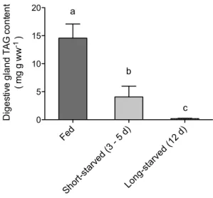
Triacylglycerol reserves in the digestive gland decreased by 72 % within 3–5 days of starvation, and were almost completely depleted by 12 days of starvation (one-way ANOVA, p < 0.05) (Fig. 1).
Enzymatic capacities for lipid and ketone body metabolism
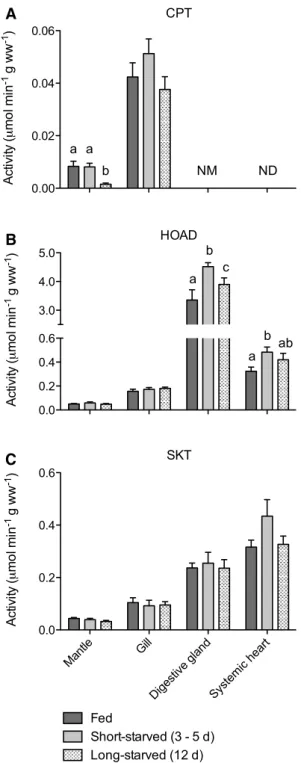
CPT activity was not detectable in systemic heart (Fig. 2a). High apparent levels of deacylase activity obscured meas-urement of CPT in digestive gland. Mantle had very low levels of CPT activity, which were maintained during short-starvation (two-way ANOVA, p > 0.05) but decreased by 82 % after long-starvation (two-way ANOVA, p < 0.01). Gill had readily detectable, albeit low, levels of CPT that were greater than mantle CPT activities (two-way ANOVA, p < 0.0001) and were unaffected by starvation (two-way ANOVA, p > 0.05).
HOAD activity was detectable in all tissues, at generally higher levels than CPT, and with the lowest levels in mantle
and the highest levels in digestive gland and systemic heart (two-way ANOVA, p < 0.0001) (Fig. 2b). HOAD activity was unaffected by starvation in mantle and gill (two-way ANOVA, p > 0.05). However, in digestive gland and sys-temic heart, short-starvation was associated with a 35 and 50 % increase in HOAD activity compared with fed indi-viduals, respectively (two-way ANOVA, p < 0.05). This increase was tempered following long-starvation, declining to activities that were 16 and 30 % above the values for fed individuals.
While d/l-β-HBDH was not detected in any tissue
(data not shown), SKT was readily detected in all tissues, with the lowest levels in mantle and the highest levels in digestive gland and systemic heart (two-way ANOVA, p < 0.0001) (Fig. 2c). SKT activity remained unchanged in all tissues throughout the starvation period (two-way ANOVA, p > 0.05).
Enzymatic capacities for glycolysis and anaerobic glycolysis
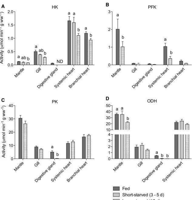
HK activity was highest in systemic heart and low-est in mantle and diglow-estive gland (two-way ANOVA, p < 0.0001) (Fig. 3a). HK activity was affected by starvation in all tissues. Mantle and gill showed grad-ual decreases with starvation duration, becoming sig-nificantly lower than fed controls following long-starvation (by 35 and 44 %, respectively) (two-way ANOVA, p < 0.05). In digestive gland, HK activity was rendered not detectable after only short-starvation. In systemic heart, HK activity was maintained at fed
Fed Short -starv ed (3 - 5 d) Long -starv ed (1 2 d) 0 5 10 15 20 a b c Diges tive gland TA G co nt en t ( mg g ww -1 )
Fig. 1 Triacylglycerol (TAG) content in digestive gland from
cut-tlefish fed daily or starved for 3–5 or 12 days. Values with different letters are significantly different (one-way ANOVA with Bonferroni, p < 0.05). Data are mean ± SEM, n = 7–8. These data were previ-ously published in Lamarre et al. (2016)
levels following short-starvation (two-way ANOVA, p > 0.05), but decreased by 28 % following long-starvation (two-way ANOVA, p < 0.05). HK activity in branchial heart was not measured in short-starved individuals, but following long-starvation levels were decreased by 21 % compared with fed controls (t test, p < 0.01).
PFK activity was higher in mantle and systemic heart compared with the low values in other tissues (two-way ANOVA, p < 0.0001) (Fig. 3b). PFK activities did not change significantly following long-starvation in any tis-sue except for mantle and systemic heart, where there was a 49 and 64 % decrease in activity, respectively (two-way ANOVA, p < 0.05).
PK activity was highest in mantle and lowest in gill and digestive gland (two-way ANOVA, p < 0.0001) (Fig. 3c). PK activities were unchanged by starvation in all tissues except digestive gland, where long-starvation was associ-ated with a 95 % decrease in activity (two-way ANOVA, p < 0.05).
ODH activity was much higher in mantle and systemic heart compared with gill or digestive gland (two-way ANOVA, p < 0.0001) (Fig. 3d). ODH activity was unaf-fected by starvation in gill and systemic heart (two-way ANOVA, p > 0.05). In mantle, ODH activity initially was unaffected by starvation but decreased by 37 % following long-starvation (two-way ANOVA, p < 0.01). In digestive gland, a decrease in ODH activity of 90 % or more per-sisted throughout the starvation period (two-way ANOVA, p < 0.05).
Enzymatic capacities for gluconeogenesis
PEPCK activity in mantle and systemic heart was unchanged following long-starvation (two-way ANOVA, p > 0.05) (Fig. 4a). PEPCK activity was not detected in gill, digestive gland, or branchial heart.
FBPase activity was found at relatively high levels in digestive gland, and at significantly lower levels in man-tle, systemic heart, and branchial heart (two-way ANOVA, p < 0.0001) (Fig. 4b). FBPase activity was not detected in gill. FBPase activities were not affected by long-starvation (two-way ANOVA, p > 0.05).
G6Pase activity was present in digestive gland and at a lower level in branchial heart (two-way ANOVA, p < 0.0005), but not found in mantle, gill, or systemic heart (Fig. 4c). Long-starvation had no effect on activ-ity in branchial heart (t test, p > 0.05). In digestive gland, G6Pase activity was retained during short-starvation (two-way ANOVA, p > 0.05) but decreased by 57 % after long-starvation (two-way ANOVA, p < 0.001).
Mantl e Gill Diges tive g land Syste mic h eart 0.00 0.02 0.04 0.06 a b a NM ND CPT Act iv ity ( mo l mi n -1 g ww -1) Mantl e Gill Diges tive g land Syste mic h eart 0.0 0.2 0.4 0.6 3.0 4.0 5.0 a b c a b ab HOAD Act iv ity ( mo l mi n -1 g ww -1) Mantl e Gill Diges tive g land Syste mic h eart 0.0 0.2 0.4 0.6 Fed Short-starved (3 - 5 d) Long-starved (12 d) SKT Ac tivi ty ( mo l mi n -1 g ww -1) A B C
Fig. 2 Activities of enzymes of fatty acid oxidation (a carnitine
palmitoyltransferase, CPT; b 3-hydroxyacyl CoA dehydrogenase, HOAD) and ketone body metabolism (c succinyl coenzyme-A ketotransferase, SKT) in tissues from cuttlefish fed daily or starved for 3–5 or 12 days. Note split y axis for HOAD. Within each tissue, values with different letters are significantly different from each other (two-way ANOVA with Bonferroni, p < 0.05) and an absence of let-ters indicate no significant difference between values. For clarity, statistical comparisons between tissues are not shown (see text for description of results). ND not detectable, NM not measured (see text). Data are mean ± SEM. n = 7–8, except for: CPT in short-starved mantle (n = 6); all enzymes in long-starved systemic heart (n = 4)
Enzymatic capacities for lipid biosynthesis and pentose phosphate pathway
Under fed conditions, G3PDH activity was highest in man-tle and digestive gland and lowest in gill (two-way ANOVA, p < 0.0001) (Fig. 5a). G3PDH activity remained unchanged in gill and systemic heart even after long-starvation (two-way ANOVA, p > 0.05). In mantle, G3PDH activity was initially maintained during starvation, but declined mod-estly but significantly by 17 % following long-starvation
(two-way ANOVA, p < 0.05). On the other hand, G3PDH activity in digestive gland plummeted by over 97 % under both short- and long-starvation (two-way ANOVA, p < 0.001).
G6PDH activity was higher in gill and digestive gland compared with systemic heart or mantle (two-way ANOVA, p < 0.0001) (Fig. 5b). G6PDH activity was unaffected by star-vation in all tissues (two-way ANOVA, p > 0.05) except diges-tive gland, where it decreased by 40–50 % following short- and long-starvation (two-way ANOVA, p < 0.001).
Mantl e Gill Diges tive g land Syste mic h eart Bran chial heart 0.0 0.5 1.0 1.5 2.0 a b ab a ab b ND a a b a b HK Act iv ity ( mo l mi n -1 g ww -1) Mantl e Gill Diges tive g land Syste mic h eart Bran chial heart 0 10 20 30 40 a b PK Ac tivi ty ( mo l mi n -1 g ww -1) Mantl e Gill Diges tive g land Syste mic h eart Bran chial heart 0 1 2 3 a b PFK a b Mantl e Gill Diges tive g land Syste mic h eart 0 1 2 3 4 10 20 30 40 50 Fed Short-starved (3 - 5 d) Long-starved (12 d) a b a a b b ODH B A D C
Fig. 3 Activities of enzymes of glycolysis (a hexokinase, HK; b
phosphofructokinase, PFK; c pyruvate kinase, PK) and anaerobic glycolysis (d octopine dehydrogenase, ODH) in tissues from cut-tlefish fed daily or starved for 3–5 or 12 days. Note, due to limited sample, short-starved samples were run only for HK (excluding digestive gland and branchial heart) and ODH. Note split y axis for ODH. Within each tissue, values with different letters are
signifi-cantly different from each other (two-way ANOVA with Bonferroni, p < 0.05) and an absence of letters indicate no significant difference between values. For clarity, statistical comparisons between tissues are not shown (see text for description of results). ND not detecta-ble. Data are mean ± SEM. n = 7–8, except for: HK in short-starved mantle and gill (n = 6), ODH in long-starved systemic heart (n = 4)
Enzymatic capacities for amino acid metabolism
AspAT was highly active in all tissues, but systemic heart had greater activity than the other tissues (two-way ANOVA, p < 0.0001) (Fig. 6). AspAT activity was unaffected by tion in mantle and gill (two-way ANOVA, p > 0.05) but starva-tion was associated with a gradual and significant increase by up to 72 % in digestive gland (two-way ANOVA, p < 0.01). In systemic heart, there was a transient 25 % significant increase in AspAT activity after short-starvation (two-way ANOVA, p < 0.001) that returned to fed levels after long-starvation.
Mantl e Gill Diges tive g land Syste mic h eart Bran chial heart 0.0 0.5 1.0 1.5 2.0 ND ND ND PEPCK Ac tivi ty ( mo l mi n -1 g ww -1) Mantl e Gill Diges tive g land Syste mic h eart Bran chial heart 0.0 0.2 0.4 0.6 0.8 1.0 ND FBPase Act iv ity ( mo l mi n -1 g ww -1) Mantl e Gill Diges tive g land Syste mic h eart Bran chial heart 0.0 0.2 0.4 0.6 b a a ND ND ND G6Pase Long-starved (12 d) Fed Short-starved (3 - 5 d) Activity ( mo l mi n -1 g ww -1) A B C
Fig. 4 Activities of enzymes of gluconeogenesis (a
phosphoe-nolpyruvate carboxykinase, PEPCK; b fructose 1,6-bisphosphatase, FBPase; c glucose-6-phosphatase, G6Pase) in tissues from cut-tlefish fed daily or starved for 3–5 or 12 days. Note, due to limited sample, short-starved samples were run only for G6Pase in digestive gland. Within each tissue, values with different letters are signifi-cantly different from each other (two-way ANOVA with Bonferroni, p < 0.05) and an absence of letters indicate no significant difference between values. For clarity, statistical comparisons between tissues are not shown (see text for description of results). ND not detectable. Data are mean ± SEM. n = 7–8
Mantl e Gill Diges tive g land Syste mic h eart 0.0 0.5 1.0 1.5 2.0 a b a a b b G3PDH Act iv ity ( mo l mi n -1 g ww -1) Mantl e Gill Diges tive g land Syste mic h eart 0.0 0.2 0.4 0.6 0.8 Fed Short-starved (3 - 5 d) Long-starved (12 d) b b a G6PDH Ac tivi ty ( mo l mi n -1 g ww -1) A B
Fig. 5 Activities of enzymes related to lipid synthesis (a glycerol
3-phosphate dehydrogenase, G3PDH) and the pentose phosphate pathway (b glucose 6-phosphate dehydrogenase, G6PDH) in tissues from cuttlefish fed daily or starved for 3–5 or 12 days. Within each tissue, values with different letters are significantly different from each other (two-way ANOVA with Bonferroni, p < 0.05) and an absence of letters indicate no significant difference between values. For clar-ity, statistical comparisons between tissues are not shown (see text for description of results). Data are mean ± SEM. n = 7–8, except for: short-starved gill (n = 6); long-starved systemic heart (n = 4)
Enzymatic capacities for the citric acid cycle
Systemic heart had much higher CS activity compared with other tissues (Fig. 7) (two-way ANOVA, p < 0.0001). CS activity in gill and systemic heart was unaffected by starvation (two-way ANOVA, p > 0.05), whereas long-starvation caused CS activity to decrease by 36 % in mantle (two-way ANOVA, p < 0.05) and to become undetectable in digestive gland. Discussion
As the first comprehensive investigation of how the enzy-matic capacities of metabolic pathways are modulated by starvation in cephalopods, our study provides novel insight into the metabolic organization of these important molluscs and their strategies for coping with food limitation. The established notion that amino acids and carbohydrates are the preferred fuels in tissues of cephalopods (Storey and Storey 1983; Hochachka 1995; Lee 1995) is confirmed by the high activities of AspAT (Fig. 6) and well-developed glycolytic capacity in all tissues of cuttlefish (Fig. 3). How-ever, lipid-based fuels, including fatty acids and the ketone body acetoacetate, appear to be more important as metabolic fuels in cephalopods than previously thought. Cuttlefish
have a previously unrecognized, albeit low, capacity for fatty acid oxidation in gill, and even in mantle, and a wide-spread capacity to catabolize acetoacetate (Fig. 2), presum-ably produced from lipids in the digestive gland (Meyer et al. 1986; Stuart and Ballantyne 1996). Furthermore, the capacity to use lipid-based fuels is largely retained during starvation, while glycolytic capacity to use glucose as fuel is decreased (Figs. 2, 3). In starved cephalopods, lipid-based fuels appear to become more heavily relied upon while glu-cose is conserved, a response similar to that generally seen in starved vertebrates and which likely explains why starved cephalopods mobilize digestive gland lipids.
Tissue‑specific variation in capacity for fatty acid oxidation in cephalopods
The CPT activities in cuttlefish tissues support the lack of mitochondrial fatty acid oxidation in systemic heart of cephalopods (Agnisola et al. 1991; Ballantyne et al. 1981; Mommsen and Hochachka 1981; Driedzic et al. 1990), yet indicate the novel presence of a capacity for fatty acid oxidation in gill and mantle. The demand for fatty acids as fuel in gill and mantle may be proportionally more signifi-cant than suggested by their relatively low CPT activities,
Mantl e Gill Diges tive g land Syste mic h eart 0 20 40 60 80 Fed Short-starved (3 - 5 d) Long-starved (12 d) a ab b b a a AspAT Ac tivi ty ( mo l mi n -1 g ww -1)
Fig. 6 Activities of aspartate aminotransferase (AspAT), an enzyme
of amino acid metabolism, in tissues from cuttlefish fed daily or starved for 3–5 or 12 days. Within each tissue, values with different letters are significantly different from each other (two-way ANOVA with Bonferroni, p < 0.05) and an absence of letters indicate no sig-nificant difference between values. For clarity, statistical comparisons between tissues are not shown (see text for description of results). Data are mean ± SEM. n = 7–8, except for: long-starved systemic heart (n = 4) Mantl e Gill Diges tive g land Syste mic h eart 0.0 0.2 0.4 0.6 0.8 1.06 8 10 12 14 Fed Short-starved (3 - 5 d) Long-starved (12 d) a b a ND CS Ac tivi ty ( mo l mi n -1 g ww -1)
Fig. 7 Activities of citrate synthase (CS), an enzyme related to
aero-bic metabolism, in tissues from cuttlefish fed daily or starved for 3–5 or 12 days. Note split y axis. Within each tissue, values with different letters are significantly different from each other (two-way ANOVA with Bonferroni, p < 0.05) and an absence of letters indicate no sig-nificant difference between values. For clarity, statistical comparisons between tissues are not shown (see text for description of results). ND not detectable. Data are mean ± SEM. n = 7–8, except for: long-starved systemic heart (n = 4)
because gill and mantle are large tissues in cephalopods (~2 and ~30–40 % of body mass in cuttlefish, respectively, vs. ~0.1 % for systemic heart; present authors, unpublished data; Gabr et al. 1999). The tissue-specific activities of CPT in cephalopods probably reflects phylogeny, because other marine molluscs have gills with mitochondria that oxidize fatty acids, gills and mantle with low but detectable CPT activities, and hearts without CPT or mitochondrial fatty acid oxidation (Ballantyne and Storey 1983; Ballantyne and Moyes 1987; Ballantyne and Berges 1991; Stewart et al. 1992). Our inability to accurately measure CPT in digestive gland matches previous failed attempts in molluscs, attrib-uted to interference from deacylases in raw homogenates (Ballantyne et al. 1981; Stuart and Ballantyne 1996). How-ever, fatty acid oxidation likely is important in cephalopod digestive gland given the very high HOAD activities, and high rates of mitochondrial fatty acid oxidation in diges-tive gland mitochondria of bivalves (Ballantyne and Storey 1984; Ballantyne and Moon 1985).
Unlike CPT, HOAD is widely distributed across cut-tlefish tissues, albeit at low activities compared with bony fishes. The presence of appreciable HOAD activ-ity in systemic heart, despite absence of CPT, is similar to other cephalopods (Ballantyne et al. 1981; Mommsen and Hochachka 1981; Mommsen et al. 1981; Driedzic et al. 1990) and also to muscles of chondrichthyan fishes (Speers-Roesch and Treberg 2010). Speculatively, in these cases, HOAD could be involved in short-chain fatty acid oxidation or peroxisomal β-fatty acid oxidation (Stewart et al. 1992; Islinger et al. 2010).
Ketone body metabolism: Widespread capacity to use acetoacetate in cuttlefish tissues
SKT activity was widely detected in cuttlefish tissues, whereas β-HBDH activity was absent, as observed in squid (Ballantyne et al. 1981) and other marine molluscs (Beis et al. 1980; Ballantyne and Moyes 1987; Ballantyne and Berges 1991; Stuart and Ballantyne 1996). Systemic hearts have the highest SKT activities in the cephalopods, suggesting that acetoacetate replaces fatty acids as a lipid-based fuel in this tissue. Unlike mammals, where SKT is exclusively for ketone body oxidation and thus is negligible in liver, molluscs, including cuttlefish (Fig. 2), have high SKT activity in the digestive gland, where it is thought to participate in ketogenesis (Zammit et al. 1979; Meyer et al. 1986; Stuart and Ballantyne 1996). Thus, the ketone body acetoacetate, but not β-HB, may be a useful metabolic fuel in cephalopods, similar to other marine molluscs where significant mitochondrial oxidation of acetoacetate also has been observed (Ballantyne and Moyes 1987).
Unfortunately, circulating acetoacetate was
unresolv-enzyme-linked spectrophotometric methods are unsuitable for use with cuttlefish hemolymph and GC–MS methods were unavailable to us. However, we made preliminary measurements of β-HB in cuttlefish hemolymph using a GC–MS method (Lamarre et al. 2014), finding that the peak corresponding to β-HB was not detected in cuttlefish hemolymph, whereas it was detected in a hemolymph sam-ple spiked with commercial β-HB (present authors, unpub-lished observation). This result agrees with the absence of β-HBDH activity and the lack of circulating β-HB in other marine molluscs (Stewart et al. 1992).
Glycolysis, tissue‑specific fuel preferences, and the aerobic/anaerobic design of cephalopod tissues
The organization of enzymatic capacities for glycolysis (HK, PFK, PK), anaerobic glycolysis (ODH), and aerobic metabolism (CS) in cuttlefish (Figs. 3, 7) corresponds with the accepted view of the aerobic vs. anaerobic design of cephalopod tissues (Hochachka 1995) and, following from the above discussion, illuminates the fuel preferences of cephalopod tissues. The mantle, which powers locomotion, had low HK and CS activities and high PFK, PK, and ODH activities that gear it towards rapid mobilization of stored glycogen to sustain anaerobic burst swimming (Storey and Storey 1983). In comparison, the continually pumping sys-temic heart relies more on aerobic glucose use, indicated by higher HK and CS activities (Driedzic et al. 1990; Lamarre et al. 2012). While carbohydrates, and amino acids, are important fuels in cephalopod muscle, we revealed a sup-plementary capacity to use lipid-based fuels, particularly acetoacetate (Fig. 2). Gills appear to use a mix of metabolic fuels, including fatty acids, acetoacetate, and amino acids, as discussed above, as well as glucose via aerobic glycoly-sis as indicated by appreciable HK activity and relatively low ODH activity (Ballantyne et al. 1981; Lamarre et al. 2012). Digestive gland had low activities of glycolytic enzymes and CS, possibly because of the biosynthetic poise of this tissue, indicated by comparatively high activities of G3PDH and G6PDH under fed conditions (Fig. 5). Amino acids may be preferred over glucose as fuel in digestive gland, given its high AspAT activity (Fig. 6) and vigorous amino acid metabolism (Hochachka et al. 1983); fatty acids may also be important fuels as previously discussed. The metabolism of branchial heart rarely has been studied, but its comparatively high glycolytic enzyme activities suggest reliance upon glucose.
Gluconeogenesis and related biosynthetic pathways Few data are available on gluconeogenesis in cephalopods (Ballantyne et al. 1981; Hochachka and Fields 1982). In
high activities (along with FBPase) (Fig. 4), providing the first evidence that the capacity for de novo glucose syn-thesis is restricted largely to the digestive gland in ceph-alopods. However, PEPCK was not reliably detectable in digestive gland (previous measurements are lacking); this could be due to analytical difficulties of determining certain enzyme activities in digestive gland (Ballantyne et al. 1981), or, speculatively, glucose synthesis in cut-tlefish digestive gland only proceeds from stored glyco-gen rather than from non-carbohydrate sources. Indeed, incorporation of radiolabelled amino acids into the gluco-neogenic pathway was poor in cephalopod digestive gland (Hochachka and Fields 1982). The possibility that glycerol derived from triacylglycerol catabolism is converted to glu-cose seems unlikely, considering that G3PDH activity is greatly decreased in digestive gland when triacylglycerol are being mobilized during starvation (Fig. 5). The notion that gluconeogenesis occurs in cephalopod gill (Fields and Hochachka 1982; Hochachka and Fields 1982) was unsup-ported by the undetectable activities of all gluconeogenic enzymes (Fig. 4).
Unlike other tissues, mantle and systemic heart of cuttle-fish showed high PEPCK activities (Fig. 4), despite absent G6Pase. Although squid muscles have low PEPCK activity (Ballantyne et al. 1981), high activities are known in mus-cle from other molluscs (Crabtree et al. 1972), which con-trasts with the absence of PEPCK in vertebrate muscle. The role of PEPCK in cuttlefish muscles is uncertain. Specula-tively, given the additional presence in muscles of FBPase (Fig. 4) and substantial glycogen stores (Storey and Storey 1979, 1983), PEPCK could funnel carbon from non-carbo-hydrate sources towards glycogen synthesis. In fact, amino acids are readily incorporated into the glycogen pool of cephalopod mantles (Hochachka and Fields 1982). Muscle PEPCK also could be involved in cataplerosis (Yang et al. 2009) or in supplying carbon to glycerol production for lipid synthesis, considering the high activities of G3PDH in mantle and systemic heart (Fig. 5) (potentially analogous to the glycerol-producing liver of rainbow smelt; Driedzic 2015).
Starvation responses: glycolytic capacity is downregulated while capacity for lipid‑based and amino acid fuel use is retained
While cuttlefish metabolic organization was mostly unchanged after short-starvation (3–5 days), a general starvation response was observed after long-starvation (12 days): enzymatic capacities for lipid-based and amino acid fuel use were retained while the enzymatic capacity to use glucose was decreased. This general response is con-sistent with radiolabel and other studies in cephalopods that have suggested that a mix of metabolic fuels is used
during early starvation, followed by a greater reliance upon lipids and amino acids in order to conserve carbohydrate stores (digestive gland glycogen is limited; mantle glyco-gen is reserved for fueling locomotion), and finally a large reliance upon amino acids once lipid stores are exhausted (O’Dor et al. 1984; Castro et al. 1992; Lamarre et al. 2012). In fact, the responses of the ammonia quotient of our study animals suggested a transitory shift to lipid catabolism dur-ing short starvation and then to protein catabolism after long starvation, when triacylglycerol became depleted (Lamarre et al. 2016). Layered upon the general response in cuttlefish was a tissue-specific response in which meta-bolic capacity in digestive gland and mantle appeared to be shut down more than in gill or systemic heart. Digestive gland showed large decreases in glycolytic, aerobic, and biosynthetic enzyme activities after only 3–5 days of star-vation, perhaps for energy savings and, given the retained capacities for amino acid, lipid, and gluconeogenic metab-olism, as part of a transformation into an exporter of glu-cose and lipid-based fuels (see below). Mantle, the largest tissue in cephalopods (~30–40 % of body mass in cuttle-fish; Gabr et al. 1999), showed decreases in many enzyme activities after long starvation (e.g. CPT, CS, G3PDH, HK, PFK, ODH), possibly resulting from starvation-induced decreases in protein synthesis (Lamarre et al. 2016). Con-versely, enzymatic capacities in gill and systemic heart were largely unaffected even after long-starvation, aside from the decreased glycolytic capacity, suggesting a greater conservation of metabolism, sustained primarily by lipid-based and amino acid fuels.
The retention of enzymatic capacity to use lipid-based fuels coincided with gradual depletion of triacylglycerol stores in digestive gland (Fig. 1). Acetoacetate may be a key starvation fuel in cuttlefish, based on the maintenance of SKT activity in all tissues, similar to starved vertebrates (Robinson and Williamson 1980). The source of acetoac-etate may be the digestive gland, where the increased HOAD activity and maintained SKT activity during star-vation is similar to the responses in the ketogenic liver of starved vertebrates (Robinson and Williamson 1980; Foster and Moon 1991) and agrees with a potential role of these enzymes in acetoacetate synthesis from digestive gland lipids in molluscs (Horst and Becker 1986; Horst et al. 1986; Meyer et al. 1986; Stuart and Ballantyne 1996).
The starvation responses of HOAD and CPT activi-ties indicate that fatty acids, presumably released from the digestive gland, are another lipid-based fuel used dur-ing starvation, in particular in the gill where HOAD and CPT activities were retained. Curiously, despite absent CPT activity, HOAD activity increased in systemic heart, suggesting some role for cardiac fatty acid oxidation dur-ing starvation, possibly involvdur-ing medium- or short-chain fatty acids (whose use does not require CPT activity). In
mantle, while HOAD activity was maintained, CPT activ-ity decreased significantly after long-starvation, perhaps because at this point protein catabolism in mantle is fully activated (Lamarre et al. 2012) and lipid stores are depleted. The importance of amino acids as fuel during starvation in mantle, and all other tissues, was supported by the univer-sal maintenance or increase of AspAT activities (Fig. 6), as found previously in cuttlefish (Lamarre et al. 2012) and vertebrates (Horio et al. 1988; Foster and Moon 1991). The retained capacity to use lipid-based fuels after long starva-tion is somewhat inconsistent with the concurrent exhaus-tion of triacylglycerol stores (Fig. 1), but we speculate that ketones continue to be synthesized from amino acids (Laf-fel 1999). Also, other lipid types, common in the digestive glands of cephalopods and mobilized during starvation in cuttlefish (e.g. free fatty acids, diacylglycerol ethers), could be additional sources of lipid fuel (Voogt 1983; Castro et al. 1992).
The starvation-induced decrease in glycolytic enzymatic capacity, especially HK, in cuttlefish tissues is similar to the well-established, rapid (days) downregulation of gly-colysis in starved mammals (Cherel et al. 1988; Pilkis et al. 1988; Gibson and Harris 2002) and certain other vertebrates (Moon and Johnston 1980; Blier and Guderley 1986; Foster and Moon 1991; Metón et al. 1999; Secor and Carey 2016). The time scale of the response in cuttlefish, with significant decreases generally occurring only after long-starvation, sug-gests that circulating glucose is used as fuel early in starva-tion but later conserved for essential tissues (e.g. brain), sim-ilar to mammals (Cahill 2006) and consistent with radiolabel studies on starved octopus (O’Dor et al. 1984). For logistical reasons, we did not measure the enzyme profile of brain, but squid have high HK activities in brain suggesting that glu-cose is an important fuel (Ballantyne et al. 1981). The early shutdown of glycolytic enzymes in digestive gland while gluconeogenic enzymatic capacity largely was retained is consistent with a strategy of emphasizing de novo glucose synthesis in the gluconeogenic tissue, as in mammals (Pilkis et al. 1988). Whereas in starving mammals multiple glyco-lytic enzymes are commonly downregulated, in cuttlefish the decreases of glycolytic enzymes downstream of HK were more tissue-specific, perhaps reflecting differences in the use of endogenous glycogen or glycerol liberated from triacyl-glycerol mobilization. Incorporation of triacyl-glycerol carbon into glycolysis just prior to the PK node during starvation would be consistent with the finding in most tissues of relatively unchanged activities of G3PDH, which will also sustain glycolytic redox balance. The exception is digestive gland, where G3PDH is greatly decreased, possibly because of this tissue’s abolished glycolytic capacity or as part of an energy-saving downregulation of lipid biosynthesis (there was also a concomitant decrease in G6PDH activity, which supplies
Note, the starvation responses of cuttlefish enzyme activities observed in the present study were unlikely to be influenced much by changes in tissue water content, which sometimes accompany food deprivation in animals. In fact, water content in digestive gland and mantle of starv-ing cuttlefish remains unchanged for at least several weeks (Castro et al. 1992), which is beyond the time period of our study. Also, as shown above, the responses we observed reveal pathway- and enzyme-specific patterns within tis-sues, rather than showing a consistent response across all enzymes that could be expected under the influence of var-ying water content.
Conclusions and perspectives
The preferences for metabolic fuels among animals are diverse, depending on the influences of phylogenetic his-tory, environmental conditions, and physiological state (e.g. starvation, exercise) (Moyes et al. 1990; Weber 2011). The use of protein (amino acids) and carbohydrates as key fuels in cephalopods has been attributed to the influences of a rapid-growth life history and the use of high-intensity jet propulsion, respectively, as well as their molluscan heritage (O’Dor and Webber 1986; Hochachka 1995; Lee 1995). Our study is consistent with this view of cephalopod energy metabolism, but strongly supports the notion that lipid-based fuels, including fatty acids and ketone bodies, are important supplements to protein and carbohydrate (Castro et al. 1992; Moltschaniwskyj and Johnston 2006; Morillo-Velarde et al. 2013). Lipid-based fuels could be used under routine conditions but appear to be particularly important during starvation, when stored lipids are mobilized from the digestive gland, putatively as acetoacetate and free fatty acids, and glycolytic capacity is decreased in most tissues, perhaps to conserve available glucose.
The sustained capacity to use mobilized lipids dur-ing starvation in cuttlefish supports the hypothesis that the lipids in cephalopod digestive glands are stored as an energy reserve (Castro et al. 1992; Seibel et al. 2000; Morillo-Velarde et al. 2013), rather than as an excretory product (Semmens 1998). Indeed, although lipids appear to be slowly catabolized in at least one cephalopod (O’Dor et al. 1984), their high mass-specific energy yield makes them a potent fuel. Thus, stored lipids in cephalopods, which are substantial in many species (Rosa et al. 2005) and might even exist outside the digestive gland in certain species (Moltschaniwskyj and Johnston 2006), may be key for surviving starvation, for example during food short-ages or during fasting accompanying prolonged spawning migrations or egg brooding (Wells and Clarke 1996; Seibel et al. 2000; Robison et al. 2014). Specifically, while amino acids are important fuels during starvation, stored lipids in
from catabolism, as seen in starved vertebrates (Lamarre et al. 2012; Bar 2014).
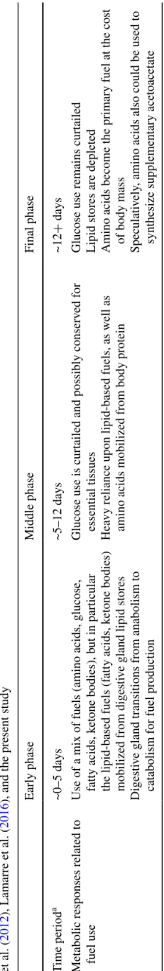
Overall, we propose a three stage metabolic response to starvation in cephalopods (Table 1) that, in general, resembles the sequential metabolic stages of starvation in mammals and fishes, in which carbohydrates are con-served, lipids sustain starvation, and protein is only used as a last resort once lipid stores fall below a critical level (Black and Love 1986; Cahill 2006; Bar 2014; Secor and Carey 2016). However, in cephalopods, the process may be accelerated, with a relatively early switch to protein catabolism. This is likely because, compared with most vertebrates (especially ectotherms), glycogen and lipid stores are small in cephalopods relative to whole-animal metabolic demand, and the largest glycogen store, in the mantle, seems to be reserved for burst exercise (O’Dor et al. 1984). Meanwhile, body protein is plentiful in cepha-lopods. However, because mobilization of body protein could impair performance, the accelerated metabolic tran-sition of starved cephalopods may ultimately reflect sen-sitivity to food deprivation, which, in turn, is a trade-off of their “live-fast, die-young” life history that emphasizes growth over energy storage (O’Dor and Webber 1986). However, exploration of the astounding functional and ecological diversity of cephalopods might provide excep-tions, for example among deep-sea species (Seibel et al. 2000; Robison et al. 2014).
Acknowledgments We thank the staff and students at Centro de
Ciências do Mar do Algarve for their hospitality and invaluable help during our research, in particular Juan Carlos Capaz, Ana Couto, Ana Oliveira, João Reis, Tania Rodríguez-González, and Cátia Silva. We also thank Dr. Matthew Nosworthy for skilled assistance during a por-tion of this study and Dr. Kurt Gamperl for loaning us certain equip-ment. Funding was provided by an NSERC Postdoctoral Fellowship to B.S.-R., an NSERC Canada Graduate Scholarship-Master’s and a New Brunswick Innovation Foundation scholarship to N.I.C., and NSERC Discovery Grants to W.R.D., T.J.M., and S.G.L. AVS was supported by Fundação para a Ciência e a Tecnologia through Pro-grama Investigador FCT 2014 (IF/00576/2014) and project SEPIAT-ECH (31-03-05-FEP-2), funded by the Portuguese Government Pro-gram PROMAR.
References
Agnisola C, Driedzic WR, Foster AR, Houlihan DF, Stewart JM (1991) Oxygen consumption, carbon dioxide production and enzyme activities of isolated working Octopus heart. J Exp Biol 157:543–549
Alegre M, Ciudad CJ, Fillat C, Guinovart JJ (1988) Determination of glucose-6-phosphatase activity using the glucose dehydroge-nase-coupled reaction. Anal Biochem 173:185–189
Ballantyne JS, Berges JA (1991) Enzyme activities of gill, hepatopan-creas, mantle, and adductor muscle of the oyster (Crassostrea virginica) after changes in diet and salinity. Can J Fish Aquat Sci 48:1117–1123 Table 1 Proposed three-stage metabolic response to starv ation in cephalopods based on findings of O’Dor et al. ( 1984 ), Boucher -Rodoni and Mangold ( 1985 ), Castro et al. ( 1992 ), Lamarre et al. ( 2012 ), Lamarre et al. ( 2016
), and the present study
a Depending on body temperature and body size
Early phase Middle phase Final phase T ime period a ~0–5 days ~5–12 days ~12 + days
Metabolic responses related to fuel use Use of a mix of fuels (amino acids, glucose, fatty acids, k
etone bodies), b
ut in particular
the lipid-based fuels (f
atty acids, k
etone bodies)
mobilized from digesti
ve gland lipid stores
Digesti
ve gland transitions from anabolism to
catabolism for fuel production
Glucose use is curtailed and possibly conserv
ed for
essential tissues
Hea
vy reliance upon lipid-based fuels, as well as
amino acids mobilized from body protein
Glucose use remains curtailed Lipid stores are depleted Amino acids become the primary fuel at the cost of body mass Speculati
vely
, amino acids also could be used to
Ballantyne JS, Moon TW (1985) Hepatopancreas mitochondria from Mytilus edulis: substrate preference and effects of pH and osmo-larity. Mar Biol 87:239–244
Ballantyne JS, Moyes CD (1987) Osmotic effects on fatty acid, pyru-vate, and ketone body oxidation in oyster gill mitochondria. Physiol Zool 60:713–721
Ballantyne JS, Storey KB (1983) Mitochondria from the ventricle of the marine clam, Mercenaria mercenaria: substrate preferences and effects of pH and salt concentration on proline oxidation. Comp Biochem Physiol B 76:133–138
Ballantyne JS, Storey KB (1984) Mitochondria from the hepatopan-creas of the marine clam Mercenaria mercenaria: substrate pref-erences and salt and pH effects on the oxidation of palmitoyl-L-carnitine and succinate. J Exp Zool 230:165–174
Ballantyne JS, Hochachka PW, Mommsen TP (1981) Studies on the metabolism of the migratory squid, Loligo opalescens: enzymes of tissues and heart mitochondria. Mar Biol Lett 2:75–85 Bar N (2014) Physiological and hormonal changes during prolonged
starvation in fish. Can J Fish Aquat Sci 71:1447–1458
Bar N, Volkoff H (2012) Adaptation of the physiological, endocrine, and digestive system functions to prolonged food deprivation in fish. In: McCue M (ed) Comparative physiology of fasting, star-vation, and food limitation. Springer, Berlin, pp 69–89
Beis A, Zammit VA, Newsholme EA (1980) Activities of 3-hydroxy-butyrate dehydrogenase, 3-oxoacid CoA-transferase and ace-toacetyl-CoA thiolase in relation to ketone-body utilisation in muscles from vertebrates and invertebrates. Eur J Biochem 104:209–215
Black D, Love RM (1986) The sequential mobilisation and restoration of energy reserves in tissues of Atlantic cod during starvation and refeeding. J Comp Physiol B 156:469–479
Blier P, Guderley H (1986) The enzymatic and metabolic effects of extended food deprivation in Rana pipiens. Physiol Zool 59:230–239
Boucher-Rodoni R, Mangold K (1985) Ammonia excretion during feeding and starvation in Octopus vulgaris. Mar Biol 86:193–197 Brooks SP (1994) A program for analyzing enzyme rate data obtained
from a microplate reader. Biotechniques 17:1154–1161
Cahill GF Jr (2006) Fuel metabolism in starvation. Annu Rev Nutr 26:1–22
Castro BG, Garrido JL, Sotelo CG (1992) Changes in composition of digestive gland and mantle muscle of the cuttlefish Sepia offici-nalis during starvation. Mar Biol 114:11–20
Cherel Y, Burnol AF, Leturque A, Le Maho Y (1988) In vivo glucose utilization in rat tissues during the three phases of starvation. Metabolism 37:1033–1039
Crabtree B, Higgins SJ, Newsholme EA (1972) The activities of pyru-vate carboxylase, phosphoenolpyrupyru-vate carboxylase and fructose diphosphatase in muscles from vertebrates and invertebrates. Biochem J 130:391–396
Driedzic WR (2015) Rainbow smelt: the unusual case of cryoprotec-tion by sustained glycerol produccryoprotec-tion in an aquatic animal. J Comp Physiol B 185:487–499
Driedzic WR, Sidell BD, Stewart JM, Johnston IA (1990) Maximal activities of enzymes of energy metabolism in cephalopod sys-temic and branchial hearts. Physiol Zool 63:615–629
Fields JHA, Hochachka PW (1982) Glucose and proline metabolism in Nautilus. Pac Sci 36:337–341
Fiorito G, Affuso A, Basil J, Cole A, de Girolamo P, D’Angelo L, Dickel L et al (2015) Guidelines for the care and welfare of cephalopods in research—a consensus based on an initiative by CephRes, FELASA and the Boyd Group. Lab Anim 49(2 suppl):1–90
Foster GD, Moon TW (1991) Hypometabolism with fasting in the yellow perch (Perca flavescens): a study of enzymes, hepatocyte
Gabr HR, Hanlon RT, Hanafy MH, El-Etreby SG (1999) Reproduc-tive versus somatic tissue allocation in the cuttlefish Sepia doll-fusi Adam (1941). Bull Mar Sci 65:159–173
Gibson DM, Harris RA (2002) Metabolic regulation in mammals. Taylor Francis, London
Grigoriou P, Richardson CA (2009) Effect of body mass, temperature and food deprivation on oxygen consumption rate of common cuttlefish Sepia officinalis. Mar Biol 156:2473–2481
Hochachka PW (1995) Oxygen efficient design of cephalopod muscle metabolism. Mar Freshw Behav Physiol 25:61–67
Hochachka PW, Fields JHA (1982) Arginine, glutamate, and proline as substrates for oxidation and for glycogenesis in cephalopod tissues. Pac Sci 36:325–335
Hochachka PW, Mommsen TP, Storey J, Storey KB, Johansen K, French CJ (1983) The relationship between arginine and proline metabolism in cephalopods. Mar Biol Lett 4:1–21
Horio Y, Tanaka T, Taketoshi M, Uno T, Wada H (1988) Rat cytosolic aspartate aminotransferase: regulation of its mRNA and contri-bution to gluconeogenesis. J Biochem 103:805–808
Horst C, Becker W (1986) Nutritive medium chain triacylglycerols cause a rapid increase of ketone bodies in the hemolymph of Biomphalaria glabrata (Gastropoda: Pulmonata). Comp Bio-chem Physiol B 85:875–878
Horst C, Becker W, Kemper A (1986) Short-term alterations of the ketone body content in the hemolymph of Biomphalaria glabrata (Gastropoda: Pulmonata). Comp Biochem Physiol B 84:555–557
Islinger M, Cardoso MJR, Schrader M (2010) Be different—the diversity of peroxisomes in the animal kingdom. Biochim Bio-phys Acta Cell Res 1803(8):881–897
Laffel L (1999) Ketone bodies: a review of physiology, pathophysiol-ogy and application of monitoring to diabetes. Diabetes Metab Res Rev 15:412–426
Lamarre SG, Ditlecadet D, McKenzie DJ, Bonnaud L, Driedzic WR (2012) Mechanisms of protein degradation in mantle muscle and proposed gill remodeling in starved Sepia officinalis. Am J Phys-iol Reg Integr Comp PhysPhys-iol 303:R427–R437
Lamarre S, Macmillan L, Morrow GP, Randell E, Pongnopparat T, Brosnan ME, Brosnan JT (2014) An isotope-dilution, GC– MS assay for formate and its application to human and animal metabolism. Amino Acids 46:1885–1891
Lamarre SG, MacCormack TJ, Sykes A, Speers-Roesch B, Calla-ghan NI, Driedzic WR (2016) Metabolic rate and rates of protein turnover in food deprived cuttlefish, Sepia officinalis (Linnaeus 1758). Am J Physiol Reg Integr Comp Physiol. doi:10.1152/ ajpregu.00459.2015
Lee PG (1995) Nutrition of cephalopods: fueling the system. Mar Freshw Behav Physiol 25:35–51
McCue MD (2010) Starvation physiology: reviewing the different strategies animals use to survive a common challenge. Comp Biochem Physiol A 156:1–18
McMurray CH, Blanchflower WJ, Rice DA (1984) Automated kinetic method for d-3-hydroxybutyrate in plasma or serum. Clin Chem 30:421–425
Metón I, Mediavilla D, Caseras A, Cantó E, Fernández F, Baanante IV (1999) Effect of diet composition and ration size on key enzyme activities of glycolysis–gluconeogenesis, the pentose phosphate pathway and amino acid metabolism in liver of gilt-head sea bream (Sparus aurata). Br J Nutr 82:223–232
Meyer R, Becker W, Klimkewitz M (1986) Investigations on the ketone body metabolism in Biomphalaria glabrata: influence of starvation and of infection with Schistosoma mansoni. J Comp Physiol B 156:563–571
Moltschaniwskyj N, Johnston D (2006) Evidence that lipid can be digested by the dumpling squid Euprymna tasmanica, but is not
Mommsen TP, Hochachka PW (1981) Respiratory and enzy-matic properties of squid heart mitochondria. Eur J Biochem 120:345–350
Mommsen TP, Ballantyne J, MacDonald D, Gosline J, Hochachka PW (1981) Analogues of red and white muscle in squid mantle. Proc Natl Acad Sci 78:3274–3278
Mommsen TP, Hochachka PW, French CJ (1983) Metabolism of argi-nine, proline, and ornithine in tissues of the squid, Illex illecebro-sus. Can J Zool 61:1835–1846
Moon TW, Johnston IA (1980) Starvation and the activities of gly-colytic and gluconeogenic enzymes in skeletal muscles and liver of the plaice, Pleuronectes platessa. J Comp Physiol 136:31–38 Morillo-Velarde PS, Valverde JC, Serra Llinares RM, García BG
(2013) Changes in lipid composition of different tissues of com-mon octopus (Octopus vulgaris) during short-term starvation. Aquacult Res 44:1177–1189
Moyes CD, Suarez RK, Hochachka PW, Ballantyne JS (1990) A com-parison of fuel preferences of mitochondria from vertebrates and invertebrates. Can J Zool 68:1337–1349
Navarro JC, Monroig O, Sykes AV (2014) Nutrition as a key factor for cephalopod aquaculture. In: Iglesias J, Fuentes L, Villanueva R (eds) Cephalopod culture. Springer, New York, pp 77–95 Newsholme EA, Crabtree B (1986) Maximum catalytic activity of
some key enzymes in provision of physiologically useful infor-mation about metabolic fluxes. J Exp Zool 239:159–167 O’Dor RK, Webber DM (1986) The constraints on cephalopods: why
squid aren’t fish. Can J Zool 64:1591–1605
O’Dor RK, Mangold K, Boucher-Rodoni R, Wells MJ, Wells J (1984) Nutrient absorption, storage and remobilization in Octopus vul-garis. Mar Behav Physiol 11:239–258
Pande SV, Lee TS, Murthy MS (1990) Freeze-thawing causes mask-ing of membrane-bound outer carnitine palmitoyltransferase activity: implications for studies on carnitine palmitoyltrans-ferases deficiency. BBA Lipid Lipid Met 1044:262–268
Pilkis SJ, El-Maghrabi MR, Claus TH (1988) Hormonal regulation of hepatic gluconeogenesis and glycolysis. Annu Rev Biochem 57:755–783
Robinson AM, Williamson DH (1980) Physiological roles of ketone bodies as substrates and signals in mammalian tissues. Physiol Rev 60:143–187
Robison B, Seibel B, Drazen J (2014) Deep-sea octopus (Granele-done boreopacifica) conducts the longest-known egg-brooding period of any animal. PLoS One 9:e103437
Rosa R, Pereira J, Nunes ML (2005) Biochemical composition of cephalopods with different life strategies, with special reference to a giant squid, Architeuthis sp. Mar Biol 146:739–751
Secor SM, Carey HV (2016) Integrative physiology of fasting. Compr Physiol 6:773–825
Seibel BA, Hochberg FG, Carlini DB (2000) Life history of Gonatus onyx (Cephalopoda: Teuthoidea): deep-sea spawning and post-spawning egg care. Mar Biol 137:519–526
Semmens JM (1998) An examination of the role of the digestive gland of two loliginid squids, with respect to lipid: storage or excre-tion? Proc Roy Soc Lond B 265:1685–1690
Sidell BD, Driedzic WR, Stowe DB, Johnston IA (1987) Biochemi-cal correlations of power development and metabolic fuel prefer-enda in fish hearts. Physiol Zool 60:221–232
Speers-Roesch B, Treberg JR (2010) The unusual energy metabolism of elasmobranch fishes. Comp Biochem Physiol A 155:417–434 Stewart JM, Brass ME, Carlin RC, Black H (1992) Maximal enzyme
activities of energy production pathways in the heart, hepato-pancreas, and white muscle of the giant scallop (Placopecten magellanicus) and lobster (Homarus americanus). Can J Zool 70:720–724
Stio M, Vanni P, Pinzauti G (1988) A continuous spectrophotomet-ric assay for the enzymatic marker glucose 6-phosphatase. Anal Biochem 174:32–37
Storey KB, Storey JM (1979) Octopine metabolism in the cuttlefish, Sepia officinalis: octopine production by muscle and its role as an aerobic substrate for non-muscular tissues. J Comp Physiol 131:311–319
Storey KB, Storey JM (1983) Carbohydrate metabolism in cephalo-pod molluscs. In: Hochachka PW, Wilbur KM (eds) The mol-lusca, vol 1. Academic Press, New York, pp 91–136
Storey KB, Storey JM, Johansen K, Hochachka PW (1979) Octopine metabolism in Sepia officinalis: effect of hypoxia and metabo-lite loads on the blood levels of octopine and related compounds. Can J Zool 57:2331–2336
Stuart JA, Ballantyne JS (1996) Correlation of environment and phy-logeny with the expression of β-hydroxybutyrate dehydrogenase in the mollusca. Comp Biochem Physiol B 114:153–160
Suarez RK (2012) Energy and metabolism. Compr Physiol 2:2527–2540
Sykes AV, Domingues P, Andrade JP (2014) Sepia officinalis. In: Iglesias J, Fuentes L, Villanueva R (eds) Cephalopod culture. Springer, New York, pp 175–204
Voogt PA (1983) Lipids: their distribution and metabolism. In: Hochachka PW, Wilbur KM (eds) The mollusca, vol 1. Aca-demic Press, New York, pp 329–370
Weber JM (2011) Metabolic fuels: regulating fluxes to select mix. J Exp Biol 214:286–294
Wells MJ, Clarke A (1996) Energetics: the costs of living and reproducing for an individual cephalopod. Phil Trans R Soc B 351:1083–1104
Yang J, Kalhan SC, Hanson RW (2009) What is the metabolic role of phosphoenolpyruvate carboxykinase? J Biol Chem 284:27025–27029
Zammit VA, Beis A, Newsholme EA (1979) The role of 3-oxo acid-CoA transferase in the regulation of ketogenesis in the liver. FEBS Lett 103:212–215