UNIVERSIDADE DE LISBOA
FACULDADE DE CIÊNCIAS
DEPARTAMENTO DE BIOLOGIA VEGETAL
Clinical and molecular characterization of Portuguese patients
with a clinical diagnosis of MODY
João Paulo de Medeiros Gomes de Mafra
Mestrado em Biologia Molecular e Genética
Dissertação orientada por:
Mafalda Bourbon
Agradecimentos
À Mafalda, pela oportunidade de ganhar experiência em ambiente laboratorial e de participar num congresso internacional. À Ana e Catarina pelo apoio inicial a lidar com assuntos que me eram estranhos. À Chora pela ajuda com os in silico e à Cibelle pelo sorriso e boa disposição. À Gisela pela colaboração no MLPA.
À Sílvia pelo bom humor e leveza. À Joana Costa pelas picardias e conversas futebolísticas. À Andreia por ser um doce de pessoa. Ao Niccolò pelo escape e pelo man time. À Inês pelo companheirismo. À Russo e Sara pela ajuda na integração à FCUL.
À Canilho por uma amizade que ultrapassa o laboratório. À Rita por me dar a mão quando caí. Ao Pedro pela irmandade e sintonia intelectual. Aos que ficam sem nome mas que não me esquecem, que acreditam em mim, nem eu sei bem porquê, mas que também não esqueço e muito estimo. Aos irmãos que por vezes sinto que perdi.
Aos meus pais, que me apoiam mais do que mereço. Obrigado.
Resumo
A diabetes mellitus, ou simplesmente diabetes, pode ser definida como um conjunto complexo de perturbações crónicas de cariz metabólico caracterizadas por hiperglicémia. A diabetes tipo MODY, do inglês Maturity-onset diabetes of the young, é uma forma monogénica de diabetes. Inicialmente descrita em 1974 por Tattersall, a diabetes tipo MODY engloba um grupo de fenótipos heterogéneos, clinica e geneticamente, caracterizados por alterações no funcionamento normal das células beta do pâncreas e por um padrão de hereditariedade autossómico dominante. De uma forma geral, a diabetes tipo MODY tem um perfil não-insulino dependente e manifesta-se em crianças e indivíduos jovens, sendo tipicamente diagnosticada antes dos 25 anos.
A diabetes tipo MODY aparenta ser rara, estimando-se que seja responsável por 0,6-2% dos casos de diabetes na Europa. No entanto, é frequente esta forma de diabetes ser equivocamente diagnosticada como diabetes tipo 1 ou tipo 2, pelo que a sua prevalência real deverá ser superior. Um dos grandes trunfos no combate a este subdiagnóstico da diabetes tipo MODY são os testes genéticos. Desde a década de 1990, foram 13 os genes associados à MODY. Mutações em heterozigotia nos genes GCK e
HNF1A são as causas mais frequentes de diabetes tipo MODY, correspondendo a cerca de 70% dos
casos. Logo a seguir estão as mutações em heterozigotia nos genes HNF4A (hepatocyte nuclear factor
4 alpha) e HNF1B, que correspondem a cerca de 15% dos casos.
O gene GCK, localizado no cromossoma 7 (7p13), codifica o enzima glicocinase (glucokinase - GCK), também conhecido como hexocinase IV. Este enzima monomérico possui três isoformas - a isoforma 1, presente nas células beta, e as 2 e 3, presentes no fígado - e, no interior das células, atua como um sensor do nível de glicose. Nos hepatócitos, este enzima intervém no desencadear da glicólise e glicogénese, enquanto facilita a exocitose de insulina nas células beta. A diabetes tipo MODY, subtipo GCK, resulta de mutações de perda de função em heterozigotia no gene GCK, diminuindo a atividade do enzima, e caracteriza-se por uma hiperglicémia moderada, assintomática e não progressiva que se manifesta desde o nascimento. Estas mutações acabam por diminuir a quantidade de glicogénio sintetizado e impedem a normal libertação de insulina, ao aumentar a concentração mínima de glicose necessária à sua secreção.
O gene HNF1A, localizado no cromossoma 12 (12q24.31), codifica um fator de transcrição (hepatocyte nuclear factor 1 alpha - HNF1A) homodimérico. Este fator de transcrição possui três isoformas, A, B e C. As três estão presentes no fígado, rins, pâncreas e intestinos mas a primeira predomina no fígado, rins e pâncreas fetal, enquanto a isoforma B predomina no pâncreas adulto. O HNF1A integra uma complexa rede de fatores de transcrição, desempenhando um papel regulador na expressão de diversos genes durante o desenvolvimento embrionário. No fígado, o HNF1A regula a expressão de vários genes hepáticos, como o gene que codifica para a albumina. Nas células beta, intervém na expressão de insulina e na proliferação e morte celular. A diabetes tipo MODY, subtipo HNF1A, resulta de mutações em heterozigotia no gene HNF1A. Estas mutações podem ter diversos efeitos, reduzindo a secreção de insulina em resposta a glicose e aminoácidos, e alterando a expressão de genes envolvidos no transporte (GLUT2) e metabolismo de glicose, afetando processos como a glicólise, gluconeogénese e derivação de aminoácidos para o ciclo de Krebs. O subtipo HNF1A está associado a defeitos na proliferação das células beta e caracteriza-se por uma incapacidade progressiva na secreção de insulina, que não acompanha o aumento de glicose em circulação, dando origem a uma hiperglicémia mais grave que o subtipo GCK.
O gene HNF1B, localizado no cromossoma 17 (17q12), codifica um fator de transcrição (hepatocyte
HNF1B produz três isoformas - 1, 2 e 3 - e é expresso no timo, pulmão, rim, fígado, pâncreas,
estômago, intestino e trato genital. Este fator de transcrição, que integra a mesma rede regulatória que HNF1A, actua no desenvolvimento embrionário do pâncreas e do rim. A diabetes tipo MODY, subtipo HNF1B, resulta de mutações em heterozigotia no gene HNF1B e em cerca de 50% dos casos caracteriza-se por uma combinação de resistência à insulina e disfunção das células beta, exibindo, à semelhança do subtipo HNF1A, uma incapacidade na secreção de insulina em resposta a concentrações crescentes de glicose. Mutações neste gene podem também resultar em anomalias extra pancreáticas e afetar tecidos como o trato genital ou o fígado, sendo quistos renais o fenómeno mais observado.
Neste estudo, foram recolhidas informações clínicas acerca de 39 indivíduos, 24 probandos e 15 familiares, através de questionários enviados por vários médicos de diferentes hospitais portugueses. Com base nestas informações, como glicémia em jejum, prova de tolerância à glicose oral e HbA1c, e no diagnóstico clínico de diabetes tipo MODY, foram-nos referenciados indivíduos com uma história familiar de diabetes que evidencie um padrão hereditário dominante; sem autoanticorpos pancreáticos; com início de sintomas antes dos 25 anos; e índice de massa corporal maioritariamente normal. Estes 24 probandos foram estudados por sequenciação de Sanger para os genes GCK e HNF1A, tendo sido também efetuada a pesquisa de inserções e deleções através da técnica MLPA (Multiplex
Ligation-dependent Probe Amplification).
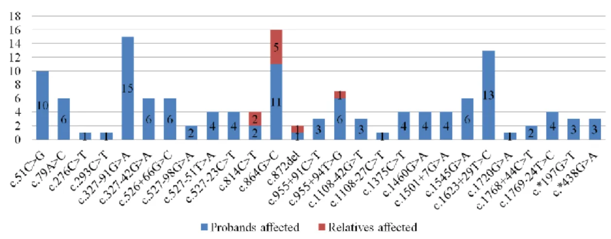
Entre substituições pontuais e pequenas deleções, a sequenciação de Sanger detetou 46 variantes genéticas, 19 no gene GCK e 27 no gene HNF1A. Entre estas, seis podem ser classificadas como patogénicas ou provavelmente patogénicas, sendo quatro no gene GCK e duas no gene HNF1A. As variantes patogénicas ou provavelmente patogénicas detetadas no gene GCK foram c.364C>T (p.(Leu122Phe)), uma alteração missense no exão 4 que cosegrega com diabetes e está associada à diabetes tipo MODY; c.579+1_579+33del, uma deleção de 33 pares de base no intrão 5 que elimina um local de splicing e também está associada à diabetes tipo MODY; c.766G>A (p.Glu256Lys), uma alteração missense no exão 7 que induz alterações conformacionais no enzima GCK, reduzindo a sua capacidade de ligação à glicose e subsequente atividade catalítica; e, finalmente, c.1268T>A (p.(Phe423Tyr)), uma alteração missense no exão 10 que cosegrega com diabetes e está associada à diabetes tipo MODY. Estas variantes foram encontradas num total de quatro probandos e cinco familiares. No gene HNF1A, as variantes patogénicas detetadas foram c.814C>T (p.(Arg272Cys)), uma alteração missense no exão 4 que cosegrega com diabetes e impede o fator de transcrição HNF1A de se ligar ao DNA, perdendo assim a sua atividade reguladora na expressão genética; e c.872del (p.(Pro291Glnfs*51)), uma deleção também localizada no exão 4 que provoca uma alteração na grelha de leitura, cosegrega com diabetes e possivelmente resulta de um fenómeno de splippage aquando da replicação de DNA, prevendo-se que resulte numa proteína truncada. Estas variantes foram encontradas num total de três probandos e três familiares.
A técnica MLPA foi aplicada na investigação de inserções e deleções em 17 probandos. Três destes probandos geraram resultados sem qualidade, sendo necessárias novas amostras para eventual repetição. Noutros 11 nenhuma inserção ou deleção foi detetada. Nos restantes três probandos foram detetadas duas deleções patogénicas: deleção em heterozigotia dos exões 5 a 8 do gene GCK (c.484-?_1019+?del) num probando; e deleção em heterozigotia do gene HNF1B (c.1-?_1674+?del) em dois probandos. A deleção no gene GCK deverá produzir uma proteína não funcional para gerar o fenótipo MODY, subtipo GCK. No entanto, não foi encontrada qualquer informação sobre esta deleção ou o seu efeito na proteína, pelo que poderemos estar na presença de uma nova mutação. Quanto à deleção do gene HNF1B, estas são frequentes e existem várias fontes que apontam deleções completas do gene como patogénicas, que muitas vezes incluem outros genes na mesma região (17q12). Um dos
probandos aparenta não ter história familiar de diabetes e, sendo mutações de novo frequentes, é possível que estejamos na presença de uma. No entanto, não haviam amostras de familiares para fazer estudos de cosegregação nestes dois probandos. A hemizigotia provocada por esta deleção deve resultar em MODY, subtipo HNF1B, por haploinsuficiência. Nenhum destes dois probandos aparenta ter anomalias renais.
No total, entre 24 probandos, este estudo identificou 10 indivíduos com MODY, cinco do subtipo GCK, três do subtipo HNF1A e dois do subtipo HNF1B, realçando assim a importância de um diagnóstico correto, com recurso a ferramentas de genética molecular, uma vez que estes indivíduos tinham diagnósticos de diabetes tipo 1 ou tipo 2. Dos restantes 14 probandos sem qualquer variante patogénica/provavelmente patogénica, foi detetada pelo menos uma variante associada a diabetes tipo 2. A utilização destas técnicas no âmbito do diagnóstico genético permite estabelecer o diagnóstico correcto, com implicações para a terapêutica a administrar e qualidade de vida dos utentes dos serviços de saúde.
Abstract
Initially described by Tattersall in 1974, maturity-onset diabetes of the young (MODY) is a form of early onset monogenic diabetes characterized by clinically heterogeneous phenotypes with autosomal dominant inheritance that generally result in β-cell dysfunction. Often misdiagnosed, MODY accounts for 0.6-2% of diabetes cases in Europe and 13 genes have been implicated in this form of diabetes, with heterozygous mutations in GCK and HNF1A being the most common etiologies, followed by heterozygous mutations in HNF4A and HNF1B.
Glucokinase (GCK) is a monomeric enzyme that acts as a cellular glucose sensor, playing a role in glycogenesis and glycolysis in hepatocytes, and in insulin release in β-cells. Heterozygous loss-of-function mutations in GCK decrease enzymatic activity and result in GCK-MODY, characterized by asymptomatic mild stable hyperglycemia present from birth. HNF1A-MODY is associated with impaired β-cell proliferation and is characterized by a progressive insulin secretory defect, resulting in a more severe hyperglycemia. This form of MODY is caused by heterozygous mutations in HNF1A, a gene that codes hepatocyte nuclear factor 1 alpha, a homodimeric transcription factor that regulates gene expression in embryonic development and plays a role in insulin expression and β-cell proliferation and death. Hepatocyte nuclear factor 1 beta (HNF1B) is also a transcription factor that acts as a homodimer, or a heterodimer with HNF1A. HNF1B is part of the same regulatory network as HNF1A and regulates embryonic pancreatic development, also playing a role in nephron development. Heterozygous HNF1B mutations result in HNF1B-MODY, characterized by a combination of β-cell dysfunction and insulin resistance in approximately 50% of mutation carriers. Like in HNF1A-MODY, insulin secretion is compromised as glucose concentrations rise. Renal cysts are common. Clinical data on 24 probands and 15 relatives was collected through questionnaires sent by physicians. Subjects with a clinical diagnosis of MODY - family history of diabetes consistent with dominant inheritance pattern, no pancreatic autoantibodies, onset before 25 years of age and typically lean - were referred to this study by physicians for genetic screening via Sanger sequencing (for GCK and
HNF1A) and Multiplex Ligation-dependent Probe Amplification (MLPA).
Between point substitutions and small deletions, Sanger sequencing detected 46 variants, 19 in GCK and 27 in HNF1A. Six of these were pathogenic or likely pathogenic, four in GCK and two in HNF1A.
GCK pathogenic or likely pathogenic variants were c.364C>T (p.(Leu122Phe)), in exon 4;
c.579+1_579+33del, in intron 5, abolishing the donor splice site; c.766G>A (p.Glu256Lys), in exon 7, which induces conformational changes, decreasing glucose binding and catalytic activity; and c.1268T>A (p.(Phe423Tyr)), in exon 10. These variants were detected in four probands and five relatives. HNF1A pathogenic variants were c.814C>T (p.(Arg272Cys)), which renders HNF1A unable to bind DNA and exert its transactivating activity; and c.872del (p.(Pro291Glnfs*51)), predicted to produce a truncated protein and likely the result of replication slippage. Both variants are located in exon 4. These variants were detected in three probands and three relatives.
MLPA analysis detected two pathogenic deletions: a heterozygous GCK exons 5 through 8 deletion (c.484-?_1019+?del) in one proband; and a heterozygous HNF1B deletion (c.1-?_1674+?del) in two probands. The seemingly unreported heterozygous GCK exons 5 through 8 deletion, which should result in a null variant that decreases overall enzymatic activity, could be a novel mutation. Heterozygous HNF1B deletions should result in HNF1B-MODY via haploinsufficiency. One proband had no apparent family history but de novo mutations are frequent. Both probands had no apparent renal abnormalities.
Among 24 probands, this study identified a total of 10 MODY cases, with five being GCK-MODY, three being HNF1A-MODY and two being HNF1B-MODY. Of the remaining 14 probands without any detected pathogenic/likely pathogenic variants, 11 had at least one type 2 diabetes associated variant. Hence, this study highlights the importance of genetic diagnosis in patients with diabetic phenotypes consistent with MODY, as a correct diagnosis impacts patient treatment and quality of life. Key words: Diabetes; MODY; GCK; HNF1A; HNF1B
Table of contents
Agradecimentos ... I Resumo ... II Abstract ... V Abbreviations, acronyms and symbols ... X
1. Introduction ... 1
1.1 MODY ... 1
1.2 Differentiating MODY from T1DM and T2DM ... 2
1.3 GCK and GCK-MODY ... 3
1.4 HNF1A and HNF1A-MODY ... 5
1.5 HNF4A and HNF4A-MODY ... 8
1.6 HNF1B and HNF1B-MODY ... 10
1.7 Rare types of MODY ... 11
2. Materials and Methods ... 12
2.1 Inclusion criteria ... 12
2.2 DNA extraction ... 12
2.3 DNA quantification and quality screen ... 12
2.4 Primer design, checks and preparation ... 12
2.5 PCR optimization ... 13
2.6 PCR reactions ... 13
2.7 Agarose gel electrophoresis ... 13
2.8 PCR product purification and Sanger sequencing reaction ... 13
2.9 Sequence analysis and variant interpretation ... 14
2.10 MLPA ... 14
3. Results ... 15
3.1 Subjects under study ... 15
3.1.1 Number of subjects, sex, ethnicity, age and age at diagnosis ... 15
3.1.2 Family history of diabetes and renal disease ... 15
3.1.3 Vascular complications ... 15
3.1.4 Birth weight and BMI ... 15
3.1.5 FPG ... 16
3.1.6 OGTT ... 16
3.1.7 HbA1c ... 16
3.1.8 Pancreatic autoantibodies ... 16
3.2 Molecular analysis: Sanger sequencing and MLPA ... 17
3.2.1 Sanger sequencing analysis: GCK ... 17
3.2.2 Sanger sequencing analysis: HNF1A ... 20
3.2.3 MLPA analysis ... 24
4. Discussion ... 26
5. References ... 31
6. Supplementary material ... 39
6.1 Comparative analysis of MODY, T1DM and T2DM ... 39
6.2 Online tools ... 40
6.3 Interpreting in silico mutation predictions ... 40
6.4 Primer sequences, PCR master mix and cycling program... 40
6.4.1 Primer sequences ... 40
6.4.2 PCR master mix and cycling program ... 41
6.5 Sanger sequencing mix and cycling program ... 42
6.6 Sequence analysis and pregap module configuration ... 42
Tables and figures
Table 6.1 Differentiating MODY from T1DM and T2DM ...39
Table 6.2: Primer data ...40
Table 6.3: Standard PCR mix and cycling program ...41
Table 6.4: Sequencing mix and cycling program ...42
Table 6.5: Detected sequence variants ...43
Figure 1.1 HNF1A structure ...7
Figure 1.2 HNF4A gene and isoform structures ...9
Figure 1.3 HNF1B structure ...11
Figure 3.1 Detected sequence variants ...17
Figure 3.2 GCK sequence variants location ...17
Figure 3.3 GCK sequence variants and affected subjects ...18
Figure 3.4 GCK c.364C>T proband chromatogram and pedigree ...18
Figure 3.5 GCK c.579+1_579+33del proband chromatogram and pedigree ...19
Figure 3.6 GCK c.766G>A proband chromatogram and pedigree ...19
Figure 3.7 GCK c.1268T>A proband chromatogram and pedigree ...20
Figure 3.8 HNF1A sequence variants location ...20
Figure 3.9 HNF1A sequence variants and affected subjects ...21
Figure 3.10 HNF1A c.814C>T proband (14-014) chromatogram and pedigree ...21
Figure 3.11 HNF1A c.814C>T proband (14-015) chromatogram and pedigree ...22
Figure 3.12 HNF1A c.872del proband chromatogram and pedigree ...22
Figure 3.13 HNF1A c.1720G>A proband chromatogram and pedigree...23
Figure 3.14 HNF1A c.79A>C proband chromatograms ...23
Figure 3.15 HNF1A c.293C>T and c.1460G>A proband chromatograms ...24
Figure 3.16: MLPA results for proband 15-014 ...25
Figure 3.17: MLPA results for probands 13-006 (A) and 14-013 (B) ...25
Abbreviations, acronyms and symbols
°C Celsius Degree
µL Microlitre (10-6 l)
µM Micromolar (10-6 M)
ABCC8 ATP Binding Cassette subfamily C member 8
ACMG American College of Medical Genetics and Genomics
ADP Adenosine Diphosphate
AE At Enrollment
AFR African population
Ala Alanine
ALT Alanine Aminotransferase
AMP Association for Molecular Pathology apo A1 Apolipoprotein A1
apo A2 Apolipoprotein A2
apo B Apolipoprotein B
apo CIII Apolipoprotein CIII
Arg Arginine
Asn Asparagine
Asp Aspartic Acid
ATP Adenosine Triphosphate
BE Before Enrollment
BLAT BLAST-like Alignment Tool
BLK B Lymphocyte Kinase, gene
BMI Body Mass Index
bp Base Pair
Ca2+ Calcium ion
CEL Carboxyl Ester Lipase, gene
CNV Copy Number Variation
Cys Cysteine
del Deletion
DKA Diabetic Ketoacidosis
DNA Deoxyribonucleic Acid
dNTP Deoxynucleotide Triphosphates EDTA Ethylenediamine Tetraacetic Acid
EUR European population
EVS Exome Variant Server
ExAC Exome Aggregation Consortium
F Forward (primer)
FPG Fasting Plasma Glucose
fs Frameshift
g Gram
G6P Glucose-6-phosphate
GADA Glutamic Acid Decarboxylase Antibodies
GCK Glucokinase, gene
GDM Gestational Diabetes
GGT Gamma-glutamyl transferase
GKRP GCK Regulatory Protein
Gln Glutamine
Glu Glutamic Acid
GLUT2 Glucose Transporter 2
Gly Glycine
HbA1c Glycated Hemoglobin
HDL High Density Lipoprotein
HNF Hepatocyte Nuclear Factor
HNF1A Hepatocyte Nuclear Factor 1 Alpha, gene
HNF1B Hepatocyte Nuclear Factor 1 Beta, gene
HNF4A Hepatocyte Nuclear Factor 4 Alpha, gene
i.e. id est
ICA Islet Cell Antibodies
IGT Impaired Glucose Tolerance
Ile Isoleucine
INS Insulin, gene kbp Kilo (103) Base Pair
KCNJ11 Potassium Voltage-gated Channel subfamily J member 11, gene
kg Kilogram
kg/m2 Kilogram per Square Metre
KLF11 Kruppel Like Factor 11, gene Km Michaelis constant
L Litre
LDL Low Density Liproprotein
Leu Leucine
Lys Lysine
MAF Minor Allele Frequency
mg/dL Milligram (10-3 g) per Decilitre (10-1 L)
MgCl2 Magnesium Chloride
min Minute
mL Millilitre (10-3 L)
MLPA Multiplex Ligation-dependent Probe Amplification mM Millimolar (10-3 M)
mmol/L Millimol (10-3 mol) per Litre
MODY Maturity-onset Diabetes of the Young mRNA Messenger Ribonucleic Acid
NA Not Applicable
NCBI National Center for Biotechnology Information
ND No Data available
NEUROD1 Neuronal Differentiation 1, gene
NH4 Ammonium
NMD Nonsense-mediated Decay
OGTT Oral Glucose Tolerance Test
OHA Oral Hypoglycemic Agent
P1 Hepatic Promoter (HNF4A)
P2 Pancreatic Promoter (HNF4A)
PAX4 Paired Box 4, gene
PCR Polymerase Chain Reaction
PDX1 Pancreatic and Duodenal Homeobox 1, gene
Phe Phenylalanine
PNDM Permanent Neonatal Diabetes Mellitus PolyPhen-2 Polymorphism Phenotyping v2
Pro Proline
PROVEAN Protein Variation Effect Analyzer
R Reverse (primer)
RCAD Renal Cysts and Diabetes RefSeq Reference Sequence
RNA Ribonucleic Acid
sec Second
Ser Serine
SGLT-2 Sodium Glucose Cotransporter 2 SIFT Sorting Intolerant From Tolerant
SNP Single Nucleotide Polymorphism T1DM Type 1 Diabetes Mellitus
T2DM Type 2 Diabetes Mellitus
TBE Tris-Borate-EDTA
TCA Tricarboxylic Acid
Thr Threonine
Tyr Tyrosine
UCSC University of California Santa Cruz
UK United Kingdom
UTR Untranslated Region
UV Ultraviolet
Val Valine
Vmax Maximal Velocity
WD Without Diagnosis
1. Introduction
1.1 MODY
Maturity-onset diabetes of the young (MODY) is a form of early onset monogenic diabetes (1). It was first described in 1974 by Tattersall, who reported on three families with seemingly dominantly inherited mild non-progressive diabetes diagnosed in their teen years or early twenties (2). Currently, MODY describes a group of clinically heterogeneous phenotypes of familial diabetes. This group is characterized by monogenic, dominantly inherited autosomal disorders that generally result in β-cell dysfunction. MODY is typically noninsulin-dependent, afflicting children and young adults. As such, its early onset usually leads to a diagnosis before individuals reach 25 years of age (3–5). MODY's heterogeneous clinical profiles result from varying features such as genetic etiology, age of onset, severity of hyperglycemia, extra pancreatic features and treatment modality (4,6).
Though seemingly rare, MODY is the most common form of monogenic diabetes, accounting for an estimated 0.6-2% of all diabetes cases in Europe (4). However, since it is often misdiagnosed as type 1 diabetes (T1DM) or type 2 diabetes (T2DM), actual prevalence should be higher, but no population-based study has been done (3,5). One study found that only 6% of MODY patients were correctly identified clinically, with 36% misdiagnosed as T1DM and 51% as T2DM (7). Another study reported that 25% of subjects with a T2DM diagnosis before 30 years of age actually had MODY, suggesting the use of widened diagnostic testing criteria to bypass this situation (8). This might reflect physicians limited awareness of the condition or cost restrictions in genetic testing. Thus, one cannot understate the importance of a correct diagnosis, which enables physicians to predict the likely clinical course and possibly impacts patient treatment and overall quality of life, as well as the cost effectiveness of the treatment process (3,7). This is attainable through molecular genetic techniques such as direct sequencing, which can diagnose MODY with up to 100% sensitivity by identifying mutations in the gene responsible for the phenotype and defining the associated subtype (3,5,9).
In the 1990s, advancements in molecular genetics allowed identification of some genes responsible for MODY (6). MODY's phenotypes result from heterozygous mutations in genes involved in β-cell development or insulin secretion. Up to 13 genes have been identified and researchers believe many more to be associated, as only 10% to 20% of MODY cases in Japan and China are attributed to known MODY genes (6). Accordingly, investigators are prompted to identify these genes and clarify their role in the pathogenesis of MODY. The 13 genes know to cause MODY are (5,6):
hepatocyte nuclear factor 4 alpha (HNF4A, * 600281), also known as MODY1;
glucokinase (GCK, * 138079), also known as MODY2;
hepatocyte nuclear factor 1 alpha (HNF1A, * 142410), also known as MODY3;
pancreatic and duodenal homeobox 1 (PDX1, * 600733), also known as MODY4;
hepatocyte nuclear factor 1 beta (HNF1B, * 189907), also known as MODY5;
neuronal differentiation 1 (NEUROD1, * 601724), also known as MODY6;
Kruppel like factor 11 (KLF11, * 603301), also known as MODY7;
carboxyl ester lipase (CEL, * 114840), also known as MODY8;
paired box 4 (PAX4, * 167413), also known as MODY9;
insulin (INS, * 176730), also known as MODY10;
ATP binding cassette subfamily C member 8 (ABCC8, * 600509), also known as MODY12;
potassium voltage-gated channel subfamily J member 11 (KCNJ11, * 600937), also known as MODY13.
Mutations in GCK and HNF1A are by far the most common causes of MODY, accounting for nearly 70% of cases (3). The relationship between the two varies according to geographical location, with
GCK mutations being the most common in Spain, France, Italy (5), Germany, Austria (10), Japan (11)
and the Czech Republic (12); and HNF1A mutations the most common in Denmark, the UK, the Netherlands (12), China (13) and Korea (14). This variation could be attributed to differences in screening strategies, as countries that seldom perform routine blood glucose tests have higher diagnostic rates for MODY3, as opposed to countries that routinely perform blood glucose tests, which present a higher prevalence of GCK mutations (3,5). Extending the focus to include HNF4A alongside
GCK and HNF1A mutations, these three account for up to 80% of all cases (3,4). The fourth most
common cause of MODY are HNF1B mutations, which comprise about 5% of cases (3,5). Each of the remaining nine genes is responsible for up to 1% of cases (5).
Several types of mutation have been identified in GCK, HNF1A and HNF4A, including missense; nonsense; splicing; promoter region variants; frameshifts; small, partial and whole gene deletions; insertions and duplications (3,7,15,16). MODY mutations have a relatively high penetrance (4).
1.2 Differentiating MODY from T1DM and T2DM
Between 50% and 80% of MODY patients are diagnosed with T1DM or T2DM at presentation (5,12). Beyond physician's lack of awareness, due to MODY's rare occurrence, lies a problem posed by overlapping clinical signs between MODY and T1DM/T2DM (5). One study found that 82% of individuals with a molecular diagnosis of MODY first presented with polyuria and polydipsia, and 44% with weight loss complaints, all classical signs of diabetes. The same study also noted that only half the MODY positive subjects had parental history of diabetes, with the same holding true for MODY negative subjects (7).
Typical MODY diagnostic criteria include onset before 25 years of age, family history of diabetes and lack of insulin dependence, and though less than half of individuals meet these criteria, they have displayed high specificity (and low sensitivity) (5,8,17). However, performing genetic tests on any and all individuals, with no regard to specific criteria is not cost-effective and can lead to inappropriate results. So how do we distinguish MODY from T1DM and T2DM? One could argue that MODY and T1DM are easily differentiable based on absence or presence of β-cell autoimmunity, respectively. This is generally true, as highlighted by a UK study (18), but it is possible to find atypical features in MODY, like positive autoantibodies. In fact, one study found that 17% of MODY individuals and 34% of T2DM individuals were positive for at least one β-cell autoantibody (10).
T2DM poses yet another problem for MODY diagnosis as its increasing prevalence in youths complicates the distinction of MODY from early onset T2DM by hampering the age of onset and family history criteria's usefulness. Also, MODY patients are typically lean, as opposed to early onset T2DM individuals. However, the increasing prevalence of adolescent and young adult obesity means more MODY patients present with increased body mass index (BMI), which could confound the diagnostic process (5,8).
Thus, genetic testing is essential for a correct diagnosis of MODY and its subtype. Downstream advantages include: 1) implementation of optimal treatment, which leads to improved quality of life and improved glycemic control, if an individual transfers from insulin therapy to oral treatment with
sulphonylurea (6,19); 2) estimation of patient prognosis; 3) prompt screening for other abnormalities associated with MODY gene mutations; 4) allow screening of proband relatives to ascertain their carrier status (3,5). For an in-depth comparison between MODY, T1DM and T2DM see table 6.1 in the supplementary material section.
1.3 GCK and GCK-MODY
GCK, also known as hexokinase IV or hexokinase D, is a product of the GCK gene, located on chromosome 7 (7p13). According to NCBI, GCK has 13 exons, nine transcript variants and generates three tissue-specific isoforms via alternative splicing. Transcript variant 1 is expressed in pancreatic β-cells, as well as multiple other β-cells, and encodes isoform 1, with 465 amino acid residues. Transcript variants 2 and 3 are expressed in the liver and encode isoforms 2 and 3, the major and minor hepatic isoforms, with 466 and 464 amino acid residues, respectively. Transcript variant 1 possesses a specific first exon with an exclusive 5' UTR, generating a unique N-terminus in isoform 1. Transcript variants 2 and 3 have a liver specific first exon, distinct from the one in transcript variant 1. Transcript variant 3 has a second exon that is also liver specific, which is absent from transcript variant 2. Thus, the three isoforms are discernible through their overall distinct N-termini, being otherwise identical among them.
GCK has two distinct promoters, separated by 30 kbp of genomic DNA, that modulate tissue-specific
expression. The upstream neuroendocrine promoter and its adjacent exon prompt transcription in β-cells, as well as enteroendocrine β-cells, like the intestinal K- (20) and L-cells (21), glucose-sensitive neurons and multiple other cells in diverse tissues. Conversely, the downstream liver promoter and its specific adjacent exon incite transcription exclusively in hepatocytes (22,23). These alternative promoters and their leader exons are at the basis of GCK isoforms' distinct N-termini.
In the liver, GCK synthesis is induced by - and solely dependent on - insulin, thereby reflecting the organism's nutritive state, either fed or fasting, with correspondingly high and very low GCK concentrations. Unlike insulin, glucagon has the opposite effect, suppressing GCK expression. However, in β-cells, GCK is constitutively expressed, regardless of the body's nutritive state and, by extension, insulin levels (22).
GCK is a monomeric protein with a small and large domain separated by a deep cleft, where the glucose binding site is located. This cleft is composed from small domain residues (Thr168 and Lys169), large domain residues (Glu256 and Glu290) and connecting region II residues (Asn204 and Asp205). GCK is allosterically regulated and seems to have three structural conformations and two catalytic cycles. Two conformations have been solved: a catalytically inactive super-open conformation, with low glucose affinity; and a catalytically active closed conformation, with high glucose affinity. Evidence suggests an intermediary active open conformation, with high glucose affinity, free of substrates and other products. The transition between the super-open conformation, where the glucose binding site is exposed to the solvent, and the open conformation corresponds to a slow catalytic cycle, due to its complex and time consuming molecular rearrangements. In contrast, due to its less complex reorganization, transition between open and closed conformations is fast and easily reversible, corresponding to a fast catalytic cycle (16,22,24).
GCK plays an important role in glucose homeostasis. Due to its low affinity towards glucose and cooperative kinetics, it acts as a highly sensitive glucose sensor in cells (25). Because it is not inhibited by its product, GCK is able to continuously carry its activity (23). This enzyme catalyzes transfer of a phosphate from ATP to glucose, the first and rate limiting step of glucose metabolism, to
generate glucose-6-phosphate (G6P). GCK activity peaks with hyperglycemia, being directly proportional to ambient glucose concentration (4,16). In hepatocytes, at low glucose concentrations, GCK regulatory protein (GKRP) binds and allosterically inhibits GCK in the nucleus, which adopts the super-open conformation. A raise in glucose concentration favors GCK release from GKRP, allowing GCK-glucose binding, accompanied by conformational change to the closed form and its translocation to the cytoplasm, where it can catalyze glucose phosphorylation (16,22,25). G6P's increasing concentration via GCK action leads to an increase in glycogenesis, as well as glycolysis and glucose oxidation, contributing to postprandial glucose level regulation (22).
In the β-cell, GCK facilitates insulin release. G6P generated by GCK undergoes glycolysis to produce pyruvate, which then enters the tricarboxylic acid (TCA) cycle to yield ATP. This raises the ATP/ADP ratio, closing ATP-sensitive potassium channels and depolarizing the plasma membrane. In turn, voltage-gated Ca2+ channels open, resulting in Ca2+ influx and, ultimately, insulin exocytosis. Like in hepatocytes, GCK controls the phosphorylation step, the first in glucose metabolism. Initiation of this pathway results in glycolysis and glucose oxidation, effectively meaning the initial reaction, catalyzed by GCK, exerts a great amount of control over glucose's metabolic flux (22,25).
Given GCK's role in glucose metabolism and insulin release, it is no surprise that GCK mutations can result in both hyper- and hypoglycemia (16). Heterozygous loss-of-function mutations lead to a decreased phosphorylation rate. In the liver, this decreases the amount of glycogen synthesized, hindering postprandial glucose regulation (26). In β-cells, insulin secretion regulation is impaired, as evidenced by a shift to the right in the glucose insulin secretion rate dose-response curve, an expression of a decrease in these cells' responsiveness to glucose (27). Although still under tight homeostatic control, this means the glycemic threshold for insulin release is set at a slightly higher concentration, when compared to healthy controls (4). The overall net result of these mutations is hyperglycemia.
Although rare, homozygous loss-of-function mutations also lead to hyperglycemia. These mutations bring about total GCK deficiency that manifests itself in the form of permanent insulin-requiring diabetes with neonatal onset. This more severe phenotype can also result from compound heterozygous loss-of-function mutations and is known as permanent neonatal diabetes mellitus (PNDM) (16). In opposition, heterozygous gain-of-function mutations, which have been increasingly reported, shift GCK to the active conformation at lower glucose concentrations, resulting in hyperinsulinemic hypoglycemia (4,16,28).
Over 600 different mutations have been identified scattered throughout the GCK gene (16). Remarkably, the heterozygous loss-of-function mutations all result in an identical phenotype, due to the compensatory action of the remaining wild type allele (4). This phenotype is known as GCK-MODY or GCK-MODY2 and is characterized by asymptomatic mild stable hyperglycemia present from birth. Being asymptomatic and non-progressive, hyperglycemia often remains undetected and individuals are usually incidentally diagnosed during routine investigations. The same applies to these individuals' relatives, who might be unaware of their carrier status and remain undiagnosed or misdiagnosed. Often, one parent has mild hyperglycemia and family history of T2DM or gestational diabetes (GDM) is common (4,5).
Notably, GCK-MODY individuals retain good homeostatic control over blood glucose, as evidenced by glucose levels' small increase at the 120 minute mark of an oral glucose tolerance test (OGTT). In fact, one study found that, at this mark, 95% of individuals display glucose levels below 83 mg/dL (4.61 mmol/L) (4,5). This, coupled with hyperglycemia's non-progressive nature, makes micro- and
macrovascular complications rare (4). Additionally, treatment with oral hypoglycemic agents (OHA) or insulin therapy may be ineffective in decreasing glycated hemoglobin (HbA1c), as administration of low dose exogenous insulin leads to a compensatory decrease in endogenous insulin secretion, meaning glucose remains unaltered and HbA1c values are near normal (3,4,29).
In pregnant women, insulin therapy may be necessary to prevent consequences for the baby (6). If the baby does not inherit a heterozygous loss-of-function GCK mutation from its afflicted mother, it will be at risk of macrosomia, due to increased insulin secretion and insulin-stimulated growth secondary to maternal hyperglycemia. However, if the baby inherits the mutation from the father and the mother is unaffected, there will not be enough glucose to stimulate the appropriate insulin secretion for normal fetal growth and the baby will be born underweight. If both mother and baby carry mutations, the baby will have the necessary glucose to stimulate the proper insulin secretion for normal fetal growth and, thus, the mutations cancel each other and the baby is born with normal weight (3–5,30).
In lean young adults, teens and children, the following features are usually indicative of GCK-MODY (see table 6.1 for additional information) (3,4):
Persistent fasting hyperglycemia on at least three separate occasions spanning months to years (fasting plasma glucose (FPG) 99-153 mg/dL or 5.5-8.5 mmol/L);
HbA1c near normal (typically under 7.5%);
OGTT increment (120 minute glucose – 0 minute glucose) under 54 mg/dL or 3.0 mmol/L;
Persistent fasting C-peptide production (stimulated serum C-peptide>200 ρmol/L);
Negative pancreatic autoantibodies;
One parent will generally have mild hyperglycemia (FPG 99-153 mg/dL or 5.5-8.5 mmol/L) unless in the presence of a de novo mutation.
For pregnant women, the diagnostic criteria are (3):
Persistent fasting hyperglycemia before, during and after pregnancy (FPG 99-144 mg/dL or 5.5-8.0 mmol/L);
At least one OGTT increment under 82.8 mg/dL or 4.6 mmol/L during or after pregnancy;
Absence of family history should not exclude GCK-MODY as one parent may have mild diabetes that remains undetected.
1.4 HNF1A and HNF1A-MODY
HNF1A is a transcription factor encoded by the HNF1A gene on chromosome 12 (12q24.31). HNF1A has 10 exons and generates multiple transcripts via alternative splicing, which encode three different isoforms (figure 1.1) (31,32). According to Uniprot, isoform A encodes the longest protein of the three, with 631 amino acid residues, while isoforms B and C encode shorter truncated versions. Isoform B has 572 amino acid residues and isoform C has 524 amino acid residues. All three isoforms are present in the liver, kidney, pancreas, isolated islets and intestines, but their expression patterns differ. Isoform A is predominant in the liver, kidney and fetal pancreas, while isoform B is the most abundant in the mature pancreas, where it seems to play a role in the continued maintenance of β-cell function. Thus, HNF1A expression differs in space and time, according not only to tissue but also with developmental stage. This differential expression pattern may reflect differences in function between isoforms, as isoform A is thought to have the lowest transactivation potential of the three, or reflect differential activation of downstream effectors (33).
HNF1A has three functional domains: a dimerization domain, a DNA-binding domain and a transactivation domain (figure 1.1). Located in the nucleus, HNF1A acts as a homodimer, the predominant form in liver and pancreas, and plays a regulatory role in the expression of several genes in multiple tissues during embryonic development. In the liver, HNF1A binds several liver-specific genes like albumin or fibrinogen's alpha and beta chains, and a total of at least 222 genes in hepatocytes (34). HNF1A also plays a role in regulating insulin expression (also binding the insulin receptor promoter) and in the development, proliferation and cell death in the mature β-cell. Unlike in GCK-MODY, HNF1A mutations do not seem to influence birth weight, as in utero β-cell function is normal, which also supports HNF1A's differential expression throughout an individual's life (4,5,34).
HNF1A shows great allelic heterogeneity (31), with over 400 pathogenic variants reported, most of
them in exons 2 and 4. One study found that missense mutations dominate, corresponding to 54.7% of reported mutations, followed by frameshifts at 21.7%, splice site mutations at 8.7%, promoter region mutations at 1.9% and partial or whole gene deletions at 1.2% (5,15). Another study found that most dimerization and DNA-binding domain mutations were missense (74%); and truncating mutations were predominant in the transactivation domain (62%). Some transactivation domain missense mutations may result in just a mild phenotype or not even result in diabetes at all. Truncating mutations tend to lead to similar phenotypes, most likely via nonsense-mediated decay (NMD), ultimately resulting in haploinsufficiency (5,15,31).
Heterozygous HNF1A mutations cause HNF1A-MODY. These mutations are highly penetrant, with 63% of carriers developing diabetes by 25 years of age and 96% by 55 years, but tend to display variable expressivity, as some individuals are normoglycemic while others are hyperglycemic. This could point to modifier gene involvement (4,31). As HNF1A is expressed in several tissues and directly or indirectly influences multiple gene expression, HNF1A mutations have a wide range of possible consequences. In humans, glucose- and amino acid-induced insulin secretion is impaired, which has been associated with defective islet-cell glycolytic flux and oxidative phosphorylation. To shed some light on the underlying processes of HNF1A-MODY, one study used knockout mouse pancreatic islets and hepatocytes and found evidence that HNF1A deficiency alters gene expression for proteins involved in glucose transport, like glucose transporter 2 (GLUT2), and glucose metabolism, like key mitochondrial enzymes. Additionally, the same study found that numerous genes with functions spanning from glycolysis to gluconeogenesis to amino acid derivation to the TCA cycle were downregulated. As glucose metabolism is affected, glucose-dependent gene expression in
HNF1A deficient islets is also affected. β-cell proliferation was also demonstrated to be impaired (35).
However, it should be noted that HNF gene expression profiles differ between humans, mouse and rat, especially for HNF1A (32), meaning conclusions from animal models should be carefully interpreted.
HNF1A mutation location has been shown to influence age of onset, with mutations in exons 1-6
associated with a lower age at onset than mutations in exons 8-10 (3). This happens because mutations in exons 1-6 affect isoforms A, B and C, having a potentially more severe phenotype, while mutations in exons 8-10 only affect isoform A, which is not the predominant isoform in the mature pancreas. One study found this genotype-phenotype relationship to only be true for missense mutations and unrelated to the protein's affected functional domain (33). However, another study found that patients with truncating mutations were, on average, diagnosed four years earlier than patients with missense mutations, reflecting the potentially more severe consequences of truncating mutations. The same study also found that individuals with dimerization and DNA-binding domain missense mutations were 10 years younger at onset than individuals with transactivation domain missense mutations. This could be due to whether one, two or three isoforms are affected or due to a particular position's importance within a domain in relation to its function (figure 1.1) (3,4,31).
Figure 1.1: HNF1A structure. Gene structure (A) and isoforms A, B and C structure (B). Numbered boxes correspond to exons and hatched boxes to incorporated intronic sequences. Numbers 1 to 631 correspond to amino acid residue positions. Adapted from (31,33).
HNF1A-MODY is characterized by a progressive insulin secretory defect that leads to a more severe hyperglycemia than GCK-MODY. Interestingly, these individuals often present normal FPG, which might result from sufficient insulin secretory capacity in HNF1A-MODY's early stages coupled with relative insulin sensitivity and low BMI. As a result, normal glucose concentrations are achieved. In fact, it has been demonstrated that non-diabetic mutation carriers have normal insulin secretion at normal glucose concentrations (36). It has also been demonstrated that non-diabetic mutation carriers already present β-cell deficiency before onset of hyperglycemia, which could be delayed by normal or increased insulin sensitivity. This means that, in the early stage of diabetes, these individuals do not require exogenous insulin, as can happen later (4,5,37). Regardless, HNF1A-MODY's progressive nature has time on its side as hyperglycemia worsens and patients ultimately tend to display poor and deteriorating glycemic control, with vascular complications – micro and macro – ensuing as frequently as they do in T1DM and T2DM. This is supported by large OGTT increments and impaired insulin secretion rate with rising glucose concentrations (4,36).
In addition to the aforementioned decreased β-cell proliferation, increased cell death through apoptosis has been proposed to explain the declining β-cell function behind HNF1A-MODY's progressive profile (4,5). Despite this, patients present normal glucose levels at birth and are generally lean, with diabetes onset typically occurring in adolescence or early adulthood, before the age of 25 years (4). These individuals are more likely to progress from a state of normal glucose tolerance to impaired glucose tolerance (IGT) and, finally, to diabetes. All of this is to be expected given HNF1A's regulatory actions in the mature β-cell.
HNF1A is expressed outside the pancreas and these patients often present extra-pancreatic features.
HNF1A plays a role in glucose reabsorption in the proximal renal tubules, by directly regulating sodium glucose cotransporter 2 (SGLT-2) expression (37). Loss-of-function HNF1A mutations hamper this process, resulting in decreased SGLT-2 expression and reduced renal glucose reabsorption, ultimately leading to glycosuria that manifests itself even before onset of hyperglycemia. In fact, 38% of non-diabetic mutations carriers were found to have glycosuria subsequent to an OGTT. In the early stage of HNF1A-MODY, OGTT will show a marked increase in glucose concentration (3). In comparison with GCK-MODY, these individuals present higher OGTT increments, with lower insulin concentrations throughout the test, illustrating their insulin secretion defect (4,36,37).
Additionally, HNF1A-MODY patients tend to have elevated HDL, unlike in T1DM and T2DM, where HDL concentrations tend to be normal and low, respectively. Despite this, cardiovascular risk does not seem to decrease, as coronary heart disease incidence sits between T1DM and T2DM (4). Interestingly, in rare cases, liver adenomatosis occurs due to upregulated expression of proliferation and cell cycle regulatory genes secondary to somatic HNF1A biallelic loss-of-function (31,35).
A
In typically lean diabetic individuals with onset before the age of 25 years and a family history of diabetes, the following features are normally indicative of HNF1A-MODY (3,4):
OGTT increment (120 minute glucose – 0 minute glucose) over 63 mg/dL or 3.5 mmol/L;
Absence of pancreatic autoantibodies;
Persistent fasting C-peptide production (stimulated serum C-peptide >200 ρmol/L);
Glycosuria at glucose levels under 180 mg/dL or 10 mmol/L;
Normal or elevated HDL levels (>1.3 mmol/L);
Marked sensitivity to the OHA sulphonylurea (with improving glycemic control).
OGTT will have higher diagnostic rate for HNF1A-MODY than GCK-MODY, due to its often normal FPG and greater OGTT increment, reflecting its insulin secretory defect and resulting impaired homeostasis capacity. In contrast, GCK-MODY is more easily diagnosed with FPG alone, as fasting hyperglycemia is persistent and OGTT increment is lower, due to relatively conserved insulin response that translates into a more capable homeostatic capability (36). Naturally, the combined use of diversified diagnostic criteria yields the highest diagnostic rates.
1.5 HNF4A and HNF4A-MODY
HNF4A is a transcription factor encoded by the HNF4A gene, located in chromosome 20 (20q13.12).
HNF4A has 13 exons, with four variants for exon 1 (1A, 1B, 1C and 1D) (figure 1.2), and two
promoters. The P2 (pancreatic) promoter is 46 kb upstream of the P1 (hepatic) promoter, controlling expression in the pancreas, though it is also used in hepatocytes (38,39), and its transcripts contain exon 1D (40). The P1 promoter seems to drive expression in the liver, kidney and fetal pancreas, but not in the mature pancreas (39), and its transcripts contain exon 1A (40). However, there are conflicting reports, with one study reporting P1-driven expression in the pancreas (40). According to NCBI, HNF4A generates multiple transcripts to encode 10 different isoforms, 1 through 10. These isoforms differ either at their N-terminus, C-terminus or both, by using alternate up- or downstream start codons and alternate splice and polyadenylation sites. Isoforms 1-6 are expressed via the hepatic promoter, while isoforms 7-9 are expressed via the pancreatic promoter, with isoform 8 being the most expressed in adult pancreas (39). Isoforms 2 and 8 are the alternatively spliced versions of isoforms 1 and 7, respectively (40). So, much like HNF1A, HNF4A is differentially expressed in space and time. Globally, HNF4A is expressed in the liver, where it is the most abundant DNA-binding protein; kidney; pancreas, where it controls approximately 11% of islet genes (41); and intestines. In the mature β-cell, HNF4A expression is negatively regulated by itself (39).
Like HNF1A, HNF4A is located in the nucleus and acts as a homodimer to bind DNA. HNF4A is part of the same complex transcription factor network that includes HNF1A and modulates gene expression in multiple tissues during embryonic development, being undoubtedly important as knockout animals are not viable (4,33). However, it seems HNF4A acts in a much larger number of hepatocyte and β-cell genes than HNF1A, by directly binding almost half of actively transcribed genes (34). One of the genes regulated by HNF4A is none other than HNF1A, which in turn also regulates
HNF4A expression. As this regulatory network is linked to several nutrient transport and metabolic
pathways, and considering the HNF4A-HNF1A regulatory loop, a disruption in these or other points in the network can result in a pleiotropic effect due to inefficient execution of cellular genetic programming, ultimately reflected in cellular dysfunction (35). In the liver, HNF4A is crucial for hepatocyte morphological and functional differentiation, glycogen storage and generation of hepatic epithelium (42), while also regulating expression of genes involved in gluconeogenesis and lipid
metabolism (41). HNF4A also plays a role in pancreatic development, β-cell differentiation and function maintenance (39), and insulin secretion, by directly activating the INS promoter (40).
Figure 1.2: HNF4A gene and isoform structures. A: alternate first exons (1A-1D) in light grey and remaining exons in dark grey. B: HNF4A isoforms' structural composition. Hatched boxes correspond to retained intronic sequences. In black, a 10 amino acid residue insertion in exon 9. P1 - hepatic promoter; P2 - pancreatic promoter. Adapted from (39).
Heterozygous HNF4A mutations cause HNF4A-MODY, which accounts for 3-10% of MODY cases, depending on the sources (4,5). These mutations can be located in the gene itself or its P2 promoter (39). HNF4A-MODY is characterized by a phenotype identical to that of HNF1A-MODY, with progressive β-cell dysfunction. However, HNF4A mutations have lower, yet variable, penetrance, as some individuals remain free of diabetes in their fourth decade (3,4). Penetrance varies with affected isoforms, as mutations affecting all isoforms show the highest penetrance (39). Over 100 mutations have been reported in HNF4A: 58.3% are missense mutations; 11.7% are frameshifts; 9.7% are nonsense mutations; 5.8% are splice site mutations; 5.8% are promoter region mutations; and 1.9% are partial and whole gene deletions. Although mutations are found throughout HNF4A, most of them are located in exons 7 and 8 (15).
Unsurprisingly, HNF4A-MODY's progressive β-cell defect translates in failure to appropriately increase insulin secretion in response to rising glucose concentrations. Like in HNF1A-MODY, mutation position is correlated with age at diagnosis, which could be explained by the differential expression patterns of HNF4A's isoforms. Mutations in exons 2-8, which affect all isoforms, are associated with earlier diagnosis, as opposed to mutations in exons 9 and 10, which affect isoforms 1, 2, 4, 5, 7 and 8, and are associated with later diagnosis. Mutations affecting P2-derived isoforms are associated with a later age at diagnosis. Mutation type and location within isoform structure do not seem to influence age at diagnosis (39).
As phenotypes are very similar, HNF4A mutations should be screened for when clinical features are highly suggestive of HNF1A-MODY but no HNF1A mutations are found (3). However, unlike HNF1A-MODY, HNF4A mutations are frequently associated with fetal macrosomia, as offspring of an affected mother or father have 50-56% risk of being born with excess weight (3,4). Increased in
utero insulin secretion is associated with increased birth weight. In the neonatal period, increased
insulin secretion can lead to hyperinsulinemic hypoglycemia, presenting in the first week of life. This condition affects approximately 10% of mutation carriers and can be transient or prolonged, ultimately transitioning to diabetes, though it is not known how nor when (4,5,39).
P2 P1
A
Due to its multi-tissue expression pattern and wide action range, HNF4A mutations also have extra-pancreatic effects like low HDL, low apolipoprotein levels (apo A1, apo A21, apo CIII, apo B), low triglycerides and elevated LDL. Unlike HNF1A-MODY, HNF4A-MODY individuals do not present glycosuria. Thus, the following features are indicative of HNF4A-MODY (4,5):
OGTT increment (120 minute glucose – 0 minute glucose) over 63 mg/dL or 3.5 mmol/L;
Persistent fasting C-peptide production (stimulated serum C-peptide >200 ρmol/L);
Negative pancreatic autoantibodies;
Macrosomia and/or neonatal hyperinsulinemic hypoglycemia;
Low HDL, low triglycerides, elevated LDL and sensitivity to the OHA sulphonylurea.
1.6 HNF1B and HNF1B-MODY
HNF1B is a transcription factor encoded by the HNF1B gene, located in chromosome 17 (17q12). According to NCBI, HNF1B has 11 exons and generates three transcripts via alternative splicing to encode three isoforms. Isoform 1 is the longest, with isoforms 2 and 3 lacking an internal segment and differing in their C-terminus, respectively. Like HNF1A, HNF1B has a dimerization domain, a DNA-binding domain and a transactivation domain (figure 1.3) (44). HNF1B is expressed in early stage embryonic development in the thymus, lung, kidney, liver, pancreas, bile ducts, stomach, intestines and genital tract, being vital for embryonic survival (4,44).
Also located in the nucleus, HNF1B acts as a homodimer or heterodimer. When acting as a heterodimer, HNF1B is coupled with HNF1A, a likely reflection of their homology, which includes the homeodomain and dimerization domain (44). HNF1B and HNF1A are actually paralogs with known interchangeable functions in several contexts, with HNF1B occupying a subset of direct HNF1A target genes to assure normal expression (35). HNF1B also binds HNF4A's P2 promoter, adding another nexus in this transcription factor network that regulates embryonic gene expression (39). HNF1B plays a role in nephron development, regulates embryonic pancreatic development and regulates expression of several genes involved in cholesterol and sphingolipid metabolism (45). One study assigns HNF1B a role in hepatic insulin sensitivity control (46). Another study suggests HNF1B functions as a classic transcriptional activator and as a bookmarking factor that marks target genes for rapid transcriptional reactivation after mitotic silencing via chromatin condensation (47).
Heterozygous HNF1B mutations result in HNF1B-MODY, which accounts for 1-5% of all MODY cases. Penetrance is highly variable, as age at diagnosis ranges from 0 to 61 years (4,5). Mutation carriers also present variable phenotypes, with a wide range of clinical features. In one case, opposing clinical features have been associated to the same mutation within the same family (44,48). This variability is likely due to HNF1B's connections within its regulatory network. Over 65 mutations have been reported and approximately 28% of individuals present with full allele deletion (4,5,49). Moreover, de novo mutations are frequent, comprising as much as half of cases, meaning family history may be absent (4). Loss-of-function, dominant-negative and gain-of-function mutations have all been reported, falling in the missense, nonsense, frameshift, insertion/deletion, and splice site categories (44). One study found that mutations were predominantly located throughout the DNA-binding domain, mostly in exons 2 and 4, but rarely affected exon 3, which is entirely included in this domain. The same study and one other report that intron 2's donor splice site was a hot spot for mutation (44,49). This second study also found the majority of mutations to be confined to the first four exons, which encode the dimerization and DNA-binding domains, with exon 2 being the most
1
affected. This region is critical for transcriptional activity, meaning mutations in this area will result in reduced target gene activation. No missense mutations were found in the transactivation domain (44).
Figure 1.3: HNF1B structure. Genomic structure and domain design are similar to those of HNF1A. Exon count refers to transcript NM_000458. Adapted from (44).
Unlike the other HNF MODY, in approximately 50% of HNF1B mutation carriers, diabetes results from a combination of β-cell dysfunction and insulin resistance. Overall pancreatic atrophy, pancreatic dysplasia and exocrine dysfunction are common. Another distinctive feature is absence of sulphonylurea sensitivity, with insulin treatment required at an early phase. In utero insulin secretion is reduced, resulting in significant birth weight decrease (800-900 g). Like in HNF1A- and HNF4A-MODY, insulin secretion is compromised as glucose concentrations rise, though initially maintained at normal glucose concentrations. As such, microvascular complications are also frequent (4,5,44). As HNF1B is expressed in several tissues during embryonic development, HNF1B mutations can also result in extra-pancreatic abnormalities. The most afflicted organ is the kidney, usually affected by renal cysts and diabetes (RCAD) syndrome, though renal structure anomalies seem to precede and be more prevalent in relation to diabetes. In fact, diabetes is not an essential feature for HNF1B mutation identification. Renal cysts are the most common abnormality, though renal dysplasia and renal tract malformations, like horseshoe kidney, have been reported. Moreover, less than 6% of HNF1B-MODY patients have normal renal function and about half have end-stage renal failure. Genital tract anomalies, like vaginal aplasia or azoospermia, have also been reported, but penetrance is incomplete. Other associated anomalies are liver dysfunction and abnormal liver function tests, specially alanine aminotransferase (ALT) and gamma-glutamyl transferase (GGT); gallbladder dysfunction; hyperuricemia and resulting gout; and hypomagnesemia (4,5,44,49). Thus, the following features are indicative of HNF1B-MODY (4):
OGTT increment (120 minute glucose – 0 minute glucose) over 63 mg/dL or 3.5 mmol/L;
Persistent fasting C-peptide production (stimulated serum C-peptide >200 ρmol/L);
Negative pancreatic autoantibodies;
Elevated creatinine and uric acid secondary to compromised renal function;
Elevated liver enzymes and low magnesium.
1.7 Rare types of MODY
The aforementioned types of MODY account for the vast majority of cases. However, a few cases have different genetic etiologies. Mutations in PDX1 (5), NEUROD1 (4,5), KLF11 (5,50), CEL (4,5,51), PAX4 (5,52), INS (5), BLK (4,5,53), ABCC8 and KCNJ11 (4,5) have all been associated with MODY, each accounting for less than 1% of all cases (5). Additionally, some individuals with MODY have no identified mutations in these genes, having what is known as MODY X.
This study's objective is to perform the molecular characterization of Portuguese patients with a clinical diagnosis of MODY through PCR amplification and Sanger sequencing of GCK and HNF1A, and through Multiplex Ligation-dependent Probe Amplification (MLPA) for copy number variation (CNV) detection. This work is part of the large study "Molecular characterization of MODY patients" and is, to our knowledge, the first of its kind in Portugal.
2. Materials and Methods
2.1 Inclusion criteria
Between September 2013 and December 2015, subjects who presented with clinical diagnosis of MODY and met the selection criteria provided in the study protocol and clinical questionnaires were enrolled. These criteria were based on best practice guidelines defined by the European Molecular Genetics Quality Network (3). All subjects had been diagnosed with diabetes at some point in their lives. Proband candidates were typically lean, with onset of diabetes before 25 years of age, a family history of diabetes consistent with a dominant inheritance pattern and no pancreatic autoantibodies. When possible, relatives were also enrolled.
The questionnaires sought to collect data on the following parameters: sex; ethnicity; age; age at diagnosis; family history of diabetes and renal disease; presence of retinopathy, coronary disease, neuropathy and nephropathy; birth weight; BMI; FPG; OGTT; HbA1c; pancreatic autoantibodies; and treatment type (diet, OHA or insulin).
2.2 DNA extraction
Subject DNA was extracted from whole blood in ethylenediamine tetraacetic acid (EDTA) using the salting-out method, based on Lahiri and Nurnberger's publication (54). Samples were stored at 4ºC until further use.
2.3 DNA quantification and quality screen
Sample DNA concentration was assessed with NanoDrop 1000 Spectrophotometer V3.7. To ensure reproducible results, each sample was measured at least twice. To ensure minimum DNA quality, all samples ran on a 1% agarose gel (1.0 g agarose in 100 mL 1X TBE buffer) for 45 minutes at 90 volt. Samples were loaded with a mix consisting of 4.5 µL loading buffer (bromophenol blue + glycerol) and 4.5 µL bidestilled water. Sample volume was 1 µL.
2.4 Primer design, checks and preparation
Primers were designed on Primer3Plus (55), with defined target regions ideally encompassing 70 (minimum 50) or more base pairs up- and downstream the coding regions. For promoter regions, forward primers were located a few hundred base pairs upstream the first exon (494 bp in GCK and 469 bp in HNF1A), with the reverse primers ending a little over 100 bp into the corresponding first exon (104 bp in GCK; 117 bp in HNF1A). Thus, primers to amplify exonic and adjacent intronic regions, and promoter regions were designed based on reference sequences for GCK (NG_008847.1; NM_000162.3) and HNF1A (NG_011731.2; NM_000545.6). All primer binding sites were checked for SNP's using the online tool SNPCheck. At the time of design, all primer binding sites were SNP free. See supplementary material for primer sequences.
In silico PCR was performed online on University of California Santa Cruz (UCSC) for all primer
pairs. Predictions showed all primer pairs amplified the intended target regions. The online UCSC BLAST-like Alignment Tool (BLAT) was used on all defined PCR products (target and included regions) to ascertain sequence similarity against genomic target regions and ensure the predicted products were the ones intended. Manual double-checks on the genomic RefSeqs were also performed for the same purpose.